Home-based preoperative rehabilitation (prehab) to improve ...

Critical Preoperative Assessment to Improve
the Outcome of Cataract Surgery
White Paper
Xiaolin Gu1, MD, PhDLi Wang2, MD, PhD
1. Alcon Medical Affairs, North America; Fort Worth, TX 2. Cullen Eye Institute, Department of Ophthalmology,
Baylor College of Medicine

Key Take-Aways1. Managing ocular surface diseases (OSD), especially dry-eye disease (DED), is critical to improve
visual outcomes and patient satisfaction after cataract surgery. The ASCRS Cornea Clinical Committee recommended algorithm for preoperative diagnosis and treatment of OSD is available for adoption
2. Performing a spectral-domain or swept-source macular optical coherence tomography (OCT) on every single cataract patient preoperatively, and not just on premium intraocular lens (IOL) patients, may prevent “visual surprise”
3. Other preoperative steps to improve outcomes include: minimizing the biometry measurement error by ensuring “no touching, no drops” before measurement, selecting highly accurate and reproducible technology, keeping consistency in measurement and technology usage, understanding the limitation of technology and utilizing additional measurements when necessary, and implementing advancements in measurement technology
IntroductionWith the advancement in IOL technology, cataract surgery has evolved to have the similar patient expectations as a refractive procedure.1 A larger proportion of the baby boomer generation, which has a greater awareness and knowledge of cataract surgery, is now in need of presbyopia correction with cataract surgery. Their visual outcome expectations, especially those who are willing to pay for premium IOLs, are much higher.2
This white paper will focus on the critical steps during preoperative assessment of cataract surgery that help surgeons to provide better visual outcomes and patient satisfaction.
Ocular surface evaluation and Dry-eye Disease (DED) managementOcular surface disease (OSD), most commonly DED, can reduce vision or visual quality and adversely affect biometric measurements before refractive corneal or cataract surgeries; all of which can lead to poor visual outcome and lower patient satisfaction.3
OSD is more common in aging population and is more prevalent in women.4 Additionally, with environmental and lifestyle changes, such as the widespread usage of computers, tablets and digital phones, more patients are now affected by DED.5 The DED incidence in cataract patients, especially asymptomatic patients, is now considered highly under-estimated.6 Signs and symptoms of DED are also been shown to be poorly correlated and patient-reported symptoms alone cannot be used to accurately assess the ocular surface.7 Trattler et al8 reported in a prospective health assessment of cataract patients’ ocular surface study (PHACO) that almost 60% of routine cataract patients were asymptomatic, but 63% patients had abnormal tear film breakup time and 77% had positive corneal staining. Gupta et al7 showed that 80% of all studied cataract patients and 85% in the asymptomatic group had at least 1 abnormal tear test result (osmolarity or MMP-9) suggestive of ocular surface dysfunction.
American Academy of Ophthalmology and the Cornea ASCRS Clinical Committee have recommended preoperative evaluation, especially for refractive cataract surgeries, to comprehend all the aspects of potential deleterious effect on post-operative outcomes, including the ocular surface.3,9 Recently, the ASCRS Cornea Clinical Committee published a consensus-based diagnostic OSD algorithm (Appendix 1) for efficiently diagnosing and treating visually significant OSD before refractive cataract or corneal refractive surgery, to improve postoperative visual outcomes and patient satisfaction.3
Any OSD that results in corneal staining or hyperosmolarity and/or irregular astigmatism is considered as visually significant OSD, which requires postponing surgery and commencing OSD treatment in

order to achieve accurate preoperative biometric measurement for accurate IOL power calculation in the case of cataract surgery to improve postoperative visual outcomes.
Based on the recommendation from ASCRS Cornea Clinical Committee3 and TFOS DEWS II (Tear film & Ocular Surface Society Dry Eye WorkShop II)10, preoperative OSD screening includes:
1. OSD symptom investigation: For example, ASCRS SPEED II pre-operative questionnaire.
2. OSD signs investigation: combination of two simple objective noninvasive screening tests, which have been shown to help identify OSD in asymptomatic preoperative cataract patients.7
3. Topography and Tomography: Koch and Wang11 recently shared their preoperative assessment recommendations and emphasized the importance of including corneal topography and tomography. They found that: 1) placido ring topography is helpful to visualize and rule out corneal surface pathology, such as DED, epithelial basement membrane dystrophy and Salzmann’s nodular degeneration (Figure 1); and 2) Tomography aids in ruling out corneal ectatic disease.
4. Clinical examination: lid, Meibomian gland, cornea/conjunctiva exam including cornea staining, fluorescein tear film breakup time test (TBUT) and Schirmer’s test.
5. Other optional objective OSD tests to help to establish OSD subtype and visual significance. Tests include meibograhy, non-invasive TBUT, ocular scatter index (OSI), aberrometry, lipid layer thickness (LLT), quantification of tear meniscus height measurement using anterior segment OCT or multipurpose corneal topographer.
Figure 1a. A case of dry eye showing irregularities on the corneal topography map and placido ring image.
Figure 1b. A case of epithelial basement membrane dystrophy treated with the epithelial debridement: before the epithelial debridement (left) and after epithelial debridement (right).
Test Positive Sign for OSD
Tear Osmolarity (mOsm/L) >307 in one eye or >7 inter-eye difference
MMP-9 (inflammatory marker) ≥40 ng/ml

Macular OCT ScanPreoperative evaluation of the retina, especially at the macula region may help rule out the conditions that could limit overall visual outcome after cataract surgery, especially in those patients considering advanced technology IOLs. It also provides a valuable baseline to monitor patients who may develop problems postoperatively. Recent studies of routine OCT in preoperative evaluation found the rate of clinically undetectable macular disease ranged from 4.6% to 13.2%.12 Performing an OCT scan of the retina is quick, safe and easy. Retinal specialist Dr. Steve Charles urges 1) performing an OCT on every single cataract patient preoperatively, not just premium IOL patients to help prevent “visual surprises”; 2) performing a spectral domain or swept source OCT; and 3) carefully reviewing multiple scan images, not just one image.13
Preoperative biometry for accurate IOL power determinationIn addition to excluding or managing any ocular pathology that may affect post-operative visual outcomes, a critical step to improve the refractive outcome is to avoid preoperative biometry error to ensure the input parameters (Table 1) for IOL power formulas are accurate. As Dr. Warren Hill stated, multiple components are involved in IOL power calculation, perfection of one individual component may not significantly affect the outcome in a series of patients. However, if one measurement is incorrect, for that individual patient, a refractive miss is guaranteed.14
Table 1: Preoperative measurements for Cataract Surgery
Efforts to minimize the biometry error include:
1. Standardizing biometry to ensure measurements are taken before any procedures that could alter the tear film or ocular surface, such as applying eye drops.15 For contact lens users, measurements should be taken at least two weeks after discontinuing soft contact lens wear or one month without rigid gas permeable lens wear and followed by topography exam to confirm stabilization.3 For visually significant OSD, biometry measurement should be repeated after OSD management.
2. Standardizing biometry technique and instrument use to ensure measurements are consistent and mostly operator independent. Noncontact optical biometry, such as LenSTAR LS900 (Haag-Streit), IOLMaster 700 (Zeiss) and ARGOS (Alcon), has become the gold-standard because of its ease of use, accuracy and reproducibility.
3. Understanding the limitation of the technologies, use additional measurements when data do not meet the validation criteria.
Ocular Optical biometers have excellent accuracy and reproducibility. However, some patients need additional measurements when the data fail the validation criteria set forth by the manufacturers (Appendix 2 and 3)16 or based on personal experience. Many successful practices adopting a “preflight checklist” proposed by Dr. Hill, to incorporate those preoperative
Axial length (AL)
Corneal power (K)
Pre-op phakic anterior chamber depth
Lens thickness
Horizontal white to white (HWTW)
Refraction
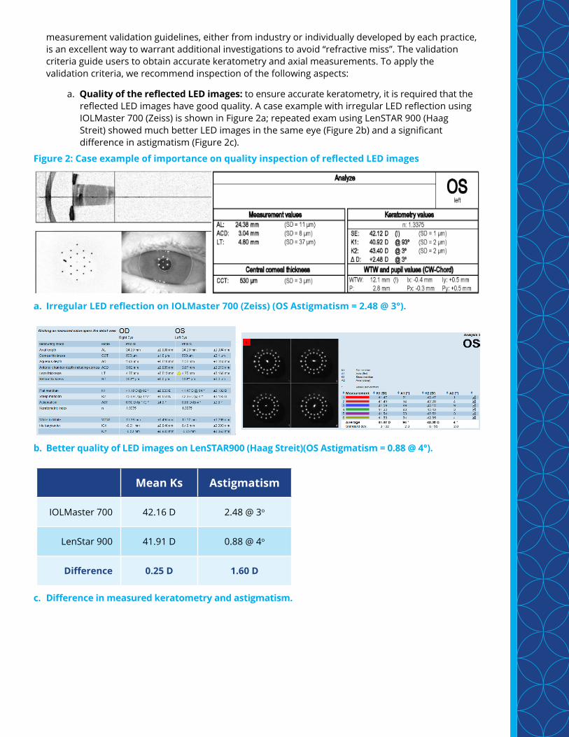
measurement validation guidelines, either from industry or individually developed by each practice, is an excellent way to warrant additional investigations to avoid “refractive miss”. The validation criteria guide users to obtain accurate keratometry and axial measurements. To apply the validation criteria, we recommend inspection of the following aspects:
a. Quality of the reflected LED images: to ensure accurate keratometry, it is required that the reflected LED images have good quality. A case example with irregular LED reflection using
IOLMaster 700 (Zeiss) is shown in Figure 2a; repeated exam using LenSTAR 900 (Haag Streit) showed much better LED images in the same eye (Figure 2b) and a significant difference in astigmatism (Figure 2c).
Figure 2: Case example of importance on quality inspection of reflected LED images
a. Irregular LED reflection on IOLMaster 700 (Zeiss) (OS Astigmatism = 2.48 @ 3°).
b. Better quality of LED images on LenSTAR900 (Haag Streit)(OS Astigmatism = 0.88 @ 4°).
c. Difference in measured keratometry and astigmatism.
Mean Ks Astigmatism
IOLMaster 700 42.16 D 2.48 @ 3o
LenStar 900 41.91 D 0.88 @ 4o
Difference 0.25 D 1.60 D

b. Individual parameters: the manufacturer’s validation criteria (Appendix 2 & 3) list the reasonable range for several parameters, such as the average keratometry, anterior chamber depth (ACD), and lens thickness. For example, if a phakic ACD is >4.5 mm for either LenSTAR 900 (Haag-Streit) or IOLMaster, additional measurements are recommended.
c. Standard deviation (SD): a smaller SD indicates more consistent repeated measurements. If the SD is greater than the criterion, inspection of raw data should be performed, outlier
data should be deleted, or additional exams should be added. Figure 3 is an example to show a large K SD adjustment.
Figure 3: Case example of large Standard Deviation (SD) adjustment
OS: original measurementsAL: SD = 0.032 mmK2: SD = 1.558 DMean K: 45.67 DAstigmatism: 2.40 @ 99°
OS: after adjustmentAL: SD = 0.009 mmK2: SD = 0.072 DMean K: 45.08 DAstigmatism: 1.52 @ 102°

d. Difference between right (OD) and left (OS) eyes: if the difference for a parameter between OD and OS exceeds certain amount as recommended by the manufacturer (Appendix 2 & 3), for example, great than 0.3 mm for axial length difference, additional exams are required. This is true for both LenSTAR 900 (Haag-Streit) and IOLMaster (Zeiss). For average K power difference between two eyes, the criterion for LenSTAR 900 (Haag-Streit) and IOLMaster (Zeiss) is 1.25D and 0.90D respectively.
4. Topography and tomography are highly recommended in addition to routine biometry for refractive cataract surgeries.11 In addition to the usage of helping to rule out OSD and confirming cornea stability after rigid contact lens wear as stated above, the corneal topography may reveal if the astigmatism is regular and may provide useful information for toric IOL selection. Topography and tomography can also help in confirming the presence of prior myopic or hyperopic corneal laser refractive surgery and IOL power calculation in those post-refractive surgical eyes.
5. Following and implementing the advancement in measurement technology. The newly developed IOLMaster 700 (Zeiss) and ARGOS (Alcon) utilize swept-source OCT technology increase the acquisition speed and can now measure AL in eyes with dense cataracts that previously would require immersion ultrasound.17, 18
ConclusionTo achieve high post-operative refractive accuracy and patient satisfaction in cataract patients, it is critical to accurately detect OSD, retina/macular pathology and any other potential ocular diseases that may affect refractive and visual outcomes, and to avoid biometry measurement errors in order to make sure the input parameters in advanced IOL formulas are accurate.

AppendixAppendix 1: The ASCRS preoperative OSD algorithm.3
ADDE= aqueous-deficient dry eye; CL= contact lens; DED= dry-eye disease; EBMD= epithelial basement membrane dystrophy; EDE= evaporative dry eye; IOL= intraocular lens; LLPP= Look, Lift, Pull, Push; LLT= lipid layer thickness; LRI= limbal relaxing incisions; LVC= laser vision correction; MGD= meibomian gland dysfunction; MMP-9= matrix metalloproteinase-9; NI-TBUT= noninvasive tear breakup time; NVS-OSD= nonvisually significant ocular surface disease; OCT= optical coherence tomography; OSD= ocular surface disease; OSI= ocular scatter index; SPEED= Standard Patient Evaluation of Eye Dryness; TBUT= tear breakup time; TMH= tear meniscus height; VS-OSD= visually significant ocular surface disease
ASCRS PREOPERATIVE OSD ALGORITHM
OSD RULED OUTNO Symptoms / NO Signs /
Normal Exam / NO Corneal Staining
SURGERY PROCEEDSCounsel patient OSD may worsen postoperatively.
Start prophylactic treatment.
NON-VISUALLY SIGNIFICANT OSD (NVS-OSD) (e.g. normal cornea, normal topography / regular
astigmatism, no corneal stain, stable vision)
To help establish OSD subtype and visual significanceOPTIONAL NONINVASIVE OSD TESTS
(e.g. meibography, topography, NI-TBUT, OCT TMH, OSI, aberrometry, LLT)
CLINICAL EXAM (LLPP)
LOOK:Blink, lids, lashes,
interpalpebral surface
LIFT:Upper lid, examine
superior surface
PULL:Assess lid laxity
‘floppy eyelids’, fornices
PUSH:Meibomian gland
expression
STAIN (dye instillation): corneal staining? TBUT? +/- Schirmer’s
NEUROPATHICPAIN
EARLY SITUATIONALOSD / DED
vs.
Symptoms / NO Signs / Normal Exam / Stain
NEUROTROPHICCORNEA
NO Symptoms / Signs / Exam / Stain
NEGATIVE SCREEN(All normal)
OSD UNLIKELY
POSITIVE SCREEN(Any abnormality)
OSD LIKELY
VISUALLY SIGNIFICANT OSD (VS-OSD) (e.g. corneal abnormality, central PEE, irregularly irregularastigmatism, abnormal osmolarity, fluctuating vision, etc.)
TREATMENT PLANBased on subtype and severity of each OSD.
Combined medical and procedural interventions.
Next office visit in 2-4 weeks.Start at beginning of algorithm.
SURGERY DELAYEDRefractive measurements unreliable.
Identify all OSD subtypes.
PREOPERATIVE VISIT≥ 2 week CL holiday / no drops
within 2 hours prior
NEXT VISIT IS SURGERY
NONINVASIVE REFRACTIVE PREOP MEASUREMENTS(e.g. keratometry, topography, optical biometry, aberrometry, etc.)
OSD SCREEN
ASCRS SPEED II PREOPQuestionnaire
SYMPTOMS?Inflammatory Marker(MMP-9)Osmolarity
SIGNS?
EXPOSURE / LIDMALPOSITION
(e.g. lagophthalmos, floppy eyelid)
DRY EYEDISEASE
EDE (MGD) > ADDE
LUMPS &BUMPS
(e.g. EBMD)
CONJUNCTIVITIS (e.g. allergic, infectious)
OPTIONAL INVASIVE REFRACTIVE TESTS(e.g. immersion and / or contact A, B scans etc.)
Finalize refractive surgical plan(e.g. IOL, LRI, LVC, etc.)
ONCE VS-OSDIS CONVERTEDTO NVS-OSD
OSD RULED IN Any combination of abnormal signs and / or symptoms.
Visual significance based on results of above.
Start / End
Diagnosis
Optional Data Collection
Essential Data Collection
Decision / Informational

Appendix 2: LenStar LS900 (Haag Steit) Validation Criteria.
LENSTAR LS 900 CalibrationWeekly nullification................................................SUCCESSFUL
KeratometryOcular surface (improvement necessary?)........................NORMAL
K1 & K2 SD (maximum value, each eye)..................................±0.25 D
Avg K power difference (between eyes)............................< 1.25 D
Avg K power (each eye).........................> 40.00 D and < 48.00 D
Steep meridian SD..............................................................< 3.5o
AST (maximum value, each eye)............................................< 4.00 D
Reflected LED images (all meridians)..................GOOD QUALITY
Soft contact lenses (at least 1 week).......................................OUT
RGP contact lenses (until topography & Rx are stable).............OUT
T-cone TopographyCalibration..............................................................SUCCESSFUL
All measurements................................CORRECTLY CENTERED
Topo maps (AC, T, E, & RINGS)...............................GOOD QUALITY
K1, K2, & A1 (all five measurements).............................CONSISTENT
Axial MeasurementsMeasurement mode (phakic, silicone oil, etc.).................CORRECT
Fixation light (confirm visualization by patient).....................STEADY
5 consistent measurements..................................CONFIRMED
Caliper placement (cornea, lens, & retina).......................CORRECT
CCT (prior myopic LASIK/PRK?).................> 480 µm and < 620 µm
Phakic ACD (each eye).........................> 1.9 mm and < 4.5 mm
Lens thickness (each eye)....................> 3.0 mm and < 6.2 mm
OD & OS axial length......................................WITHIN 0.30 mm
AL consistent with oldest Rx SphEq.....................CONFIRMED
Outliers (either eye).........................................DELETE & REPEAT
White to WhiteLimbus ring..............................................ADJUST AS REQUIRED
Avg WTW (unusual Ks, ACD, or AL?)....> 10.0 mm and < 13.0 mm
Avg WTW (each eye).............................................WITHIN 0.1 mm
Avg WTW (between eyes)......................................WITHIN 0.2 mm
Additional Validation/StudiesPhakic ACD > 4.5 mm or < 1.9 mm...................MD CONFIRMS
OD/OS AL difference > 0.30 mm......................MD CONFIRMS
OD/OS avg K power > 1.25 D...........................MD CONFIRMS
AST > 4.25 D (KCN or PMD?)...............TOPOGRAPHIC AXIAL MAP
Avg K power > 48.00 D or < 40.00 D.................MD CONFIRMS
Warren E. Hill, MD - LENSTAR LS 900

Appendix 3: IOLMaster (Zeiss) Validation Criteria.
IOLMaster - CalibrationTest block (AL, K, ACD)....................CORRECT, PRINTED, & FILED
Axial LengthCorrect setting (phakic, acrylic, silicone oil, etc.)...............CONFIRM
Patient able to see red fixation light.........................CONFIRM
Double peaks (anterior peak is likely the ILM)....................DELETED
Poorly formed primary maxima.................................DELETED
Significant Outliers (look at primary maxima)...................DELETED
At least 5 measurements within 0.05 mm................CONFIRM
Composite SNR > 10 (typically > 100)................................CONFIRM
OD & OS AL within 0.30 mm......................................CONFIRM
AL consistent with oldest Rx......................................CONFIRM
AutokeratometryOcular surface (postpone measurements?)........................NORMAL
K1 & K2 within 0.25 D in each meridian....................CONFIRM
Astigmatism lines up with Rx cyl & axis....................CONFIRM
Astigmatism for each eye < 3.50 D..........................CONFIRM
Avg K power for both eyes within 0.90 D..................CONFIRM
Avg K power < 47.00 D or > 41.00 D........................CONFIRM
No “x” appearing in any LED location........................CONFIRM
Soft contact lenses.............................OUT 1 WEEK MINIMUM
RGP contact lenses..............OUT UNTIL TOPO & Rx STABLE
Optical ACD MeasurementAphake & pseudophake.............................DO NOT MEASURE
5 consistent measurements (green light)....................CONFIRM
ACD < 4.5 mm and > 2.0 mm....................................CONFIRM
White to White3 measurements within 0.2 mm...............................CONFIRM
OD & OS within 0.2 mm (check arc position).................CONFIRM
Exceptions & Additional StudiesAL < 22.0 mm (ACD & LT for H2)...................IMMERSION A-SCAN
AL > 30.0 mm (is there reduced BCVA?)..................STAPHYLOMA?
OD/OS AL difference > 0.30 mm......................MD CONFIRMS
Astigmatism > 3.50 D (KCN, PMD?)....................TOPO AXIAL MAP
Avg Ks > 0.90 difference....................................MD CONFIRMS
Avg K power > 47.00 D or < 41.00 D...............MD CONFIRMS
ACD < 2.0 mm or > 4.5 mm.............................MD CONFIRMS
White-to-White < 10.2 mm or > 13.0 mm........MD CONFIRMS
Warren E. Hill, MD - IOLMaster software version 5.4

References1. T. Olsen, “Calculation of intraocular lens power: a review,” Acta Ophthalmol. Scand., vol. 85, pp. 472-
485, 2007.
2. C. Kent, “Cataract Patients: Younger Every Year,” REVIEW of Opthalmomology, 15 March 2013.
3. C. E. Starr, P. K. Gupata , M. Farid, K. A. Beckman, C. C. Chan, E. Yeu, J. A. Gomes, B. D. Ayers , J. P. Berdahl, E. J. Holland, T. Kim and F. S. Mah, “An algorythm for the preoperative diagnosis and treatment of ocular surface disorders,” J Cataract Refract Surg, vol. 45, pp. 669-684, 2019.
4. F. Stapleton , M. Alves , V. Bunyya, I. Jalbert, K. Lekhanont, F. Malet, K.-S. Na, D. Schaumberg, M. Uchino, J. Vehof, E. Viso, S. Vitale and L. Jones, “TFOS DEWS II Epidemiology Report,” The Ocular Surface, vol. 15, pp. 334-365, 2017.
5. A. L. Sheppard and J. S. Wlffsohn, “Digital eye strain: prevalence, measurement and amelioration,” BMJ Open Ophth, vol. 3, p. e000146, 2018.
6. N. Shamie, “The status of dry eye care in surgical practices,” ASCRS Eye World CME Supplement, October 2013.
7. P. K. Gupta, O. J. Drinkwater, K. W. VanDusen, A. R. Brissette and C. E. Starr, “Prevalence of ocular surface dysfunction in patients presenting for catarct surgery evaluation,” J Cataract Refract Surg, vol. 44, pp. 1090-1096, 2018.
8. W. B. Trattler, P. A. Majmudar, E. D. Donnenfeld, M. B. McDonald, K. G. Stonecipher and D. F. Goldberg, “The prospective health assessment of cataract patients’ ocualr surface (PHACO) study: the effect of dry eye,” Clinical Ophthalmology, vol. 11, pp. 1423-1430, 2017.
9. T. W. Olsen , A. Al-Rajhi, A. Ambrus, . R. Lastra, L. C. Lum and D. Mizuiri, “Dry Eye Syndrome Preferred Practice Pattern,” Elservier Inc, 2018.
10. J. S. Wolffson , R. Arita, R. Chalmers, A. Djalilian, M. Dogru, K. Dumbleton , P. K. Gupta, P. Karpecki, S. Lazreg, H. Pult, B. D. Sullivan, A. Tomlinson, L. Tong, E. Villani, K. C. Yoon , L. Jones and J. P. Craig, “TFOS DEWS II Diagnostic Methodology report,” The Ocular Surface, vol. 15, pp. 539-574, 2017.
11. D. D. Koch and L. Wang , “Step--by-step to the right IOL power, Cataract surgery preoperatie assessment and IOL power selection for your patient,” Ophthalmology Management, vol. 23, no. March, pp. 28, 31, 32, 34, 1 March 2019.
12. M. McKeague, P. Sharma and A. C. Ho, “Evaluation of the macula prior to cataract surgery,” Curr Opin Ophthalmol, vol. 29, no. 1, pp. 4-8, 2018.
13. S. Charles, “OCT before cataract surgery: not optional,” Review of Ophthalmology, 7th August 2018.
14. W. E. Hill, A. Abulafia, L. Wang and D. D. Koch, “Pursuing perfection in IOL calculations. II. Measuremetn foibles: Mesurement errors, validation criteria, IOL constants, and lanelenght,” J Cataract Refract Surg, vol. 43, no. 7, pp. 869-870, 2017.
15. J. T. Holladay, “Accurate corneal powe measurements for IOL calculations,” Ocular Surgery News, 10 September 2016.
16. “Biometry validation guidelines _IOLMAster and Lenstar,” [Online]. Available: https://www.doctor-hill.com/biometry_validation.html. [Accessed 28 July 2019].
17. H. J. Shammas, S. Ortiz, M. C. Shammas, S. H. Kim and C. Chong, “Biometry measurements usign a new large-coherence-length swept-source opticl coherence tomographer,” J Cataract Refract Surg, vol. 42, pp. 50-61, 2016.
18. M. Kurian , N. Neglur, S. Das, N. K. Puttaiah, D. Haria and M. M. Thakkar , “Biometry with a new swept-source optical coherence tomography biometer: repeatabilkity and agreement with an Optical low coherence reflectometry device,” J Cataract Refract Surg, vol. 42, pp. 577-581, 2016.

© 2019 Alcon Inc. 11/19 US-CAT-1900002
Alcon Medical Affairs