
P ROCEDURES F OR C REDIT BY E XAMINATION A ND C OLLEGE L EVEL E XAMINATION P ROGRAM.

CRANIAL TRAUMA PROCEDURES Op320 (1)
Cranial Trauma Procedures Last updated: September 5, 2017
General principles ............................................................................................................................ 1
SCALP WOUNDS ....................................................................................................................................... 1
SKULL FRACTURES .................................................................................................................................. 3 DEPRESSED SKULL FRACTURES .............................................................................................................. 3
Ping Pong fracture ............................................................................................................................ 3 Leptomeningeal cyst (s. growing fracture) ...................................................................................... 3
TWIST DRILL CRANIOSTOMY (FOR CHRONIC SUBDURAL HEMATOMA) .................................................. 6
BURR HOLE WASHOUT, S. CRANIOSTOMY (FOR CHRONIC SUBDURAL HEMATOMA) .............................. 6 Indications ........................................................................................................................................ 6
Procedure .......................................................................................................................................... 6 Postoperatively ................................................................................................................................. 6
ACUTE EPIDURAL HEMATOMA EVACUATION .......................................................................................... 7 IMMEDIATE EXPLORATORY BUR HOLES .................................................................................................. 7
CRANIECTOMY ....................................................................................................................................... 9
CRANIOTOMY....................................................................................................................................... 10
ACUTE SUBDURAL HEMATOMA EVACUATION ....................................................................................... 11 IMMEDIATE EXPLORATORY BUR HOLES ................................................................................................ 11 CRANIOTOMY....................................................................................................................................... 11
Malignant cerebral edema .............................................................................................................. 11
Postoperatively ............................................................................................................................... 11
DECOMPRESSIVE CRANIECTOMIES ....................................................................................................... 11 Indications ...................................................................................................................................... 11 Types .............................................................................................................................................. 12
Before surgery ................................................................................................................................ 12
Principles ........................................................................................................................................ 12 Postop ............................................................................................................................................. 12
DECOMPRESSIVE HEMICRANIECTOMY (“TRAUMA FLAP”) / FRONTOTEMPORPARIETAL CRANIOTOMY . 12 Planning .......................................................................................................................................... 12
Procedure ........................................................................................................................................ 13
Complications ................................................................................................................................. 17
DECOMPRESSIVE BIFRONTAL CRANIECTOMY (KJELLBERG) ................................................................ 18
Indications ...................................................................................................................................... 18 Procedure ........................................................................................................................................ 18
PENETRATING BRAIN INJURIES .............................................................................................................. 20 GUNSHOT WOUNDS .............................................................................................................................. 20 STAB INJURY ........................................................................................................................................ 21
DURAL VENOUS SINUS INJURIES ............................................................................................................ 21
Subdural tap through fontanel – see p. TrH13 >>
GENERAL PRINCIPLES
INDICATIONS FOR SURGERY
See p. TrH1 >>
PREOPERATIVELY
correct coagulopathy - fresh frozen plasma (for prolonged PT/aPTT), cryoprecipitate (for
fibrinogen < 1.5 g/L), thrombocyte transfusions (for platelets < 100,000).
consider antiepileptic (AED)
antibiotic prophylaxis.
tetanus immunization status should be checked and updated (esp. lacerations, contaminated
wounds).
if suspect vascular lesion (e.g. young person with deep bleed):
1) order CTA preop
2) plan craniotomy so will have proximal control (e.g. by dissecting Sylvian fissure).
3) start evacuating blood clot farthest from suspicious area and may leave small clots
ANESTHESIA
even if patient is in coma it is unwise to begin without full anesthetic support (only exception is
patient thought likely to expire during time taken to organize these precautions, which can usually
be done while theater is prepared).
if head is rotated for surgery:
- make sure neck veins are not twisted
- head above heart level (if BP permits)
- 3-point head fixation device is used if unstable C-spine fracture is present
drape to allow extension of surgical incision beyond actual confines of wound or to allow possible
scalp rotation procedures.
patients with high ICP are very vulnerable to incidents of respiratory obstruction, hypercarbia,
systemic hypotension.
N.B. once intracranial hematoma begins to be removed blood pressure may fall precipitously
(esp. if multiple injury has produced hypovolemia, masked by effects of raised ICP).
ICP monitor (or ventricular drain) usually is placed intraoperatively in patients with GCS ≤ 8.
TECHNIQUE
whole head is shaved (e.g. for placement of ICP monitor on contralateral side).
never make small trauma craniotomy (brain may swell and may need to leave bone flap out); big
trauma flap is never wrong choice (postoperative brain swelling may strangulate brain against tight
craniotomy bone edges).
Dr. Mathern likes dural tuck-ups placed before opening dura (if time permits) – place stitch under
bone – helps with hemostasis and dural closure!
after evacuating mass lesion, leave ICP monitor in (even if removed bone flap).
4) next to suspected lesion (to avoid brisk bleeding).
send selected removed blood clots for pathology (may find tumor, vascular lesion).
malignant cerebral edema – see below >>
POSTOPERATIVE
CT is obtained ≤ 24 hours postoperatively.
COMPLICATIONS
Infection (cranial osteomyelitis, subdural empyema, meningitis, brain abscess).
risk factors – open scalp fractures, violated paranasal sinuses (esp. with CSF leak).
SCALP WOUNDS
CRANIAL TRAUMA PROCEDURES Op320 (2)
SCALP ABRASIONS
often contaminated with pieces of dirt - should be thoroughly cleaned and inspected for puncture
wounds to ensure removal of unsuspected foreign bodies.
SCALP LACERATIONS
Because of scalp's rich vascular supply:
1) scalp lacerations may be source of significant bleeding.
2) most uncomplicated lacerations can be closed (after cleansing and debridement) and antibiotics
are usually not needed.
3) even very large scalp avulsions can survive:
– if avulsion remains attached to rest of scalp by tissue bridge, it should be reattached
to surrounding tissue.
– if avulsion is completely detached from scalp it should be treated as any other
amputated part and reimplanted ASAP.
– small scalp deficit is repaired by rotating portion of scalp.
– large scalp deficit requires skin graft or vascularized free flap.
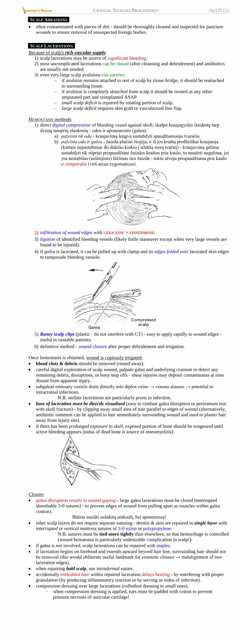
HEMOSTASIS methods:
1) direct digital compression of bleeding vessel against skull; skalpo kraujagyslės išsidėstę tarp
dviejų tamprių sluoksnių - odos ir aponeurozės (galea):
a) pažeista tik oda - kraujavimą lengva sustabdyti spaudžiamuoju tvarsčiu.
b) pažeista oda ir galea - žaizda plačiai žiojėja, o iš jos kraštų profūziškai kraujuoja
(kartais nepastebimai iki didelio kiekio į uždėtą storą tvarstį) - kraujavimą galima
sustabdyti tik stipriai prispaudžiant žaizdos kraštus prie kaulo; to naudoti negalima, jei
yra nestabilus (nelinijinis) lūžimas ties žaizda - tokiu atveju prispaudžiama prie kaulo
a. temporalis (virš arcus zygomaticus).
22)) infiltration of wound edges with LLIIDDOOCCAAIINNEE + EEPPIINNEEPPHHRRIINNEE.
3) ligation of identified bleeding vessels (likely futile maneuver except when very large vessels are
found to be injured).
4) if galea is lacerated, it can be pulled up with clamp and its edges folded over lacerated skin edges
to tamponade bleeding vessels:
5) Raney scalp clips (plastic - do not interfere with CT) - easy to apply rapidly to wound edges -
useful in unstable patients.
6) definitive method - wound closure after proper debridement and irrigation.
Once hemostasis is obtained, wound is copiously irrigated:
blood clots & debris should be removed (rinsed away).
careful digital exploration of scalp wound, palpate galea and underlying cranium to detect any
remaining debris, disruptions, or bony step offs - shear injuries may deposit contaminants at sites
distant from apparent injury.
subgaleal emissary vessels drain directly into diploe veins → venous sinuses → potential to
intracranial infections.
N.B. stellate lacerations are particularly prone to infection.
base of laceration must be directly visualized (easy to confuse galea disruption or periosteum tear
with skull fracture) - by clipping away small area of hair parallel to edges of wound (alternatively,
antibiotic ointment can be applied to hair immediately surrounding wound and used to plaster hair
away from injury site).
if there has been prolonged exposure to skull, exposed portion of bone should be rongeured until
active bleeding appears (nidus of dead bone is source of osteomyelitis):
Closure
galea disruption results in wound gaping - large galea lacerations must be closed (interrupted
absorbable 3-0 sutures) - to prevent edges of wound from pulling apart as muscles within galea
contract.
Būtina susiūti sužalotą antkaulį, bei aponeurozę!
other scalp layers do not require separate suturing - dermis & skin are repaired in single layer with
interrupted or vertical mattress sutures of 3-0 nylon or polypropylene.
N.B. sutures must be tied more tightly than elsewhere, so that hemorrhage is controlled
(wound hematoma is particularly undesirable complication in scalp!).
if galea is not involved, scalp lacerations can be repaired with staples.
if laceration begins on forehead and extends upward beyond hair line, surrounding hair should not
be removed (this would obliterate useful landmark for cosmetic closure → malalignment of two
laceration edges).
when repairing bald scalp, use intradermal suture.
accidentally embedded hair within repaired laceration delays healing - by interfering with proper
granulation (by producing inflammatory reaction or by serving as nidus of infection).
compression dressing over large lacerations (collodion dressing to small ones).
– when compression dressing is applied, ears must be padded with cotton to prevent
pressure necrosis of auricular cartilage!
CRANIAL TRAUMA PROCEDURES Op320 (3)
SKULL FRACTURES
DEPRESSED SKULL FRACTURES
Used sources:
Sekhar “Atlas of Neurosurgical Techniques - Brain” (2006), ch. 78 (pages 909-910)
indications – see p. TrH >>
lazy "S" or horseshoe-shaped incision over depression; bicoronal incision is preferred for forehead
depressions; in open fractures, scalp wound is debrided and incorporated into incision.
burr hole near fracture (and over intact dura to prevent brain injury) → bony fragments are
elevated → soaked in antibiotic and isotonic saline solution (if wound seems clean and occurred in
< 48 hours).
o cultures of wound and bone and devitalized tissue should be sent for later tailoring of
antibiotic coverage should an infection develop.
dural tears are repaired (thus, entire lacerated dura must be exposed).
bony fragments are reassembled (larger pieces may be wired together); titanium mesh is applied to
cover larger skull defects; methyl methacrylate can be used as substitute for bone fragments (avoid
in children; H: absorbable bone plates and screws).
PING PONG FRACTURE
there are reports of using obstetric vacuum extractor at bedside to elevate fracture; worsk for
prematures.
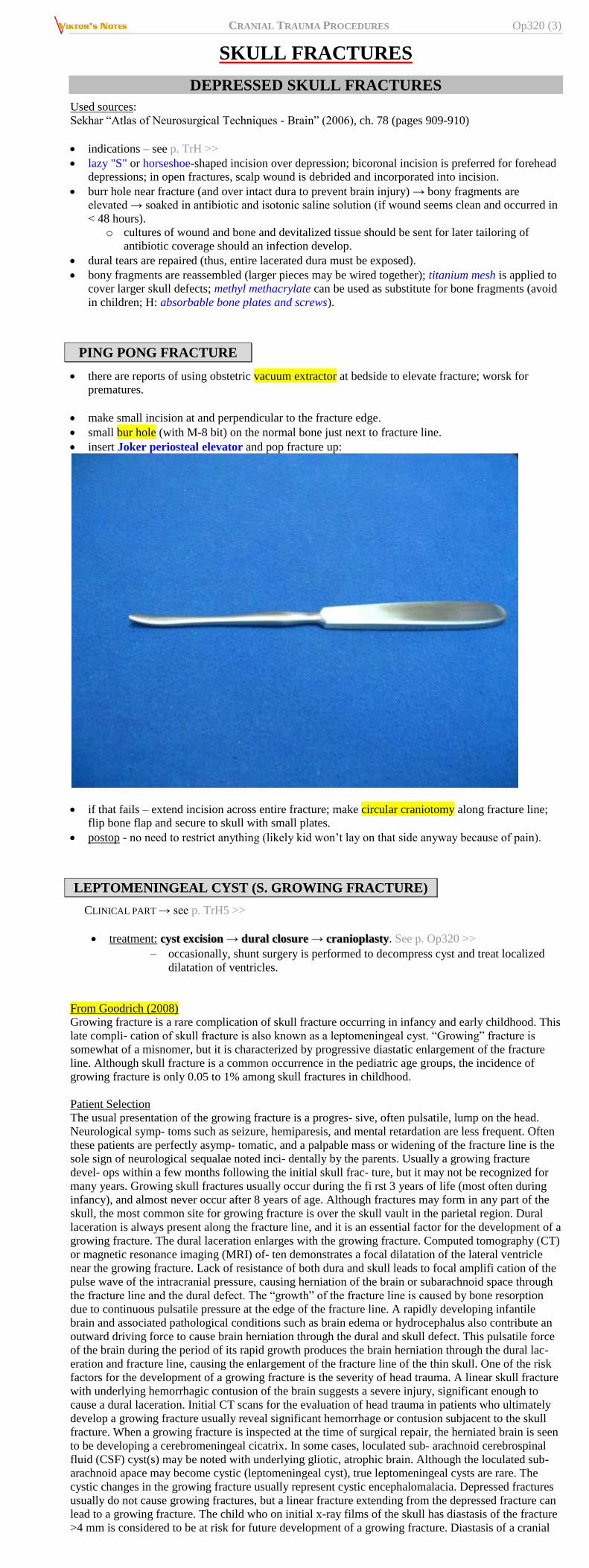
make small incision at and perpendicular to the fracture edge.
small bur hole (with M-8 bit) on the normal bone just next to fracture line.
insert Joker periosteal elevator and pop fracture up:
if that fails – extend incision across entire fracture; make circular craniotomy along fracture line;
flip bone flap and secure to skull with small plates.
postop - no need to restrict anything (likely kid won’t lay on that side anyway because of pain).
LEPTOMENINGEAL CYST (S. GROWING FRACTURE)
CLINICAL PART → see p. TrH5 >>
treatment: cyst excision → dural closure → cranioplasty. See p. Op320 >>
– occasionally, shunt surgery is performed to decompress cyst and treat localized
dilatation of ventricles.
From Goodrich (2008)
Growing fracture is a rare complication of skull fracture occurring in infancy and early childhood. This
late compli- cation of skull fracture is also known as a leptomeningeal cyst. “Growing” fracture is
somewhat of a misnomer, but it is characterized by progressive diastatic enlargement of the fracture
line. Although skull fracture is a common occurrence in the pediatric age groups, the incidence of
growing fracture is only 0.05 to 1% among skull fractures in childhood.
Patient Selection
The usual presentation of the growing fracture is a progres- sive, often pulsatile, lump on the head.
Neurological symp- toms such as seizure, hemiparesis, and mental retardation are less frequent. Often
these patients are perfectly asymp- tomatic, and a palpable mass or widening of the fracture line is the
sole sign of neurological sequalae noted inci- dentally by the parents. Usually a growing fracture
devel- ops within a few months following the initial skull frac- ture, but it may not be recognized for
many years. Growing skull fractures usually occur during the fi rst 3 years of life (most often during
infancy), and almost never occur after 8 years of age. Although fractures may form in any part of the
skull, the most common site for growing fracture is over the skull vault in the parietal region. Dural
laceration is always present along the fracture line, and it is an essential factor for the development of a
growing fracture. The dural laceration enlarges with the growing fracture. Computed tomography (CT)
or magnetic resonance imaging (MRI) of- ten demonstrates a focal dilatation of the lateral ventricle
near the growing fracture. Lack of resistance of both dura and skull leads to focal amplifi cation of the
pulse wave of the intracranial pressure, causing herniation of the brain or subarachnoid space through
the fracture line and the dural defect. The “growth” of the fracture line is caused by bone resorption
due to continuous pulsatile pressure at the edge of the fracture line. A rapidly developing infantile
brain and associated pathological conditions such as brain edema or hydrocephalus also contribute an
outward driving force to cause brain herniation through the dural and skull defect. This pulsatile force
of the brain during the period of its rapid growth produces the brain herniation through the dural lac-
eration and fracture line, causing the enlargement of the fracture line of the thin skull. One of the risk
factors for the development of a growing fracture is the severity of head trauma. A linear skull fracture
with underlying hemorrhagic contusion of the brain suggests a severe injury, significant enough to
cause a dural laceration. Initial CT scans for the evaluation of head trauma in patients who ultimately
develop a growing fracture usually reveal significant hemorrhage or contusion subjacent to the skull
fracture. When a growing fracture is inspected at the time of surgical repair, the herniated brain is seen
to be developing a cerebromeningeal cicatrix. In some cases, loculated sub- arachnoid cerebrospinal
fluid (CSF) cyst(s) may be noted with underlying gliotic, atrophic brain. Although the loculated sub-
arachnoid apace may become cystic (leptomeningeal cyst), true leptomeningeal cysts are rare. The
cystic changes in the growing fracture usually represent cystic encephalomalacia. Depressed fractures
usually do not cause growing fractures, but a linear fracture extending from the depressed fracture can
lead to a growing fracture. The child who on initial x-ray films of the skull has diastasis of the fracture
>4 mm is considered to be at risk for future development of a growing fracture. Diastasis of a cranial

CRANIAL TRAUMA PROCEDURES Op320 (4)
suture, however, is an unusual site for a growing fracture. A growing fracture at the skull base can
occur in an older age group, especially where the bone is thin such as in the orbital roof, if a linear
fracture is accompanied by a dural laceration. Growing fracture and a meningoencephalocele can
develop with a similar mechanism as those occurring in the skull vault of the young patient.
Radiological Studies
X-ray films of the skull show wide diastases of the fracture line. If initial skull films are available, one
can compare the films to confirm “growth” of the fracture line during the interval. When multiple
fractures are noted in the same patient, healing of the fracture in one area may be noted as opposed to a
growing fracture in another area. The fracture line can cross the coronal or lambdoid sutures but is
usually limited to one parietal bone. Neuroimagings such as CT and MRI provide information
regarding the sequelae within the growing fracture and any intracranial pathological changes.
Furthermore, if they are available from the time of initial trauma, it should be possible to demonstrate
progressive changes. It is not unusual that the initial neuroimagings show hemorrhagic contusion, or
subarachnoid or extraparenchymal hemorrhage. At the time of discovery of the growing fracture,
neuroimag ings demonstrate the diastasis of the fracture line and of- ten cystic lesions near the fracture
site. These cystic lesions represent encephalomalacia, a loculated arachnoidal cyst, or cortical atrophy.
The ipsilateral ventricle tends to show focal porencephalic dilatation with ipsilateral shift of the
midline structure. This phenomenon may be due not only to lack of dural resistance but also to cerebral
atrophy.
Management
Surgical intervention is indicated with a growing fracture line, seizure disorder, or progressive
neurologic deficits. A progressive cystic degeneration in the brain that has herniated through the dural
and cranial defects can occur; there- fore, surgical correction is recommended in young children even
when seizures or other neurological symptoms or signs are absent. However, incidental, asymptomatic,
and stable fractures in late childhood or adulthood probably do not require surgery. The goal of
surgery for growing skull fractures is to repair the dural laceration and cranial defect, and to resect
seizure foci. Growth of the growing fracture may arrest after CSF diversion shunting by a decrease of
the CSF pulse pressure, but this does not correct a seizure disorder. Placing a shunt for primary
treatment of these patients is not advised unless hydrocephalus is present. Shunting for
nonhydrocephalic patients creates undesirable shunt de- pendency.
OPERATIVE PROCEDURE
The scalp incision should be large enough to expose the entire length of the skull defect. An S-shaped
or semicircular skin in- cision is made, and the scalp fl ap is turned subgaleally, leaving the underlying
periosteal tissue intact ( Fig. 7–1A ). By palpa- tion, the entire length of the cranial defect covered by
pericra- nium is exposed in surgical view. The site of the cranial defect is often bulging and may be
accompanied by blush appearance due to an underlying subarachnoid cyst. As the cranial defect is
dissected by incising the pericranium along the edge of the bony defect ( Fig. 7–1B ), soft tissues
adherent to the edge of the cranium defect are scraped off by a sharp dissector. The surgeon should
remember that the dural edge is invar- iably larger than the cranial defect, and that the pericranium is
directly adherent to the underlying cerebral tissue at the cranial defect. An effort to expose the dural
edge by removing the cranial edge should not be undertaken, as this procedure is often complicated by
removing the dura simultaneously with the skull bone due to the adhesive nature of the du- ral edge.
To identify the dura, several bur holes are made away from the skull defect with a distance of at least
50% of the width of the cranial defect. At this time, a large enough amount of pericranium is removed
from the neighboring skull to use it for repair of the dural defect. Once the dura is identifi ed at each
bur hole site, the dura is separated from the inner table of the skull toward the defect ( Fig. 7–1C ). A
crani- otomy is made around the skull defect by connecting the bur holes with a craniotomy. Two
pieces of the craniotomy fl ap are obtained, one from each side of the growing fracture. After the
craniotomy is completed ( Fig. 7–2A ), reactive pe- riosteal tissue and the cerebromeningeal cicatrix
are identi- fi ed in the dural defect. Under magnifi ed vision by means of surgical loupes, the cicatrix
including the periosteal tissue is lifted, and all abnormal tissue is separated and transected us- ing a
bipolar cautery until normal white matter is exposed ( Fig. 7–2B ). The edge of the dura is separated
from the cer- ebral tissue, carefully avoiding trauma to the cerebral blood vessels. In this region,
abnormal tissue such as cystic changes or xanthochromic discoloration due to previous hemorrhage is
often noted. After adequate debridement of the cicatrix at the growing fracture and freeing of the intact
dural edge from the corti- cal surface, the dural defect is closed using the periosteal graft ( Fig. 7–2C ).
Autologous pericranium is preferable to cadaver dura. A watertight closure of the dura is important to
avoid a recurrence of the growing fracture or postoperative CSF leakage. Each of the obtained
craniotomy fl aps is split at the diploic space with an osteotome, separating it into inner and outer
tables ( Fig. 7–2D ). The cranial defect is then repaired by laying in the split autologous skull grafts.
Usually four pieces are laid next to each other side by side to fill the cranial defect. These flaps are
secured to each other with either nylon sutures or stainless steel wires through drill holes ( Fig. 7–2E ).
These fl aps are further secured to the craniotomy edge. If the defect of the skull is too large or the
skull is too thin to separate into inner and outer tables, one may consider autologous rib grafts. These
autologous bone grafts are well incorporated, and healing is excellent. Foreign materials such as
methyl methacrylate should be avoided for cranioplasty in the growing skull.
Figure 7–1 (A) The scalp fl ap is turned subperiosteally. The cranial defect is usually covered by the
pericranium. (B) The pericranium is in- cised along the edge of the cranial defect. Then, the edge of
the cranial defect is exposed by scraping off the soft tissues adherent to it. (C) The pericranium is
removed from the surrounding skull surface and pre- served for dural repair. Four bur holes are made
in the surrounding skull for a craniotomy. After the confi rmation of intact dura matter under the bur
hole, the dura is separated from the bur hole toward the cranial defect. The surgeon should not attempt
to identify the dura by remov- ing the bone from the edge of the cranial defect. The craniotomy is
performed on both sides of the growing fracture. The two bone fl aps are removed and preserved for
autologous bone cranioplasty.

CRANIAL TRAUMA PROCEDURES Op320 (5)
Figure 7–2 (A) After the craniotomy, the intact dura mater is exposed around the dural defect, which is
covered by the periosteum. Underneath the overgrowing periosteum is a cerebromenin- geal cicatrix
that is removed using bipolar cautery and sharp dissection until healthy white matter is exposed. (B)
After all pathological tissues have been removed, the edge of the surrounding dura is separated from
the intact cortical surface. (C) The previously removed periosteum is used to repair the dural defect. A
watertight closure is achieved with 4–0 sutures. (D) The bone grafts are split at the diploic space
between the inner and outer ta- bles by means of an osteotome. (E) The obtained split bone fl aps are
used to repair the cranial de- fect. The bone fl aps are secured to each other and to the edge of the
cranial defect with nylon sutures or stainless steel wires.
Specific Considerations
The growing fracture may extend toward a dural venous sinus such as the superior sagittal or lateral
sinus. Although these venous sinuses were spared from direct injury at the initial trauma, direct
exposure of them is not advised or necessary. When the fracture line extends perpendicularly to these
sinuses, the closest end to the sinus does not need dural repair. However, if the growing fracture is
parallel and near to the sinus, dural repair may be difficult due to the lack of enough dural edge next to
the sinus. In these cases, one may repair the dural defect with a periosteal graft sutured to the
periosteum of the skull above the sinus.
Postoperative Management
Including Possible Complications CSF diversion shunting has been recommended for persist- ent
postoperative CSF leakage from the craniotomy wound. It is justified if coexisting hydrocephalus is
evident, or if CSF leakage occurs despite adequate repair of the growing fracture. A lumboperitoneal
shunt or temporary lumbar CSF drainage is to be considered under these circumstances.

CRANIAL TRAUMA PROCEDURES Op320 (6)
TWIST DRILL CRANIOSTOMY (FOR CHRONIC
SUBDURAL HEMATOMA)
hole is drilled at 45° angle to skull over thickest part of hematoma (unless this lies over motor
strip); possible under local anesthesia at bedside.
twist drill is used to perforate dura and to release subdural hematoma.
thin rubber catheter is gently guided* into subdural space, tunneled under scalp, and brought out
through stab incision (connect to closed drainage system without suction for 24-72 hours).
*e.g. on stylet bent like hockey stick
postop – see below >>
BURR HOLE WASHOUT, s. CRANIOSTOMY (FOR
CHRONIC SUBDURAL HEMATOMA)
Principles of chronic SDH treatment (incl. indications, surgery types) → see p. TrH13 >>
INDICATIONS
- subacute / chronic SDH see p. TrH13 >>
sometimes even acute SDH can be evacuated through burr holes (e.g. unstable patient with low Hb
– nonclotting hematoma); but usually, if patient is minimally symptomatic (e.g. headaches), – may
wait until hematoma liquefies (usually 10-14 days).
PROCEDURE
POSITION
supine position ± towel roll under ipsilateral shoulder.
head on gel donut / horseshoe / subdural head holder slightly rotated to the contralateral side.
Dr. Graham likes subdural head holder – keeps head straight vertical (helps with
drainage during procedure) and permits access to posterior region.
ANESTHESIA
a) local anesthesia at bedside
b) monitored local anesthesia or general anesthesia in OR (best results!)
PROCEDURE
burr holes are placed over thickest aspects of hematoma; ideally, at superior temporal line (just
above temporalis muscle; if needed, OK to go through muscle)
make 2 (1-3) burr holes.
– e.g. frontal and parietal burr holes - if needed, can be incorporated into craniotomy;
– studies show that patients do well with just 1 burr hole but difficult to irrigate through
1 hole!
– if holes are placed too near extremes of hematoma, drainage may be blocked by brain
(H: depress surface of brain with patty or spatula).
coagulate dura with bipolar cautery
incise dura in cruciform manner* with a #15 or #11 blade (*do not cut to the edge of bone – leave
some dura – easier to stop bleeding if it happens) – watch for motor oil color thin fluid.
cut with Bovie to completely open up burr holes.
– if clearly see blood through dura, may open dura with Bovie (avoiding bipolar & blade
step)
if thick dark clot is found → thorough copious washout; attempt to scrape (mobilize) clot with
Penfield #3, #4, hockey-stick
– may need additional bur holes or even craniotomy; alternative – if significant part of
clot was evacuated and brain reexpanded, - may finish and follow with CT (repeat
BHWO if needed – less morbidity than craniotomy)
attempt to fenestrate (with bipolar) visible pathologic membranes (but they may bleed!)
irrigate with sterile saline via Becker catheter inserted subdurally in all directions (except
superiorly towards sagittal suture – not to disrupt bridging veins)
irrigate until returning fluid is clear and free of old clot fragments.
see for brain re-expansion.
– often, brain does not re-expand right away → tunnel ventriculostomy catheter* under
skin and leave as subdural drain (direct drain towards frontal pole; connect to closed
drainage system [“transfer pack’] without suction).
*Drs. Graham, Holloway use round 7F JP drain
drain drastically reduces subdural hematoma recurrence – 8% (vs. 20% without drain).
– insert through posterior* bur hole and advance anteriorly (when brain reexpands
postop, last fluid to disappear is frontal; if drain sits posteriorly, it becomes blocked
early by reexpanding brain)
*Dr. Graham – through anterior bur hole (and also direct anteriorly)
– do not force drain (stop with slightest resistance encounter – drain goes to parenchyma
very easily!); if brain reexpanded, do not place subdural drain, rather place drain under
galea and running over bur holes.
– drain complications: puncturing cortex*, causing hematoma, subdural empyema.
*H: Becker catheter is used after the stylet is bent in shape of hockey
stick – so that catheter slides not into the brain!
CLOSURE
both burr holes are covered with cranial plates and screws.
galea is approximated with 2-0 Vicryl in interrupted fashion except small gap in upper (anterior)
incision → sterile saline is gently injected through Becker catheter allowing intracranial air to be
expelled through galeal gap (→ table is brought into Trendelenburg position, anesthesia applies
Valsalva maneuver) → galea closed completely
Becker catheter is connected to transfer pack (or JP bulb with thumbprint gentle suction – Dr.
Graham).
staples, bacitracin ointment.
POSTOPERATIVELY
postoperative seizures are reported in 3-10% patients (many surgeons use prophylactic
anticonvulsants for 7-30 days after operation).
hCT next day
– CT often (≈ 92%) shows residual subdural collection - should be left alone (unless it
continues to exert significant mass effect); thus, recommendation is not to do postop
head CT until ≥ 3 days postop (unless patient deteriorates)
flat bed regimen as long as subdural drain is present (to prevent sucking air intracranially)* +
adequate IV hydration (to promote brain re-expansion).

CRANIAL TRAUMA PROCEDURES Op320 (7)
*alternatively, subdural drain may be connected to JP bulb (with minimal suction –
“dimple on bulb”) – patient may sit up to 30 degrees - much better tolerated.
keep drain 1-3 days; studies show that patients do best if drain is left for ≥ 72 hours and patient
remains flat all that time; Dr. Graham keeps longer (with daily stripping of JP drain) if on CT brain
is not reexpanding.
if drainage is sub-optimal (< 50 cc) TTPPAA can be instilled through drain. Neils D, Singanallur P, Huaping W, et al. Recurrence-Free Chronic Subdural hematomas: A
Retrospective Analysis of the Instillation of Tissue Plasminogen Activator in Addition to Twist
Drill or Burr Hole Drainage in the Treatment of Chronic Subdural Hematomas
In this study intrathecal tPA lead to zero cSDH recurrences, zero complications
IF SDH RECURS AFTER BURR HOLE DRAINAGE
repeat bur hole washout.
look for causes:
1) MRI, CTA of brain – for vascular abnormalities
2) MRI of spine, myelogram – for occult CSF leak (Dr. Holloway would do blood patch
even if spine looks unremarkable)
medications (TTXXAA, AAPPRROOTTIINNIINN, etc) → see p. TrH13 >>
craniotomy with partial removal of enveloping membranes (membranectomy*) or shunting of
subdural cavity into pleural or peritoneal cavity.
REOPERATION RATES (for hematoma reaccumulation) ≈ 12-22%.
*if you see clearly brain surface, leave that “membrane” alone – do not violate
arachnoid (→ CSF leak)
ACUTE EPIDURAL HEMATOMA EVACUATION
IMMEDIATE EXPLORATORY BUR HOLES
INDICATION
- conservatively uncontrollable ICP↑ with rapid patient deterioration in ED (consciousness↓ +
asymmetric findings) → think of brain compression* → emergency BURR HOLES.
*if skull fracture is present – most commonly epidural hematoma.
It is life-saving procedure - unless burr hole is done patient will die or be damaged: you and patient have nothing to lose
and everything to gain - inelegant burr hole now will do much more good than elegant operation one hour or more later.
STRATEGY
If CT is unavailable – side of burr hole is chosen:
Feature Hematoma Side
First (or only) dilated pupil Ipsilateral 94%
Most abnormal motor response Contralateral 82%
Skull fracture Ipsilateral 66%
– also can be helpful - scalp wound side, M-echo signal dislocation, pineal gland
dislocation (on X-ray).
position patient supine – allows approach to both skull sides.
1. First burr hole - low in temporal region, just above zygoma (one finger’s breadth anterior to tragus
and one finger’s breadth above zygomatic process).
– skin incision is started perpendicular to zygomatic process and curved so it can be
incorporated into scalp flap.
– inexperienced surgeons often make this burr hole too high, near temporal crest (this
may fail to disclose hematoma in temporal region and is also difficult to incorporate
into suitable bone flap).
– superficial temporal artery will likely be transected (cauterize or pick up with
hemostat & tie).
Source of picture: Paul W. Roberts “Useful Procedures in Medical Practice” (1986);
Lea & Febiger; ISBN-13: 978-0812109856 >>

CRANIAL TRAUMA PROCEDURES Op320 (8)
2. If initial temporal burr hole is negative, other openings are made:
a) close to fracture-line
b) in region of any scalp wound (in absence of fracture)
c) frontal (1 cm anterior to coronal suture [still behind hairline] and 3 cm from
midline; skin incision is parallel to midline; periosteum here is directly
beneath galea) and parietal (at parietal boss – most prominent point in
parietal bone).
N.B. incision & burr holes should, if possible, be made in such sites that can be incorporated
into “question mark” flap; burr hole must be adjacent to, but not over, skull fracture.
Source of picture: Paul W. Roberts “Useful Procedures in Medical Practice” (1986);
Lea & Febiger; ISBN-13: 978-0812109856 >>
3. If no hematoma is found on one side → proceed in analogous order on other skull side.
4. If no hematoma is found on supratentorially ± occipital fracture → bilateral occipital burr holes.
if epidural hematoma is not found – consider subdural hematoma. see p. TrH13 >>
TECHNIQUE
on occasion, procedure may be performed in ED (under sterile conditions!)
shave entire scalp (if time permits).
usually no local anaesthetic is necessary.
3-4 cm scalp incision; incise right down to bone (if available, cutting cautery is used to incise galea
and temporalis muscle); do not stop to stop scalp bleeding.
scrape back pericranium (periosteum) using periosteal elevator (or similar instrument, e.g.
handle of scalpel) to expose skull.
insert self-retaining retractor - this will stop all bleeding.
– persisting bleeding (from skin or muscle) must be controlled before proceeding.

CRANIAL TRAUMA PROCEDURES Op320 (9)
perforate bone using perforator - do no more than just perforate skull - this will create conical hole
- dark clot will ooze out; dura will not be seen as it is stripped away by clot:
enlarge perforation using burr (hole becomes nearly cylindrical); clot will immediately ooze out:
suck clot away by applying sucker to burr hole:
N.B. do not insert sucker into cavity - that will cause more bleeding and might
damage brain.
Source of picture: Paul W. Roberts “Useful Procedures in Medical Practice” (1986);
Lea & Febiger; ISBN-13: 978-0812109856 >>
to remove clot adequately, it may be necessary to enlarge hole with rongeur (i.e. to do
craniectomy).
be ready to cope with BP drop (as Cushing reflex is relieved).
middle meningeal artery (if identified to be torn) is cauterized, waxed into bone, or secured in some
manner.
bone edges are waxed.
Gelfoam is placed in opening to prevent blood from subgaleal space from entering cranial cavity.
it is now safe to transfer patient for definite neurosurgical help.
– leave SCALP RETRACTOR in with voluminous sterile dressing (but some surgeons suture
scalp before transfer).
– leave in endotracheal tube and leave drip up.
– send CT scans and any blood that has been cross matched with patient.
CRANIECTOMY
- unusual (method best suited for non-neurosurgeons)

CRANIAL TRAUMA PROCEDURES Op320 (10)
CUSHING incision and craniectomy:
CRANIOTOMY
APPROACH
body is supine, head is placed on donut.
avoid head clamps - may propagate existing skull fractures.
– occipital / posterior fossa EDH requires positioning in lateral, semiprone, or prone position
(three-point head clamps are used for stable head fixation).
calvarium is opened over site of hemorrhage;
– if patient presents with clinical signs of herniation, rapid BURR HOLE is made over hematoma
→ hematoma is partially evacuated (pressure relief until entire epidural blood clot can be
evacuated).
“Question Mark” skin flap (frontotemporoparietal craniotomy) - best for classical anterior temporal
hematoma:
incision is started at zygoma, 1 cm anterior to auricle; continued parietally upward and
backward (“question mark”); finished frontally not far from midline at hairline.
initial burr hole is made over zygoma:
“Horse Shoe” skin flap - for more posterior hematoma:
PROCEDURE
hematoma evacuation.
inspection of dura → bleeding control:
arterial sites - coagulation/ligation/clipping; it may be necessary to follow middle meningeal
artery to foramen spinosum, where it can be controlled by plugging foramen with cotton,
bone wax, or swab stick.
dural venous sinus - tamponade with muscle + head-of-the-bed elevation;
diploic vein bleeding is controlled with bone waxing.
if dark subdural hematoma shines through dura (or preoperative CT demonstrated contusions
beneath EDH) → open dura to explore subdural space.
epidural tack-up sutures are placed from dura to craniotomy bone edge ± to center of craniotomy
flap - to tamponade epidural bleeding from areas beyond craniotomy edges and to prevent
recurrence.
occasionally, epidural Jackson-Pratt drains are employed for as long as 24 hours.
POSTOPERATIVELY
unusual to require ICP monitoring & treatment.
follow-up CT scans (in ≈ 7% cases EDH recurs).

CRANIAL TRAUMA PROCEDURES Op320 (11)
ACUTE SUBDURAL HEMATOMA EVACUATION
IMMEDIATE EXPLORATORY BUR HOLES
dura at burr hole is cauterized and cut in cruciate manner* (using SMALL SCALPEL).
*cutting dura in cruciate manner produces four corners of dura
that may be shrunk using cautery to expose what lies beneath
if only outer dural layer is incised initially, SHARP HOOK is placed between dural leaves → dura is
elevated away from brain → inner dural layer is incised (avoid injury to underlying brain).
– if subdural hematoma has been present for some time, there will be membrane (just deep to
dura) that must also be incised carefully.
solid subdural blood is cautiously aspirated (liquid blood is allowed to escape).
– burr hole may be enlarged with rongeur, but craniotomy is required to adequately remove
fresh subdural blood (pressure release via burr holes serves only as primary treatment).
cautious irrigation (with saline) to rinse out blood clots.
CRANIOTOMY
make initial burr hole (even before opening entire incision), open dura → early decompression
(gentle suction + irrigation) to forestall herniation.
burr hole evacuation is insufficient for acute SDH → large question mark-shaped flap
CRANIOTOMY (should encompass nearly extent of hematoma).
– exposure should include sylvian fissure (likely source of ruptured cortical vessel).
– if hematoma extends near temporal fossa, bone flap may be hinged on temporalis muscle,
but dura is turned toward sagittal sinus!
– if exact SDH location is known, LINEAR SCALP INCISION may be used (reduced surgical
time).
may fenestrate dura in mesh-like fashion rather than opening it (to prevent brain herniation through
wound).
remove blood (biopsy forceps, gentle suction, irrigation) and surrounding "subdural membranes".
– organized hematoma peels off surface of brain.
– segments of hematoma, not directly exposed, can be irrigated and gently loosened with
suction and Penfield #3.
– explore areas under dura along entire perimeter by depressing brain gently* with widest
ribbon (narrow ribbon cuts into brain).
*beware pulling on bridging veins
stop any active bleeding:
1) arterial bleeding → bipolar electrocautery.
2) venous oozing → Gelfoam, Surgicel; bridging veins → tamponade with muscle pieces.
intraoperative ultrasound may locate INTRAPARENCHYMAL CLOTS (also may require evacuation).
if associated brain injury and edema are present → place ICP monitor.
subgaleal (±subdural) drains is placed.
MALIGNANT CEREBRAL EDEMA
ominous sign!
may happen if patient had period of hypotension / hypoxia – maximally dilated and brain vessels
with paralyzed autoregulation.
MANAGEMENT
make sure no venous outflow obstruction, elevate HOB.
avoid high SBP
increase depth of anesthesia
additional mannitol
pace EVD
suspect contralateral hematoma.
subtotal temporal and/or frontal lobectomies may be necessary
bone flap management:
a) bone flap is replaced loosely (“hinge craniectomy”).
b) leave bone flap out (craniectomy)
N.B. craniectomy is never wrong with slightest anticipation of (postoperative) brain
swelling
POSTOPERATIVELY
obtain CT within 24 hours of removing acute SDH (symptomatic recurrent / residual hematoma →
repeat operative intervention).
DECOMPRESSIVE CRANIECTOMIES
- allow brain to swell → reduced ICP → prevention of cerebral herniation and death.
INDICATIONS
1. Elevated ICP refractory to medical treatment (e.g. in severe TBI, malignant MCA stroke) –
surgery is lifesaving but not restorative!
2. Prophylactic measure during emergency evacuation of traumatic subdural or epidural mass lesion
when bone is not replaced in anticipation of postoperative elevated ICP.
Two issues to talk to family:
1) primary brain injury is so severe that, if patients survive, they are most likely to remain
severely disabled.
2) how do people feel about survival with severe neurological disability for themselves? “How do
you feel about surgical intervention that may leave you severely disabled?”
it is important for family members to know dire prognosis to avoid unrealistic expectations (older
patients, patients with limited brainstem reflexes and low GCS from time of injury are at greatest
risk for poor outcome).
RREESSCCUUEEiiccpp ttrriiaall
- results are pending.
DDEECCRRAA ((DDeeccoommpprreessssiivvee CCrraanniieeccttoommyy iinn PPaattiieennttss wwiitthh SSeevveerree TTrraauummaattiicc BBrraaiinn IInnjjuurryy)) ssttuuddyy -
randomly assigned patients between early decompressive craniectomy and standard medical
management.

CRANIAL TRAUMA PROCEDURES Op320 (12)
patients undergoing hemicraniectomy had lower ICPs and shorter ICU stays; however, their
neurological outcomes at 6 months were worse than those patients who received standard medical
therapy.
obtaining lower ICP is not necessarily translated into improvement in clinical outcome (i.e. having
lower ICP after procedure cannot be considered as “substantial benefit” to patient).
benefit provided by lowering ICP was likely offset by surgical morbidity.
TYPES
1. Frontotemporoparietal
2. Bifrontal
3. Suboccipital
UNILATERAL VS. BIFRONTAL
even in cases of bifrontal contusions* good ICP control can be obtained with unilateral surgery.
*surgery on side of larger intraparenchymal injury
in unilateral surgery, larger decompression can be obtained without manipulation or exposure of
sagittal sinus.
unilateral surgery enables more extensive decompression low in temporal region.
opening into frontal sinuses (look at CT) can be more easily avoided in unilateral decompression.
cranioplasty after unilateral decompression is simpler and safer.
bifrontal approach is required only for bifrontal extraaxial mass lesions.
BEFORE SURGERY
check coags!; order pRBC, FFP, platelets
give AAEEDD,, MMAANNNNIITTOOLL,, AANNTTIIBBIIOOTTIICC (add 500 mg of MMEETTRROONNIIDDAAZZOOLLEE if air sinus involved)
ventilate to PaCO2 25-30 mmHg
ask to have hemostatic agents ready (before incision!) – Surgicel, Surgifoam/FloSeal (it takes
time to mix them!!!), Avitene, peroxide soaked cotton balls or cottonoids.
PRINCIPLES
exposure of internal carotid artery in neck for proximal control is always considered when
injury to intracranial portion of artery is suspected.
large craniectomies - to prevent brain strangulation over bone edges
minimal brain debridement.
deep bone fragments should not be chased.
adequate brainstem decompression - squamous portion of temporal bone and lateral greater wing
of sphenoid are removed; anterior temporal lobectomy is performed if needed.
adequate venous drainage decompression – craniectomy extends over vein of Labbé, over
parasagittal bridging veins.
dural onlay substitutes (e.g. DuraGuard) for dural closure – cover entire craniectomy defect (not
just dural gaps) – prevents dura scarring to scalp flap (easier plane dissection during cranioplasty).
bone flap management:
a) wash with bacitracin irrigation, sterilely wrap, and store at −30°F
b) implant into left lower quadrant to avoid contamination by feeding tube placement and to
decrease confusion with appendectomy scar
HISTORY
treatment of wartime penetrating head injury has evolved over the last century from Cushing's
recommendation of aggressive brain debridement and watertight dural closure in World War I to
minimal brain debridement with maximal bone removal instituted by Israeli physicians during the
Lebanon War of 1982 with improvement in long-term seizure outcomes.
POSTOP
after decompressive craniectomy, the new ICP threshold is > 15 mmHg.
DECOMPRESSIVE HEMICRANIECTOMY (“TRAUMA FLAP”) /
FRONTOTEMPORPARIETAL CRANIOTOMY
Panaudota literatūra:
R. Jandial “Core Techniques in Operative Neurosurgery” (2011), procedure 12
E. Sander Connolly “Fundamentals of operative techniques” (2002), no info in this book
E. Sander Connolly “Fundamentals of operative techniques” (2010), ch. 20
Suggested readings:
1. Bullock M.R., Chesnut R., Ghajar J., et al: Guidelines for the surgical management of
traumatic brain injury. Neurosurgery 2006; 58(Suppl. 3):s2-1–62.
2. Coplin W.M., Cullen N.K., Policherla P.N., et al: Safety and feasibility of craniectomy with
duraplasty as the initial surgical intervention for severe traumatic brain injury. J Trauma 2001;
50:1050-1059.
3. Munch E., Horn P., Schurer L., et al: Management of severe traumatic brain injury by
decompressive craniectomy. Neurosurgery 2000; 47:315-322.
4. Schneider G.H., Bardt T., Lanksch W.R., et al: Decompressive craniectomy following
traumatic brain injury: ICP, CPP and neurological outcome. Acta Neurochir Suppl 2002;
81:77-79.
5. Valadka A.B., Robertson C.S.: Surgery of cerebral trauma and associated critical care.
Neurosurgery 2007; 61:203-220
PLANNING
patient is positioned supine.
towel roll under ipsilateral shoulder
head on foam headrest / gel donut - allows for repositioning of head intraoperatively if venous
outflow obstruction is suspected.
head turned to contralateral side (do not compress jugular veins!).
N.B. in setting of trauma, it is important to position patient with cervical spine
precautions (if cervical spine fracture – position patient on bean bag)

CRANIAL TRAUMA PROCEDURES Op320 (13)
Source of picture: R. Jandial “Core Techniques in Operative Neurosurgery: Expert Consult - Online and Print”, 1st ed (2011), Saunders;
ISBN-13: 978-1437709070 >>
Prepping
it is easy to get off midline in emergency setting (especially if head is not pinned); H: mark skin in
midline → place drapes up to midline so that you are always oriented to midline; watch for sagittal
suture intra-operatively.
prepping contralateral Kocher point for EVD can save some time after case.
Pins JRC likes to pin the patient!
N.B. frame pins are contraindicated unless certain that there are no skull fractures.
PROCEDURE
SKIN INCISION
protect superficial temporal artery - to preserve blood supply to flap.
incorporate scalp lacerations if feasible.
A. Large (REVERSE) QUESTION MARK:
— starts 1-2 cm anterior to tragus at temporal root of zygoma, curves posteriorly above and gently
behind ear (or just superior to pinna) to asterion;
— sweeps around parietal boss to midline (alternatively - few centimeters lateral to sagittal suture)
and forward to widow's peak;
— may cross over to opposite frontal region in curvilinear fashion along hairline for 3-4 cm.
Source of picture: Brian T. Ragel et al. “Wartime decompressive craniectomy: technique and lessons learned”. Neurosurg Focus /
Volume 28 / May 2010 >>
B. Ludwig G. Kempe hemispherectomy incision (midline sagittal incision with “T-bar” extension)
- spares posterior auricular and occipital arteries, unlike large reverse question mark incision:
— starts at widow's peak and is carried posteriorly along the sagittal suture to inion;
— “T-bar” extension is started 2 cm anterior to tragus at temporal root of zygoma, curving slightly
above ear and then incised superiorly to meet midline sagittal suture (approximately 1 cm
behind the coronal suture)
— advantages over “?” craniotomy - much better healing (preserved vascular supply, no pressure
on posterior incision), also allows for easy surgical access to contralateral side by placing
second “T-bar” extension if bilateral surgical access is needed

CRANIAL TRAUMA PROCEDURES Op320 (14)
Source of picture: Brian T. Ragel et al. “Wartime decompressive craniectomy: technique and lessons learned”. Neurosurg Focus /
Volume 28 / May 2010 >>
In rapidly deteriorating patient with acute SDH/EDH, immediate temporal decompression is performed
by incising skin and temporalis muscle down to bone just anterior to ear and above zygoma → burr
hole and, if necessary, small craniectomy are created to partially decompress temporal lobe, before
entire skin incision is completed.
MUSCLE AND SOFT TISSUE DISSECTION
skin incision is carried down to cranium; incise temporalis muscle with Bovie along skin incision.
expose superior portion of temporalis fascia (along temporal line) and make incision leaving
fascial cuff for reconstruction.
musculocutaneus flap is reflected anteriorly (i.e. temporalis muscle is elevated off bone using
periosteal / Bovie) and fixed with scalp hooks/towel clamps.
ideally, muscle dissection extends down to root of zygoma and as far below keyhole as possible, to
maximize temporal decompression achieved.
Source of picture: R. Jandial “Core Techniques in Operative Neurosurgery: Expert Consult - Online and Print”, 1st ed (2011), Saunders;
ISBN-13: 978-1437709070 >>
BURR HOLES AND BONE FLAP
bur holes placed at pterion (exposing frontal and temporal dura), at root of zygoma, about 2 cm
above asterion (to avoid transverse sinus) inferior to parietal boss, and superior to parietal boss and
additional one or two bur holes 2 cm off midline on ipsilateral side of craniectomy.
when turning bone flap, it is important to hug floor of middle fossa above mastoid air cells to get as
low as possible, extending back and around parietal boss toward midline, leaving 2-cm lip of bone
adjacent to sagittal suture.

CRANIAL TRAUMA PROCEDURES Op320 (15)
Source of picture: Brian T. Ragel et al. “Wartime decompressive craniectomy: technique and lessons learned”. Neurosurg Focus /
Volume 28 / May 2010 >>
several burr holes (at least three) are made to create bone flap that is at least 12 cm × 15 cm
(smaller bone flaps would not sufficiently decompress brain but may be sufficient for EDH or SDH
evacuation when brain edema is not expected) – use ruler to measure back from keyhole to ensure
anteroposterior extent of bone flap is 15 cm
large burr hole is placed in temporal squamosa at zygoma root; additional burr holes can be placed
posterior (parietal) and 1.5 cm off midline (frontal):
Source of picture: R. Jandial “Core Techniques in Operative Neurosurgery: Expert Consult - Online and Print”, 1st ed (2011), Saunders;
ISBN-13: 978-1437709070 >>
flap should extend 1.5 cm from midline, 1-2 cm from transverse sinus – to decompress vein of
Labbé, parasagittal bridging veins
CRANIAL TRAUMA PROCEDURES Op320 (16)
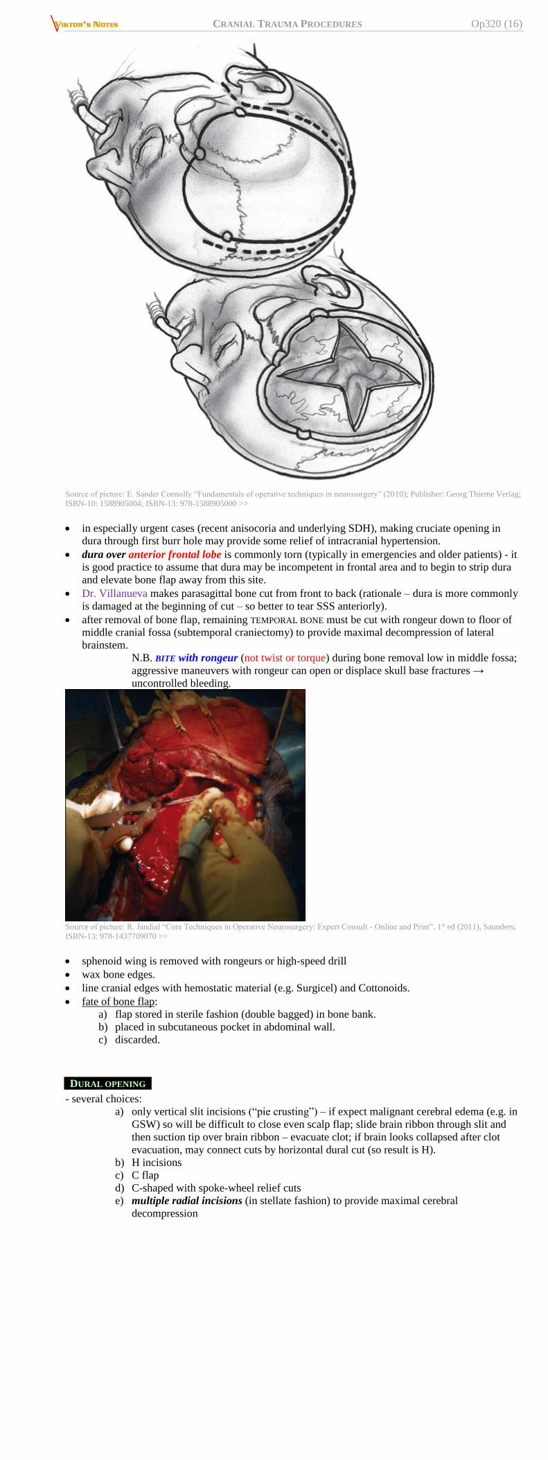
Source of picture: E. Sander Connolly “Fundamentals of operative techniques in neurosurgery” (2010); Publisher: Georg Thieme Verlag;
ISBN-10: 1588905004; ISBN-13: 978-1588905000 >>
in especially urgent cases (recent anisocoria and underlying SDH), making cruciate opening in
dura through first burr hole may provide some relief of intracranial hypertension.
dura over anterior frontal lobe is commonly torn (typically in emergencies and older patients) - it
is good practice to assume that dura may be incompetent in frontal area and to begin to strip dura
and elevate bone flap away from this site.
Dr. Villanueva makes parasagittal bone cut from front to back (rationale – dura is more commonly
is damaged at the beginning of cut – so better to tear SSS anteriorly).
after removal of bone flap, remaining TEMPORAL BONE must be cut with rongeur down to floor of
middle cranial fossa (subtemporal craniectomy) to provide maximal decompression of lateral
brainstem.
N.B. BITE with rongeur (not twist or torque) during bone removal low in middle fossa;
aggressive maneuvers with rongeur can open or displace skull base fractures →
uncontrolled bleeding.
Source of picture: R. Jandial “Core Techniques in Operative Neurosurgery: Expert Consult - Online and Print”, 1st ed (2011), Saunders;
ISBN-13: 978-1437709070 >>
sphenoid wing is removed with rongeurs or high-speed drill
wax bone edges.
line cranial edges with hemostatic material (e.g. Surgicel) and Cottonoids.
fate of bone flap:
a) flap stored in sterile fashion (double bagged) in bone bank.
b) placed in subcutaneous pocket in abdominal wall.
c) discarded.
DURAL OPENING
- several choices:
a) only vertical slit incisions (“pie crusting”) – if expect malignant cerebral edema (e.g. in
GSW) so will be difficult to close even scalp flap; slide brain ribbon through slit and
then suction tip over brain ribbon – evacuate clot; if brain looks collapsed after clot
evacuation, may connect cuts by horizontal dural cut (so result is H).
b) H incisions
c) C flap
d) C-shaped with spoke-wheel relief cuts
e) multiple radial incisions (in stellate fashion) to provide maximal cerebral
decompression

CRANIAL TRAUMA PROCEDURES Op320 (17)
Source of picture: R. Jandial “Core Techniques in Operative Neurosurgery: Expert Consult - Online and Print”, 1st ed (2011), Saunders;
ISBN-13: 978-1437709070 >>
Source of picture: Brian T. Ragel et al. “Wartime decompressive craniectomy: technique and lessons learned”. Neurosurg Focus /
Volume 28 / May 2010 >>
open dura slowly (!!! – risk of sudden cardiovascular collapse; H: adequate resuscitation by
anesthesia - central line is a must).
remove hematomas (gentle scraping and sucking), obtain hemostasis (bipolar), irrigate copiously.
N.B. no attempt should be made to chase elusive pieces of clot in subdural space!
arterial blood exiting from middle fossa in large amounts warrants exploration and often arises
from middle meningeal artery or sphenoid wing. H: slightly more conservative temporal
craniectomy provides bone to which temporal dura can be tacked, which may stop bleeding;
consider waxing foramen spinosum, peroxide soaked cotton balls or cottonoids.
large frontal and temporal contusions can be removed with gentle suction and bipolar cautery.
ultrasound can be used to identify intracerebral hematomas that do not come to surface.
CLOSURE
Options
A. Duraplasty
B. Leave durotomy open; cover brain with dural substitute (DuraGuard, SepraFilm) to protect
brain surface and reduce adhesions - leaves of dura are folded over dural substitute (do not
suture patch to dura!)
epidural tack-up sutures: bone edge and central.
drains are placed over surface of dural substitute and tunneled externally; at least two Jackson-
Pratt drains (patients often do not clot properly, and without tamponading effect provided by bone
flap, risk of EDH is high)
N.B. routine prolonged use of drains is needed!?
galea is closed with numerous, closely spaced interrupted 2-0 absorbable braided sutures.
skin is closed with running 4-0 absorbable monofilament suture / staples – to prevent CSF leak
N.B. to ensure watertight closure, sutures are placed very close together!
sterile head wrap (not tight if bone removed and label side of head without bone)
COMPLICATIONS
venous sinus injury – see below >>
skull fracture contralateral to side of decompression is significant risk factor for postoperative
EDH - routine early postop CT should be considered in cases with skull fracture remote to site of
decompression.
CRANIAL TRAUMA PROCEDURES Op320 (18)
precipitous external herniation intraoperatively (soon after decompression) → empiric surgical
exploration on other side without interim CT (esp. in setting of contralateral skull fracture)
— make sure no tapes around neck
— avoid volatile anesthetics
— repeat mannitol
— primary scalp closure maybe difficult (if this possibility is suspected in advance, it is wise
to obtain hemostasis and be prepared to close before dura is opened)
— consider temporal lobectomy.
POSTOP
wound complications are most common source of surgical morbidity:
a) traumatic injury to skin at incision
b) CSF egress (open durotomy + CSF absorption problems)
DECOMPRESSIVE BIFRONTAL CRANIECTOMY (KJELLBERG)
pronounced “Shellberg”
INDICATIONS
Level II A recommendation
bifrontal DC is not recommended to improve outcomes (as measured by GOS-E score at 6 months
post-injury in severe TBI patients with diffuse injury and with ICP > 20 mm Hg for > 15 minutes
within a 1-hour period that are refractory to first-tier therapies).
demonstrated to reduce ICP and to minimize days in ICU.
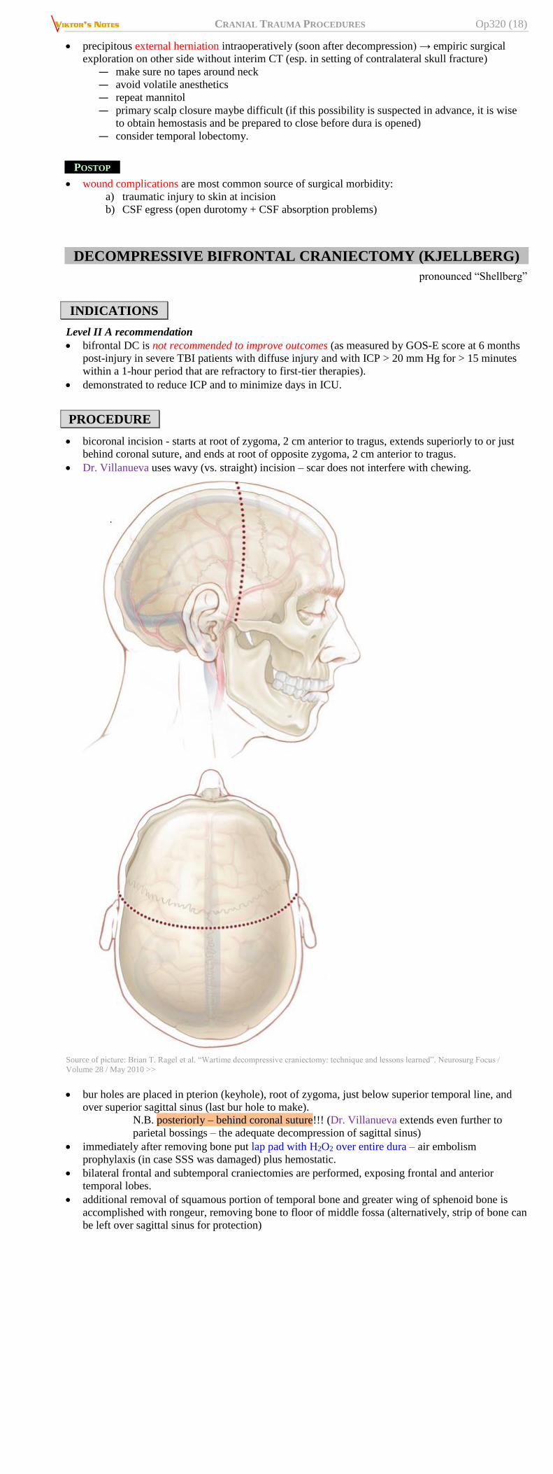
PROCEDURE
bicoronal incision - starts at root of zygoma, 2 cm anterior to tragus, extends superiorly to or just
behind coronal suture, and ends at root of opposite zygoma, 2 cm anterior to tragus.
Dr. Villanueva uses wavy (vs. straight) incision – scar does not interfere with chewing.
Source of picture: Brian T. Ragel et al. “Wartime decompressive craniectomy: technique and lessons learned”. Neurosurg Focus /
Volume 28 / May 2010 >>
bur holes are placed in pterion (keyhole), root of zygoma, just below superior temporal line, and
over superior sagittal sinus (last bur hole to make).
N.B. posteriorly – behind coronal suture!!! (Dr. Villanueva extends even further to
parietal bossings – the adequate decompression of sagittal sinus)
immediately after removing bone put lap pad with H2O2 over entire dura – air embolism
prophylaxis (in case SSS was damaged) plus hemostatic.
bilateral frontal and subtemporal craniectomies are performed, exposing frontal and anterior
temporal lobes.
additional removal of squamous portion of temporal bone and greater wing of sphenoid bone is
accomplished with rongeur, removing bone to floor of middle fossa (alternatively, strip of bone can
be left over sagittal sinus for protection)

CRANIAL TRAUMA PROCEDURES Op320 (19)
Source of picture: Brian T. Ragel et al. “Wartime decompressive craniectomy: technique and lessons learned”. Neurosurg Focus /
Volume 28 / May 2010 >>
dural cuts:
a) standard Kjellberg open fish-mouth cuts made along floor of anterior fossa with release of
inferior aspect of interhemispheric falx.
b) mitral valve–type dural incisions - parallel to superior sagittal and parallel to posterior bone
edge:

CRANIAL TRAUMA PROCEDURES Op320 (20)
Source of picture: Brian T. Ragel et al. “Wartime decompressive craniectomy: technique and lessons learned”. Neurosurg Focus /
Volume 28 / May 2010 >>
PENETRATING BRAIN INJURIES
systemic antibiotics + tetanus prophylaxis + seizure prophylaxis!!!
rapid local debridement - clean from bone fragments, necrotic brain, debris, foreign objects.
– debridement of devitalized brain is gentle (use combination of suction and irrigation).
extensive exploration for hemorrhage and necrotic tissue (recent data question practice of pursuing
bone chips spread deeply into brain).
N.B. retained fragments have not been associated strongly with infection, most authors remove
fragments only if they are accessible! (secondary removal is performed only for unusually large
retained fragments - complication rate for repeat exploration may exceed rate of complications
of retained fragments)
after hemostasis, all layers of wound are closed tightly (drains are added when hemostasis is not
absolute).
GUNSHOT WOUNDS
first surgery often represents “damage control” surgery - quick removal of mass lesions
(hematomas), parenchymal debridement without overly aggressive pursuit of deep and small bone
or foreign fragments hemostasis, and quick decision with respect to decompressive craniectomy;
deep imploded bone fragments and foreign bodies are not chased.
second or third operations are sometimes necessary for further debridement of necrotic brain
tissue; deep imploded bone fragments and foreign bodies would often deliver themselves to surface
at this time.
1) kraujavimo sustabdymas, intrakranijinių hematomų pašalinimas.
2) šautinio kanalo išvalymas (pasiekiamų kaulinių ar metalinių fragmentų pašalinimas → nekrotinių
smegenų detrito išplovimas).
ultrasound is helpful in finding retained fragments or hematomas.
N.B. bullet is not removed unless it is easily accessible (because risk of brain injury
from bullet retrieval exceeds benefit of its removal).
3) kietojo dangalo užsiuvimas ar plastika (pericranium* or temporalis fascia; avoid artificial
synthetic or biological dural substitutes).
*pericranium is positioned so that surface which has been against
bone is directed away from brain.
4) įėjimo ir išėjimo angų pirminis chirurginis sutvarkymas.
5) cranioplasty is delayed for ≈ 1 year (when patient is medically stable and risk of infection is low).
scalp incision (during primary craniotomy) is better to locate away from penetration wound so
that least possible scarring will overlie site of any future cranioplasty.
Current standard is debridement of first few centimeters of tract → watertight dural closure*
*to prevent centripetal infection and CSF fistula
– reports from Middle East conflicts in which patients were treated without any debridement
but with only simple skin closure of bullet hole did not indicate worse outcome.
Chirurginė taktika:

CRANIAL TRAUMA PROCEDURES Op320 (21)
stabili sąmonės būklė (GCS > 10), praėję < 6 val nuo sužeidimo, nėra kaulinių skeveldrų žaizdoje,
nėra kulkos išėjimo angos, nėra intrakranijinės hematomos, reikalingos chirurginio gydymo →
pakanka atlikti tik pirminį chirurginį žaizdų sutvarkymą.
3 balų koma, nėra intrakranijinės hematomos - chirurginis gydymas neprasmingas.
intrakranijinė hematoma → hematomos šalinimas ir šautinio kanalo išvalymas.
4-5 balų koma, išlikęs vyzdžių reaktyvumas → šautinės žaizdos sutvarkymas.
visiems ligoniams su išlikusiu koordinuotu reagavimu į dirginimą turi būti atliekamas šautinio
kanalo sutvarkymas.
STAB INJURY
a) jei ligonis pateko jau išėmus žalojančią priemonę, jei sužeidimas įvykęs prieš < 12 valandų, žaizda
nėra stipriai užteršta, nėra intrakranijinės hematomos ar impresinio lūžio → pakanka pirminio
chirurginio odos žaizdos sutvarkymo.
b) jei šių reikalavimų neišlaikoma → kraniektomija, įėjimo angos išvalymas, kietojo smegenų
dangalo plastika;
– į galvą įsmigęs svetimkūnis šalinamas tik po CT, operacinėje, atvėrus dura (removal
under direct vision) pasiruošus galimoms komplikacijoms (atsinaujinęs ar
suintensyvėjęs kraujavimas).
– pašalinimas atliekamas atkartojant ginklo trajektoriją retrogradiškai.
– ypač pavojingos medžio liekanos (sunkiai identifikuoja rentgenas) → meningitas,
smegenų pūlinys.
DURAL VENOUS SINUS INJURIES
Traumatic dural venous sinus injuries carry high mortality (vs. slow sinus occlusions by tumor) –
hard to control bleeding, ligation is dangerous
– may be caused by depressed skull fractures overlying any of major intracranial venous
sinuses.
– maintain hydration to prevent thrombosis.
– if major venous sinus injury is suspected, approach by exposing intact sinus above and
below, opening dura on both sides so that it can be clamped; EETTOOMMIIDDAATTEE for
neuroprotection.
– anterior 1/3 of superior sagittal sinus can be ligated without any clinical sequelae;
tears of posterior 2/3 of sinus need repair* (primary repair or patching with galea or
pericranium; alternatively, piece of muscle or Gelfoam may be sutured over sinus).
*because ligation may cause lethal venous congestion
Viktor’s Notes℠ for the Neurosurgery Resident
Please visit website at www.NeurosurgeryResident.net