Cranial nerves Not directly associated with the eye
42
CRANIAL NERVES NOT DIRECTLY RELATED TO THE EYE Othman Al-Abadi, M.D
-
Upload
othman-al-abbadi -
Category
Education
-
view
47 -
download
2
Transcript of Cranial nerves Not directly associated with the eye

CRANIAL NERVES NOT DIRECTLY RELATED TO THE EYE
Othman Al-Abadi, M.D
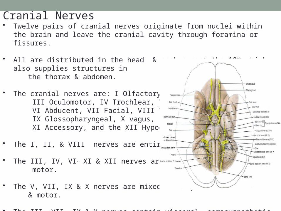
Cranial Nerves• Twelve pairs of cranial nerves originate from nuclei within the brain and leave the cranial
cavity through foramina or fissures.
• All are distributed in the head & neck except the 10th which also supplies structures in the thorax & abdomen.
• The cranial nerves are: I Olfactory, II.Optic, III Oculomotor, IV Trochlear, V Trigeminal, VI Abducent, VII Facial, VIII Vestibulocochlear, IX Glossopharyngeal, X vagus, XI Accessory, and the XII Hypoglossal
• The I, II, & VIII nerves are entirely sensory.
• The III, IV, VI, XI & XII nerves are entirely motor.
• The V, VII, IX & X nerves are mixed sensory & motor.
• The III, VII, IX & X nerves contain visceral parasympathetic component supplying smooth muscles & glands.

Olfactory Nerve • Is a purely sensory nerve.
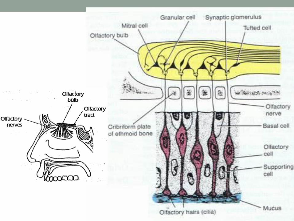
• It arises from receptors of olfactory neurons located in the upper posterior part of the nasal cavity above the superior concha.
• The olfactory neurons are bipolar cells with coarse peripheral dendrites and long central processes forming olfactory nerves which pass through the cribriform plate of ethmoid bone and ending in the olfactory bulb.
• The olfactory bulb consists mainly of large mitral cells with few small tufted & granular cells.
• The dendrites of mitral cells synapse with the incoming olfactory nerve fibers forming rounded areas called synaptic glomerulus. Also the tufted & granular cells synapse with mitral cells.
• The mitral cells also synapse with axons from contralateral olfactory bulb through the olfactory tract.
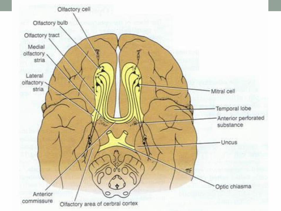
• Axons of ipsilateral mitral & tufted cells and few axons form contralateral olfactory bulb form the olfactory tract located in the olfactory sulcus on the orbital surface of frontal lobe.
• The olfactory tract proximal to the anterior perforated substance divides into medial & lateral olfactory striae.
• The lateral olfactory stria terminates in the olfactory areas of temporal cerebral cortex. These areas represent the primary olfactory cortex.
• The primary & secondary olfactory areas are responsible for appreciation of olfactory sensations.
• The olfactory pathway consists of two order neurons & has no connection with the thalamus.

• The primary olfactory cortex has connections with brain centers which evoke emotional & autonomic responses to olfactory stimuli.
• Anosmia is loss of olfactory sensation.
• Bilateral temporary anosmia usually occurs in common cold & allergic rhinitis.
• Unilateral anosmia results from damage to olfactory nerve, bulb or tract… fractures to the ant cranial fossa involving the cribriform plate can result in tear of the olfactory nerve.
• Unilateral lesion of cortical olfactory area usually has no impairment of olfactory sensation as the olfactory pathway from each nasal cavity is bilaterally presented in the cerebral cortex.
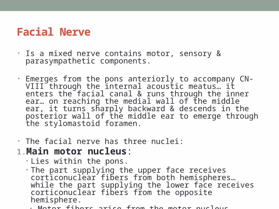
Facial Nerve
• Is a mixed nerve contains motor, sensory & parasympathetic components.
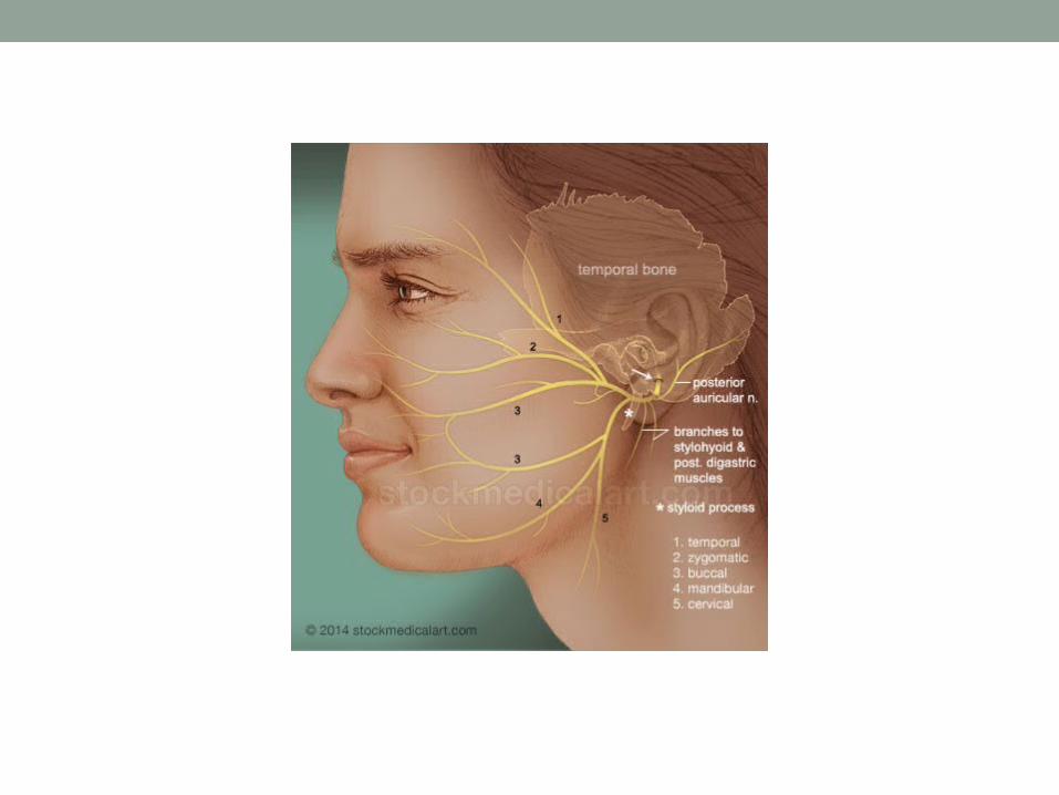
• Emerges from the pons anteriorly to accompany CN-VIII through the internal acoustic meatus… it enters the facial canal & runs through the inner ear… on reaching the medial wall of the middle ear, it turns sharply backward & descends in the posterior wall of the middle ear to emerge through the stylomastoid foramen.
• The facial nerve has three nuclei:1.Main motor nucleus:
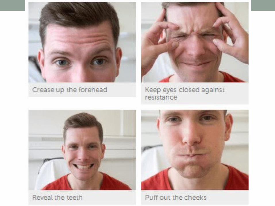
• Lies within the pons.• The part supplying the upper face receives corticonuclear fibers from both
hemispheres… while the part supplying the lower face receives corticonuclear fibers from the opposite hemisphere. • Motor fibers arise from the motor nucleus located in the pons and
supplies muscles of facial expression, stapedius, stylohyoid & posterior belly of digastric.
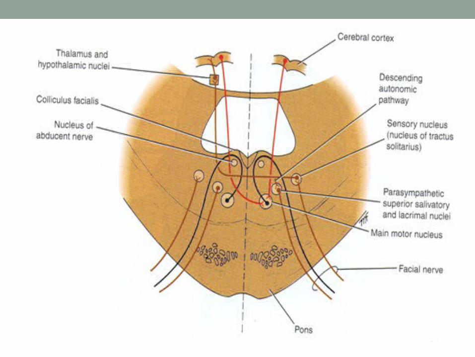

2. Parasympathetic Nuclei: • They are the superior salivatory and lacrimal nuclei. • The superior salivatory nucleus receives afferent fibers from the
hypothalamus, & supplies the submandibular & sublingual glands• The lacrimal nucleus supplies the lacrimal gland… it receives afferent
fibers from the hypothalamus for emotional responses and from the sensory nucleus of the trigeminal nerve for reflex lacrimation secondary to irritation of the cornea or conjunctiva.
3. Sensory Nucleus : • This is the upper part of the nucleus of the tractus solitarius.• Sensations of taste travel from the anterior 2/3rd of the tongue & the floor
of the mouth & palate through the peripheral axons of cells situated in the geniculate ganglion of the seventh cranial nerve. Efferent fibers cross the midline and ascend to the thalamus to the taste area of the cortex.


TheVestibulocochlear Nerve
• Is a pure sensory nerve which consists of two distinct parts, the cochlear nerve & the vestibular nerve.

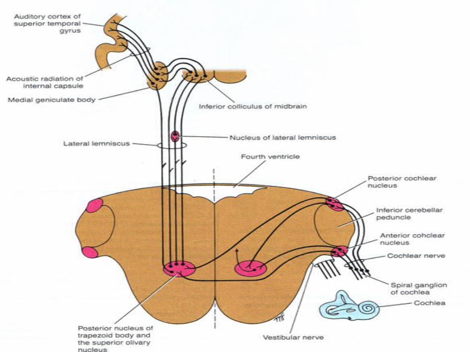
The Cochlear Nerve • It conducts nerve impulses concerned with sound from the organ of corti in the
cochlea.
• The fibers of the cochlear nerve are the central processes of nerve cells located in the spiral ganglion of the cochlea.
• On entering the pons, the nerve fibers divide with one branch enter the posterior cochlear nucleus and the other enter the anterior cochlear nucleus.
• The majority of the fibers of 2nd order neuron of cochlear nuclei cross the median plane and pass to the trapezoid body and superior olivary nucleus and ascend as tract called the lateral lemniscus, while few fibers remain uncrossed ascending in ipsilateral lateral lemniscus.
• On reaching the midbrain, the fibers either terminate in the nucleus of the inferior colliculus or are relayed in the medial geniculate body and pass to the auditory cortex of the cerebral hemisphere in the superior temporal gyrus.
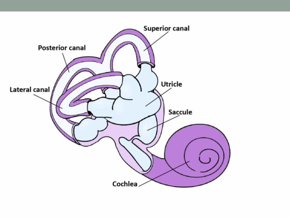
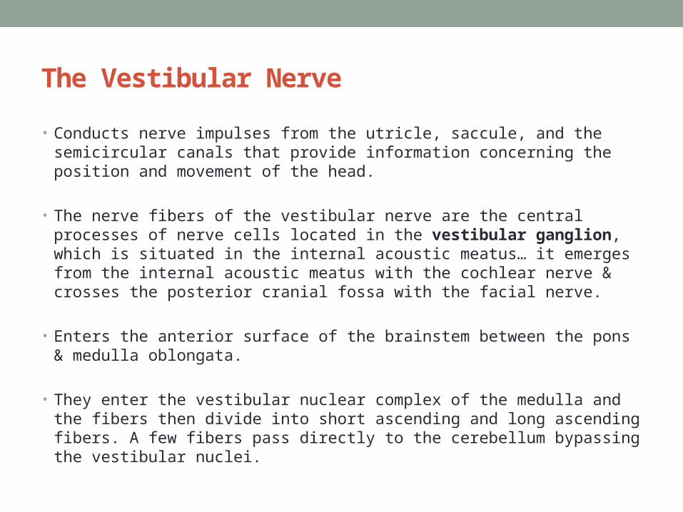
The Vestibular Nerve
• Conducts nerve impulses from the utricle, saccule, and the semicircular canals that provide information concerning the position and movement of the head.
• The nerve fibers of the vestibular nerve are the central processes of nerve cells located in the vestibular ganglion, which is situated in the internal acoustic meatus… it emerges from the internal acoustic meatus with the cochlear nerve & crosses the posterior cranial fossa with the facial nerve.
• Enters the anterior surface of the brainstem between the pons & medulla oblongata.
• They enter the vestibular nuclear complex of the medulla and the fibers then divide into short ascending and long ascending fibers. A few fibers pass directly to the cerebellum bypassing the vestibular nuclei.
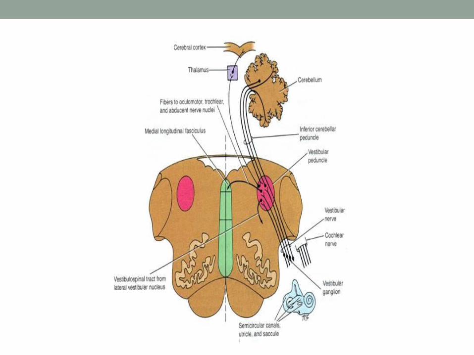

The Vestibular Nerve • The vestibular nuclear complex consists of a group of nuclei situated below
the floor of the 4th ventricle. Four nuclei may be recognized: the superior, inferior, medial and lateral vestibular nuclei.
• These nuclei receive afferent fibers from the vestibular nerve and from the cerebellum. Efferent fibers from the nuclei pass to the cerebellum while other fibers descend from the lateral nucleus uncrossed to the spinal cord forming the vestibulospinal tract.
• In addition, efferent fibers pass to the nuclei of the oculomotor, trochlear, and abducent nerve through the medial longitudinal fasciculus. These connections enable the movements of the head and the eyes to be coordinated so that visual fixation on an object can be maintained.
• Ascending fibers also pass upward from the vestibular nucleus to the post-central gyrus of the cerebral cortex after first synapsing in the thalamus… they consciously orient the individual in space.

• Testing vestibular function• Integrity Caloric tests: inc. & dec. temp of the external
auditory meatus (through endolymph of the semicircular canals.
• Function vertigo & nystagmus
• Testing cochlear function• Integrity whispered voice or vibrating tuning fork
(weber & rinne tests)• Function deafness & tennitus
The Glossopharyngeal Nerve
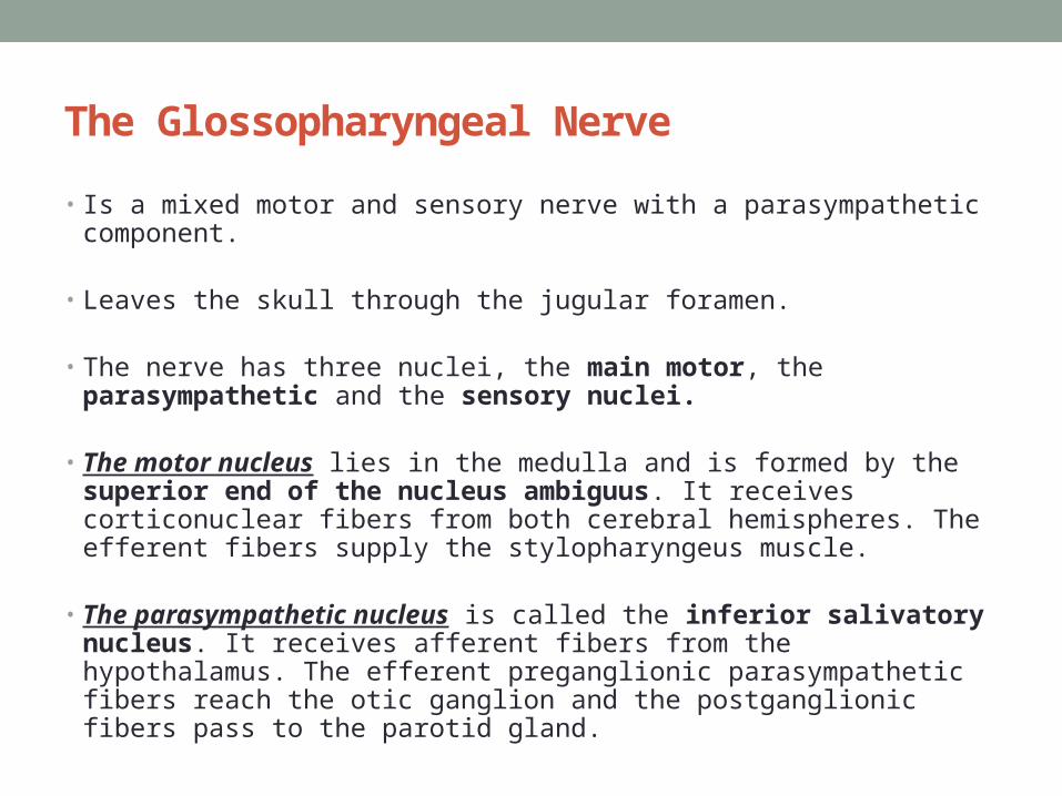
• Is a mixed motor and sensory nerve with a parasympathetic component.
• Leaves the skull through the jugular foramen.
• The nerve has three nuclei, the main motor, the parasympathetic and the sensory nuclei.
• The motor nucleus lies in the medulla and is formed by the superior end of the nucleus ambiguus. It receives corticonuclear fibers from both cerebral hemispheres. The efferent fibers supply the stylopharyngeus muscle.
• The parasympathetic nucleus is called the inferior salivatory nucleus. It receives afferent fibers from the hypothalamus. The efferent preganglionic parasympathetic fibers reach the otic ganglion and the postganglionic fibers pass to the parotid gland.


The Glossopharyngeal Nerve
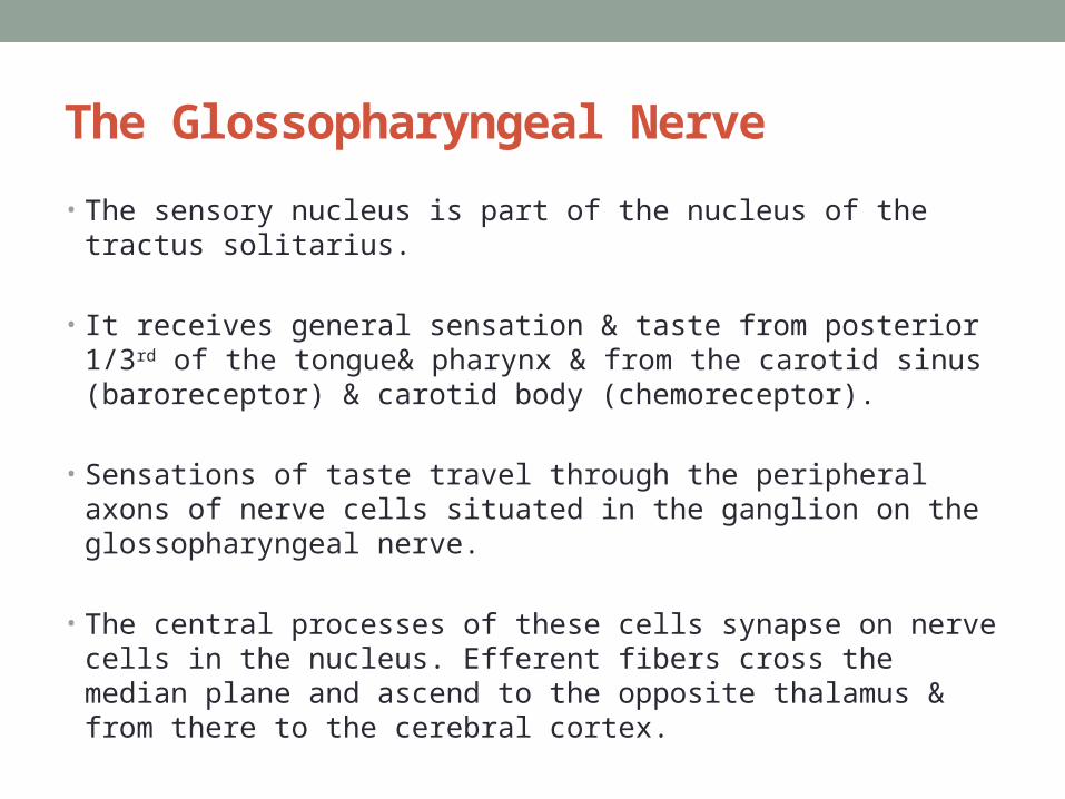
• The sensory nucleus is part of the nucleus of the tractus solitarius.
• It receives general sensation & taste from posterior 1/3rd of the tongue& pharynx & from the carotid sinus (baroreceptor) & carotid body (chemoreceptor).
• Sensations of taste travel through the peripheral axons of nerve cells situated in the ganglion on the glossopharyngeal nerve.
• The central processes of these cells synapse on nerve cells in the nucleus. Efferent fibers cross the median plane and ascend to the opposite thalamus & from there to the cerebral cortex.

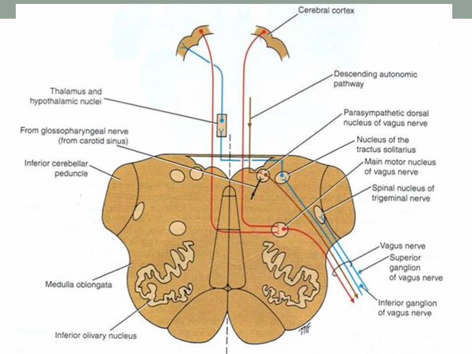
The Glossopharyngeal Nerve• Afferent information concerning common sensation enters the
brainstem through the superior ganglion of the glossopharyngeal nerve but ends in the spinal nucleus of the trigeminal nerve.
• Afferent impulses from the carotid sinus also travel with the glossopharyngeal nerve and terminate in the nucleus of the tractus solitarius and are connected to the dorsal motor nucleus of the vagus nerve.
• The carotid sinus reflex assists in the regulation of arterial blood pressure.

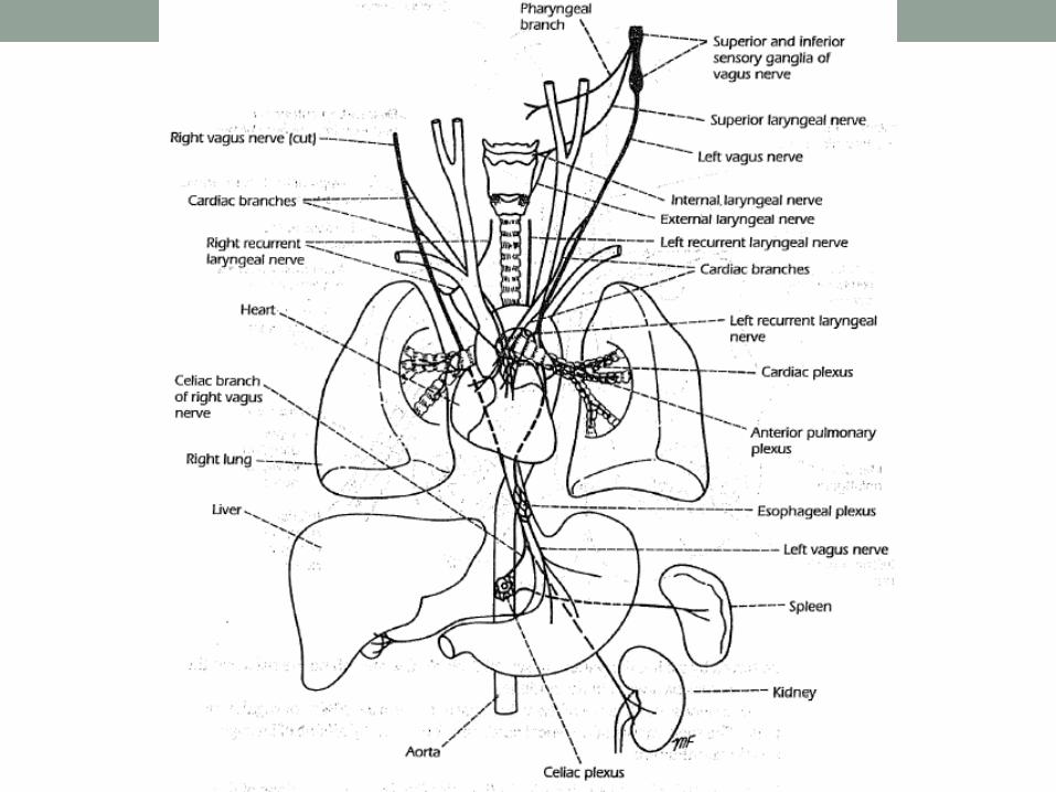
The Vagus Nerve
• Is a mixed motor and sensory nerve with a parasympathetic component.
• Exit the skull through the jugular foramen.
• It supplies the heart, great thoracic vessels, larynx, trachea, bronchi, lungs, alimentary tract from the pharynx to 2/3rd of colon, liver, kidneys & pancreas.
• Has three nuclei;
• The motor nucleus lies within the medulla and is formed by the nucleus ambiguus. It receives corticonuclear fibers from both cerebral hemispheres.
• The efferent fibers supply the constrictor muscles of the pharynx and the intrinsic muscles of the larynx.

The Vagus Nerve
• The parasympathetic nucleus forms the dorsal nucleus of vagus… It receives afferent fibers from the hypothalamus.
• The efferent fibers are distributed to the involuntary muscles along its course.
• The sensory nucleus is the lower part of the nucleus of the tractus solitarius. Sensations of taste from most of the posterior part of the tongue travel through the peripheral axons of nerve cells located in the inferior vagal ganglion on the vagus nerve. The central processes synapse with nerve cells of the nucleus.
• Efferent fibers cross the median plane and ascend to the opposite thalamus & the to the postcentral cortex.
The Vagus Nerve
• The function can be tested by the gag reflex (pharyngeal)… the soft palate can be tested by sayn “aaah” while noticing the soft palate rise & the uvula moving back in the midline.

The Accessory Nerve• Is a purely motor nerve that is formed by union of a cranial and a
spinal root.
• The cranial root is formed from the axons of the nucleus ambiguus.
• Receives corticonuclear fibers from both cerebral hemispheres.
• Distributed with the vagus nerve to supply muscles of soft palate, pharynx & larynx.
• Fibers of spinal accessory nerve arise from the spinal nucleus, which is situated in the anterior gray column of the upper five cervical segments of the spinal cord (C1- C5) and supply the trapezius & sternocleidomastoid muscles.


The Accessory Nerve• Receives corticonuclear fibers from both cerebral hemispheres.
• The nerve fibers emerge from the spinal cord to form a trunk that ascends into the skull through the foramen magnum… it joins the cranial root & both exit through the jugular foramen then they re-separate.
• The cranial root joins the vagus & is distributed in its pharyngeal & recurrent laryngeal branches to the muscles of the soft palate, pharynx & larynx.
• The spinal runs downward & laterally to supply the sternocleidomastoid & trapezius

The Hypoglossal Nerve• Is a pure motor nerve that arises from the hypoglossal nucleus
in the medulla at the level of the lower part of the 4th ventricle.
• Leaves the skull through hypoglossal canal.
• The hypoglossal nerve supplies all intrinsic & extrinsic muscles of the tongue except ??
• Paralysis of the hypoglossal nerve result in atrophy of ipsilateral half of tongue and once the tongue is protruded it deviates to the side of lesion due the unopposed action of the opposite normal genioglossus.

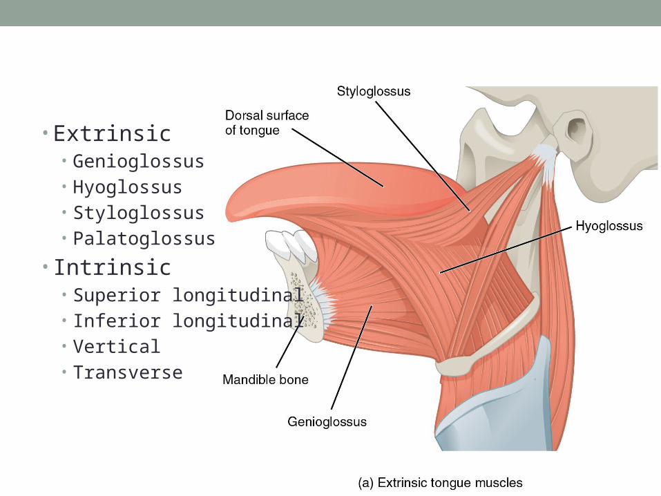
• Extrinsic• Genioglossus• Hyoglossus• Styloglossus• Palatoglossus
• Intrinsic• Superior longitudinal• Inferior longitudinal• Vertical• Transverse