CRANIAL NERVE TESTING FOR OPTOMETRISTS by Rachel A …
26
CRANIAL NERVE TESTING FOR OPTOMETRISTS by Rachel A Copley Brigid C Roney This paper is submitted in pa1iial fulfillment of the requirements for the degree of Doctor of Optometry Ferris State University Michigan College of Optometry May,2019
Transcript of CRANIAL NERVE TESTING FOR OPTOMETRISTS by Rachel A …
CRANIAL NERVE TESTING FOR OPTOMETRISTS
by
Rachel A Copley Brigid C Roney
This paper is submitted in pa1iial fulfillment of the requirements for the degree of
Doctor of Optometry
Ferris State University Michigan College ofOptometry
May,2019

CRANIAL NERVE TESTING FOR OPTOMETRISTS
by
Rachel A Copley Brigid C Roney
Has been approved
'1- May, 2019
APPROVED:
Fa~
CCEPTED: ~'J
~ ·
Ferris State University Doctor of Optometry Senior Paper
Library Approval and Release
CRANIAL NERVE TESTING FOR OPTOMETRISTS
We, Rachel A Copley and Brigid C Roney, hereby release this Paper as described above to Ferris State University with the understanding that it will be accessible to the general public. This release is required under the provisions of the Federal Privacy Act.
Date
ABSTRACT
Background: This research study examines if education and exposure to cranial nerve
testing improves practitioner comfort and confidence in performing and interpreting such
testing. The study is geared toward the primary care optometry setting, where
practitioners may not be as familiar with cranial nerve testing and interpretation due to
perceived infrequency of need in this modality. Methods: A survey was administered
questioning participants' ability and comfort in performing and interpreting cranial nerve
testing prior to and then after watching cranial nerve instructional videos. A total of48
participants identifying as optometrists, optometry students, or optometry faculty were
surveyed. Results: Statistical analysis using a Mann Whitney U-test showed no
statistically significant difference in subject confidence in performing or interpreting
cranial nerve testing by viewing the instructional videos. Conclusions: Providing
educational materials for cranial nerve testing does not improve practitioner comfort and
confidence in perfom1ing and interpreting testing. Despite this, providing educational
materials may increase the number of times practitioners perfom1 cranial nerve testing
during patient examination when such testing is indicated.

TABLE OF CONTENTS
Page
LIST OF TABLES.......... ..... ......................... ... .............. .... ... .... ........... iii
CHAPTER
INTRODUCTION............................................ ..... .... ... .. .
2 METHODS.................... ................................................. 4
3 RESULTS..................... ..................... . .... .... .... ...... .......... 5
4 DISCUSSION... ..... ......... .... ........ ......... ........ .... .......... ...... 7
APPENDIX
A. PRELIMINARY QUESTIONS............................................. 17
B. PRE-VIDEOS SURVEY INSTRUMENT............... ....... .... . .... 18
C. POST-VIDEOS SURVEY INSTRUMENT..................... .... .... 19
ii
LIST OF TABLES
Table Page
Participant Profile.......................................................... l 0
2 Practitioners Performing Testing Pre and Post Videos..... .... ..... 11
3 Confidence and Competence Performing Testing . . . . . . . . . . . . . . . .... 12
4 Confidence and Competence Interpreting Testing.................... 13
5 Mann Whitney U Test: Performing Testing.. . . . ..................... 14
6 Mann Whitney U Test: Interpreting Testing... .. .......... . .......... l 5
7 Critical Values of the Mann-Whitney U Test........................ 16
lll
CHAPTER I
INTRODUCTION
Cranial nerve testing is taught by most schools of optometry and included in the
optometric scope ofpractice. Cranial nerves JI, III, JV, and VI, are routinely evaluated in
a primary care optometric evaluation. While there is little data indicating how often
primary care optometrists incorporate evaluation of the remaining cranial nerves, one can
postulate that potential underutilization may stem from a lack of practitioner confidence
in performing and interpreting cranial nerve testing. Depending on practice modality and
patient demographics, optometrists may practice days or weeks before having a patient
encounter that wan-ants the need for cranial nerve testing. Despite this, it is crucial to
recall the role cranial nerves play in potential systemic and neurological pathologies. The
varying intracranial courses and distinct functions of each cranial nerve makes them very
useful in detecting potential locations oflesions within the nervous system, sometimes
long before a patient becomes severely ill (Bombard, 2013). Additionally, the close
proximity ofcranial nerve nuclei in the brainstem may aid in diagnosis; a large lesion
affecting multiple cranial nerve nuclei may present with multiple symptoms and therefore
help localize a lesion (Gutien-ez, 2017). Considering this, a basic understanding of
cranial nerve testing and interpretation is certainly warranted in the optometrist's office.
As primary care providers, accessibility is in the optometrist's favor. A study by
US News notes that 97% of the population lives within five miles ofan optometrist' s
office. This puts optometrists at the forefront of diagnosing and treating various
pathologies (Howley, 2018). Furthermore, according to the Association ofAmerican
Medical Colleges, the United States is estimated to lose as many as 100,000 primary care
doctors by 2025. This will have a particularly devastating effect in rural areas where
primary care doctors are already in short supply. Highlighting this discrepancy of care, a
recent survey revealed that 41 % ofmen in Nevada do not even have a primary care
doctor. This is much higher than the national average of 28% (Finnegan, 2017).
Considering these statistics and projections, it is necessary that optometrists play a role to
fill this void in primary care by utilizing the skills allowed in their scope ofpractice to
detect pathologies and initiate appropriate referrals.
Each year, more than 795,000 people in the United States suffer from a stroke
(Benjamin, et al, 2017). Many healthcare providers may be familiar with the acute signs
of a stroke, such as difficulty speaking, difficulty walking, upper limb paralysis, and
unilateral facial weakness. Acute loss of vision may also occur with a stroke, which is
often what brings a patient into the optometrist's office. Solid understanding ofhow to
perfonn and interpret cranial nerve testing will allow the optometrist to detect other signs
and symptoms indicative of a stroke and will facilitate prompt and appropriate referrals to
manage the patient. This process has the potential to be life-saving for a patient
considering the underlying systemic disease that causes a stroke.
Multiple sclerosis is yet another condition optometrists have the potential to be at
the forefront of diagnosing. According to the national Multiple Sclerosis Society, an
estimated one million individuals in the United States suffer from multiple sclerosis (MS
Prevalence, 2019). For 20% ofmultiple sclerosis patients, optic neuritis is the initial sign
2
oftheir disease. Moreover, 50% of established multiple sclerosis patients will experience
at least one episode of optic neuritis during their lives (Kale, 2016).These patients are
typically young females who present with acute, unilateral, and painful vision loss. By
perfonning a cranial nerve examination, optometrists may be able to more easily
distinguish multiple sclerosis from other neurological disorders. Careful examination and
data collection in such cases allows the optometrist to make an educated referral ,
streamlining the patient's care toward the most appropriate specialist.
Another disease, neurosyphilis, has the potential to masquerade as various other
neurological diseases. In the early stages, neurosyphilis commonly appears in the basilar
meninges; this meningeal inflammation has the potential to spread to neighboring cranial
nerves and can result in vaiious cranial nerve palsies. Ocular manifestations of
neurosyphilis may include virtually any structures in the eye. Some of those include
nongranulomatous anterior uveitis, Argyll-Robertson Pupil, and optic neuritis (Wender,
2008).
3
CHAPTER2
METHODS
Instructional videos demonstrating how to test for cranial nerves I, V, Vll, and
VIII - XII were created. At the end of each video, a brief summary with possible
interpretations for testing was included. Videos were uploaded to a YouTube channel
and made freely available to any members of the public. A survey using a Likert scale
was created to question individuals on their views on the necessity of cranial nerve
testing in a primary care optometry setting, as well as comfort performing and
interpreting said testing prior to and after watching instructional videos. An example item
from this survey is "Prior to watching the instructional videos, I felt comfortable and
competent perfo1ming cranial nerve testing" where 1 = "Strongly disagree", 2 =
"Disagree", 3 = "Neutral", 4 =" Agree", and 5 = "Strongly agree" (see Appendix
B). Participants were sought out using social media and e-mail. Individuals taking the
survey included optometry students, optometry faculty members, and optometrists.
Statistical analysis was conducted using a Mann-Whitney U Test. In this study
the independent variable was the instructional videos and the dependent variable was
comfort and competence in performing and interpreting cranial nerve testing. The null
hypothesis (Ho) was that there was no statistically significant difference in subject
confidence in performing or interpreting cranial nerve testing by viewing the instructional
videos.
4

CHAPTER3
RESULTS
The research survey had 48 participants total. Ofthese 48 subjects, 52.1 % of
participants identified as female, 43. 7% as male, and 4.2% preferred not to state (Table
1 ). To fm1her delve into the demographics of the subject population, the majority of
participants were licensed optometrists not associated with academia (64.6%) while
others identified as optometry students, optometry faculty, or other (25%, 6.25%, 4.15%)
(Table 1).
Prior to viewing the instructional videos the majority ofparticipants "rarely"
perfonned cranial nerve testing when indicated (45.8%) and many subjects admitted that
they "never" utilized cranial nerve testing in their practice (27.1 %) (Table 2). However,
after viewing the instructional videos the majority of participants anticipated that they
would "sometimes" perform cranial nerve testing in the future (31.35%) (Table 2).
Overall there was a shift in pai1icipants' perception ofperfo1ming cranial nerve testing
from underutilization to increased incorporation in the optometric setting (Table 2).
There was a shift in reported participant confidence and competence in
performing and interpreting cranial nerve testing toward improved confidence and
competence (Tables 3-4). However, the Mann-Whitney U test revealed that the
introduction of cranial nerve videos produced failed to produce a statistically significant
change in participants' reported confidence and competence in both performing (U =
5

10.5) and interpreting (U = 10) cranial nerve testing. Thus, the study is unable to reject
Ho in favor ofH1 (alpha = 0.05) (Tables 5-7).
6

CHAPTER4
DISCUSSION
Cranial nerve testing, while included in optometrist' s scope ofpractice, is largely
underutilized. This was demonstrated when 72.9% ofparticipants for this study stated
they either "rarely" or "never" performed cranial nerve testing in practice. While the
instructional videos provided in this study did not produce a statistically significant
improvement in practitioner confidence in performing and interpreting such testing, there
was still a shift toward improved confidence when instructional materials were provided.
Additionally, there was a shift toward increased practitioner utilization of cranial nerve
testing following watching the instructional videos with the most participants (30.25%)
saying they would perform cranial nerve testing "sometimes". Despite the cun-ent
underutilization of cranial nerve testing in the optometric realm, it remains an important
part of optometric examination when indicated. The potential for nervous system lesion
localization and both time appropriate and practitioner appropriate referrals provide
sound reasoning for the role of these simple and quick examination elements.
This study did have some limitations. First, the sample size was small (48
participants); this potentially affected outcomes depending on the members of the
population who were polled. Second, because the study mainly targeted practicing
optometrists, it may have been more beneficial to include only licensed optometrists. By
not including optometry students, before and after video analysis may be affected. A
future study could poll and analyze the difference among healthcare providers (primary
7

care physicians, neurologists, registered nurses, etc.) when given a similar survey and
instructional videos.
8
REFERENCES
Benjamin, E. J., Blaha, M. J., Chiuve, S. E., Cushman, M., Das, S. R., Deo, R., ... Muntner, P. (2017). Heart Disease and Stroke Statistics- 2017 Update: A Report From the American Heart Association. Circulation,135(10). doi: 1 O. l l 61 /cir.0000000000000485
Bombard, T., NREMT-P, MD. (2013, November 22). Neurotrauma Review Series-Part 1: Why Evaluate the Cranial Nerves? Retrieved January 27, 2019, from https://www.emsworld.com/article/11245200/neurotrauma-review-series-part-l-why-evaluate-cranial-nerves
Finnegan, J. (2017, January 12). Many Americans don't have a primary care doctor. Retrieved January 27, 2019, from https://www.fiercehealthcare.com/practices/many-americans-don-t-have-a-primary-care-doctor
Gutierrez, M. (2009, September 2). Save a Life; Neuro-Optometry Primer: The Brain. Review ofOptometry. Retrieved from: https://www.reviewofoptometry.com/CMSDocuments/2009/9/Neuro-Optom-Supp_R0-09.02.09.pdf
Howley, E. K. (2018, December 3). What's the Difference Between Ophthalmologists, Optometrists and Opticians? Retrieved January 27, 2019, from https:/ /health.usnews.com/health-care/patient-advice/articles/2018-12-03/whats-the-di fference-between-ophthalmologists-optometrists-and-opticians
Kale, N. (2016). Optic neuritis as an early sign ofmultiple sclerosis. Eye and Brain,Volume 8, 195-202. doi:10.2147/eb.s54131
LaMorte, W.W. (2017, May 4). Mann Whitney U Test (Wilcoxon Rank Sum Test). Retrieved January 27, 2019, from http://sphweb.bumc.bu.edu/otlt/mph-modules/bs/bs704_nonparametric/BS704_Nonparametric4.html
MS Prevalence. (n.d.). Retrieved January 27, 201 9, from https://www.nationalmssociety.org/About-the-Society/MS-Prevalence
Wender, J. D. (2008, November 20). How to Recognize Ocular Syphilis. Retrieved January 27, 2019, from https://www.reviewofophthalrnology.com/article/how-to-recognize-ocular-syphilis
9

Table 1. Participant Profile
Gender
• Female • Male r Other
Professional Level
• Student Faculty Member • Optometrist Other
JO

25
Table 2. Practitioners perfonning testing before and after videos
Frequency of Cranial Nerve Testing Before and After Instructional Videos
20
15
10
5
I .I I0 NIA Nevt:r Rarely Sometimes Often Always
Before A ftcr
(Pertaining to item 3 in Appendix A and item 7 in Appendix C)
11

Table 3. Practitioner confidence and competence interpreting cranial nerve testing before and after videos
Confidence and Competence Interpreting Cranial Nerve Testing Before and After Instructional Videos
25
20
15
10
5
0 I I I Strong ly Disagree Disag ree Neutral Agree Strongly Agree
Before After
(Pertaining to item 1 in Appendix B and item 1 in Appendix C)
12
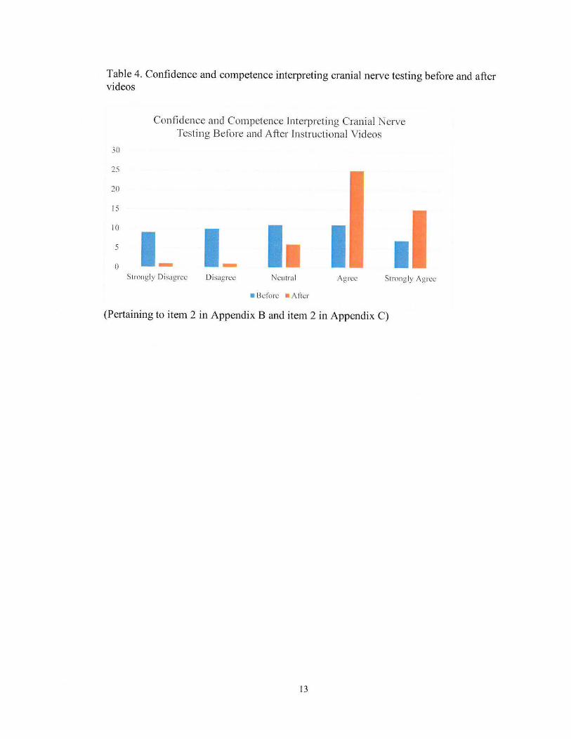
Table 4. Confidence and competence interpreting cranial nerve testing before and after videos
Confidence and Competence Interpreting Cranial Nerve Testing Before and After Instructional Videos
30
25
20
15
10
5
0 I_ I I I
Strongly Disagree Disagree Neutral Agree Strongly Agree
Bdore Atier
(Pertaining to item 2 in Appendix B and item 2 in Appendix C)
13

Table 5 Mann Whitney U Test: Confidence and competence performing cranial nerve testing before and after instructional videos
rotal Sample (Ordered Ranks
~rl ~· . ~h {,llll
~r l
I 9 I I 1.5 2 IO I I 1.5 3 I I 6 2 3 4 I I 25 3 4 5 7 15 4 5
5 6 6 7.5 6 7.5
7 9 8 IO
RI =29.5 R2 =25.5 UI = 10.5 U2 = 14.5
(alpha = 0.05)
14
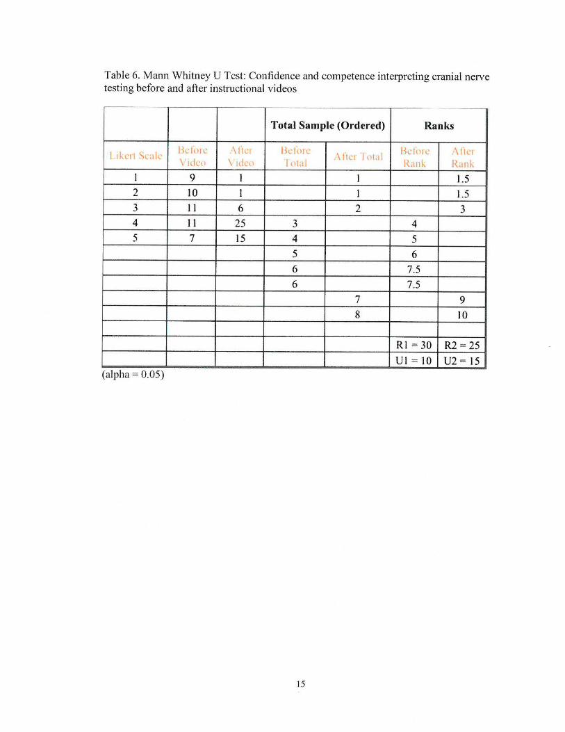
Table 6. Mann Whitney U Test: Confidence and competence interpreting cranial nerve testing before and after instructional videos
Total Sample (Ordered) Ranks
I~ l'rl ~l 1 11 bclorl \lie lkh1rc \It r Iot 1
lkfo1 \ ll \ dc11 \ du I01 Ran~ {rnk
I 9 l l l.5 2 IO I I l.5 3 l l 6 2 3 4 l I 25 3 4 5 7 15 4 5
5 6 6 7.5 6 7.5
7 9 8 10
RI = 30 R2 = 25 Ul = I0 U2 = 15
(alpha = 0.05)
15

Table 7. Critical values of the Mann Whitney U Test
n1 n2 a • • - · 0
3 . 4 5__ _6 ·-·-7 __i __ 8 _ · _9 _ '. I_O - _11 _· 12 . 13_ _ _14 ~--15 - _16_t 1_7 . 18 _; 19 _20 .----•-- --· -··--r ----- 1 3 . .05 . 0 , 0 I I , 2 : 2 , 3 4 ! 4 ! 5 5 '. 6 7 . 7 '. 8 9 . 9 : JO I 11
1.01 0 0 0 0 0 1 I 2 2 1 2 3 3 1 4 4 1 4 5 2 3 4 5 6 7 1 8 9 l 10 ti II 12 14 15 16 [ 17 l 18 ,I .95 . Q4 0 I 2 3 3 4 5 j 5 6 7
1
. 7 f 8 9 I 9 I 10·-- .01 4 5 6 8 9 I I 1_2 . 13 I I 5 1 I6 ' 18
1 19 ; 20 22 i 23 25
15 .05 •I 2 I 2 3 4 5 6 7 8 9 i 10 · 11 12 13 14 ! 15 i 16 · I .01 I 0
2 3 5 7 8 JO 12 , 14 16 17 I 19 . 21 . 23 ! 25 1 26 . 28 , 30 I 32 , 2 3 4 6 7 : 8 ! 9 11 12 13 15 : I 6 I I 8 19 : 20 I 22-- I I 1
2 4 6 8 11 13 15 : 17 , 19 . 21 1 24 ' 26 . 28 . 30 33 . 35 ! 37 397 l.,_Q~ 13 4 6 7 9 1 11 12 14 t 16 i 17 19 21 I 23 ' 24 i 26 281.01 o I 1
.
' 058 r.C-• .01 3 5. -0 ' 2'
9 ~ 92__ __4__L _§_ -.01 I I 3
IO ~Qi_ ±-\- J_ ·--···· .0 I I , 3
11 ~ - 5 I 8 .01 1 -1 4
12 .05 . 2 +2 _ .01 2 5
13 Lll2.._ _Q_ t 10 I .0) 2 5
14 ,!)p__ 7 11 .01 2 6
15 .05 7 12 .0 I 3 7
16 .05 8 14 .Ol 3 7--
17 I .05 9 15 i .01 4 I 8
18 I .05 9 16
.01 4 9 19 .05 10 I 17
I .01 4 9
20 t o5 11 18 I .01 5 10- -
- ' l ' l • - -! . . . 1 I
4 6 7 9 II 13 15 17 ' 20 22 24 26 28 30 ' 32 34 .2. _ l_l . ! 5 _J] _ U 1 1=1 ; 27 • 30 " iJ... '. 36 _ 39 . 42 ] 42 J 48 I I 51 _ 5 7 1 9 1 11 14 i 16 • 18 21 ; 23 I 26 28 I 31 I 33 36 l 38 1 40
8 10 13 15 18 20 23 26 28 31 33 36 I 39 41 44 47
.lL:11. !? t 20 24 l 21 .1. 31 _ 34 ..J_37 -1 4t . 44 ~ .i 51 . 55 J 58 62 ..., 6 8 , 11 13 16 j )9 22 24 1 27 I 30 33 I 36 : 38 . 41 I 44 i 47 12 · 16 I I9 ! 23 27 I 3 I 1 34 38 ' 42 1 46 50 54 r- 1:~ i~j · 1 9 · 12 1 15 · 18 I 22 · 25 - 28 1 31 J'34 · 31 l 41 1.3 . 17 : 21 _j_ 26 . l Q .11 1- 38 - 42 i ±7 I 2!_ _ 1 60 . 64 t _6l I 22 t 77 J
8 11 : 14 ; 17 21 24 28 3) I 35 ! 38 42 46 I 49 53 '. 56 l 60 _15 J. 19 _ 24 + 28 3i _ ]]_ I 42 _ 47 i L 56 61 I 65 _IQ_,_ 7i -~ 80 ; 84 1-
9 . 12 16 • 20 23 27 , 31 35 39 I 43 47 I 51 55 59 63 67 16 , 21 . 2_§ ; 31 . J6 41 ; 46 51 5_6
1 QI . 66 ; 7 I l. 77 _ 82 [ 87 ! 92 ;
- - .... 10 13 17 ! 22 26 30 34 38 43 47 51 I 56 ! 60 65 ' 69 73 t8 23 -~ 28.l 33 -·· 39 __44.. L50 55 ! 61 : 66 72 UJ. --} 83 88 94 1J OO 11 15 19 I 24 28 33 I 37 42 I 47 ; 51 56 I 61 i 66 70 75 l 80 19 . 25 I 30 ! 36 42 48 I 54 60 65 I 71 , 77 I 83 89 • 95 IO I I 107 12 16 I 21 ! 26 31 36 , 41 46 51 i 56 , 61 I 66 ! 11 76 : 82 1 87 20 26
1 33 I 39 45 i 51 57 64 70 I 77 83 ! 89 96 102 109 : 115
13 , I 8 • 23 1 28 33 ' 38 44 49 55 60 66 1 71 , 77 82 ' 88 93 22 28 35 I 41 48 I 55 - 61 68 I 75 82 88 I 95 I I 02 109 !116 123 14 ' 19 , 24 30 36 41 47 53 I 59 , 65 70 I 7 6 I 82 . 88 i 94 ' I00 23 30 37 44 51 58 65 72 80 I 87 94 ; IOI 109 116 1123 130 15 20 T26 32 38 44 50 56 , 63 I 69 : 75 1 82 88 ..l 94 IO I I I 07 -
54 : 62 i 6925 I 32 39 47 77 I 84 I 92 100 I 107 115 123 ' 130 I 138 16 I 22 I 28 : 34 40 i 47 I 53 60 J 67 ; 73 80 l 87 t 93 , 100 !107 . 114
• J
(LaMorte, 2017).
16

APPENDIX A. PRELIMINARY QUESTIONS
Cranial Nerve Testing Instructional Videos Survey Thank you for participating in our survey for our senior project! After viewing the
instructional videos, please answer the following items
1. Are you an optometry student, optometrist, or optometry faculty member? a. Optometry Student b. Optometrist c. Optometry Faculty Member d. Other
2. Are you female, male, or prefer not to state? a. Female b. Male c. Prefer no to state
3. On average, how often do you perform cranial nerve testing on your patients? a. Never b. Rarely - less than 30% of patients when indicated c. Sometimes - between 30% and 50% of patients when indicated d. Often - between 50% and 75% ofpatients when indicated e. Always - greater than 90% of patients when indicated
17
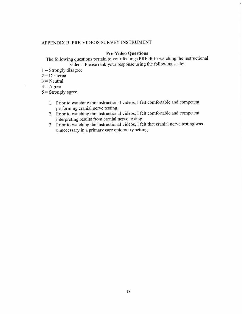
APPENDIX B: PRE-VIDEOS SURVEY INSTRUMENT
Pre-Video Questions The following questions pertain to your feelings PRIOR to watching the instructional
videos. Please rank your response using the following scale: 1 = Strongly disagree 2 = Disagree 3 = Neutral 4 = Agree 5 = Strongly agree
1. Prior to watching the instructional videos, I felt comfortable and competent performing cranial nerve testing.
2. Prior to watching the instructional videos, I felt comfortable and competent interpreting results from cranial nerve testing.
3. Prior to watching the instructional videos, I felt that cranial nerve testing was unnecessary in a primary care optometry setting.
18

APPENDIX C. POST-VIDEOS SURVEY INSTRUMENT
Post-Video Questions The following questions pertain to your feelings AFTER watching the instructional
videos. Please rank your response using the following scale: 1 = Strongly disagree 2 = Disagree 3 = Neutral 4 = Agree 5 = Strongly agree
1. After watching the instructional videos, I feel comfortable and competent performing cranial nerve testing.
2. After watching the instructional videos, I feel comfortable and competent interpreting results from cranial nerve testing.
3. After watching the instructional videos, I feel comfortable and competent communicating the results from cranial nerve testing to my patients.
4. After watching the instructional videos, I feel that I can make an educated referral for further testing depending on the cranial nerve testing results.
5. After watching the instructional videos, I plan to incorporate cranial nerve testing into my primary care exam when indicated.
6. After watching the instructional videos, I feel that cranial nerve testing is necessary in a primary care optometry setting.
7. After watching the instructional videos, how often do you expect to perfonn cranial nerve testing on your patients?
Amended Likert scale: never, rarely, sometimes, often, always a. Never b. Rarely - less than 30% of patients when indicated c. Sometimes - between 30% and 50% ofpatients when indicated d. Often - between 50% and 75% of patients when indicated e. Always - greater than 90% ofpatients when indicated
8. If you would like to provide additional comments, please comment below
19