cr ka inal
27
CASE REPORT Medical Record Number : 22907/ 96.44.49 Admission Date : 21-10-2013 Admission Time : 18.15 wib Name : Mr S Gender : Male Age : 50 Occupation : Farmer Address : Jabung, East Lampung Anamnesis Chief Complaint : chest pain Secondary Complaint : progressive shortness of breathe, cough. History of Present Illness The patient came to the hospital with shortness of breathe he already felt for about a year. The shortness of breathe occured gradually then suddenly developed rapidly into severe breathlessness and get worse for the past 2 weeks, so that the shortness of breathe felt in rest position. It occurs for the whole day, and there is no marked worsening in any particular time of the day. He also felt chest pain in the left side o f his chest. The pain is not radiating to the shoulder, arm, nor the n eck. He also had productive cough for the last 8 months. He also had night sweats, loss of appetite which cause significant weight lost. The patient used to be an active smoker, which he could smoke more than 4 cigarettes in a day. History of Past Illness His past illness is unremarkable. He never had asthma or severe breathlessness before. He also never took any 6 months regiments / antituberculosis drug.
Transcript of cr ka inal

7/27/2019 cr ka inal
http://slidepdf.com/reader/full/cr-ka-inal 1/27
CASE REPORT
Medical Record Number : 22907/ 96.44.49
Admission Date : 21-10-2013
Admission Time : 18.15 wib
Name : Mr S
Gender : Male
Age : 50
Occupation : Farmer
Address : Jabung, East Lampung
Anamnesis
Chief Complaint : chest pain
Secondary Complaint : progressive shortness of breathe, cough.
History of Present Illness
The patient came to the hospital with shortness of breathe he already felt for about a year.
The shortness of breathe occured gradually then suddenly developed rapidly into severe
breathlessness and get worse for the past 2 weeks, so that the shortness of breathe felt in rest
position. It occurs for the whole day, and there is no marked worsening in any particular time
of the day. He also felt chest pain in the left side of his chest. The pain is not radiating to the
shoulder, arm, nor the neck. He also had productive cough for the last 8 months. He also had
night sweats, loss of appetite which cause significant weight lost. The patient used to be an
active smoker, which he could smoke more than 4 cigarettes in a day.
History of Past Illness
His past illness is unremarkable. He never had asthma or severe breathlessness before. He
also never took any 6 months regiments / antituberculosis drug.

7/27/2019 cr ka inal
http://slidepdf.com/reader/full/cr-ka-inal 2/27
History of Family Illness
There was no family member who diagnosed as tuberculosis, having wet cough
more than 2 weeks, nor present any symptoms like the patients.
Physical Examination
General appearance : Looks ill
Consciousness : Compos mentis, E4V5M6
Height : 158 cm
Blood Pressure : 90/50 mmHg
Pulse : 86 bpm , regular
Temperature : 37.20 C
Respiration Rate : 28x/minute
Head : Normocephali, atraumatic, normal hair distribution,
hair not easily revoked
Eye : isochor pupils, anemic conjuctiva +/+, icteric sclera -/-
visual field intact,
Nose : Symmetrical, septum deviation (-), discharge (-),
concha oedem (-)
Mouth : caries , stomatitis (-)
Throat : tonsil T1-T1 calm, hyperemis pharing (-)
Neck : thyroid gland normal size, lymph nodes not palpable,
deviation of trachea (-)
Thorax
Lung

7/27/2019 cr ka inal
http://slidepdf.com/reader/full/cr-ka-inal 3/27
Inspection : symmetrical shape, asymetrical chest movement, decreased
left hemithorax movement, accessory muscle use (-),
Palpation : absent vocal fremitus on the left hemithorax, no tenderness.
Percussion : marked dullness on the left hemithorax,
Auscultation : absent breathe sounds of the left hemithorax, vesicular breath
sound on the right hemithorax. Wheezing (-), Crackles (-)
Abdomen
Inspection : abdomen flat, no tension, no dilated veins
Palpation : no percussion pain, no defense muscular, no enlarged liver
Percussion : timpanic, percussion pain (-), shifting dullness (-)
Auscultation : bowel movement (+), normal
Extemity : warm , oedem (-), cyanosis (-)
Laboratory Findings
- Hematology
Hemoglobin : 11,5 gr %
WBC counts : 9600 / μl
Diff-count : 0 / 0 / 0 / 73 / 12 / 15
Platelet counts : 280.000/ul
Random blood glucose : 116 mg/dl

7/27/2019 cr ka inal
http://slidepdf.com/reader/full/cr-ka-inal 4/27
Ureum : 25 mg/dl
Creatinin : 0,7 mg/dl
DIAGNOSIS
Lung carcinoma
DIFFERENTIAL DIAGNOSIS
Left pleural effusion et causa tuberculosis
Management
Bed rest
Pharmacological Intervention :
IVFD RL xx gtt/minute
Roborantia
Expectorant
Another WorkUp (Recommended)
Posteroanterior chest Xray
ECG
Pleural fluid analysis : Cytology
PROGNOSIS

7/27/2019 cr ka inal
http://slidepdf.com/reader/full/cr-ka-inal 5/27
Quo ad vitam : dubia ad malam
Quo ad functionam : dubia ad malam
FOLLOW UP
DATE October 21, 2013
Subjective : - Dyspneu, which worsen when the body slant in left-side
position
- Productive Cough +
Objective
Vital Sign
- BP
- Pulse
- RR
- T
100/70 mmHg
108 x/mnt
28 x/mnt
38,3
C
7/27/2019 cr ka inal
http://slidepdf.com/reader/full/cr-ka-inal 6/27
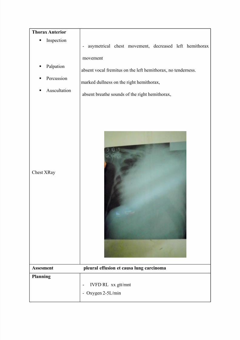
Thorax Anterior
Inspection
Palpation
Percussion
Auscultation
Chest XRay
- asymetrical chest movement, decreased left hemithorax
movement
absent vocal fremitus on the left hemithorax, no tenderness.
marked dullness on the right hemithorax,
absent breathe sounds of the right hemithorax,
Assesment pleural effusion et causa lung carcinoma
Planning
- IVFD RL xx gtt/mnt
- Oxygen 2-5L/min
7/27/2019 cr ka inal
http://slidepdf.com/reader/full/cr-ka-inal 7/27
Work Up
- Ceftriaxone 1 gr/ 12 hour, IV
- Dextrometorphan Syr ( 3 x 1C )
- Glyceryl Guaiacolat tab ( 3 x 1 )
- B1, B6, B12 2 x 1 tab
Conclusion No Improvement
date October 22, 2013
Subjective - Dyspneu
- Chest pain when the body slant to the right sideway
- Tightness of chest
- Cough (-)
Objective
Vital Sign
- BP
- Pulse
- RR
90/70 mmHg
108 x/min
28 x/min

7/27/2019 cr ka inal
http://slidepdf.com/reader/full/cr-ka-inal 8/27
- T 39 oC
Thorax Anterior
Inspection
Palpation
Percussion
Auscultation
asymetrical chest movement, decreased left hemithorax
movement
absent vocal fremitus on the left hemithorax, no tenderness.
marked dullness on the right hemithorax,
right hemithorax breath sound> left hemithorax. Crackles (-)
Wheezing (-),
Assesment Pleural Effusion et causa lung tuberculosis
Planning Antituberculosis drug
Carry on other medication
Conclusion Slight Improvement

7/27/2019 cr ka inal
http://slidepdf.com/reader/full/cr-ka-inal 9/27
Date October 23, 2013
Subjective - Improvement in symptoms : less shortness of breath and
chest tightness
- Cough (+)
Objective
Vital Sign
- BP
- Pulse
- RR
- T
75/50 mmHg
100 x/mnt
24 x/mnt
38,1 oC
Pleural fluid analysis : No malignancy. Pleuritis
7/27/2019 cr ka inal
http://slidepdf.com/reader/full/cr-ka-inal 10/27
Thorax Anterior
Inspection
Palpation
Percussion
Auscultation
asymetrical chest movement, decreased left hemithorax
movement
absent vocal fremitus on the left hemithorax, no tenderness.
marked dullness on the left hemithorax,
right hemithorax breath sound > left hemithorax. Crackles (-
) Wheezing (-),
Assesment Pleural effusion et causa tuberculosis
Planning Carry on previous therapy
Conclusion Marked Improvement
Date October 23, 2013
Subjective - Dyspneu
- Less chest thightness
- Cough

7/27/2019 cr ka inal
http://slidepdf.com/reader/full/cr-ka-inal 11/27
- Mild increase of the appetite
Objective
Vital Sign
- BP
- Pulse
- RR
- T
110/70 mmHg
92 x/mnt
24 x/mnt
36,2 C

7/27/2019 cr ka inal
http://slidepdf.com/reader/full/cr-ka-inal 12/27
Thorax Anterior
Inspection
Palpation
Percussion
Auscultation
asymetrical chest movement, decreased left hemithorax
movement
decrease vocal fremitus on the left hemithorax, absent vocal
fremitus from ICS 3 to basal left hemithorax ,no tenderness.
marked dullness on the left hemithorax,
Absent breath sound in basal left hemithorax to third
intercostal space. Coarse crackles in right hemithorax
Planning - Carry on previous treatment
- WSD Pleural fluid : 500 cc
- Serous with mild hemorrhage (drained every 24 hours)
Conclusion Slight Improvement
DATE October 24, 2013
Subjective - Less dyspneic

7/27/2019 cr ka inal
http://slidepdf.com/reader/full/cr-ka-inal 13/27
- Less tightness of breathe
- Less cough
- Good appetite
Objective
Vital Sign
- BP
- Pulse
- RR
- T
100/70 mmHg
100 x/mnt
24 x/mnt
36,3 C

7/27/2019 cr ka inal
http://slidepdf.com/reader/full/cr-ka-inal 14/27
Thorax Anterior
Inspection
Palpation
Percussion
Auscultation
asymetrical chest movement, decreased left hemithorax
movement
decrease vocal fremitus on the left hemithorax, absent vocal
fremitus from ICS 3 to basal left hemithorax ,no tenderness.
marked dullness on the left hemithorax,
Absent breath sound in basal left hemithorax to third
intercostal space. Coarse crackles in right hemithorax
Planning
- Carry on previous treatment
- Isoniazid tab 300 mg ( 1 x 1 )
- Rifampicin tab 450 mg ( 1 x 1 )
- Pyrazinamid tab 500 mg ( 2 x 1 )
- Etambutol tab 500 mg ( 1 x 1,5 )
- WSD Pleural fluid : 350 cc (drained every 24 hour)
7/27/2019 cr ka inal
http://slidepdf.com/reader/full/cr-ka-inal 15/27
Conclusion Improvement
DATE October 26, 2013
Subjective - Less dyspneic
- Chest tightness (-)
- Cough (-)
- Nausea (+)
Objective
Vital Sign
- BP
- Pulse
- RR
- T
100/70 mmHg
88 x/mnt
20 x/mnt
35,8 C

7/27/2019 cr ka inal
http://slidepdf.com/reader/full/cr-ka-inal 16/27
Thorax Anterior
Inspection
Palpation
Percussion
Auscultation
asymetrical chest movement, decreased left hemithorax
movement
decreased vocal fremitus on left hemithorax, no tenderness.
Dullness on left hemithorax: from basal to ICS 3
Coarse crackles in left hemithorax, absent breath sounds in
the basal left hemithorax to ICS 3.
Planning
- Carry on previous treatment
- WSD Pleural fluid : 250 cc
serohemorrhagic (drained every 24 hour)
Conclusion Marked Improvement
DATE October 27, 2013
Subjective - Dyspneu (-)
- Chest pain (-)
- Cough (-)

7/27/2019 cr ka inal
http://slidepdf.com/reader/full/cr-ka-inal 17/27
Good appetite (nausea (-) )
Objective
Vital Sign
- BP
- Pulse
- RR
- T
110/60 mmHg
80 x/mnt
24 x/mnt
36,1 C
Thorax Anterior
Inspection
Palpation
Percussion
Auscultation
asymetrical chest movement, decreased left hemithorax
movement
decreased vocal fremitus on left hemithorax, no tenderness.
Dullness on left hemithorax: from basal to ICS
Coarse crackles in both hemithorax, absent breath sounds in
the basal left hemithorax to ICS 3.
Planning
- carry on previous therapy
7/27/2019 cr ka inal
http://slidepdf.com/reader/full/cr-ka-inal 18/27
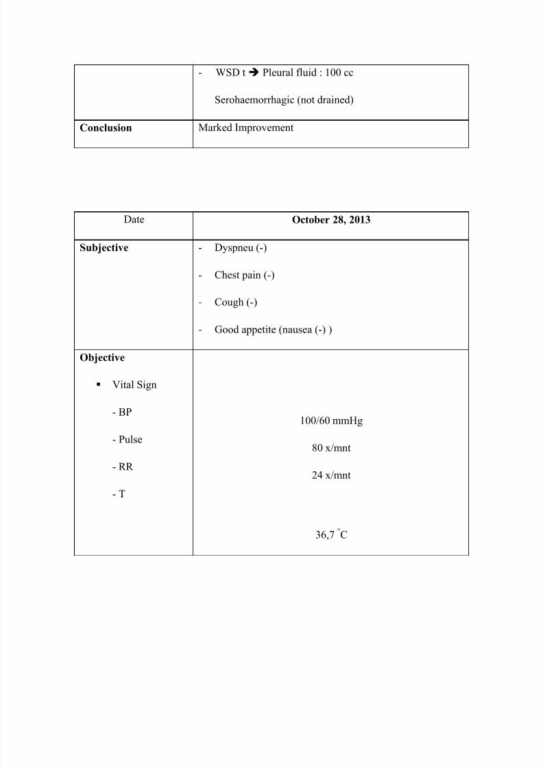
- WSD t Pleural fluid : 100 cc
Serohaemorrhagic (not drained)
Conclusion Marked Improvement
Date October 28, 2013
Subjective - Dyspneu (-)
- Chest pain (-)
- Cough (-)
- Good appetite (nausea (-) )
Objective
Vital Sign
- BP
- Pulse
- RR
- T
100/60 mmHg
80 x/mnt
24 x/mnt
36,7
C
7/27/2019 cr ka inal
http://slidepdf.com/reader/full/cr-ka-inal 19/27
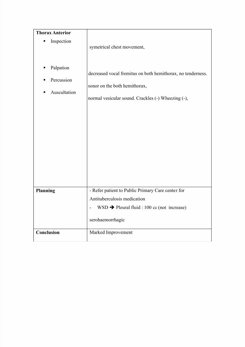
Thorax Anterior
Inspection
Palpation
Percussion
Auscultation
symetrical chest movement,
decreased vocal fremitus on both hemithorax, no tenderness.
sonor on the both hemithorax,
normal vesicular sound. Crackles (-) Wheezing (-),
Planning - Refer patient to Public Primary Care center for
Antituberculosis medication
- WSD Pleural fluid : 100 cc (not increase)
serohaemorrhagic
Conclusion Marked Improvement
7/27/2019 cr ka inal
http://slidepdf.com/reader/full/cr-ka-inal 20/27
PLEURAL EFFUSION
DEFINITION
Pleural effusion is a condition of buildup of fluid in the pleural cavity. Pleural
effusion can be either a transudate or exudate. ¹)
The transudate effusion is caused by diseases that usually not found primarily in the lung,
such as congestive heart failure, liver cirrhosis, nephrotic syndrome, peritoneal dialysis,
albumin deficiency by various circumstances, constrictive pericarditis, malignancy,
pneumothorax and pulmonary atelectasis. ¹)
Exudate effusion occurs when there is an inflammatory process that causes blood vessels in
pleural capillary permeability increased then affect mesotelial cells that turned into squamous
or cuboidal cell that produce fluid into the pleural cavity. Exudative pleural fluid is most
often caused by Mycobacterium tuberculosa that called Tuberculous Exudative Pleuritis.
INCIDENCY
In Indonesia pulmonary tuberculosis is the leading cause of pleural effusion , followed by
malignancy . Pleural effusion found more in women than men . Pleural effusion caused by
lung tuberculosis is more prevalent in men than women . Most affected ages are from 21 to
30 years of age .
Pathophysiology

7/27/2019 cr ka inal
http://slidepdf.com/reader/full/cr-ka-inal 21/27
In normal people , the fluid in pleural cavity is as much as 1-20 ml . Amount of fluid in the
pleural cavity is constant because there is a balance between production by the parietal pleura
and absorption by the visceral pleura . This situation can be maintained because of the
balance between hydrostatic pressure of the parietal pleura of 9 cm H2O and colloid osmotic
pressure of the visceral pleura of 10 cm H2O.
Pleural fluid accumulation can occur if :
1 . Colloid osmotic pressure in the blood decreases , for example in hipoalbuminemia .
2 . Or condition that cause increase in :
• Capillary permeability ( inflammation , neoplasm )
• Hydrostatic pressure in the blood vessels to the heart / pulmonary vein ( left heart failure )
• Negative pressure inside the pleura ( atelectasis )
Etiology
Pleural fluid is divided into :
1 . Transudate , can be caused by :
• Congestive heart failure ( left heart failure )
• Nephrotic Syndrome
• Ascites
• superior vena cava syndrome
• Tumor
• Meig”s Syndrome
2 . Exudate , can be caused by :
• Infections : tuberculosis , pneumonia , and other infective disease
• Tumor

7/27/2019 cr ka inal
http://slidepdf.com/reader/full/cr-ka-inal 22/27
• Pulmonary Infarction
• Radiation
• Collagen Diseases
3 . Hemorrhagics effusion , can be caused by :
• Tumor
• Trauma
• Pulmonary Infarction
• Tuberculosis
Difference between transudate and Exudate
Jenis pemeriksaan Transudate Exudate
Rivaltra - / + (weak) +
Berat jenis < 1,016 > 1,016
Protein < 3 gr / dl > 3 gr / dl
Pleural pritein ratio with
serum proteins
< 0,5 > 0,5
LDH (Lactic
Dehydrogenase)
< 200 IU > 200 IU
Ratio of pleural fluid LDH
with serum LDH
< 0,6 >0,6
White blood cells < 1000 / mm > 1000 / mm

7/27/2019 cr ka inal
http://slidepdf.com/reader/full/cr-ka-inal 23/27
Pleural Fluid Analysis
Macam cairan pleura Makroskopis
Transudate Clear, yellowish
Eksudate Yellow to yellow-green
Chylothorax Milky white
Empyema Thick and murky
Anaerobic empyema Foul smell
Malignant
mesothelioma
Very viscous with
hemorrhage
Cell Count And Cytology
Leukocytes 25,000 / mm3 : Empyema
High amount of neutrophils : pneumonia , pulmonary infarction , pancreatitis , early
pulmonary tuberculosis .
High amount of of lymphocytes : Tubarkulosis , lymphoma , malignancy .
CHEMICAL TEST
a. Glucose
Glucose levels < 30 mg / 100 cc : Pleurutis rheumatoid
< 60 mg / 100 cc : Tuberculosis , malignancy , or the empyema
Decreased glucose levels caused by : Glycolysis extracellular
Diffuse pleural disorders due to damage

7/27/2019 cr ka inal
http://slidepdf.com/reader/full/cr-ka-inal 24/27
b. Amylase
Obtained when the amylase levels increased several times higher than serum amylase is
possibly due to pancreatitis or esophageal rupture .
Some disease that complication is Pleural Effusion
1 . Tuberculosis
Pleural effusion due to tuberculosis is one of the most often encountered in practice .
Diagnosis is made on the basis of positive acid fast bacilli found in the pleural fluid or in
sputum or tissue obtained from pleural biopsy .
2 . Neoplasms
The most common neoplasm caused pleural effusion is cancer metastases from the primary
tumor of breast to the pleura.
3 . Meig’s syndrome
Meig’s syndrome is a disease with :
• benign solid ovarian tumors
• Ascites
• Pleural effusion
4 .Heart Failure
Left heart failure often leads to bilateral pleural effusion .
DIAGNOSIS
1 . Clinical

7/27/2019 cr ka inal
http://slidepdf.com/reader/full/cr-ka-inal 25/27
Asymmetrical hemithorax movement , decrease of vocal fremitus of the affected area , Barrel
chest , egophony ( if the fluid does not fill the entire pleural cavity ) , decreased to absent
breath sounds , the deviation of mediastinal organ to healhy side.
2 . Radiology
Blunting of the costophrenic angle and elevated diaphragm .
3 . Laboratory
Pleural fluid analysis with clinical chemistry test methods
4 . Pathology
Obtained from the pleural biopsy and pleural fluid
DIFFERENTIAL DIAGNOSIS
1 . lung tumors
2 . Schwarte or pleural thickening
3 . Lower lobe atelectasis
4 . Diaphragm high position
MANAGEMENT
Management of pleural effusion is aimed at treat the underlying disease and to evacuate the
excess fluid (by thoracosintesis) .
Indications for thoracocentesis is
1 . Eliminate dyspneu caused by fluid accumulation pleural cavity
2 . When specific therapy for the primary disease is not effective or fail
3 . If there is fluid reaccumulation

7/27/2019 cr ka inal
http://slidepdf.com/reader/full/cr-ka-inal 26/27
At first, evacuate pleural fluid not more than 1000 cc , because the sudden decrease of
pleural fluid can cause swollen lungs marked by coughing and tightness .
Complications
1 . Thoracocentesis can causes loss of protein
2 . Infection in the pleural cavity
3 . Pneumothorax can occur

7/27/2019 cr ka inal
http://slidepdf.com/reader/full/cr-ka-inal 27/27
REFERENCES
1.
Abrahamian, Fredrick M, DO, FACEP, June 27, 2005. pleural effusion.
www.emedicine.com
2. Bambang Kisworo, Efusi pleura keganasan in Cermin Dunia Kedokteran No. 99. 1995.
Hal 40
3. Hadi Halim. 2006. Penyakit-Penyakit Pleura in Buku Ajar Ilmu Penyakit Dalam FKUI.
Jilid II. Edisi IV. Jakarta. Pp 1066-68.
4. Light, Richard W., 1995. Kelainan pada pleura, mediastinum dan difragma in Harrison
Prinsip-prinsip Ilmu Penyakit Dalam. Volume 3. Edisi 13. Jakarta, Pp1385-87.