CpG oligodeoxynucleotides activate dendritic cells in vivo and induce a functional and protective...
9
Vaccine 24 (2006) 1880–1888 CpG oligodeoxynucleotides activate dendritic cells in vivo and induce a functional and protective vaccine immunity against a TERT derived modified cryptic MHC class I-restricted epitope S´ ebastien Cornet a , Jeanne Menez-Jamet a , Franc ¸ois Lemonnier b , Kostas Kosmatopoulos a,∗ , Isabelle Miconnet a a Vaxon Biotech, G´ enopole bat G2, 2 rue Gaston Cr´ emieux, 91057 Evry, France b Unit´ ed Immunit´ e Cellulaire Antivirale, Institut Pasteur, 28 rue du docteur Roux, 75015 Paris, France Received 25 August 2005; received in revised form 18 October 2005; accepted 18 October 2005 Available online 28 October 2005 Abstract The use of synthetic peptides derived from tumor-associated Ags is attractive for the development of antitumoral vaccines as far as strong adjuvants are found to render them immunogenic. Here, we investigated the possibility to enhance the CD8 response against the human and mouse shared TERT 572Y HLA-A*0201 restricted modified cryptic peptide by using ODN-CpG as adjuvant. Humanized transgenic mice were immunized with the TERT 572Y modified cryptic peptide in the presence of ODN-CpG and compared to mice immunized in IFA. By contrast with IFA, we first showed that, in vivo, ODN-CpG leads to the recruitment of dendritic cells in the lymph nodes draining the injection site. Those cells and especially the CD11c + CD11b − CD8a + lymphoid and the CD11c + B220 + plasmacytoid dendritic cells were activated as shown by up-regulation of CD40 at their cell surface. Immunization against TERT 572Y peptide in the presence of ODN-CpG rather than IFA led to a strong CD8 response and can delayed mortality in an induced tumor model. Study of the CD8 response obtained after antigenic challenge suggested that a functional memory response is induced upon vaccination with ODN-CpG. Thus, MHC class I-restricted epitope in combination of ODN-CpG is a promising and rather simple cancer vaccine formulation. © 2005 Elsevier Ltd. All rights reserved. Keywords: ODN-CpG; Cryptic peptides; Dendritic cells; Plasmacytoid; Activation 1. Introduction The identification of CTL-defined tumor-associated Ag has allowed for the development of new strategies for can- cer therapy based on the use of synthetic peptides. Since numerous tumor Ag are self proteins involved in the negative selection of the T cell repertoire [1], the efficiency of this approach depends on the capacity of the vaccination strategy to bypass the immune tolerance against self tumor antigens. In this regard, the tumor Ag-derived peptide has to be care- fully selected and the conditions for vaccination optimized. ∗ Corresponding author. Tel.: +33 160789210; fax: +33 160789219. E-mail address: [email protected] (K. Kosmatopoulos). We previously found that low affinity tumor Ag-derived peptides (so-called cryptic peptides) which are weakly presented at the cell surface and thus weakly involved in the T cell negative selection are better candidates than high affinity ones (so-called dominant peptides) [2,3]. The substi- tution of the first amino acid of these low affinity peptides, involved in their interaction with the MHC class I molecule, importantly increased their immunogenicity [4]. The CD8 T cells generated against the modified cryptic peptides are able to cross recognize their low affinity native counterpart [2]. Among the different actors involved in the expan- sion of a functional CD8 T cell response, the appropri- ate presentation of antigen by activated dendritic cells (DC) is required. DC mature upon different danger sig- 0264-410X/$ – see front matter © 2005 Elsevier Ltd. All rights reserved. doi:10.1016/j.vaccine.2005.10.036
-
Upload
sebastien-cornet -
Category
Documents
-
view
215 -
download
1
Transcript of CpG oligodeoxynucleotides activate dendritic cells in vivo and induce a functional and protective...
Vaccine 24 (2006) 1880–1888
CpG oligodeoxynucleotides activate dendritic cells in vivo and inducea functional and protective vaccine immunity against a TERT
derived modified cryptic MHC class I-restricted epitope
Sebastien Corneta, Jeanne Menez-Jameta, Francois Lemonnierb,Kostas Kosmatopoulosa,∗, Isabelle Miconneta
a Vaxon Biotech, Genopole bat G2, 2 rue Gaston Cremieux, 91057 Evry, Franceb Unite d′Immunite Cellulaire Antivirale, Institut Pasteur, 28 rue du docteur Roux, 75015 Paris, France
Received 25 August 2005; received in revised form 18 October 2005; accepted 18 October 2005Available online 28 October 2005
Abstract
r as stronga human andm e werei ontrastw tion site.T ass IFAl r antigenicc ted epitopei©
K
1
hcnsatIf
(
edaklyd inhigh
-es,le,
s arepart
an-pri-
cellssig-
0d
The use of synthetic peptides derived from tumor-associated Ags is attractive for the development of antitumoral vaccines as fadjuvants are found to render them immunogenic. Here, we investigated the possibility to enhance the CD8 response against theouse shared TERT572Y HLA-A*0201 restricted modified cryptic peptide by using ODN-CpG as adjuvant. Humanized transgenic mic
mmunized with the TERT572Y modified cryptic peptide in the presence of ODN-CpG and compared to mice immunized in IFA. By cith IFA, we first showed that, in vivo, ODN-CpG leads to the recruitment of dendritic cells in the lymph nodes draining the injechose cells and especially the CD11c+ CD11b− CD8a+ lymphoid and the CD11c+ B220+ plasmacytoid dendritic cells were activatedhown by up-regulation of CD40 at their cell surface. Immunization against TERT572Y peptide in the presence of ODN-CpG rather thaned to a strong CD8 response and can delayed mortality in an induced tumor model. Study of the CD8 response obtained aftehallenge suggested that a functional memory response is induced upon vaccination with ODN-CpG. Thus, MHC class I-restricn combination of ODN-CpG is a promising and rather simple cancer vaccine formulation.
2005 Elsevier Ltd. All rights reserved.
eywords: ODN-CpG; Cryptic peptides; Dendritic cells; Plasmacytoid; Activation
. Introduction
The identification of CTL-defined tumor-associated Agas allowed for the development of new strategies for can-er therapy based on the use of synthetic peptides. Sinceumerous tumor Ag are self proteins involved in the negativeelection of the T cell repertoire[1], the efficiency of thispproach depends on the capacity of the vaccination strategy
o bypass the immune tolerance against self tumor antigens.n this regard, the tumor Ag-derived peptide has to be care-ully selected and the conditions for vaccination optimized.
∗ Corresponding author. Tel.: +33 160789210; fax: +33 160789219.E-mail address: [email protected]
K. Kosmatopoulos).
We previously found that low affinity tumor Ag-derivpeptides (so-called cryptic peptides) which are wepresented at the cell surface and thus weakly involvethe T cell negative selection are better candidates thanaffinity ones (so-called dominant peptides)[2,3]. The substitution of the first amino acid of these low affinity peptidinvolved in their interaction with the MHC class I molecuimportantly increased their immunogenicity[4]. The CD8T cells generated against the modified cryptic peptideable to cross recognize their low affinity native counter[2].
Among the different actors involved in the expsion of a functional CD8 T cell response, the approate presentation of antigen by activated dendritic(DC) is required. DC mature upon different danger
264-410X/$ – see front matter © 2005 Elsevier Ltd. All rights reserved.oi:10.1016/j.vaccine.2005.10.036
S. Cornet et al. / Vaccine 24 (2006) 1880–1888 1881
nals and adjuvants are thought to mimic these danger sig-nals. Besides IFA which has been so far commonly usedin experimental models of vaccination, new potent bacte-rial derived adjuvants such as ODN-CpG have been iden-tified [5]. Moreover, their strong DC activating effect[6]suggests that a concomitant T helper CD4 T cell responsecould be avoided which would be of particular inter-est in humans in view of simplifying vaccine formula-tion.
In this paper, we investigated the possibility of enhanc-ing the CD8 response against the human and mouse sharedTERT572Y HLA-A*0201 restricted modified cryptic peptideby using ODN-CpG as an adjuvant. HLA-A*0201 trans-genic HHD mice[7] were immunized with the TERT572Ymodified cryptic peptide in the presence of ODN-CpGand compared to HHD mice immunized in IFA. We firstassessed the influence of the two different adjuvants onthe recruitment and activation status of APC in the lymphnode draining the injection site. A strong activation of lym-phoid and plasmacytoid dendritic cells was only observedin mice injected with ODN-CpG. Then, we directly mon-itored the peptide-specific CD8+ T cell response in bothconditions of vaccination by tetramer staining ex vivo andexamined the functionality of specific T cells by measur-ing their IFN-� production. ODN-CpG are more effectivethan IFA to recruit specific CD8+ T cells against TERTp Thisc nedi onso -n ithI
2
2
v ris,F ntedw cti-v
2
a yE yn-t hemw sys,H fur-m DNw solu-t
2.3. Immunization
HHD transgenic mice were kindly provided by Pr Franc¸oisLemonnier (Institut Pasteur, Paris, France). HHD mice wereimmunized subcutanously (s.c.) at the base of the tail with100�g peptide emulsified in IFA or mixed with 50�g ODN-CpG in a volume of 100�l. To study the secondary response,mice were boosted under the same conditions 1 month afterthe first immunization.
2.4. Tumor challenge
HHD transgenic mice were immunized s.c. with 100�gpeptide emulsified in IFA or mixed with 50�g ODN-CpG ina volume of 100�l. Fourteen days later, they were injecteds.c. with 25000 EL4/HHD tumor cells. Tumor growth wasmonitored weekly. For ethical reasons and according to thegood laboratory practices defined by the animal experimenta-tion rules in France, mice were euthananized when the tumorsize reached 300 mm2. Statistical analysis performed usingthe logrank test of the Kaplan–Meier model (p ≤ 0.05).
2.5. Flow cytometry immunofluorescence analysis
For tetramer staining, cells from peripheral blood, inguinaland paraaortic lymph nodes (LN) and spleen from immu-n -A ibed[ ) in2 erew anti-C eH s,L na pre-s afteri ento ticL tain-i nos-t .D steels FCS.C one2 singM rer’si no-t wasd nti-C ingt nt-d aledw es,L PBS2 ur(
572Yeptide and to induce a functional memory response.onclusion is confirmed in vivo thanks to results obtain a tumoral challenge which prove that two vaccinatif HHD mice with ODN-CpG and TERT572Y delayed sigificatively mortality as compared with vaccinations w
FA.
. Materials and methods
.1. Cell lines
Previously described EL4/HHD cells[7] were kindly pro-ided by Pr. Franc¸ois Lemonnier (Institut Pasteur, Parance) and maintained in DMEM medium supplemeith 1% HEPES, 1% strepto-penicillin and 10% heat inaated fetal calf serum (FCS).
.2. Synthetic peptides, ODN-CpG
TERT572 (RLFFYRKSV), TERT572Y (YLFFYRKSV)nd the control gp100209 (ITDQVPFSV) were made bpytop (Nımes, France). The immunostimulatory s
hetic ODN-CpG 1826 optimized for stimulation of touse immune system (TCCATGACGTTCCTGACGTT)ere used (CpG motifs are underlined) (Sigma-Genoaverhill, UK). The backbone for these ODN was sulodified phosphorothioate to protect it from nucleases. Oere formulated as a sterile phosphate buffered saline
ion and stored at−20◦C.
ized mice were stained with 15�g/ml of PE-coupled HLA2/TERT572Y tetramer synthesized as previously descr
8] in the presence of anti-Fc receptor Ab (clone 2.4 G20�l PBS 2% FCS for 1 h at room temperature. Cells washed once in PBS 2% FCS and then stained withD44-FITC (clone 1M.178), anti-TCR�-Cychrome (clon57) and anti-CD8�-APC (clone 53.6.7) (BD Biosciencee Pont de Claix, France) in 50�l of PBS 2% FCS 30 mit 4◦C. The phenotype and activation status of antigenenting cells recruited in the draining lymph nodes 18 hmmunization were also studied before and after enrichmf CD11c+ dendritic cells. Briefly, inguinal and paraaorN were cut in small pieces and incubated in PBS con
ng 0.5% FCS and 1 mg/ml collagenase A (Roche Diagics GmbH, Mannheim, Germany) at 37◦C for 45–60 minigested fragments were filtered through a stainless-ieve and cell suspensions washed in PBS 0.5%ells were preincubated with anti-Fc receptor Ab (cl.4 G2) and submitted to CD11c enrichment by uACS CD11c micro beads according to the manufactu
nstructions (Miltenyi Biotec, Paris, France). The pheype of LN cells obtained before and after enrichmentetermined by using anti-F4/80-FITC, anti-CD11c-PE, aD11b-biotin Abs and their activation status by stain
hem with anti-CD40-biotin Ab (BD Biosciences, Le Poe-Claix, France). The biotin conjugated Abs were reveith FITC- or Cy5-conjugated streptavidin (BD Bioscience Pont-de-Claix, France). Cells were washed once in% FCS and immediately analyzed using a FACSCalib®
Becton Dickinson, San Jose, CA, USA).
1882 S. Cornet et al. / Vaccine 24 (2006) 1880–1888
2.6. Intracellular IFN-γ staining
Splenocytes or lymph node cells (2× 10⁶) from immu-nized HHD mice were stimulated with 1�M of TERT572Ymodified cryptic peptide or third-party gp100209 modi-fied cryptic peptide in the presence of 10�g/ml brefeldinA (Sigma, Oakville, Canada). Six hours later, cells werewashed, stained with anti-CD8a-PE mAb (BD Biosciences,Le Pont-de-Claix, France) in PBS for 25 min at 4◦C, washedand fixed with 4% paraformaldehyde. Then cells were perme-abilized with PBS containing 0.2% saponin and 0.5% BSAand stained with anti-IFN-�-APC (BD Biosciences, Le Pont-de-Claix, France). Cells were analyzed on a FACSCalibur®
(Becton Dickinson, San Jose, CA, USA).
2.7. ELISpot assay
Peptide-specific T cells from immunized mice were eval-uated in IFN-� ELISpot assay as previously described[21].Mice were immunized twice at 1 month intervals with 100�gof TERT572Y in the presence of either IFA or ODN-CpG asmentioned above. Mouse IFN-� ELISpot PVDF-Enzymatickit (Diaclone, Besanc¸on, France) was used according to themanufacturer’s recommendation. After activation of PVDFplates and coating with IFN-� capture antibody, 3× 10⁵
splenocytes from immunized mice were distributed in eachwpt ativec hreet withb nw ocheM ered -4,3-i orp.,M atedi ben,G
3
3at
d intC ei eanv aluep eeni ithO teda ells.
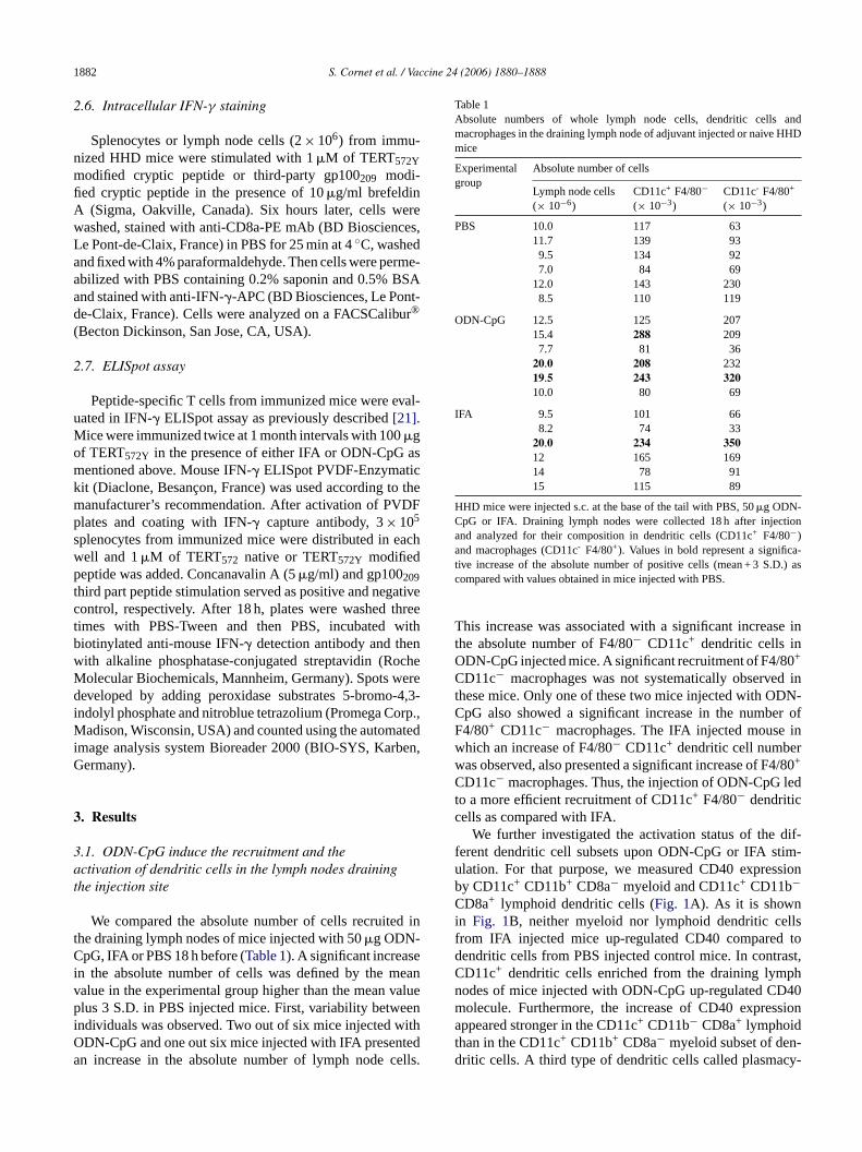
Table 1Absolute numbers of whole lymph node cells, dendritic cells andmacrophages in the draining lymph node of adjuvant injected or naive HHDmice
Experimentalgroup
Absolute number of cells
Lymph node cells(× 10−6)
CD11c+ F4/80−(× 10−3)
CD11c- F4/80+
(× 10−3)
PBS 10.0 117 6311.7 139 939.5 134 927.0 84 69
12.0 143 2308.5 110 119
ODN-CpG 12.5 125 20715.4 288 2097.7 81 36
20.0 208 23219.5 243 32010.0 80 69
IFA 9.5 101 668.2 74 33
20.0 234 35012 165 16914 78 9115 115 89
HHD mice were injected s.c. at the base of the tail with PBS, 50�g ODN-CpG or IFA. Draining lymph nodes were collected 18 h after injectionand analyzed for their composition in dendritic cells (CD11c+ F4/80−)and macrophages (CD11c- F4/80+). Values in bold represent a significa-tive increase of the absolute number of positive cells (mean + 3 S.D.) ascompared with values obtained in mice injected with PBS.
This increase was associated with a significant increase inthe absolute number of F4/80− CD11c+ dendritic cells inODN-CpG injected mice. A significant recruitment of F4/80+
CD11c− macrophages was not systematically observed inthese mice. Only one of these two mice injected with ODN-CpG also showed a significant increase in the number ofF4/80+ CD11c− macrophages. The IFA injected mouse inwhich an increase of F4/80− CD11c+ dendritic cell numberwas observed, also presented a significant increase of F4/80+
CD11c− macrophages. Thus, the injection of ODN-CpG ledto a more efficient recruitment of CD11c+ F4/80− dendriticcells as compared with IFA.
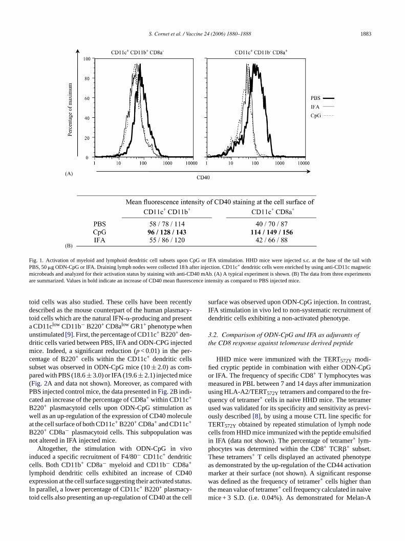
We further investigated the activation status of the dif-ferent dendritic cell subsets upon ODN-CpG or IFA stim-ulation. For that purpose, we measured CD40 expressionby CD11c+ CD11b+ CD8a− myeloid and CD11c+ CD11b−CD8a+ lymphoid dendritic cells (Fig. 1A). As it is shownin Fig. 1B, neither myeloid nor lymphoid dendritic cellsfrom IFA injected mice up-regulated CD40 compared todendritic cells from PBS injected control mice. In contrast,CD11c+ dendritic cells enriched from the draining lymphnodes of mice injected with ODN-CpG up-regulated CD40molecule. Furthermore, the increase of CD40 expressionappeared stronger in the CD11c+ CD11b− CD8a+ lymphoidthan in the CD11c+ CD11b+ CD8a− myeloid subset of den-dritic cells. A third type of dendritic cells called plasmacy-
ell and 1�M of TERT572 native or TERT572Y modifiedeptide was added. Concanavalin A (5�g/ml) and gp100209
hird part peptide stimulation served as positive and negontrol, respectively. After 18 h, plates were washed times with PBS-Tween and then PBS, incubatediotinylated anti-mouse IFN-� detection antibody and theith alkaline phosphatase-conjugated streptavidin (Rolecular Biochemicals, Mannheim, Germany). Spots weveloped by adding peroxidase substrates 5-bromo
ndolyl phosphate and nitroblue tetrazolium (Promega Cadison, Wisconsin, USA) and counted using the autom
mage analysis system Bioreader 2000 (BIO-SYS, Karermany).
. Results
.1. ODN-CpG induce the recruitment and thectivation of dendritic cells in the lymph nodes draininghe injection site
We compared the absolute number of cells recruitehe draining lymph nodes of mice injected with 50�g ODN-pG, IFA or PBS 18 h before (Table 1). A significant increas
n the absolute number of cells was defined by the malue in the experimental group higher than the mean vlus 3 S.D. in PBS injected mice. First, variability betw
ndividuals was observed. Two out of six mice injected wDN-CpG and one out six mice injected with IFA presenn increase in the absolute number of lymph node c
S. Cornet et al. / Vaccine 24 (2006) 1880–1888 1883
Fig. 1. Activation of myeloid and lymphoid dendritic cell subsets upon CpG or IFA stimulation. HHD mice were injected s.c. at the base of the tail withPBS, 50�g ODN-CpG or IFA. Draining lymph nodes were collected 18 h after injection. CD11c+ dendritic cells were enriched by using anti-CD11c magneticmicrobeads and analyzed for their activation status by staining with anti-CD40 mAb. (A) A typical experiment is shown. (B) The data from three experimentsare summarized. Values in bold indicate an increase of CD40 mean fluorescence intensity as compared to PBS injected mice.
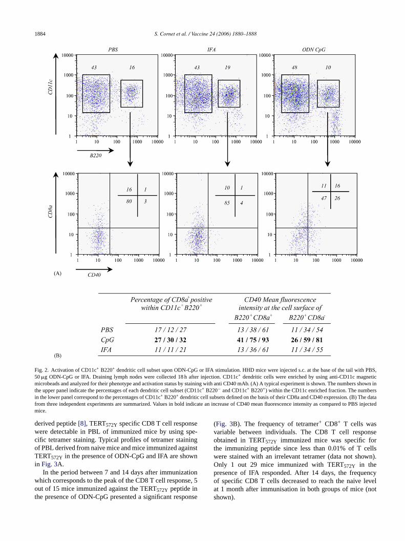
toid cells was also studied. These cells have been recentlydescribed as the mouse counterpart of the human plasmacy-toid cells which are the natural IFN-�-producing and presenta CD11clow CD11b− B220+ CD8alow GR1+ phenotype whenunstimulated[9]. First, the percentage of CD11c+ B220+ den-dritic cells varied between PBS, IFA and ODN-CPG injectedmice. Indeed, a significant reduction (p < 0.01) in the per-centage of B220+ cells within the CD11c+ dendritic cellssubset was observed in ODN-CpG mice (10± 2.0) as com-pared with PBS (18.6± 3.0) or IFA (19.6± 2.1) injected mice(Fig. 2A and data not shown). Moreover, as compared withPBS injected control mice, the data presented inFig. 2B indi-cated an increase of the percentage of CD8a+ within CD11c+
B220+ plasmacytoid cells upon ODN-CpG stimulation aswell as an up-regulation of the expression of CD40 moleculeat the cell surface of both CD11c+ B220+ CD8a+ and CD11c+
B220+ CD8a− plasmacytoid cells. This subpopulation wasnot altered in IFA injected mice.
Altogether, the stimulation with ODN-CpG in vivoinduced a specific recruitment of F4/80− CD11c+ dendriticcells. Both CD11b+ CD8a− myeloid and CD11b− CD8a+
lymphoid dendritic cells exhibited an increase of CD40expression at the cell surface suggesting their activated status.In parallel, a lower percentage of CD11c+ B220+ plasmacy-toid cells also presenting an up-regulation of CD40 at the cell
surface was observed upon ODN-CpG injection. In contrast,IFA stimulation in vivo led to non-systematic recruitment ofdendritic cells exhibiting a non-activated phenotype.
3.2. Comparison of ODN-CpG and IFA as adjuvants ofthe CD8 response against telomerase derived peptide
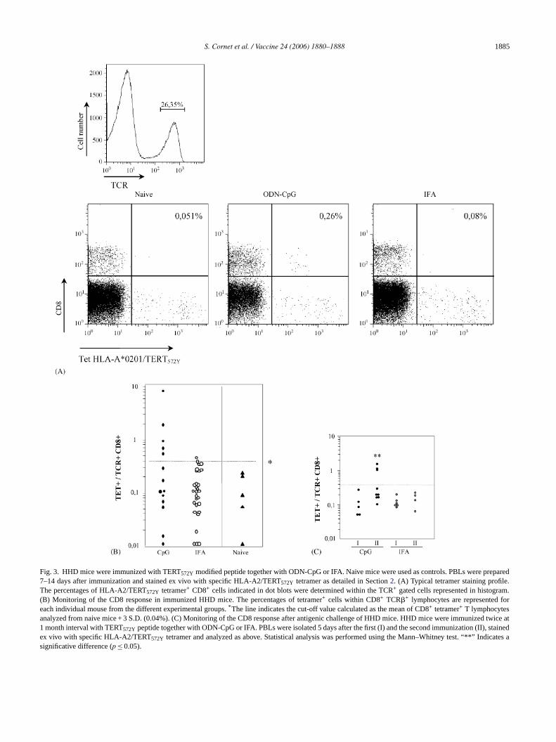
HHD mice were immunized with the TERT572Y modi-fied cryptic peptide in combination with either ODN-CpGor IFA. The frequency of specific CD8+ T lymphocytes wasmeasured in PBL between 7 and 14 days after immunizationusing HLA-A2/TERT572Y tetramers and compared to the fre-quency of tetramer+ cells in naive HHD mice. The tetramerused was validated for its specificity and sensitivity as previ-ously described[8], by using a mouse CTL line specific forTERT572Y obtained by repeated stimulation of lymph nodecells from HHD mice immunized with the peptide emulsifiedin IFA (data not shown). The percentage of tetramer+ lym-phocytes was determined within the CD8+ TCR�+ subset.These tetramers+ T cells displayed an activated phenotypeas demonstrated by the up-regulation of the CD44 activationmarker at their surface (not shown). A significant responsewas defined as the frequency of tetramer+ cells higher thanthe mean value of tetramer+ cell frequency calculated in naivemice + 3 S.D. (i.e. 0.04%). As demonstrated for Melan-A
1884 S. Cornet et al. / Vaccine 24 (2006) 1880–1888
Fig. 2. Activation of CD11c+ B220+ dendritic cell subset upon ODN-CpG or IFA stimulation. HHD mice were injected s.c. at the base of the tail with PBS,50�g ODN-CpG or IFA. Draining lymph nodes were collected 18 h after injection. CD11c+ dendritic cells were enriched by using anti-CD11c magneticmicrobeads and analyzed for their phenotype and activation status by staining with anti CD40 mAb. (A) A typical experiment is shown. The numbers showninthe upper panel indicate the percentages of each dendritic cell subset (CD11c+ B220− and CD11c+ B220+) within the CD11c enriched fraction. The numbersin the lower panel correspond to the percentages of CD11c+ B220+ dendritic cell subsets defined on the basis of their CD8a and CD40 expression. (B) The datafrom three independent experiments are summarized. Values in bold indicate an increase of CD40 mean fluorescence intensity as compared to PBS injectedmice.
derived peptide[8], TERT572Y specific CD8 T cell responsewere detectable in PBL of immunized mice by using spe-cific tetramer staining. Typical profiles of tetramer stainingof PBL derived from naive mice and mice immunized againstTERT572Y in the presence of ODN-CpG and IFA are shownin Fig. 3A.
In the period between 7 and 14 days after immunizationwhich corresponds to the peak of the CD8 T cell response, 5out of 15 mice immunized against the TERT572Y peptide inthe presence of ODN-CpG presented a significant response
(Fig. 3B). The frequency of tetramer+ CD8+ T cells wasvariable between individuals. The CD8 T cell responseobtained in TERT572Y immunized mice was specific forthe immunizing peptide since less than 0.01% of T cellswere stained with an irrelevant tetramer (data not shown).Only 1 out 29 mice immunized with TERT572Y in thepresence of IFA responded. After 14 days, the frequencyof specific CD8 T cells decreased to reach the naive levelat 1 month after immunisation in both groups of mice (notshown).
S. Cornet et al. / Vaccine 24 (2006) 1880–1888 1885
Fig. 3. HHD mice were immunized with TERT572Y modified peptide together with ODN-CpG or IFA. Naive mice were used as controls. PBLs were prepared7–14 days after immunization and stained ex vivo with specific HLA-A2/TERT572Y tetramer as detailed in Section2. (A) Typical tetramer staining profile.The percentages of HLA-A2/TERT572Y tetramer+ CD8+ cells indicated in dot blots were determined within the TCR+ gated cells represented in histogram.(B) Monitoring of the CD8 response in immunized HHD mice. The percentages of tetramer+ cells within CD8+ TCR�+ lymphocytes are represented foreach individual mouse from the different experimental groups.* The line indicates the cut-off value calculated as the mean of CD8+ tetramer+ T lymphocytesanalyzed from naive mice + 3 S.D. (0.04%). (C) Monitoring of the CD8 response after antigenic challenge of HHD mice. HHD mice were immunized twice at1 month interval with TERT572Y peptide together with ODN-CpG or IFA. PBLs were isolated 5 days after the first (I) and the second immunization (II), stainedex vivo with specific HLA-A2/TERT572Y tetramer and analyzed as above. Statistical analysis was performed using the Mann–Whitney test. “**” Indicates asignificative difference (p ≤ 0.05).
1886 S. Cornet et al. / Vaccine 24 (2006) 1880–1888
3.3. A peptide-specific secondary T cell response isinduced following immunization in the presence ofODN-CpG
One month after the first immunization, HHD mice weresubmitted to antigenic challenge under the same conditions.The kinetics of the specific CD8 T cell response after anti-genic challenge was different than the one obtained after oneimmunization with a peak of CD8+ T cell response at day 5(data not shown). Therefore, we analyzed the specific CD8T cell response 5 days after recall with TERT572Y peptidein the presence of IFA or ODN-CpG (Fig. 3C). The CD8 Tcell response specific for the TERT572Y peptide observed inthe blood 5 days after antigenic challenge in the presence ofODN-CpG was stronger than the one observed at day 5 afterthe first immunization (according to Student’s testp ≤ 0.05).By contrast, the frequencies of HLA-A2/TERT572Ytetramer+
CD8+ within T lymphocytes measured 5 days after antigenicchallenge in the presence of IFA were not different from thefrequencies observed 5 days after the first immunization. Thisdata suggest the development of a memory CTL responseagainst TERT572Y peptide in the presence of ODN-CpG.
3.4. Functionality of peptide-specific T cell responseinduced following immunization in the presence ofO
thep essedb y( hal-l
F mu-n erO uceIpf ativeo eS
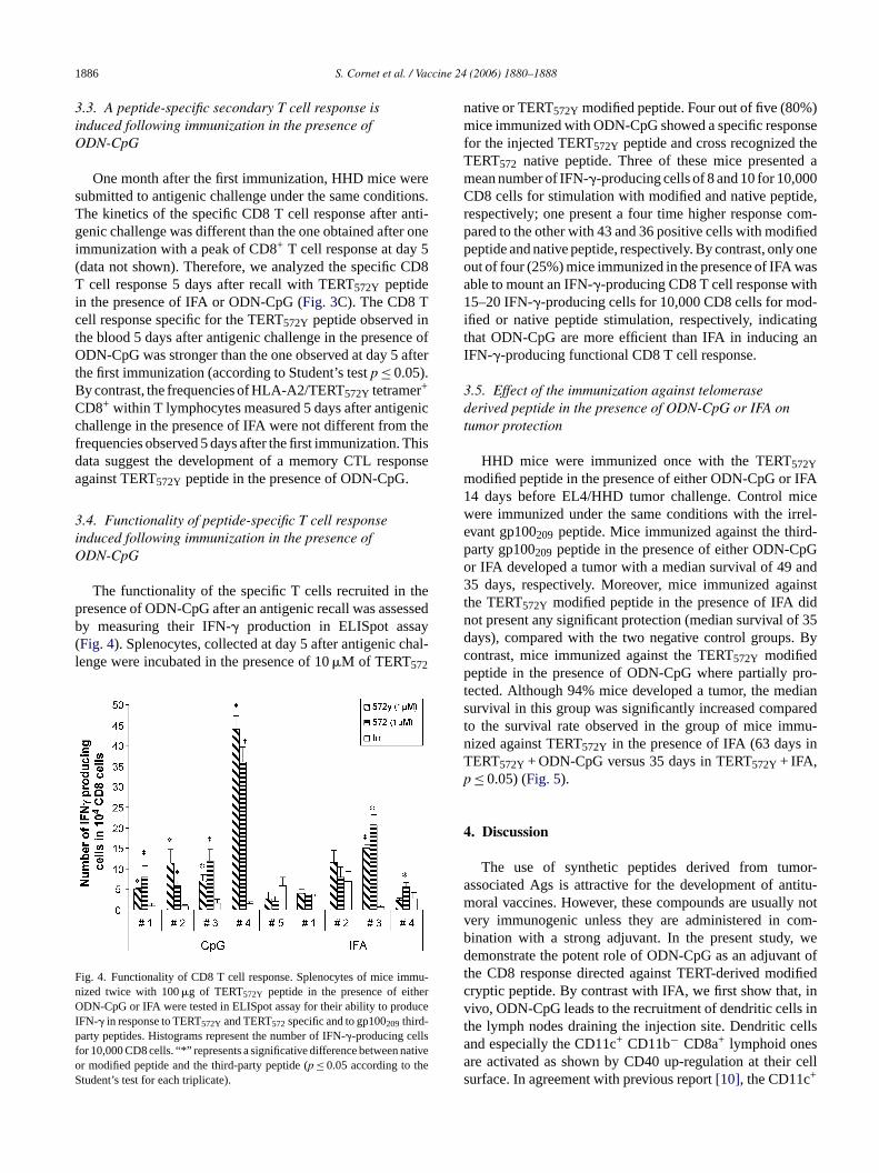
native or TERT572Y modified peptide. Four out of five (80%)mice immunized with ODN-CpG showed a specific responsefor the injected TERT572Y peptide and cross recognized theTERT572 native peptide. Three of these mice presented amean number of IFN-�-producing cells of 8 and 10 for 10,000CD8 cells for stimulation with modified and native peptide,respectively; one present a four time higher response com-pared to the other with 43 and 36 positive cells with modifiedpeptide and native peptide, respectively. By contrast, only oneout of four (25%) mice immunized in the presence of IFA wasable to mount an IFN-�-producing CD8 T cell response with15–20 IFN-�-producing cells for 10,000 CD8 cells for mod-ified or native peptide stimulation, respectively, indicatingthat ODN-CpG are more efficient than IFA in inducing anIFN-�-producing functional CD8 T cell response.
3.5. Effect of the immunization against telomerasederived peptide in the presence of ODN-CpG or IFA ontumor protection
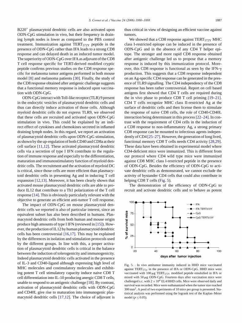
HHD mice were immunized once with the TERT572Ymodified peptide in the presence of either ODN-CpG or IFA14 days before EL4/HHD tumor challenge. Control micewere immunized under the same conditions with the irrel-evant gp100209 peptide. Mice immunized against the third-party gp100 peptide in the presence of either ODN-CpGo and3 instt idn f 35d . Bycp pro-t dians redt mu-n inTp
4
or-a titu-m ly notv om-b wed nt oft ifiedc , inv s int ellsaa cells
DN-CpG
The functionality of the specific T cells recruited inresence of ODN-CpG after an antigenic recall was assy measuring their IFN-� production in ELISpot assaFig. 4). Splenocytes, collected at day 5 after antigenic cenge were incubated in the presence of 10�M of TERT572
ig. 4. Functionality of CD8 T cell response. Splenocytes of mice imized twice with 100�g of TERT572Y peptide in the presence of eithDN-CpG or IFA were tested in ELISpot assay for their ability to prod
FN-� in response to TERT572Y and TERT572specific and to gp100209 third-arty peptides. Histograms represent the number of IFN-�-producing cells
or 10,000 CD8 cells. “*” represents a significative difference between nr modified peptide and the third-party peptide (p ≤ 0.05 according to thtudent’s test for each triplicate).
209r IFA developed a tumor with a median survival of 495 days, respectively. Moreover, mice immunized aga
he TERT572Y modified peptide in the presence of IFA dot present any significant protection (median survival oays), compared with the two negative control groupsontrast, mice immunized against the TERT572Y modifiedeptide in the presence of ODN-CpG where partially
ected. Although 94% mice developed a tumor, the meurvival in this group was significantly increased compao the survival rate observed in the group of mice imized against TERT572Y in the presence of IFA (63 daysERT572Y + ODN-CpG versus 35 days in TERT572Y + IFA,≤ 0.05) (Fig. 5).
. Discussion
The use of synthetic peptides derived from tumssociated Ags is attractive for the development of anoral vaccines. However, these compounds are usual
ery immunogenic unless they are administered in cination with a strong adjuvant. In the present study,emonstrate the potent role of ODN-CpG as an adjuva
he CD8 response directed against TERT-derived modryptic peptide. By contrast with IFA, we first show thativo, ODN-CpG leads to the recruitment of dendritic cellhe lymph nodes draining the injection site. Dendritic cnd especially the CD11c+ CD11b− CD8a+ lymphoid onesre activated as shown by CD40 up-regulation at theirurface. In agreement with previous report[10], the CD11c+
S. Cornet et al. / Vaccine 24 (2006) 1880–1888 1887
B220+ plasmacytoid dendritic cells are also activated uponODN-CpG stimulation in vivo, but their frequency in drain-ing lymph nodes is lower as compared to the PBS controltreatment. Immunization against TERT572Y peptide in thepresence of ODN-CpG rather than IFA leads to a strong CD8response and can delayed death in an induced tumor model.The superiority of ODN-CpG over IFA as adjuvant of the CD8T cell response specific for TERT-derived modified crypticpeptide confirms previous studies on the CD8 response spe-cific for melanoma tumor antigens performed in both mousemodel[8] and melanoma patients[30]. Finally, the study ofthe CD8 response obtained after antigenic challenge suggeststhat a functional memory response is induced upon vaccina-tion with ODN-CpG.
ODN-CpG interact with Toll-like receptor (TLR) 9 presentin the endocytic vesicles of plasmacytoid dendritic cells andthus can directly induce activation of those cells. Althoughmyeloid dendritic cells do not express TLR9, we observedthat these cells are recruited and activated upon ODN-CpGstimulation in vivo. This could be explained by an indi-rect effect of cytokines and chemokines secreted in inflameddraining lymph nodes. In this regard, we report an activationof plasmacytoid dendritic cells upon ODN-CpG stimulation,as shown by the up-regulation of both CD40 and CD8a at theircell surface[11,12]. These activated plasmacytoid dendriticcells via a secretion of type I IFN contribute to the regula-t tion,m en-d DCi acy-t elle hata pro-d cellr heo nse.
en-d e ane . Plasm iginpe riticc db sedb va-t nceb city.I enceo l ofM ibit-i 8 Tc lls,u ,a pGa las-m is
thus critical in view of designing an efficient vaccine againsttumors.
We showed that a CD8 response against TERT572Y MHCclass I-restricted epitope can be induced in the presence ofODN-CpG and in the absence of any CD4 T helper epi-tope. The stronger and more rapid CD8 response obtainedafter antigenic challenge led us to propose that a memoryresponse is induced by this immunization protocol. More-over, this CD8 response is functional as seen by the IFN-�production. This suggests that a CD8 response independenton an Ag-specific CD4 response can be generated in the pres-ence of TLR9 signalling. The CD4 independency of the CD8response has been rather controversial. Report on cell basedantigens first showed that CD4 T cells are required duringthe in vivo phase to produce CD8 T cell priming[19–21].CD4 T cells recognize MHC class II-restricted Ag at thesurface of dendritic cells and then license them to stimulatethe response of naive CD8 cells, the role of CD40-CD40Linteraction being determinant in this process[22–24]. In con-trast with the requirement of CD4 cells in the induction ofa CD8 response to non-inflammatory Ag, a strong primaryCD8 response can be mounted to infectious agents indepen-dently of CD4[25–27]. However, the generation of long lived,functional memory CD8 T cells needs CD4 activity[28,29].These data have been obtained in experimental model whereCD4-deficient mice were immunized. This is different fromo eda enceo cti-v e thea e inh
tor otent
F teda rev rm erec nds ached3 Sta-t eierm
ion of immune response and especially to the differentiaaturation and immunostimulatory function of myeloid dritic cells. The recruitment and the activation of myeloid
s critical, since those cells are more efficient than plasmoid dendritic cells in presenting Ag and in inducing T cxpansion[12,13]. Moreover, it has been clearly shown tctivated mouse plasmacytoid dendritic cells are able touce IL12 that contribute to a Th1 polarization of the Tesponse[14]. This is obviously particularly relevant with tbjective to generate an efficient anti-tumor T cell respo
The impact of ODN-CpG on mouse plasmacytoid dritic cells we reported is also of particular interest, sincquivalent subset has also been described in humansacytoid dendritic cells from both human and mouse orroduce high amounts of type I IFN (reviewed in[15]). How-ver, the production of IL12 by human plasmacytoid dendells has been controversial[16,17]. This may be explainey the differences in isolation and stimulation protocols uy the different groups. In line with this, a proper acti
ion of plasmacytoid dendritic cells is critical in the balaetween the induction of tolerogenicity and immunogeni
ndeed plasmacytoid dendritic cells activated in the presf IL-3 and CD40-ligand although expressing high leveHC molecules and costimulatory molecules and exh
ng potent T cell stimulatory capacity induce naive CDell differentiation into IL-10 producing anergic CD8 T cenable to respond to an antigenic challenge[18]. By contrastctivation of plasmacytoid dendritic cells with ODN-Cnd CD40L give rise to maturation into immunogenic pacytoid dendritic cells[17,12]. The choice of adjuvant
-
ur protocol where CD4 wild type mice were immunizgainst CD8 MHC class I-restricted peptide in the presf ODN-CpG. Besides the efficiency of ODN-CpG to aate dendritic cells as demonstrated, we cannot excludctivity of bystander CD4 cells that could also contributelping CD8 T cells (Fig. 5).
The demonstration of the efficiency of ODN-CpGecruit and activate dendritic cells and to behave as p
ig. 5. . In vivo antitumor immunity induced in HHD mice vaccinagainst TERT572y in the presence of IFA or ODN-CpG. HHD mice weaccinated with 100�g TERT572Y modified peptide emulsified in IFA oixed with 50�g ODN-CpG. Fourteen days after vaccination mice w
hallenged s.c. with 2× 10⁴ EL4/HHD cells. Mice were observed daily aurvival was recorded. Mice were euthananized when the tumor size re00 mm2. A pool of two experiments of 10 mice per group is presented.
istical analysis was performed using the logrank test of the Kaplan–Model (p ≤ 0.05).
1888 S. Cornet et al. / Vaccine 24 (2006) 1880–1888
adjuvant for a functional CD8 response against TERT derivedmodified cryptic peptide has important implications for thedesign of a cancer vaccine. The fact that MHC class I-restricted epitope combined with ODN-CpG is able to inducea functional CD8 response without the requirement of a Thelper CD4 epitope is promising in view of simplifying vac-cine formulation.
References
[1] Sebzda E, Mariathasan S, Ohteki T, Jones R, Bachmann MF,Ohashi PS. Selection of the T cell repertoire. Annu Rev Immunol1999;17:829–74.
[2] Scardino A, Gross DA, Alves P, Schultze JL, Graff-Dubois S,Faure O, et al. HER-2/neu and hTERT cryptic epitopes as noveltargets for broad spectrum tumor immunotherapy. J Immunol2002;168(11):5900–6.
[3] Gross DA, Graff-Dubois S, Opolon P, Cornet S, Alves P, Bennaceur-Griscelli A, et al. High vaccination efficiency of low-affinity epitopesin antitumor immunotherapy. J Clin Invest 2004;113(3):425–33.
[4] Tourdot S, Oukka M, Manuguerra JC, Magafa V, Vergnon I, Riche N,et al. Chimeric peptides: a new approach to enhancing the immuno-genicity of peptides with low MHC class I affinity: application inantiviral vaccination. J Immunol 1997;159(5):2391–8.
[5] Krieg AM. The role of CpG motifs in innate immunity. Curr OpinImmunol 2000;12(1):35–43.
[6] Sparwasser T, Koch ES, Vabulas RM, Heeg K, Lipford GB, EllwartJW, et al. Bacterial DNA and immunostimulatory CpG oligonu-
ells.
ia Z,atileera-
JC,inst
.itiveing
[ Kir-ty ofctiveMed
[ den-ony-
[ N,ells
unol
[ u J,en-
dritic cells with interleukin (IL)-3 and CD40-ligand. J Exp Med1997;185(6):1101–11.
[14] Martin P, Del Hoyo GM, Anjuere F, Arias CF, Vargas HH, Fer-nandez LA, et al. Characterization of a new subpopulation ofmouse CD8alpha+ B220+ dendritic cells endowed with type 1interferon production capacity and tolerogenic potential. Blood2002;100(2):383–90.
[15] Liu YJ. IPC: professional type 1 interferon-producing cellsand plasmacytoid dendritic cell precursors. Annu Rev Immunol2005;23:275–306.
[16] Rissoan MC, Soumelis V, Kadowaki N, Grouard G, Briere F, de WaalMalefyt R, et al. Reciprocal control of T helper cell and dendriticcell differentiation. Science 1999;283(5405):1183–6.
[17] Krug A, Towarowski A, Britsch S, Rothenfusser S, Hornung V, BalsR, et al. Toll-like receptor expression reveals CpG DNA as a uniquemicrobial stimulus for plasmacytoid dendritic cells which synergizeswith CD40 ligand to induce high amounts of IL-12. Eur J Immunol2001;31(10):3026–37.
[18] Gilliet M, Liu YJ. Generation of human CD8 T regulatory cellsby CD40 ligand-activated plasmacytoid dendritic cells. J Exp Med2002;195(6):695–704.
[19] Keene JA, Forman J. Helper activity is required for the in vivo gen-eration of cytotoxic T lymphocytes. J Exp Med 1982;155(3):768–82.
[20] Mitchison NA, O’Malley C. Three-cell-type clusters of T cells withantigen-presenting cells best explain the epitope linkage and noncog-nate requirements of the in vivo cytolytic response. Eur J Immunol1987;17(11):1579–83.
[21] Wang B, Norbury CC, Greenwood R, Bennink JR, Yewdell JW,Frelinger JA. Multiple paths for activation of naive CD8+ T cells:CD4-independent help. J Immunol 2001;167(3):1283–9.
[22] Ridge JP, Di Rosa F, Matzinger P. A conditioned dendritic cell cancell.
[ eathsig-
[ elief40-
[ rdnce
[ b-n ofack-
[ n-D8+
[ ating.
[ ute–42.
[ e-es to909.
cleotides trigger maturation and activation of murine dendritic cEur J Immunol 1998;28(6):2045–54.
[7] Firat H, Garcia-Pons F, Tourdot S, Pascolo S, Scardino A, Garcet al. H-2 class I knockout HLA-A2.1-transgenic mice: a versanimal model for preclinical evaluation of antitumor immunothpeutic strategies. Eur J Immunol 1999;29(10):3112–21.
[8] Miconnet I, Koenig S, Speiser D, Krieg A, Guillaume P, Cerottiniet al. CpG are efficient adjuvants for specific CTL induction agatumor antigen-derived peptide. J Immunol 2002;168(3):1212–8
[9] Prakash A, Smith E, Lee CK, Levy DE. Tissue-specific posfeedback requirements for production of type I interferon followvirus infection. J Biol Chem 2005;280(19):18651–7.
10] Shah JA, Darrah PA, Ambrozak DR, Turon TN, Mendez S,man J, et al. Dendritic cells are responsible for the capaciCpG oligodeoxynucleotides to act as an adjuvant for protevaccine immunity against Leishmania major in mice. J Exp2003;198(2):281–91.
11] Bjorck P. Isolation and characterization of plasmacytoiddritic cells from Flt3 ligand and granulocyte-macrophage colstimulating factor-treated mice. Blood 2001;98(13):3520–6.
12] Asselin-Paturel C, Boonstra A, Dalod M, Durand I, YessaadDezutter-Dambuyant C, et al. Mouse type I IFN-producing care immature APCs with plasmacytoid morphology. Nat Imm2001;2(12):1144–50.
13] Grouard G, Rissoan MC, Filgueira L, Durand I, BanchereaLiu YJ. The enigmatic plasmacytoid T cells develop into d
be a temporal bridge between a CD4+ T-helper and a T-killerNature 1998;393(6684):474–8.
23] Bennett SR, Carbone FR, Karamalis F, Flavell RA, Miller JF, HWR. Help for cytotoxic-T-cell responses is mediated by CD40nalling. Nature 1998;393(6684):478–80.
24] Schoenberger SP, Toes RE, van der Voort EI, Offringa R, MCJ. T-cell help for cytotoxic T lymphocytes is mediated by CDCD40L interactions. Nature 1998;393(6684):480–3.
25] Buller RM, Holmes KL, Hugin A, Frederickson TN, Morse IIIHC. Induction of cytotoxic T-cell responses in vivo in the abseof CD4 helper cells. Nature 1987;328(6125):77–9.
26] Rahemtulla A, Fung-Leung WP, Schilham MW, Kundig TM, Samhara SR, Narendran A, et al. Normal development and functioCD8+ cells but markedly decreased helper cell activity in mice ling CD4. Nature 1991;353(6340):180–4.
27] Wu Y, Liu Y. Viral induction of co-stimulatory activity on antigepresenting cells bypasses the need for CD4+ T-cell help in CT-cell responses. Curr Biol 1994;4(6):499–505.
28] Shedlock DJ, Shen H. Requirement for CD4 T cell help in generfunctional CD8 T cell memory. Science 2003;300(5617):337–9
29] Sun JC, Bevan MJ. Defective CD8 T cell memory following acinfection without CD4 T cell help. Science 2003;300(5617):339
30] Speiser DE, Lienard D, Rufer N, Rubio-Godoy V, Rimoldi D, Lejune F, et al. Rapid and strong human CD8+ T cell responsvaccination with peptide, IFA, and CpG oligodeoxynucleotide 7J Clin Invest 2005;115(3):739–46.