CPAP and sleep disorders - World Allergy Organization and sleep...of obstructive sleep apnea and its...
136
CPAP and sleep disorders Workshop Fulvio Braido Allergy and Respiratory Diseases Department University of Genoa
Transcript of CPAP and sleep disorders - World Allergy Organization and sleep...of obstructive sleep apnea and its...
CPAP and sleep disorders
Workshop
Fulvio Braido Allergy and Respiratory Diseases Department
University of Genoa
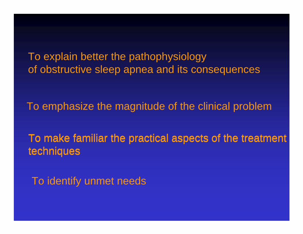
To explain better the pathophysiologyof obstructive sleep apnea and its consequences
To emphasize the magnitude of the clinical problem
To make familiar the practical aspects of the treatmenttechniques
To identify unmet needs
To make familiar the practical aspects of the treatmenttechniques
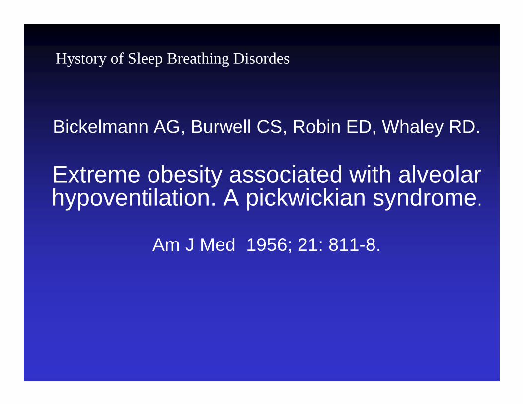
Bickelmann AG, Burwell CS, Robin ED, Whaley RD.
Extreme obesity associated with alveolar hypoventilation. A pickwickian syndrome.
Am J Med 1956; 21: 811-8.
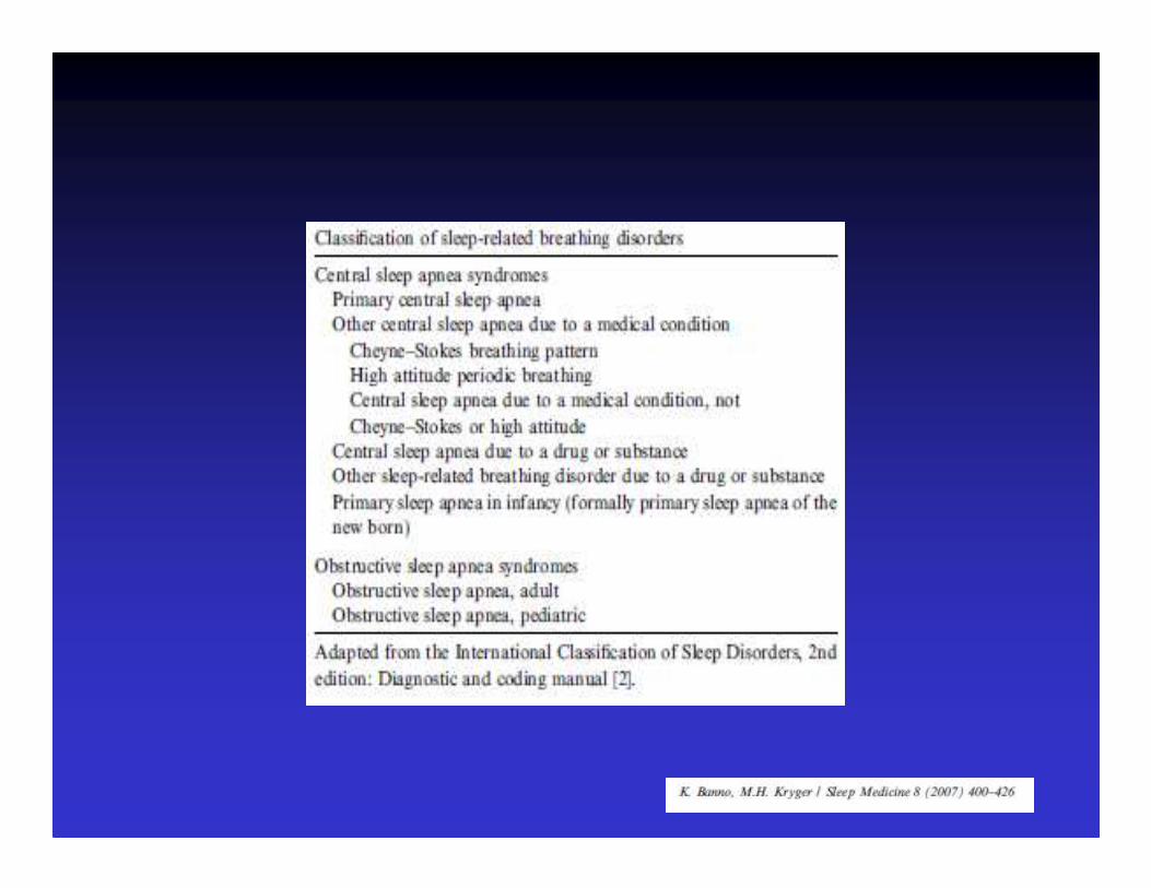
Hystory of Sleep Breathing Disordes

1956 – Bickelmann AG, Burwell CS, Robin ED, Whaley RD. Extreme obesityassociated with alveolar hypoventilation. A pickwickian syndrome. Am J Med 21: 811-8.
1965 - Gastaut H, Tassinari CA, Duron B. Etude polygraphique des manifestations episodiques (hypniques et respiratoires) du syndrome de Pickwick. Rev Neural 112: 568-79.
1965 - Jung R, Kuhlo W. Neurophysiological studiesof abnormal night sleep and the pickwickian syndrome. Prog Brain Res 18: 140-59.
1970 - Lugaresi E, Coccagna G, Mantovani M, Brignani F. Effets de la trachéotomiedans les hypersomnies avec respiration périodique. Rev Neurol 123: 267-8.
1972 - Lugaresi E. Organizer Symposium: Hypersomniawith Periodic Breathing. Rimini, Italy, May 25-27. Bull Physiopath Resp 8:967.
1981 - Sullivan CE, Issa FG, Berthon-Jones M, Eves L. Reversal of obstructive sleep apnoea by continuous positive airway pressureapplied through the nares. Lancet i: 862-5.
OSA Mile Stones

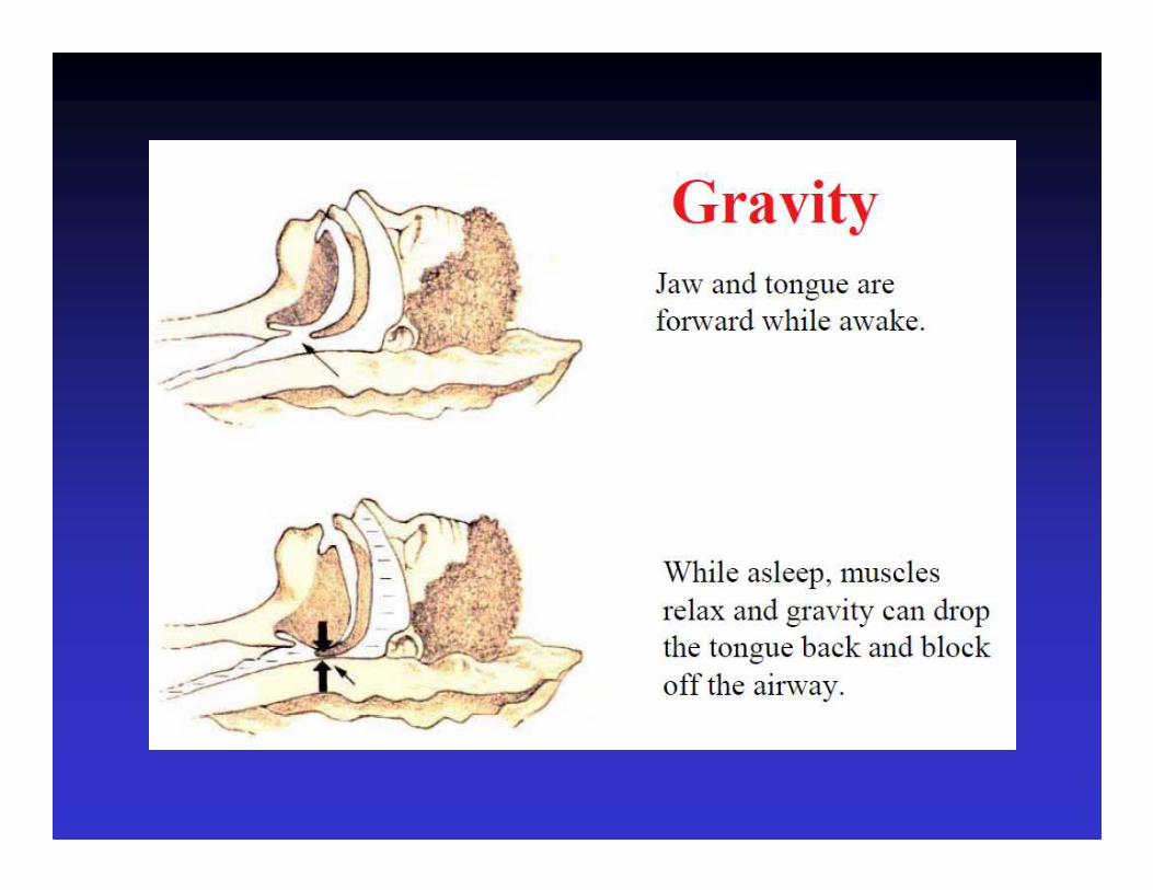
It is caracterized by repetitive collapseof the upper airways during sleep
OSA

Hpopnea: reduction in the airflow > 30%SO2 decrease >3%at least 10 second
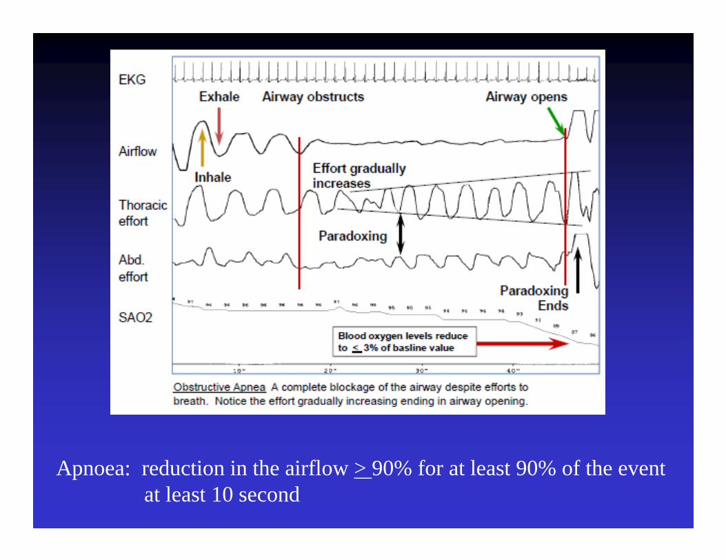
Apnoea: reduction in the airflow > 90% for at least 90% of the eventat least 10 second
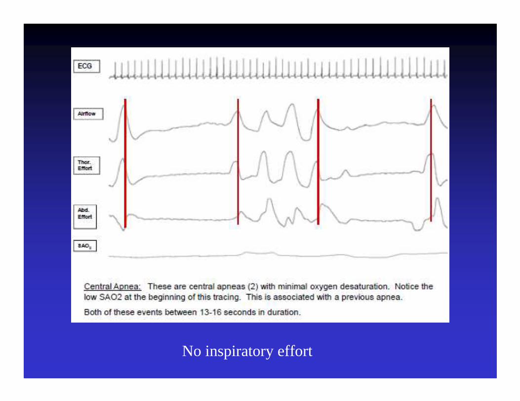
No inspiratory effort

Obstructive Sleep Apnea (OSA)
• Snoring
• Recurrent episodes of upper airway obstructionduring sleep (apneas, hypopneas)
• Arousals
• Excessive and disabling daytime sleepiness
• OSA Syndrome– features of OSA on sleep study
+
symptoms of daytime sleepiness

OSA � OSAS

OSASIt is characterised by repetitive collapseof the upper airways during sleep
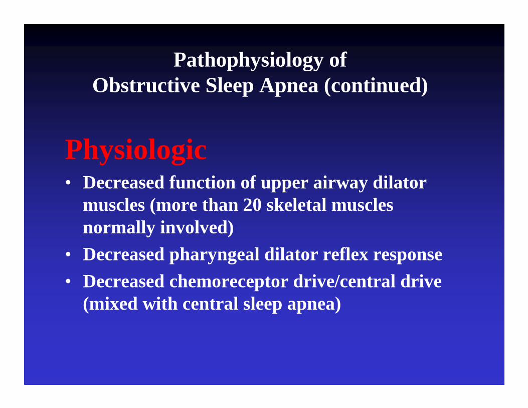
Pathophysiology of Obstructive Sleep Apnea (continued)
Physiologic• Decreased function of upper airway dilator
muscles (more than 20 skeletal muscles normally involved)
• Decreased pharyngeal dilator reflex response
• Decreased chemoreceptor drive/central drive (mixed with central sleep apnea)

Impact of sleep on ventilation
Fall in phrenic and hypoglossal activityReduce response to hypercapnia and hypoxiaReduction in upper ariways protective reflexes
Minute ventlation fall (16%)PaCO2 increase 4-6 mmHgPaO2 decrease (So2 decrease 2%)Irregular breathing during light and fragmented sleepUpper airways caliber reduction

SLEEP
Cortical imputs
Chemoreceptor sensitivity
Respiratory motor
neurones
Respiratory muscle
contraction
Lung mechanics: Airflow resistance
FRC V/Q relationships
Hypoventilation
Hypoxemia
Hypercapnia

Pathophysiology of Obstructive Sleep Apnea
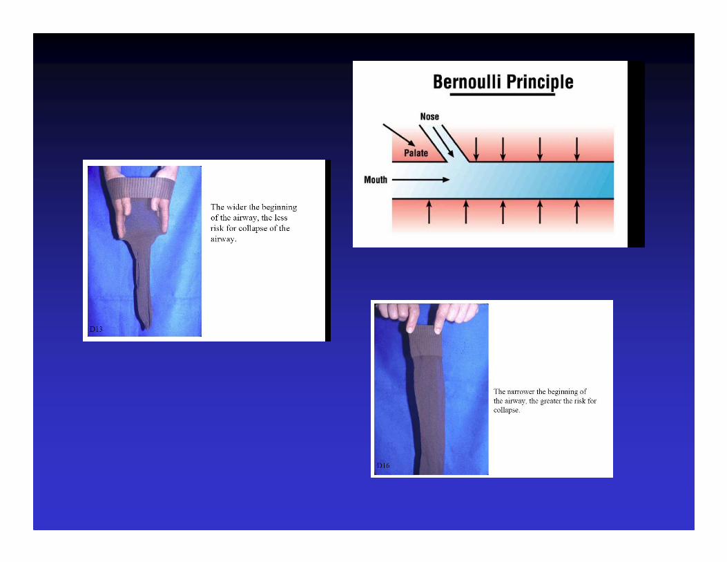
Mechanical• Short, thick neck• Neck flexion, supine position• Nasal obstruction, congestion, polyps

Pathophysiology of Obstructive Sleep Apnea (continued)
Anatomic• Enlarged tonsils and adenoids (esp. ages 3-5), enlarged
uvula• Macroglossia• Retrognathia, craniofacial abnormalities• Compliant (floppy) pharynx, especially soft palate• Fat deposition in lateral walls of pharynx, pharyngeal
dilator muscles (obesity)• Submucosal edema in lateral walls of pharynx

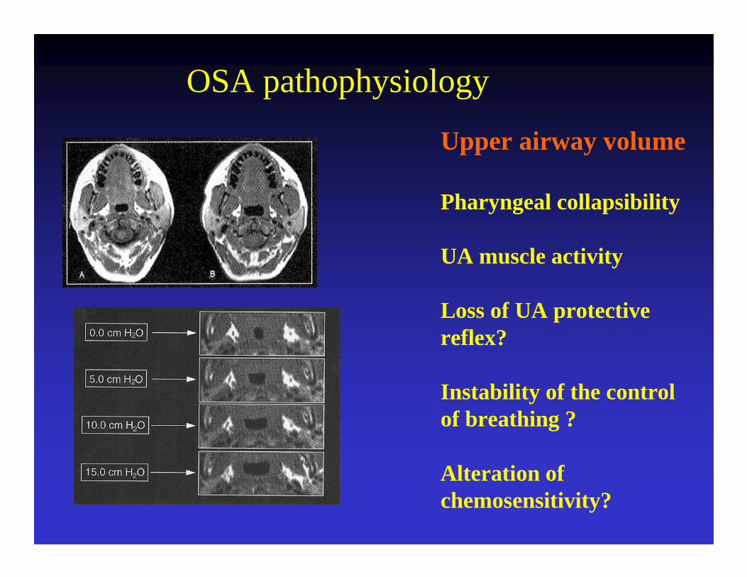
OSA pathophysiology
Upper airway volume
Pharyngeal collapsibility
UA muscle activity
Loss of UA protective reflex?
Instability of the controlof breathing ?
Alteration of chemosensitivity?

Craniofacial size and Obesity can infuence upper airway caliber.
Watanabe et al, AJRCCM 165:260, 2002
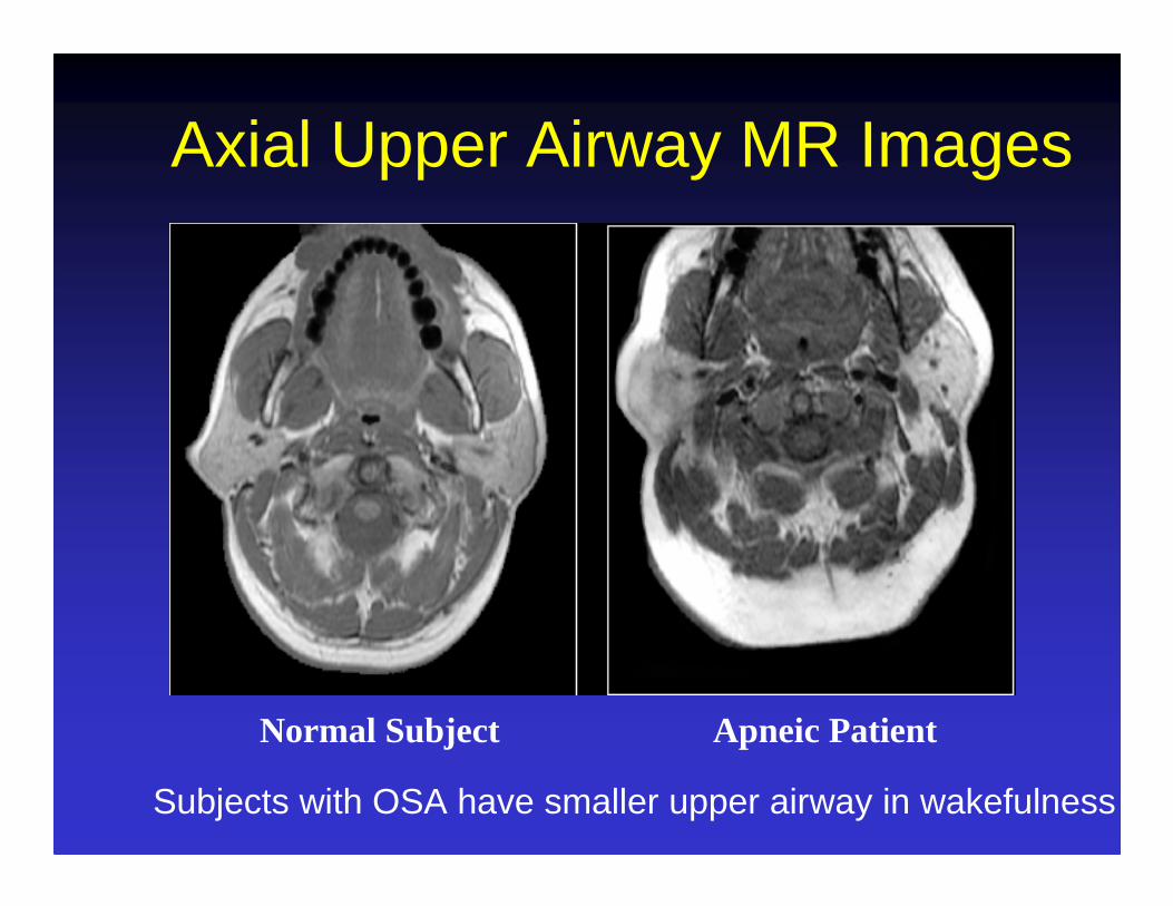
Axial Upper Airway MR Images
Normal Subject Apneic Patient
Subjects with OSA have smaller upper airway in wakefulness

OSA Patients Have Elevated Activity of Their Genioglossus MuscleDuring Wakefulness (Lost during sleep)
Mezzanotte et al, JCI 89:1571, 1992
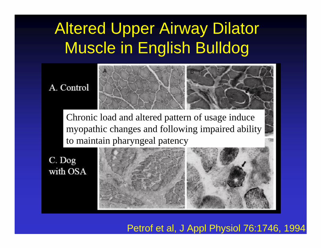
Altered Upper Airway DilatorMuscle in English Bulldog
Petrof et al, J Appl Physiol 76:1746, 1994
Chronic load and altered pattern of usage induce myopathic changes and following impaired abilityto maintain pharyngeal patency

Alveolar pressure:0 cm H2O
Alveolar pressure:0 cm H2O
END EXPIRATION
Intrapleural pressure:-5 cmH2O
Transmural pressure= -1 cmH2O - (-8cmH2O)= +7 cmH2O
DURING INSPIRATION
Alveolar pressure:0 cm H2O
Outward recoil of chest wall
Inward recoil of alveoli
Alveolar pressure:-1 cm H2O
Transmural pressure= 0 cmH2O - (-5cmH2O)= +5 cmH2O
Intrapleural pressure:-8 cmH2O
No flow Inspiratory force
Flow in
Atmospheric Pressure : 0 cm H2O Atmospheric Pressure : 0 cm H2O
Eupneic Inspiration(Revised from Fig. 2-1 in Levitzky’s Pulmonary Physiology)

END EXPIRATION DURING INSPIRATION
Atmospheric Pressure : 0 cm H2O Atmospheric Pressure : 0 cm H2O
Alveolar pressure:0 cm H2O
Alveolar pressure:0 cm H2O
Intrapleural pressure:-5 cmH2O
Transmural pressure= -23 cmH2O - (-30 cmH2O)= +7 cmH2O
Alveolar pressure:0 cm H2O
Outward recoil of chest wall
Inward recoil of alveoli
Alveolar pressure:-23 cm H2O
Transmural pressure= 0 cmH2O - (-5cmH2O)= +5 cmH2O
Intrapleural pressure:-30 cmH2O
No flow Inspiratory force
Flow in
Forced Inspiration
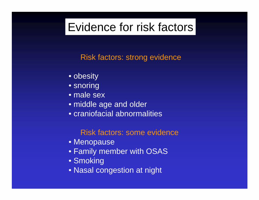
Evidence for risk factors
Risk factors: strong evidence
• obesity• snoring• male sex• middle age and older• craniofacial abnormalities
Risk factors: some evidence• Menopause• Family member with OSAS• Smoking• Nasal congestion at night
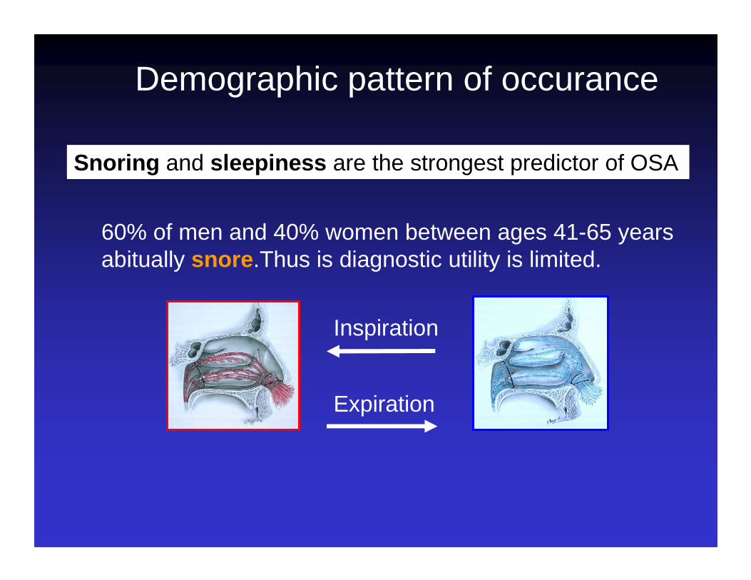
Demographic pattern of occurance
Snoring and sleepiness are the strongest predictor of OSA
60% of men and 40% women between ages 41-65 yearsabitually snore .Thus is diagnostic utility is limited.
Inspiration
Expiration

Obesity is a very strong risk factor for OSA
all measure of obesity - neck and waist girths, weight, skin folds -predict OSA
An increase of 1 Kg/m2 in BMI yelds an estimated 30% increased in the odds of developing OSA
An increase (decrease) of 1 kg/m2 in BMI yields and estimated 9% increase (decrease) in AHI
Demographic pattern of occurance
male: female ratio for OSA prevalence is 2:1
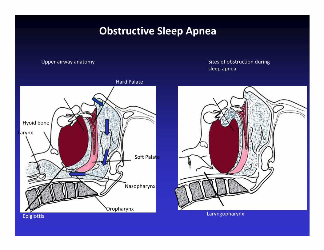
Upper airway anatomy
Hard Palate
Soft Palate
Nasopharynx
Hyoid bone
Larynx
Epiglottis
Oropharynx
Sites of obstruction during
sleep apnea
Laryngopharynx
TongueTongue
Obstructive Sleep Apnea
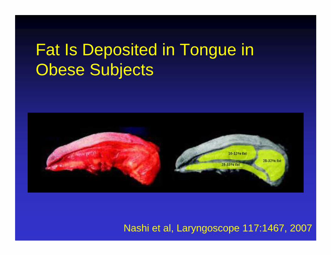
Fat Is Deposited in Tongue in Obese Subjects
Nashi et al, Laryngoscope 117:1467, 2007
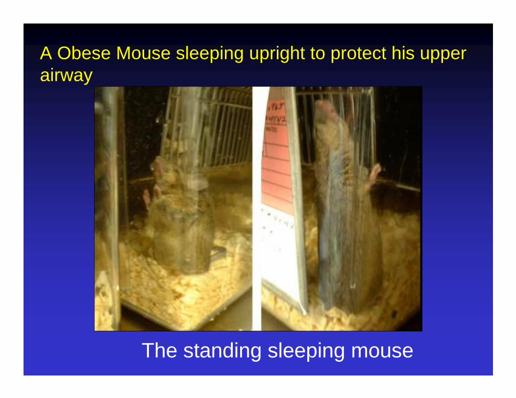
The standing sleeping mouse
A Obese Mouse sleeping upright to protect his upper airway
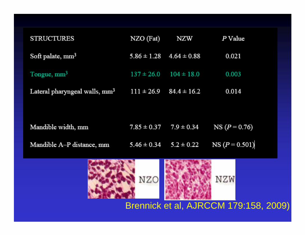
Brennick et al, AJRCCM 179:158, 2009)
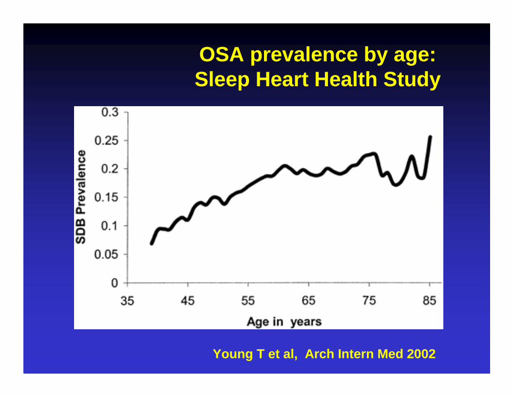
Young T et al, Arch Intern Med 2002
OSA prevalence by age:Sleep Heart Health Study

Tishler et al, JAMA 2003
Cleveland Family Study: interaction of age with gender and
BMI
By age 50 ys, incidence rates among men and women are similar
The effect of BMI deacreases with age and may be irrilevant in elderly
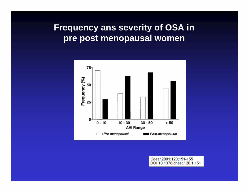
Frequency ans severity of OSA in pre post menopausal women
Guilleminault, et al 1978Sleep Apnea Syndrome
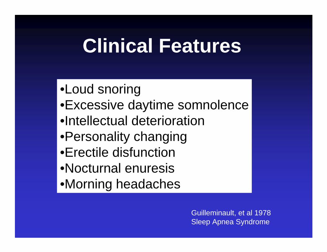
•Loud snoring•Excessive daytime somnolence•Intellectual deterioration •Personality changing•Erectile disfunction•Nocturnal enuresis•Morning headaches
Clinical Features

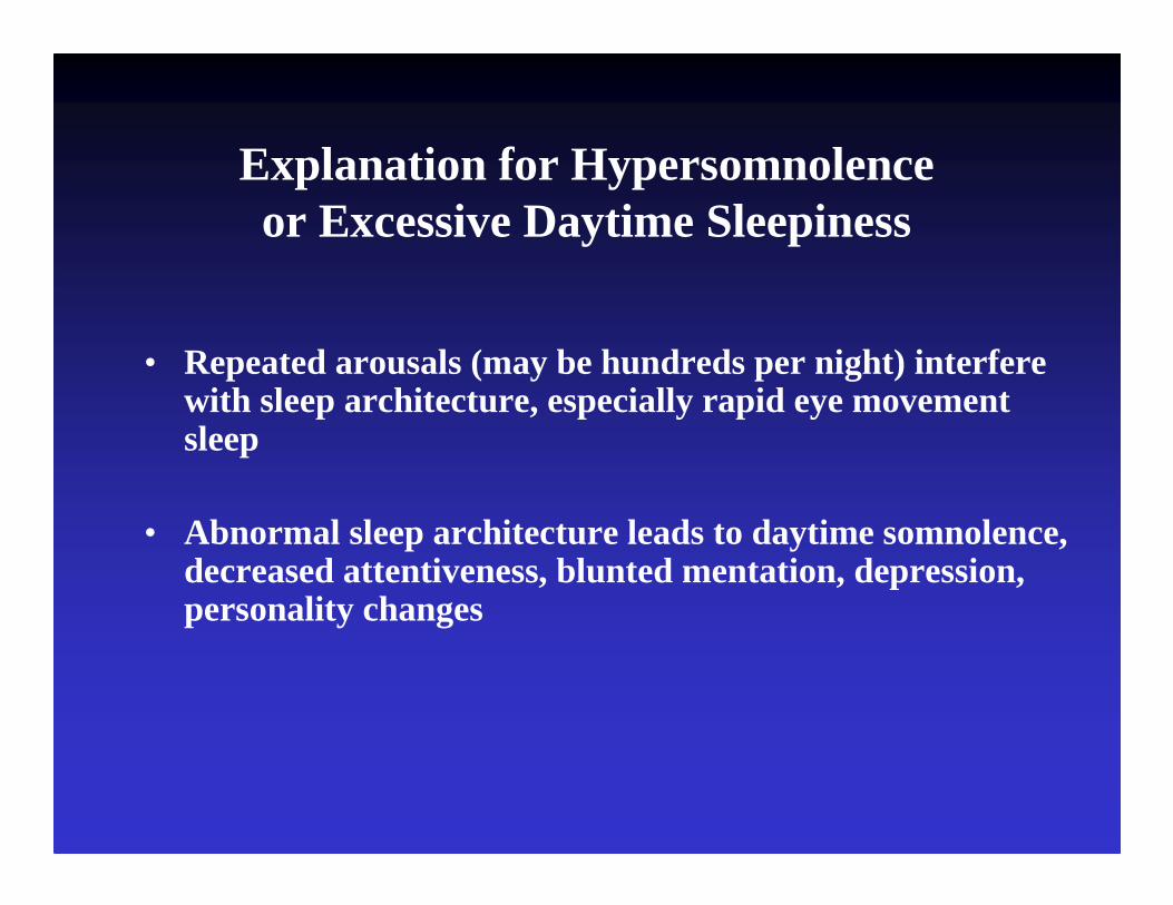
Explanation for Hypersomnolence or Excessive Daytime Sleepiness
• Repeated arousals (may be hundreds per night) interfere with sleep architecture, especially rapid eye movement sleep
• Abnormal sleep architecture leads to daytime somnolence, decreased attentiveness, blunted mentation, depression, personality changes
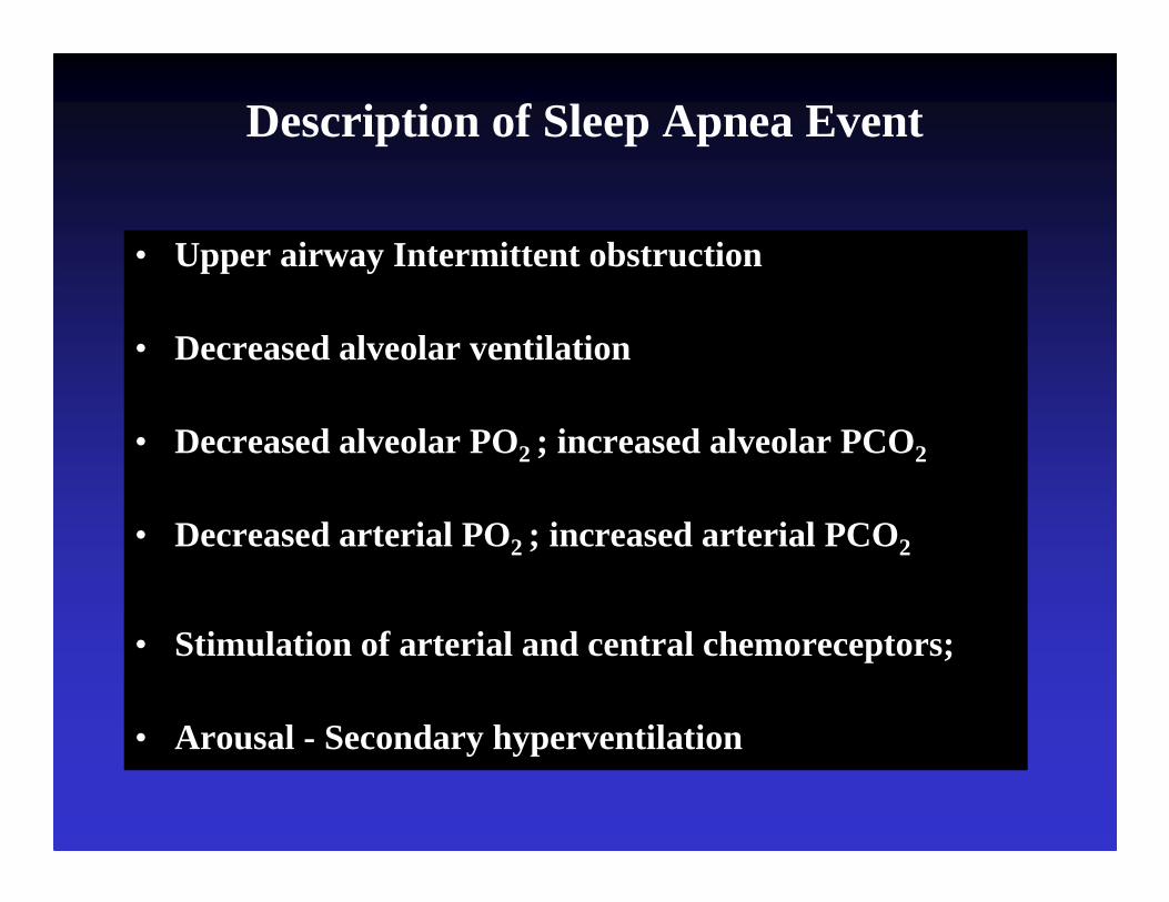
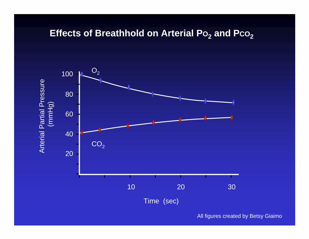
Description of Sleep Apnea Event
• Upper airway Intermittent obstruction
• Decreased alveolar ventilation
• Decreased alveolar PO2 ; increased alveolar PCO2
• Decreased arterial PO2 ; increased arterial PCO2
• Stimulation of arterial and central chemoreceptors;
• Arousal - Secondary hyperventilation
10 20 30
100
80
40
20
Time (sec)
O2
CO2
Art
eria
l Par
tial P
ress
ure
(mm
Hg)
Effects of Breathhold on Arterial P O2 and PCO2
60
All figures created by Betsy Giaimo
0.2 0.4 0.6 0.8
Hematocrit
8
6
4
2
Rel
ativ
e V
isco
sity
Effects of Hematocrit on Human Blood Viscosity

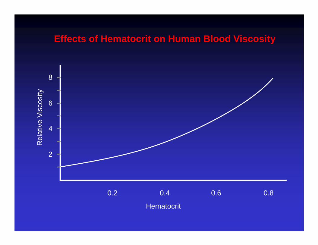
Possible Explanation for Nocturia
• Increased blood viscosity and arterial hypertension• Increased right ventricular afterload• Increased right ventricular end diastolic pressure and
volume• Increased right atrial volume• Increased secretion of atrial natriuretic peptide from
atrial myocytes, • Increases sodium excretion, and stretches receptors
that suppress ADH secretion from the posterior pituitary gland
40 100
Arterial PO2 (mm Hg)
100
75
50
25
Cer
ebra
l Blo
od F
low
(m
l/100
mg/
min
)
Arterial PCO2 (mm Hg)
20 60 80
40 10020 60 80
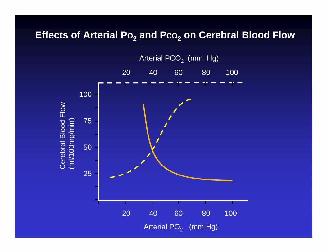
Effects of Arterial P O2 and PCO2 on Cerebral Blood Flow
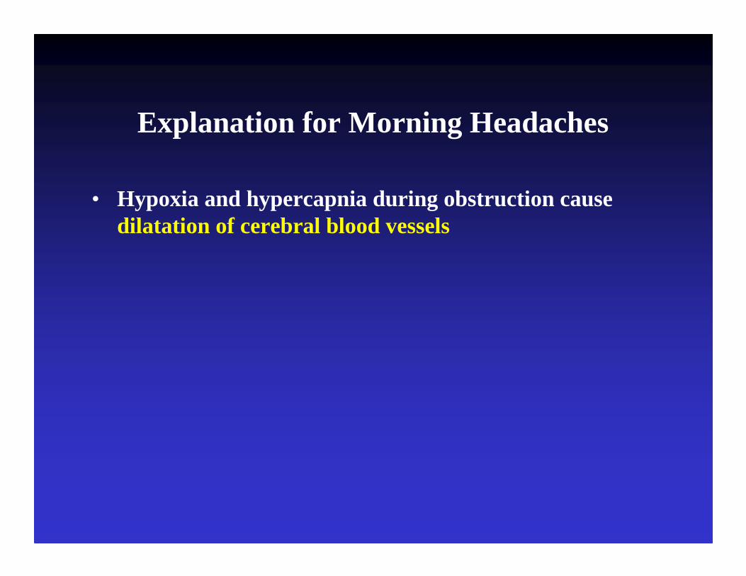
Explanation for Morning Headaches
• Hypoxia and hypercapnia during obstruction cause dilatation of cerebral blood vessels

CONDITIONS ASSOCIATED TO OSA
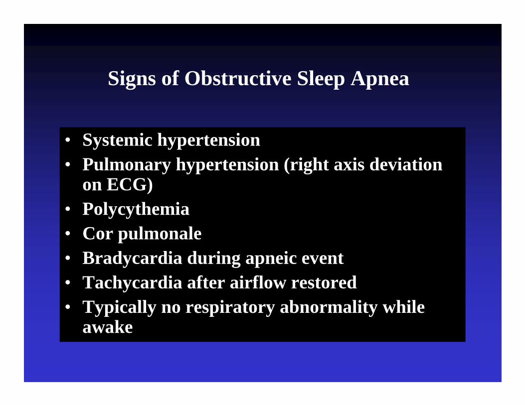
Signs of Obstructive Sleep Apnea
• Systemic hypertension• Pulmonary hypertension (right axis deviation
on ECG)• Polycythemia• Cor pulmonale• Bradycardia during apneic event• Tachycardia after airflow restored• Typically no respiratory abnormality while
awake
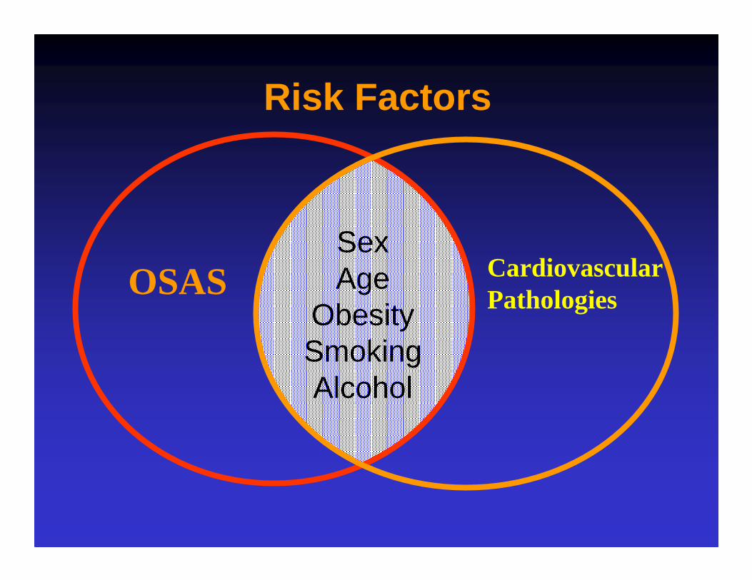
SexAge
ObesitySmokingAlcohol
Risk Factors
OSAS CardiovascularPathologies
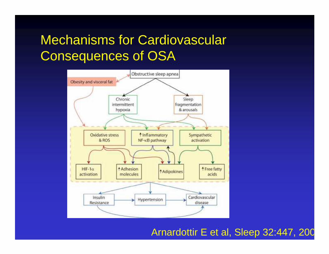
Mechanisms for Cardiovascular Consequences of OSA
Arnardottir E et al, Sleep 32:447, 2009

Possible Explanation for Systemic Hypertension
• Repeated increases in sympathetic tone and systemic blood pressure during arousals may cause vascular remodeling and changes in endothelial function

OSAS & Arterial Hypertension
Arterial hypertension is especially frequent in patients Suffering from OSAS
Partisen M et Al. Sleep Res 1983;12:273Kryger M et Al. WB Saunders 1989Pekkarinen T et Al. Clin Endocrinol 1987; 27 649-654.Cozzi Fet Al Pediatrics 1985; 75:836-843Bliwise D et Al Am J Public Haelth 1988:78:544-547
25 - 38%
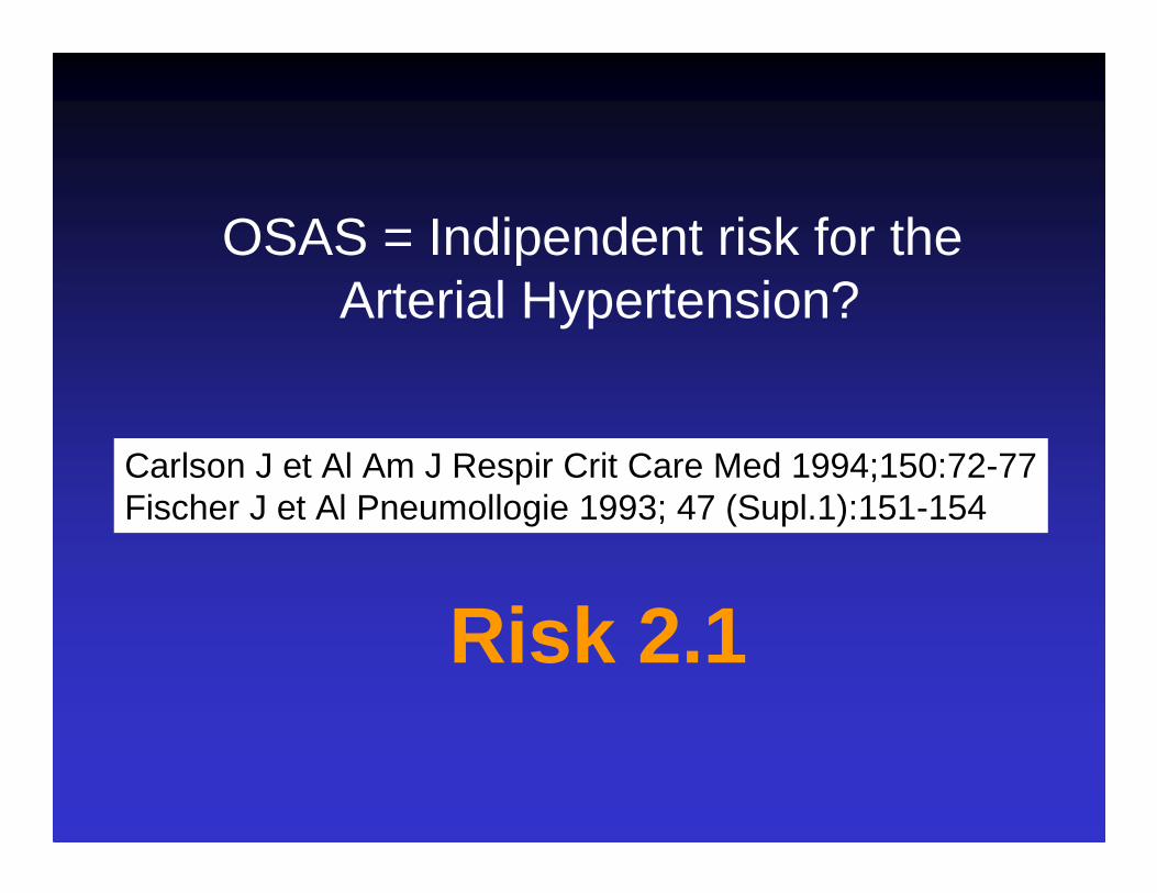
OSAS = Indipendent risk for the Arterial Hypertension?
Carlson J et Al Am J Respir Crit Care Med 1994;150:72-77Fischer J et Al Pneumollogie 1993; 47 (Supl.1):151-154
Risk 2.1

Prospective Study of the Association betweenSleep-Disordered Breathing and Hypertension
Paul E. Peppard, Ph.D., Terry Young, Ph.D., Mari Palta, Ph.D., and James Skatrud, M.D.
Volume 342:1378-1384 May 11, 2000 Number 19

NEJM Volume 342:1378-1384 May 11, 2000 N°19
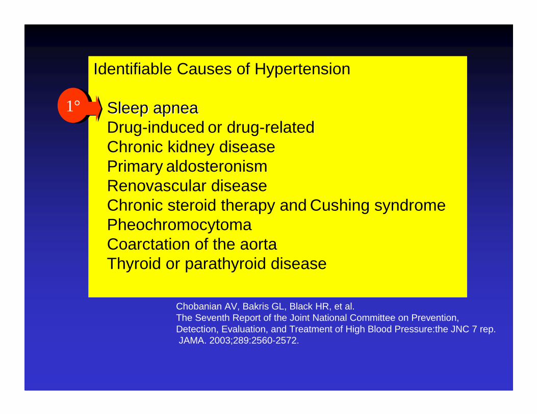
Identifiable Causes of Hypertension
Sleep apneaSleep apneaDrug-induced or drug-related Chronic kidney diseasePrimary aldosteronismRenovascular diseaseChronic steroid therapy and Cushing syndromePheochromocytomaCoarctation of the aortaThyroid or parathyroid disease
1°
Chobanian AV, Bakris GL, Black HR, et al. The Seventh Report of the Joint National Committee on Prevention, Detection, Evaluation, and Treatment of High Blood Pressure:the JNC 7 rep.JAMA. 2003;289:2560-2572.
1°
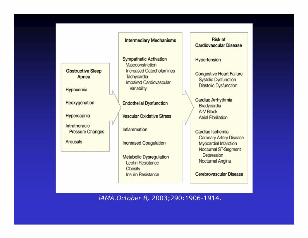
JAMA.October 8, 2003;290:1906-1914.
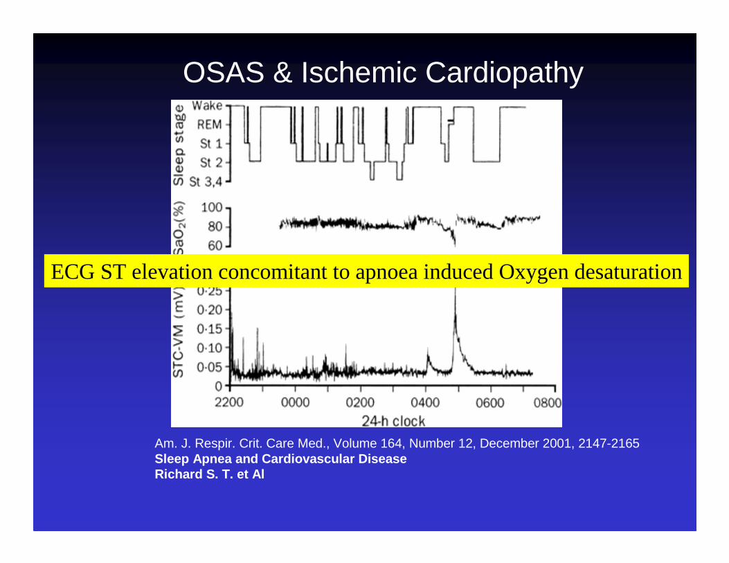
Am. J. Respir. Crit. Care Med., Volume 164, Number 12, December 2001, 2147-2165Sleep Apnea and Cardiovascular Disease Richard S. T. et Al
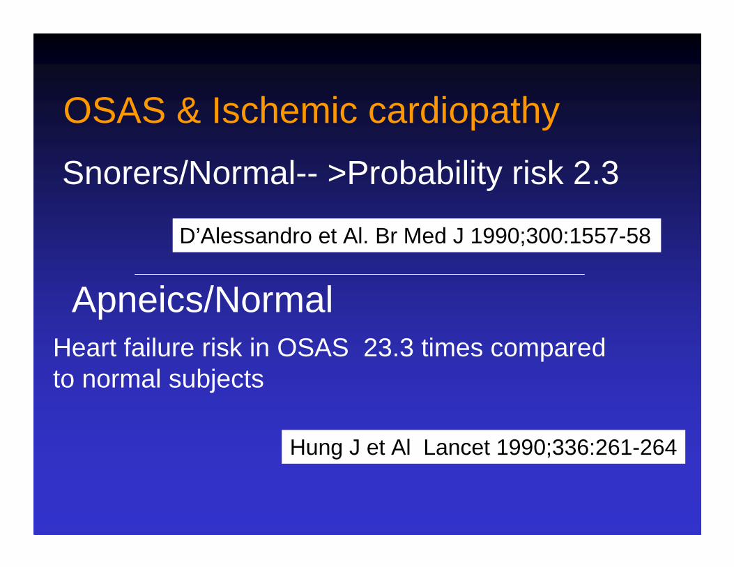
OSAS & Ischemic Cardiopathy
ECG ST elevation concomitant to apnoea induced Oxygen desaturation
OSAS & Ischemic cardiopathy
D’Alessandro et Al. Br Med J 1990;300:1557-58
Snorers/Normal-- >Probability risk 2.3
Heart failure risk in OSAS 23.3 times comparedto normal subjects
Hung J et Al Lancet 1990;336:261-264
Apneics/Normal

OSAS & Ischemic Cardiopathy
OSAS minimum prevalence among Coronaropathic subjects is about 16%
Andreas S et Al Coron Artery Dis 1996;7:541-545

Peker Y, et AlAm J Respir Crit Care Med. 2000;162:81-86
During the follow-up period (post-myocardial infarction),cardiovascular death occurred in six of 16 OSA patients
(37.5%) compared with 4 (9.3%) in the non-OSA group(p = 0.018)
62 patients with established CAD
OSA is represents a risk factor for death in post-yocardial infarction
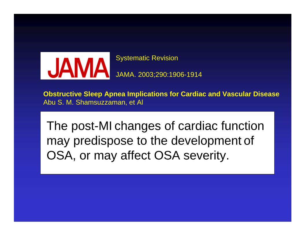
Obstructive Sleep Apnea Implications for Cardiac an d Vascular Disease Abu S. M. Shamsuzzaman, et Al
Systematic Revision
JAMA. 2003;290:1906-1914
The post-MI changes of cardiac function may predispose to the development of OSA, or may affect OSA severity.

Effects of Obstruction on Pulmonary Circulation and Right Ventricle
• Hypoxic and hypercapnic pulmonary vasoconstriction cause pulmonary hypertension
• Chronic nighttime hypoxia may cause erythropoiesis and polycythemia
• Increased hematocrit increases blood viscosity• Hypoxic pulmonary vasoconstriction (HPV), increased
blood viscosity, pulmonary hypertension increase right ventricular afterload
• Increased right ventricular afterload may lead to right ventricular hypertrophy and eventually cor pulmonale

Possible Explanations for Bradycardia During Obstruction, Tachycardia after Airflow Restored
• Stimulation of arterial chemoreceptors usually increases heart rate because it increases tidal volume (lung inflation reflex)
• Stimulation of arterial chemoreceptors without stretching the lungs causes bradycardia
• After arousal leads to restoration of airflow, large tidal volumes stretch lungs and cause tachycardia
• May hyperventilate immediately after arousal, then hypoventilate until CO2 is restored
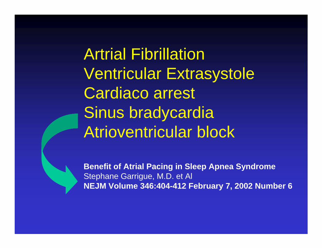
Artrial FibrillationVentricular ExtrasystoleCardiaco arrest Sinus bradycardiaAtrioventricular block
Benefit of Atrial Pacing in Sleep Apnea SyndromeStephane Garrigue, M.D. et AlNEJM Volume 346:404-412 February 7, 2002 Number 6

Cardiac arrhythmias, snoring, and sleep apneaV Hoffstein and S Mateika Department of Medicine, St. Michael's Hospital, Toronto, Canada.
458 patients
. 82 % of patients with mean nocturnal oxygen saturation< 90 % had arrhythmias vs 40 % of patients with mean nocturnal oxygen saturation > 90 %
70 % of patients with AHI > 40 had arrhythmias vs 42% with AHI < or 40
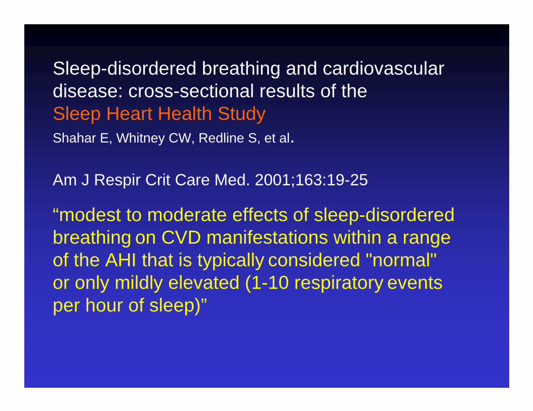
Sleep-disordered breathing and cardiovasculardisease: cross-sectional results of the Sleep Heart Health StudyShahar E, Whitney CW, Redline S, et al.
Am J Respir Crit Care Med. 2001;163:19-25
“modest to moderate effects of sleep-disordered breathing on CVD manifestations within a range of the AHI that is typically considered "normal" or only mildly elevated (1-10 respiratory eventsper hour of sleep)”

Sleep-disordered breathing and cardiovasculardisease: cross-sectional results of the Sleep Heart Health Study Shahar E, Whitney CW, Redline S, et al.
Am J Respir Crit Care Med. 2001;163:19-25
modestly elevated risk coupled with a high prevalence of mild sleep-disordered breathing might have considerable public health implications
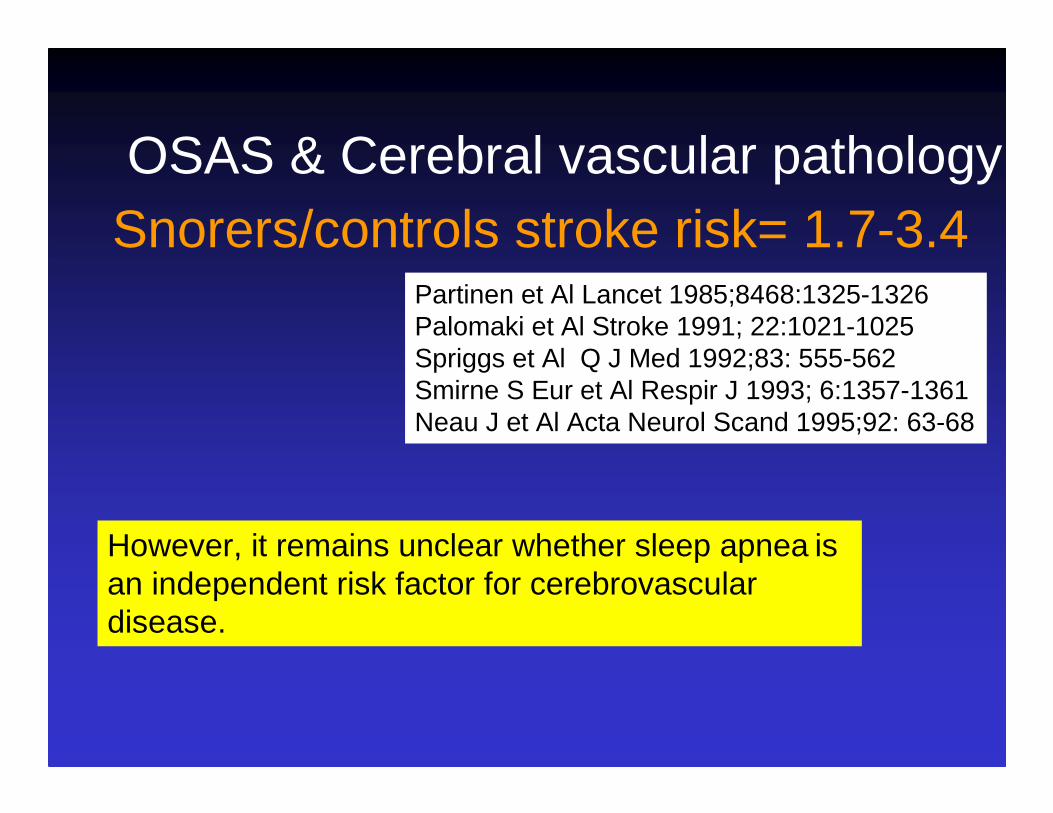
OSAS & Cerebral vascular pathologySnorers/controls stroke risk= 1.7-3.4
Partinen et Al Lancet 1985;8468:1325-1326Palomaki et Al Stroke 1991; 22:1021-1025Spriggs et Al Q J Med 1992;83: 555-562Smirne S Eur et Al Respir J 1993; 6:1357-1361Neau J et Al Acta Neurol Scand 1995;92: 63-68
However, it remains unclear whether sleep apnea is an independent risk factor for cerebrovascular disease.
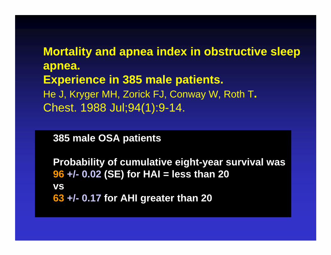
Mortality and apnea index in obstructive sleep apnea. Experience in 385 male patients.He J, Kryger MH, Zorick FJ, Conway W, Roth T.Chest. 1988 Jul;94(1):9-14.
385 male OSA patients
Probability of cumulative eight-year survival was 96 +/- 0.02 (SE) for HAI = less than 20 vs63 +/- 0.17 for AHI greater than 20
EFFECTS of OSAS TREATMENT ON OSAS-RELATED
CARDIOCIRCULATORY DISEASES
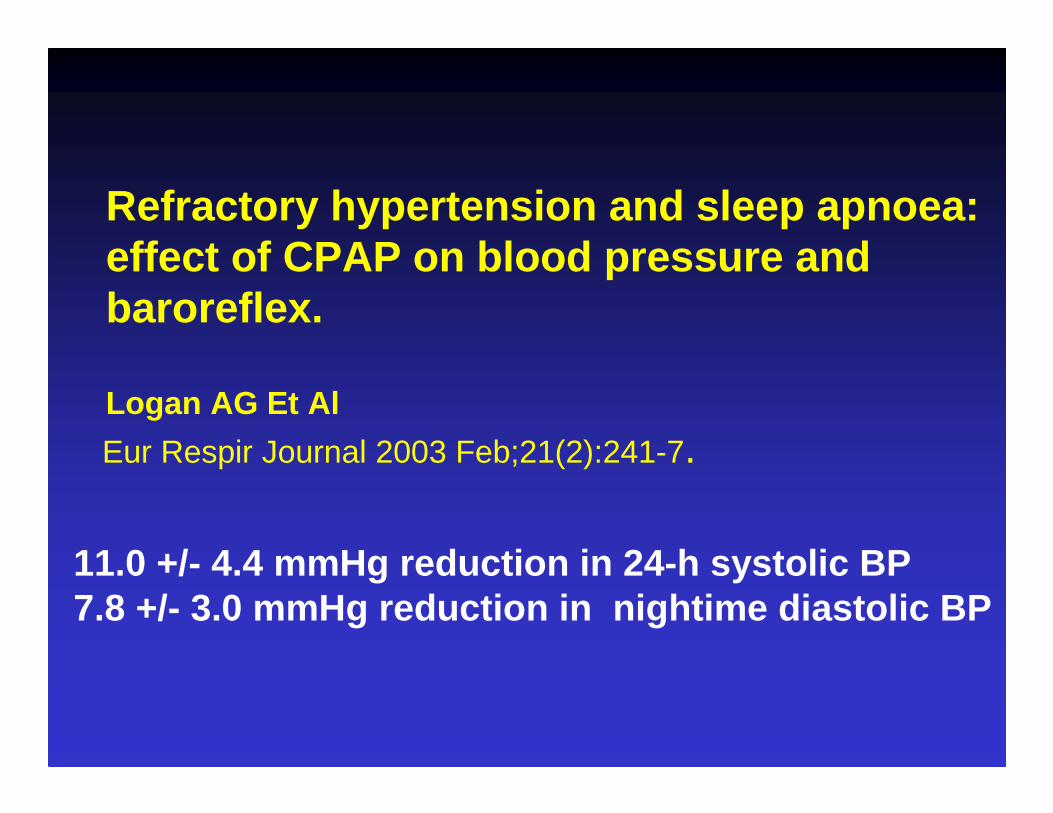
Refractory hypertension and sleep apnoea: effect of CPAP on blood pressure and baroreflex.
Logan AG Et Al
Eur Respir Journal 2003 Feb;21(2):241-7.
11.0 +/- 4.4 mmHg reduction in 24-h systolic BP7.8 +/- 3.0 mmHg reduction in nightime diastolic BP

Pepperell JC,, et al. Ambulatory blood pressure after therapeutic and subtherapeutic nasal continuous positive airway pressure for obstructive sleep apnoea: a randomised parallel trial. Lancet. 2002;359:204-210
Becker HF, Jerrentrup A, Ploch T, et al. Effect of nasal continuous positiveairway pressure treatment on blood pressure in patients with obstructivesleep apnea. Circulation. 2003;107:68-73
“several months of CPAP therapy resulted in a small but significant reduction of daytime blood pressureof between 1.3 and 5.3 mm Hg”

Treatment with continuous positive airwaypressure significantly ameliorated the nocturnalST depression time from 78 min to 33 min (p<0.001)
Nocturnal ischemic events in patients with obstruct ive sleep apnea syndrome and ischemic heart disease: effects of con tinuous positive air pressure treatment.
Peled N, Abinader EG, Pillar G, Sharif D, Lavie Am Coll Cardiol. 1999 Nov 15;34(6):1744-9

The Cardiomyopathy of Obstructive Sleep Apnea Robert Joseph Thomas, MD Annals of Internal Medicine
1 September 1996 | Volume 125 Issue 5 | Page 425
Cardiovascular Effects of Continuous Positive Airwa y Pressure in Patients with Heart Failure and Obstructive Slee p Apnea
Kaneko et al. 348 (13): 1233 NEJM, March 27, 2003
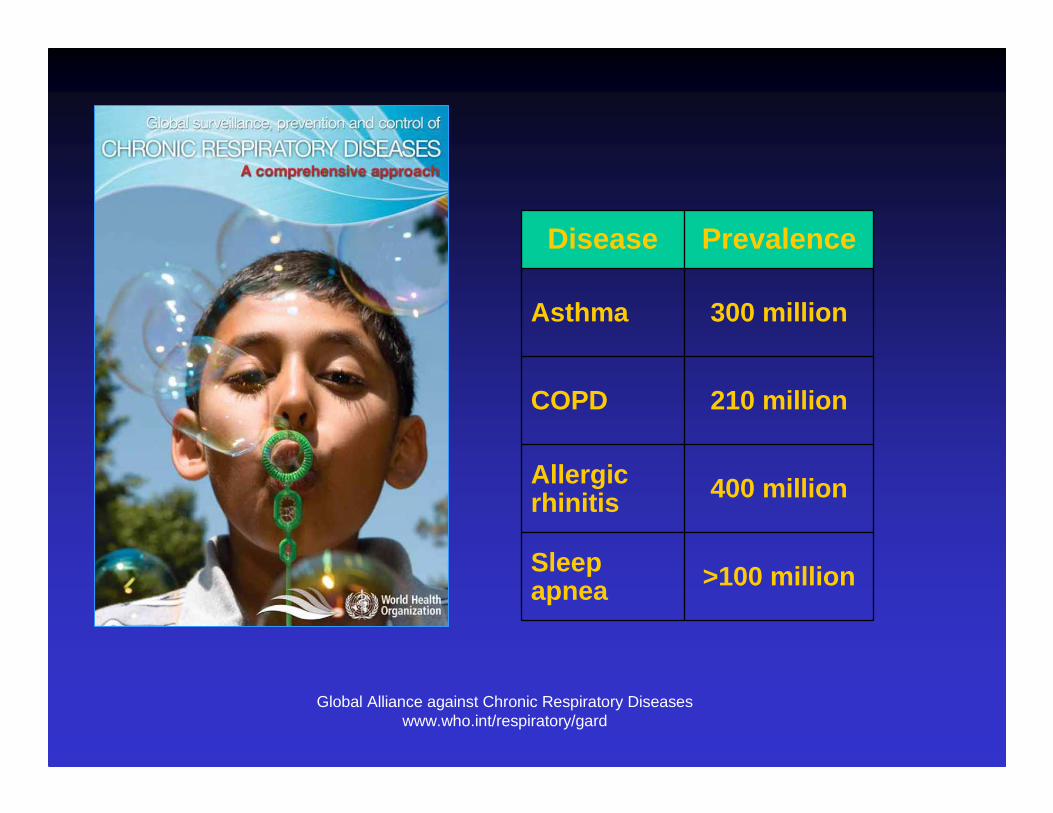
Global Alliance against Chronic Respiratory Diseaseswww.who.int/respiratory/gard
Disease Prevalence
Asthma 300 million
COPD 210 million
Allergic rhinitis 400 million
Sleep apnea >100 million
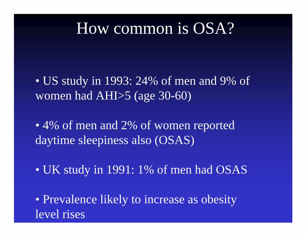
How common is OSA?
• US study in 1993: 24% of men and 9% ofwomen had AHI>5 (age 30-60)
• 4% of men and 2% of women reporteddaytime sleepiness also (OSAS)
• UK study in 1991: 1% of men had OSAS
• Prevalence likely to increase as obesitylevel rises

Ferini-Strambi L et al, 2004
• Minimally symptomatic or asymptomatic OSA Minimally symptomatic or asymptomatic OSA Minimally symptomatic or asymptomatic OSA Minimally symptomatic or asymptomatic OSA is estimated to occur in 1 of 5 adultsis estimated to occur in 1 of 5 adultsis estimated to occur in 1 of 5 adultsis estimated to occur in 1 of 5 adults
• OSA with daytime impairment (OSA OSA with daytime impairment (OSA OSA with daytime impairment (OSA OSA with daytime impairment (OSA syndrome) occur in 1 of 20 adults and is rarely syndrome) occur in 1 of 20 adults and is rarely syndrome) occur in 1 of 20 adults and is rarely syndrome) occur in 1 of 20 adults and is rarely recognizedrecognizedrecognizedrecognized
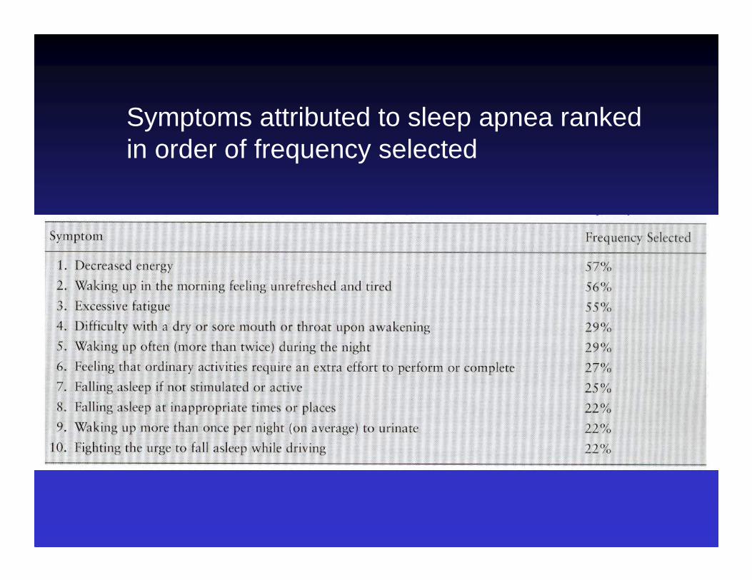
Symptoms attributed to sleep apnea ranked in order of frequency selected
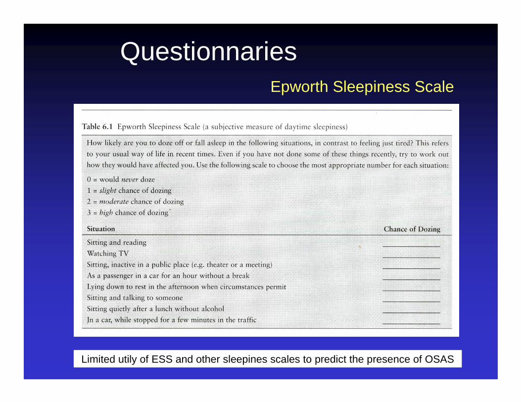
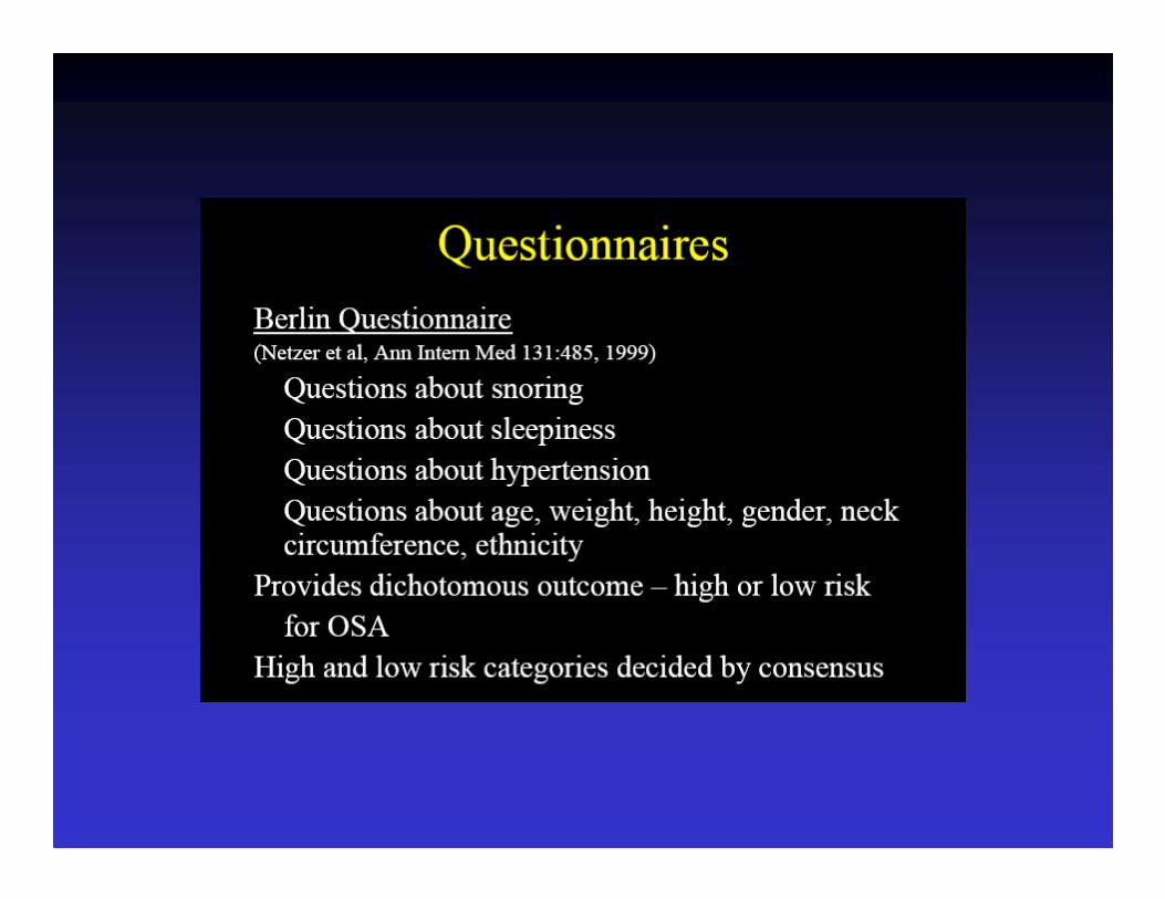
QuestionnariesEpworth Sleepiness Scale
Limited utily of ESS and other sleepines scales to predict the presence of OSAS

Clinical Impressions
Upper airways abnormalities
Morphometric measurements
High-arched palateLarge tongheTonsillar hypertrophyRedundant soft palatal tissueRetrognathiaMicrognathiaAllergic Rhinitis Features
Measuring neck sizePerforming skin fold thickness measurements

Assessment of the Upper Airways
Direct visualisationEndoscopyRhinometryRhinomanometryImaging

Clinical Impressions
Bed partner report of apnea an snoring:Sensitivity 78%Specificity 64%Positive predictive value 64%
Kapuniai et al Sleep 1988
History + Physical examination: sensitivity 50%
Hoffstein & Szalai Sleep 1993

Integration of Multiple Factors
Witnessed apneaSnoringNocturnal chokingExcessive daytime sleepinessMotor vehicle accidentsMale sexObesityHypertension
80%
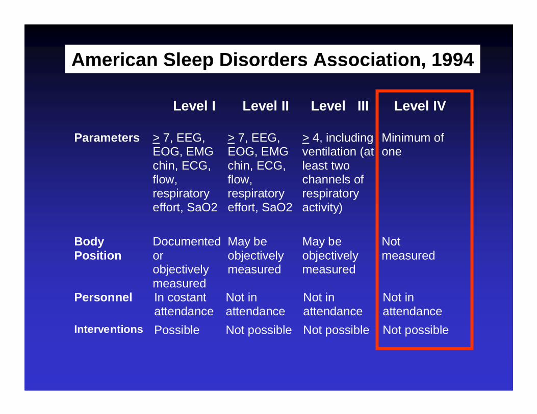
American Sleep Disorders Association, 1994
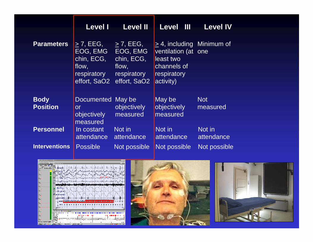
Level I Level II Level III Level IV
Parameters > 7, EEG,EOG, EMGchin, ECG,flow,respiratoryeffort, SaO2
> 7, EEG,EOG, EMGchin, ECG,flow,respiratoryeffort, SaO2
> 4, includingventilation (atleast twochannels ofrespiratoryactivity)
Minimum ofone
BodyPosition
Documentedorobjectivelymeasured
May beobjectivelymeasured
May beobjectivelymeasured
Notmeasured
Personnel In costantattendance
Not inattendance
Not inattendance
Not inattendance
Interventions Possible Not possible Not possible Not possible
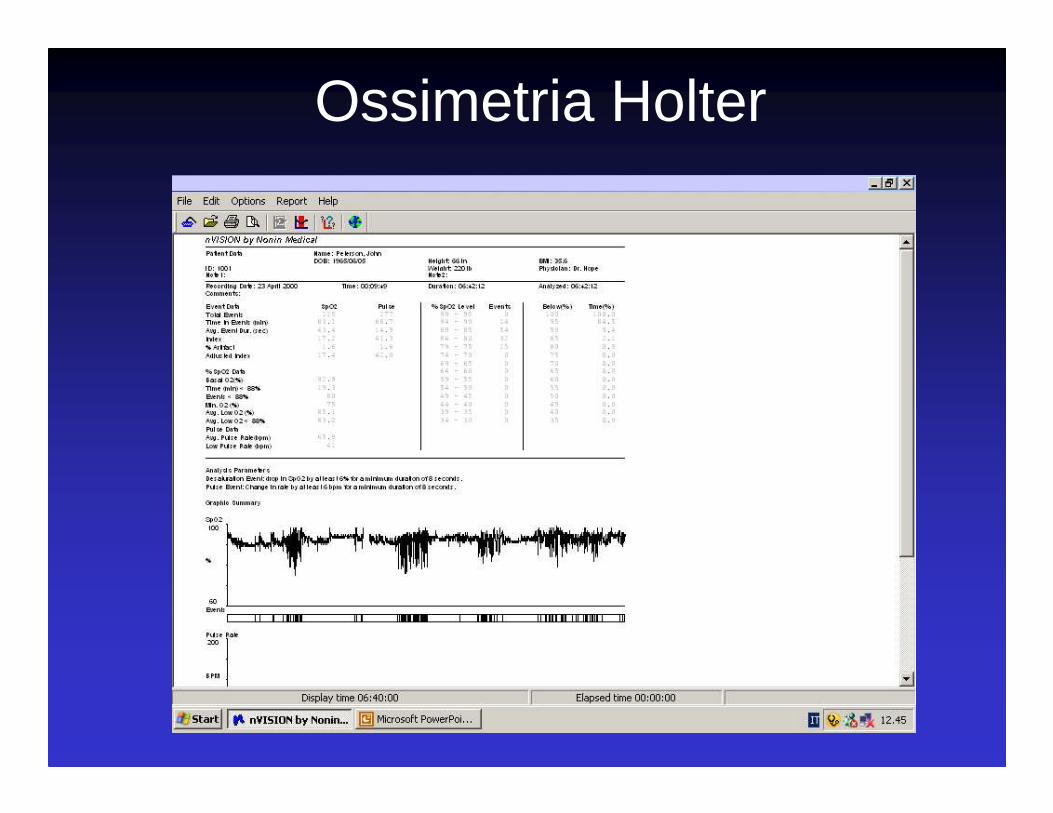
Ossimetria Holter
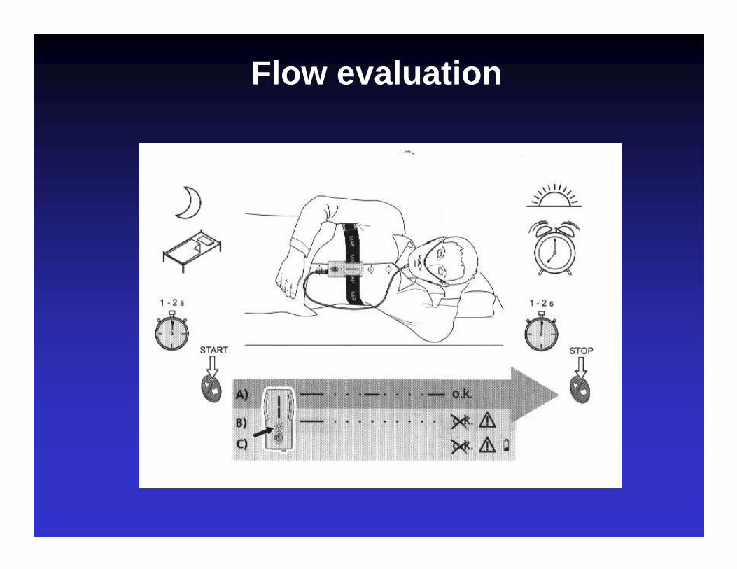
Flow evaluation
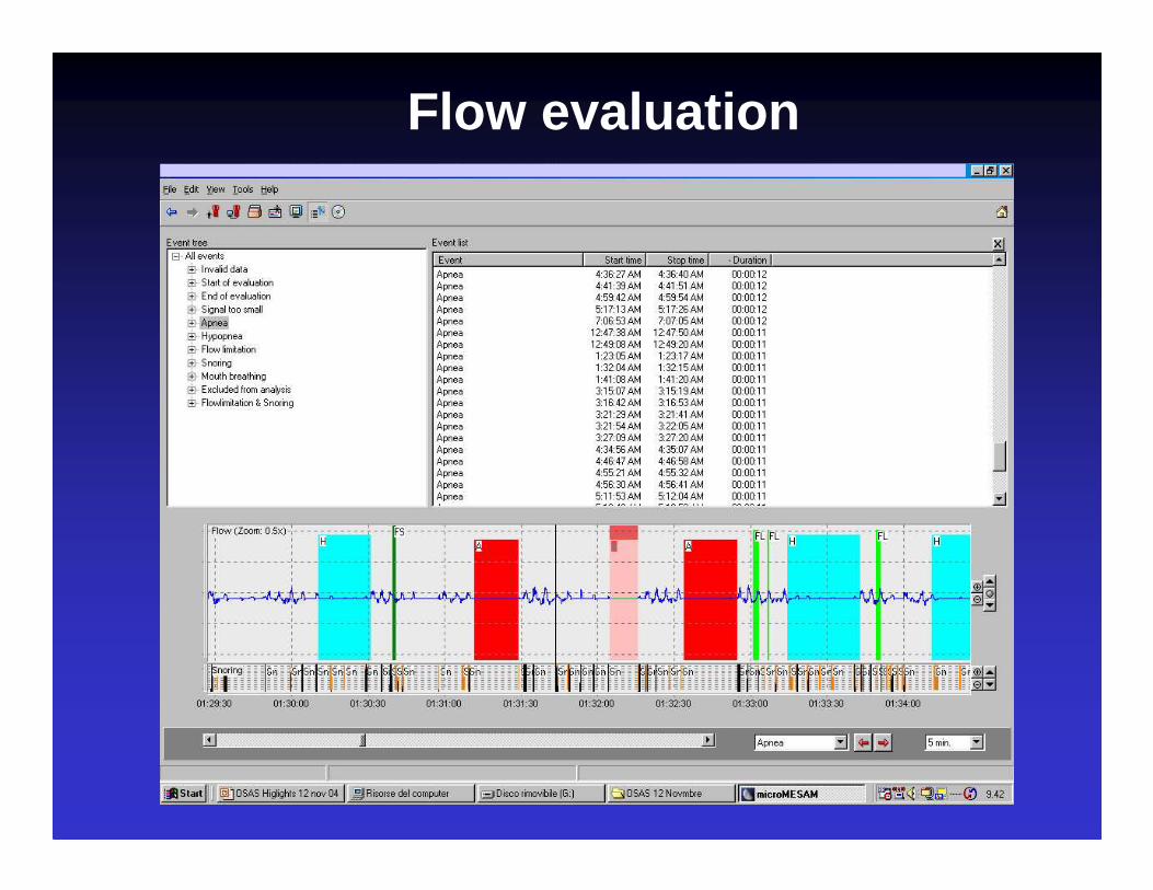
Flow evaluation
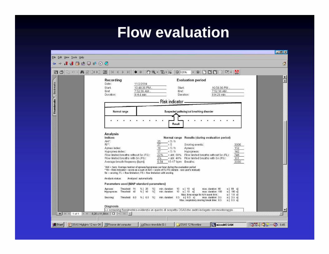
Flow evaluation

Level I Level II Level III Level IV
Parameters > 7, EEG,EOG, EMGchin, ECG,flow,respiratoryeffort, SaO2
> 7, EEG,EOG, EMGchin, ECG,flow,respiratoryeffort, SaO2
> 4, includingventilation (atleast twochannels ofrespiratoryactivity)
Minimum ofone
BodyPosition
Documentedorobjectivelymeasured
May beobjectivelymeasured
May beobjectivelymeasured
Notmeasured
Personnel In costantattendance
Not inattendance
Not inattendance
Not inattendance
Interventions Possible Not possible Not possible Not possible

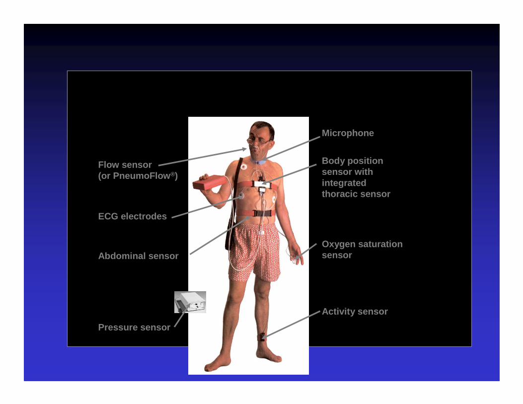
Flow sensor(or PneumoFlow ®)
ECG electrodes
Activity sensor
Oxygen saturation sensor
Body position sensor with integrated thoracic sensor
Microphone
Abdominal sensor
Pressure sensor
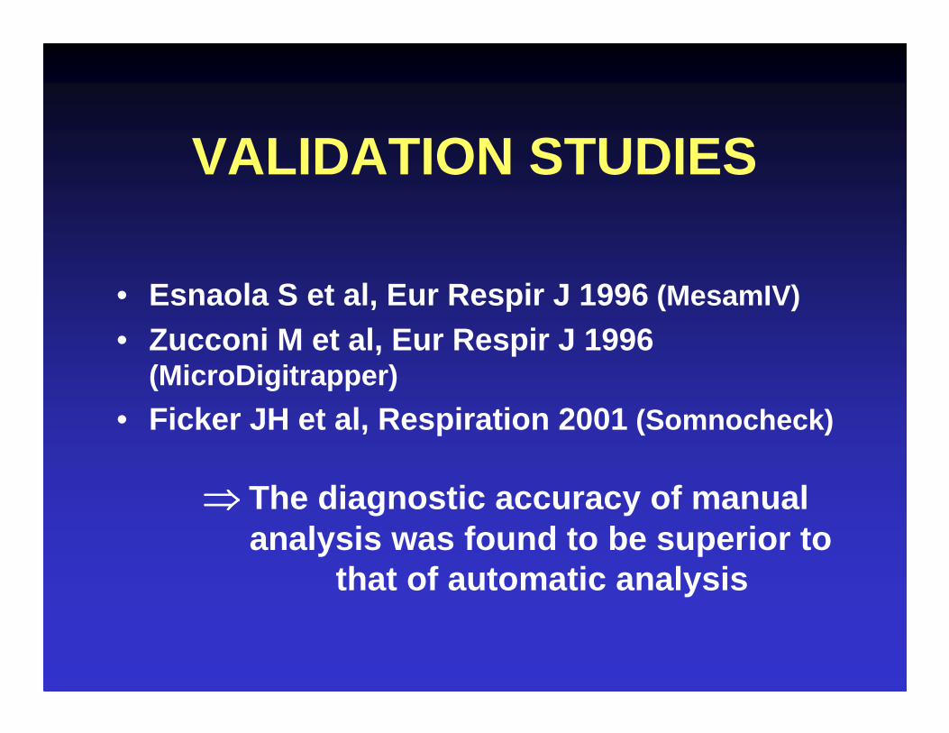
VALIDATION STUDIES
• Esnaola S et al, Eur Respir J 1996 (MesamIV)
• Zucconi M et al, Eur Respir J 1996(MicroDigitrapper)
• Ficker JH et al, Respiration 2001 (Somnocheck)
⇒⇒⇒⇒ The diagnostic accuracy of manual analysis was found to be superior to
that of automatic analysis
Level I Level II Level III Level IV
Parameters > 7, EEG,EOG, EMGchin, ECG,flow,respiratoryeffort, SaO2
> 7, EEG,EOG, EMGchin, ECG,flow,respiratoryeffort, SaO2
> 4, includingventilation (atleast twochannels ofrespiratoryactivity)
Minimum ofone
BodyPosition
Documentedorobjectivelymeasured
May beobjectivelymeasured
May beobjectivelymeasured
Notmeasured
Personnel In costantattendance
Not inattendance
Not inattendance
Not inattendance
Interventions Possible Not possible Not possible Not possible
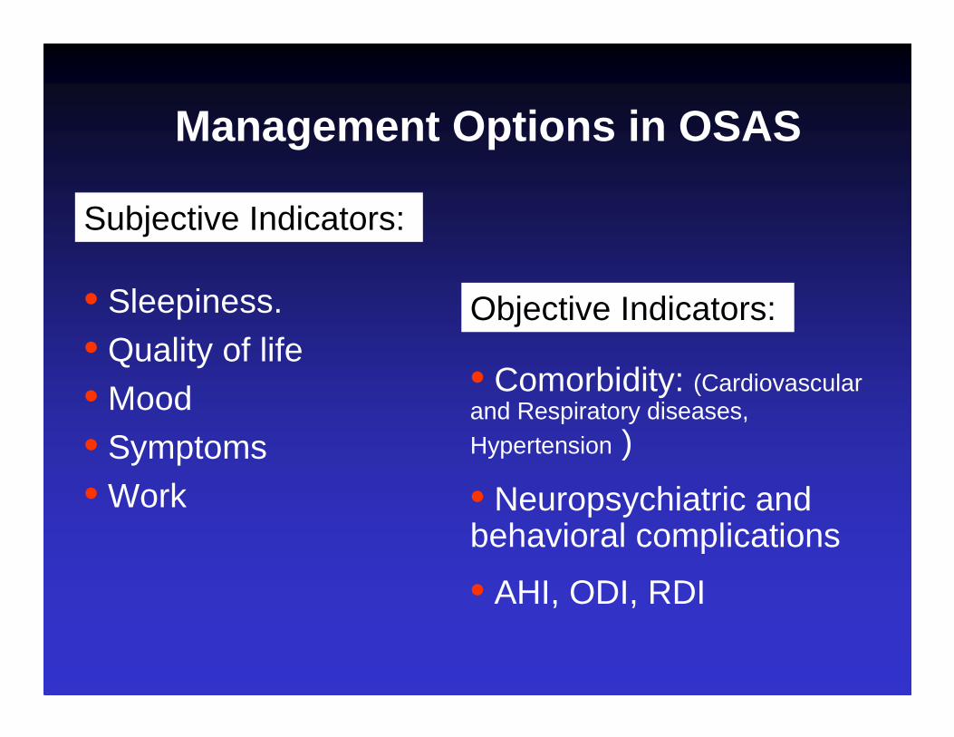
Management Options in OSAS
Subjective Indicators:
• Sleepiness. • Quality of life• Mood
• Symptoms• Work
Objective Indicators:
• Comorbidity: (Cardiovascular and Respiratory diseases, Hypertension )
• Neuropsychiatric and behavioral complications
• AHI, ODI, RDI

CPAP for OSAS
• AHI >20 with or without symptoms
• AHI 5-19 with sleepiness, behavioral complications
Modified ACCP Statement
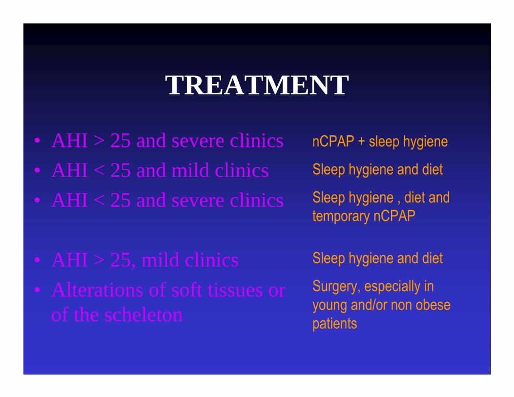
TREATMENT
• AHI > 25 and severe clinics
• AHI < 25 and mild clinics
• AHI < 25 and severe clinics
• AHI > 25, mild clinics
• Alterations of soft tissues or of the scheleton
nCPAP + sleep hygiene
Sleep hygiene and diet
Sleep hygiene , diet and
temporary nCPAP
Sleep hygiene and diet
Surgery, especially in
young and/or non obese
patients
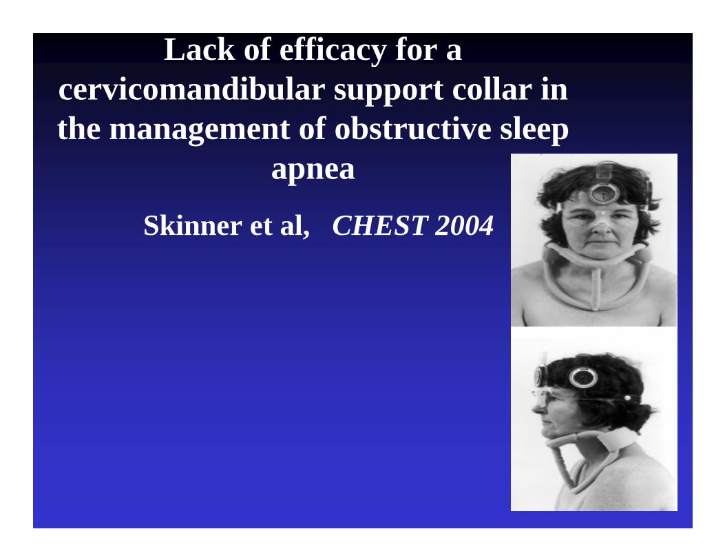
Lack of efficacy for a cervicomandibular support collar in the management of obstructive sleep
apnea
Skinner et al, CHEST 2004
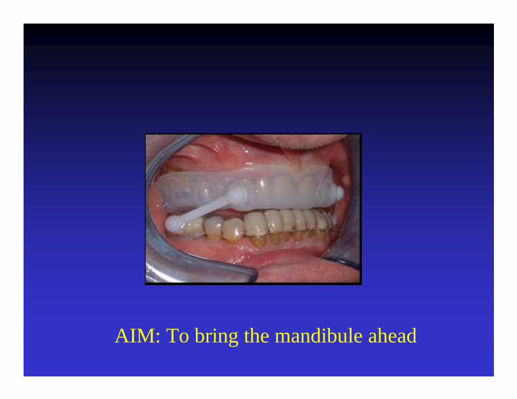
AIM: To bring the mandibule ahead

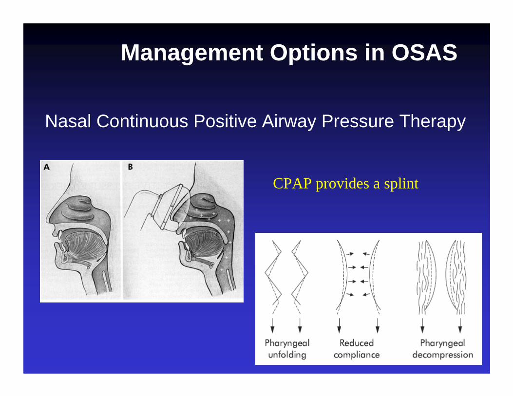
Management Options in OSAS
Nasal Continuous Positive Airway Pressure Therapy
CPAP provides a splint
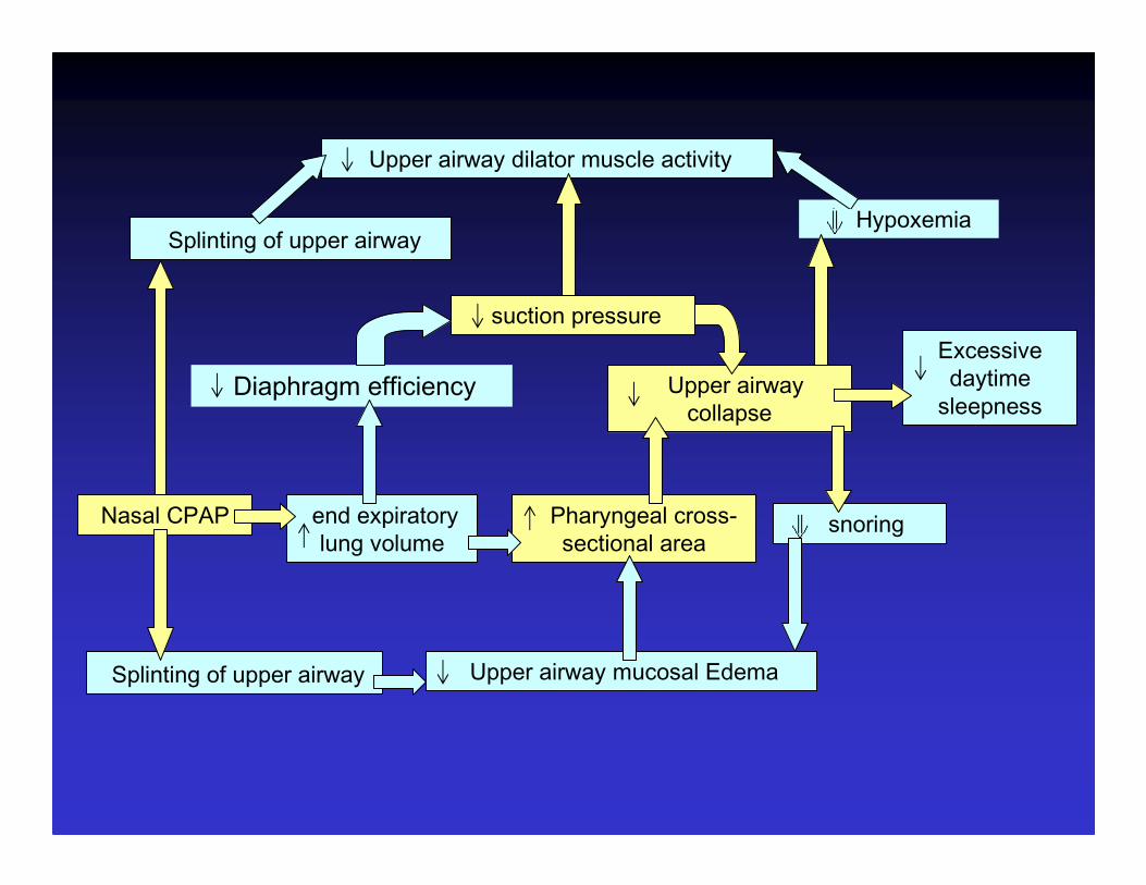
snoring
Upper airway dilator muscle activity
suction pressure
Splinting of upper airwayHypoxemia
Nasal CPAP
Diaphragm efficiency
end expiratory lung volume
Upper airway collapse
Pharyngeal cross-sectional area
Excessive daytime
sleepness
Upper airway mucosal EdemaSplinting of upper airway
0
10
20
30
40
50
60
Placebo CPAP
RDI
N°di risvegli
P<0,001
P<0,001
0
10
20
30
40
50
60
Placebo CPAP
Baseline1 day7 days
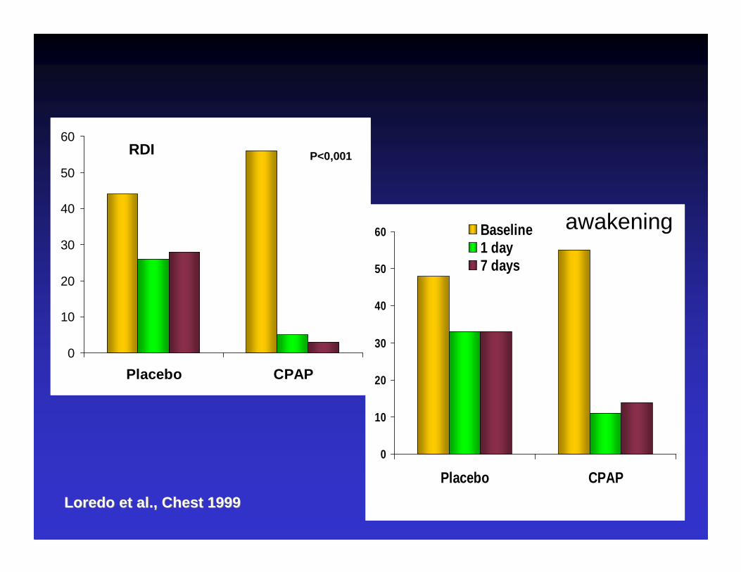
Loredo et al., Chest 1999Loredo et al., Chest 1999
awakening
0
5
10
15
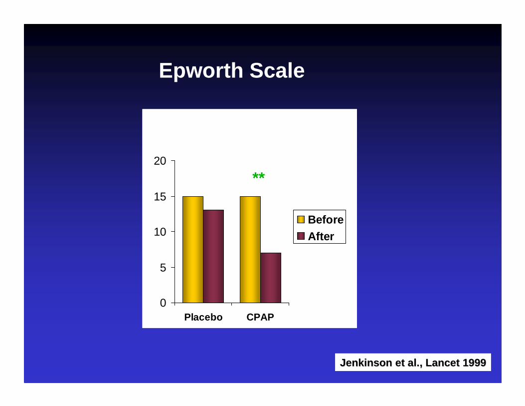
20
Placebo CPAP
BeforeAfter
****
Jenkinson et al., Lancet 1999Jenkinson et al., Lancet 1999
Epworth Scale
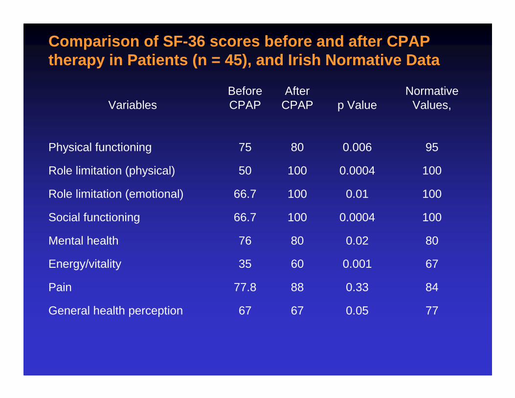
Comparison of SF-36 scores before and after CPAP therapy in Patients (n = 45), and Irish Normative D ata
VariablesBefore CPAP
After CPAP p Value
Normative Values,
Physical functioning 75 80 0.006 95
Role limitation (physical) 50 100 0.0004 100
Role limitation (emotional) 66.7 100 0.01 100
Social functioning 66.7 100 0.0004 100
Mental health 76 80 0.02 80
Energy/vitality 35 60 0.001 67
Pain 77.8 88 0.33 84
General health perception 67 67 0.05 77

SFSF--3636
0
10
20
30
40
50
60
Placebo CPAP
Prima
Dopo
****
Mental Componenent Summary
Mental Componenent Mental Componenent SummarySummary
Physical Component Summary
Physical Component Physical Component SummarySummary
0
10
20
30
40
50
Placebo CPAP
Jenkinson et al. Lancet 1999Jenkinson et al. Lancet 1999
****

Impact of Nasal Continuous Positive Airway Pressure Therapy on the Quality of Life of Bed Partners of Patients With Obstructive Sleep Apnea Syndrome
Doherty LS, Kiely JL, Lawless G, McNicholas WT.
Chest, 2003

0.14 (1–5)4 (2–6)Depression
0.027 (4–8)7 (5–11)Anxiety
0.0072 (1–5)4 (1–8.5)ESS
Partner
0.024 (2–6)5 (3–8)Depression
0.0016 (3–8)8 (5–9)Anxiety
0.0018 (4–15)16 (11–20)ESS
Patient
p ValueAfter CPAPBefore CPAPVariables
Comparison between Partners ESS scores before and after CPAP therapy (n=45)

0
10
20
30
40
50
60
70
Baseline after 1 month After onenight withoutCPAP CPAP
AHI
Kribbs et al. Am Rev Respir Dis 1993
AHIAHIAHI

Daytime SleepenessDaytime SleepenessDaytime Sleepeness
0
2
4
6
8
10
12
14
Baseline 3 months 12 months
Munoz et al. Eur Respir J 2000Munoz et al. Eur Respir J 2000
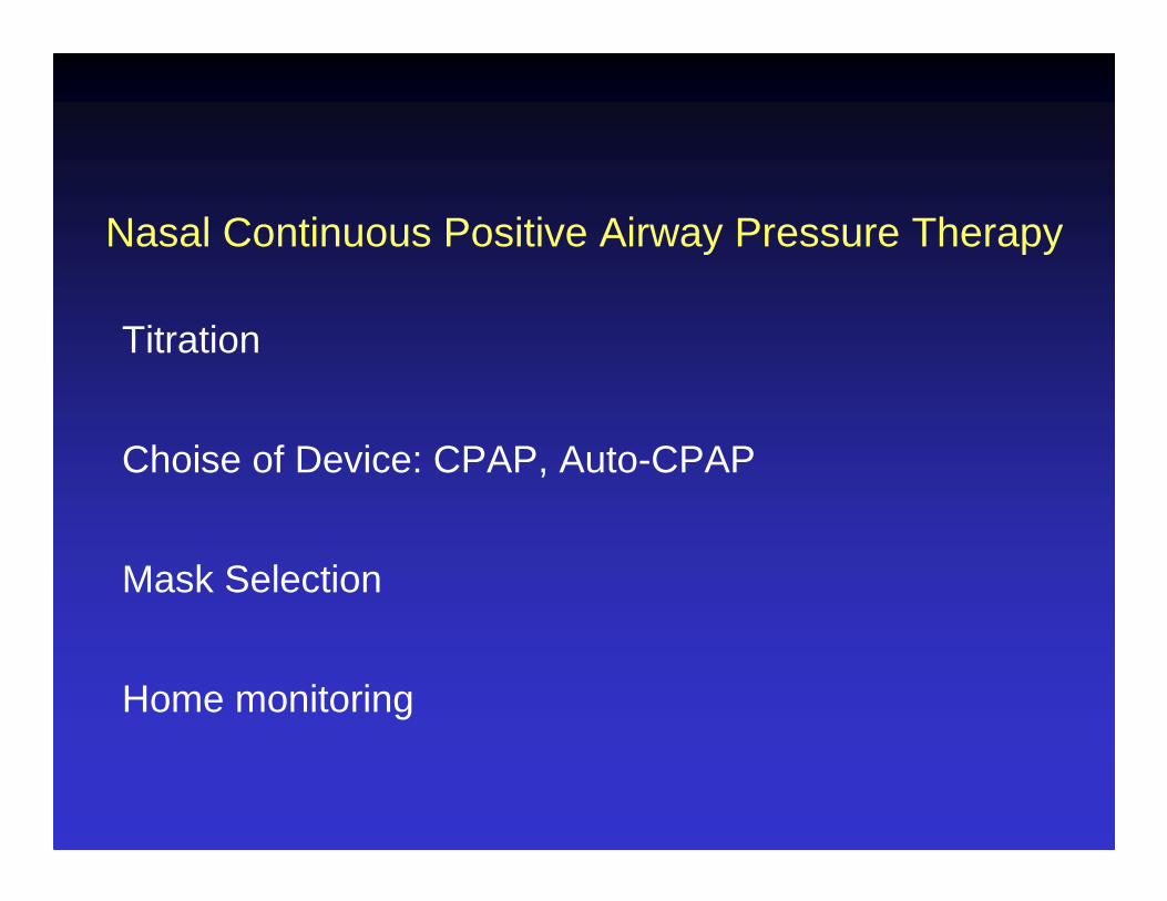
Titration
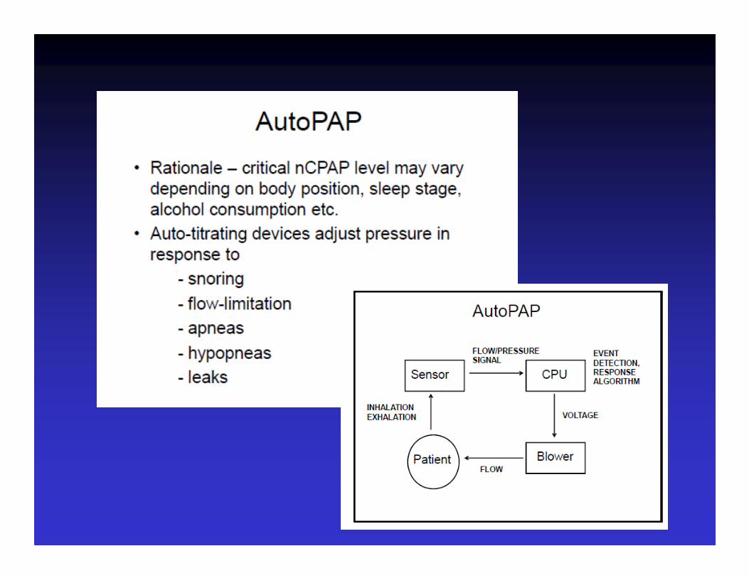
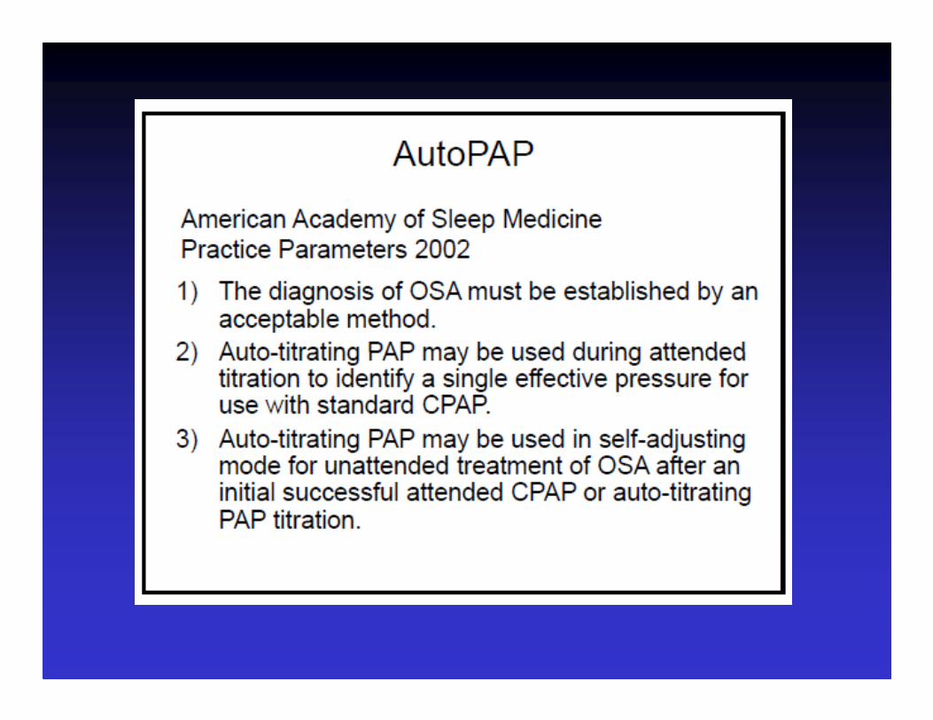
Choise of Device: CPAP, Auto-CPAP
Mask Selection
Home monitoring
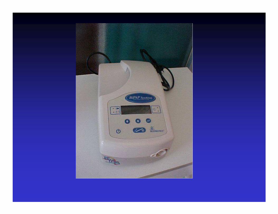
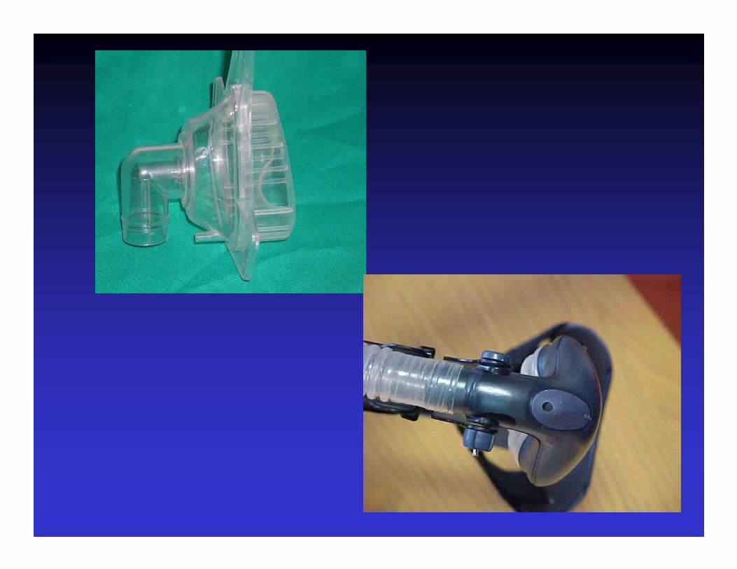
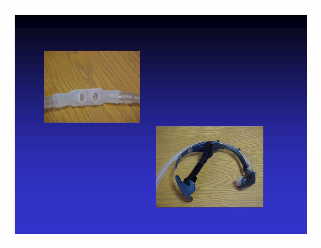
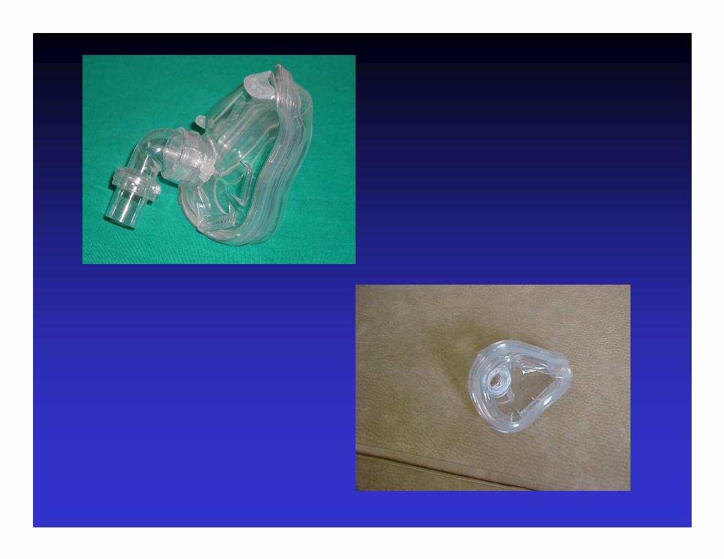
Nasal Continuous Positive Airway Pressure Therapy
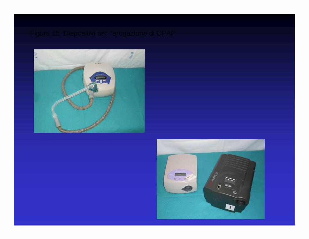
Figura 15. Dispositivi per l’erogazione di CPAP


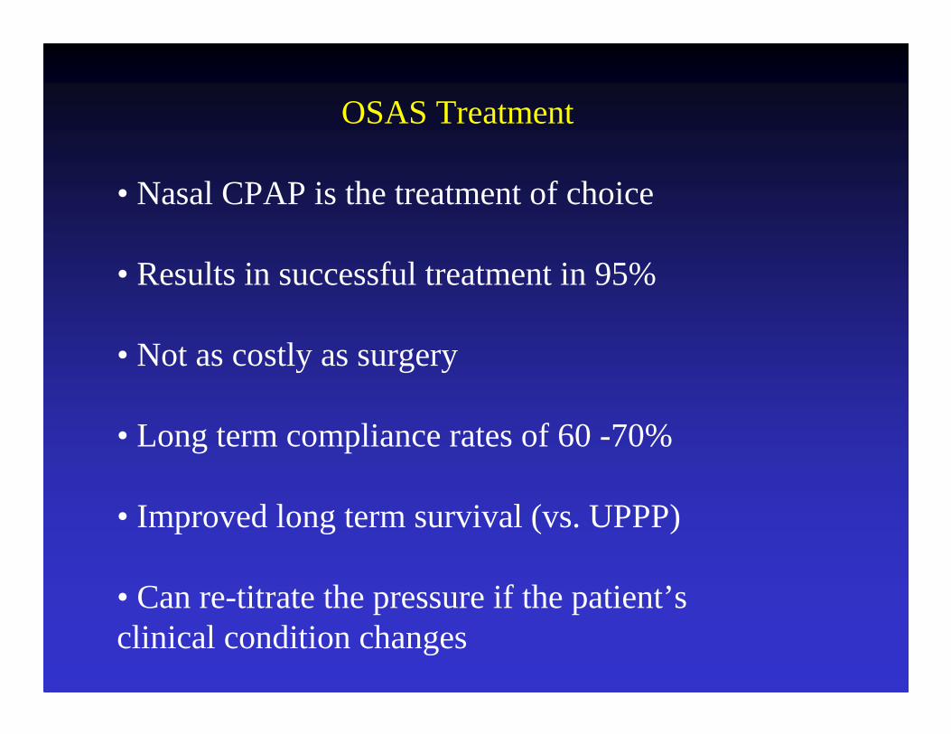
OSAS Treatment
• Nasal CPAP is the treatment of choice
• Results in successful treatment in 95%
• Not as costly as surgery
• Long term compliance rates of 60 -70%
• Improved long term survival (vs. UPPP)
• Can re-titrate the pressure if the patient’sclinical condition changes

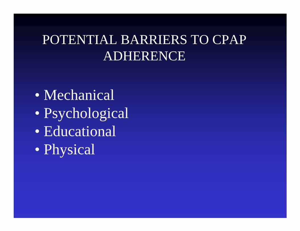
POTENTIAL BARRIERS TO CPAP ADHERENCE
• Mechanical• Psychological• Educational• Physical
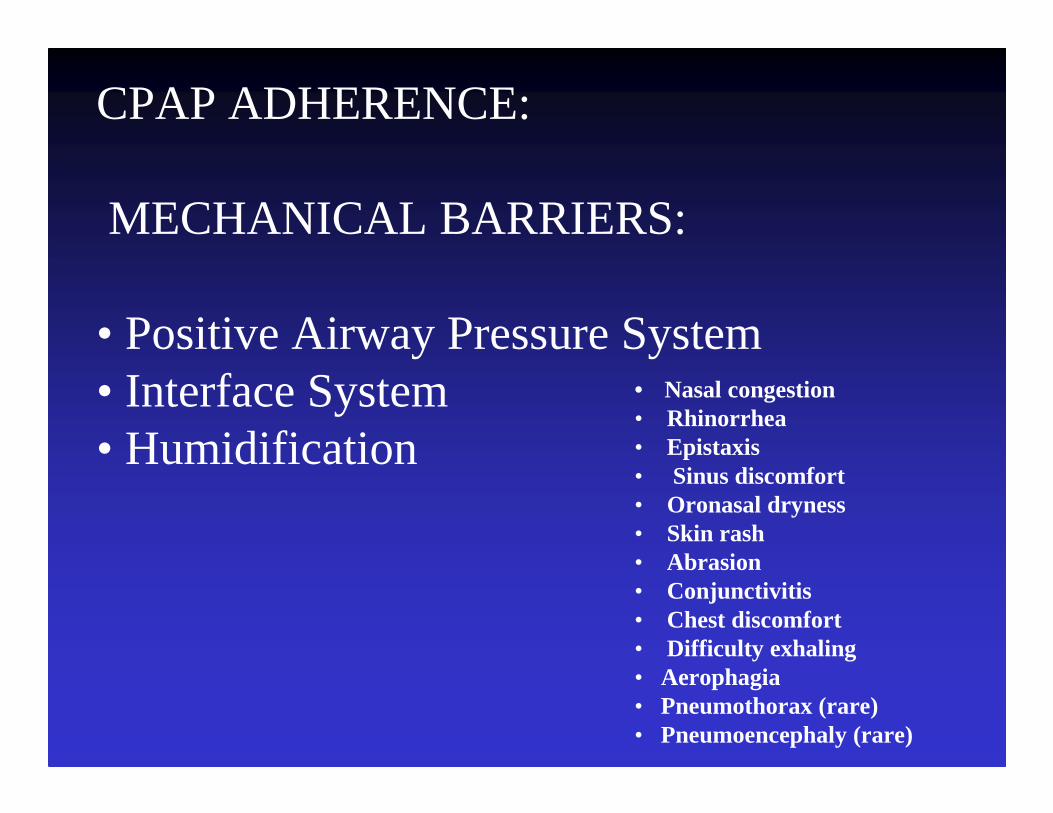
CPAP ADHERENCE:
MECHANICAL BARRIERS:
• Positive Airway Pressure System• Interface System• Humidification
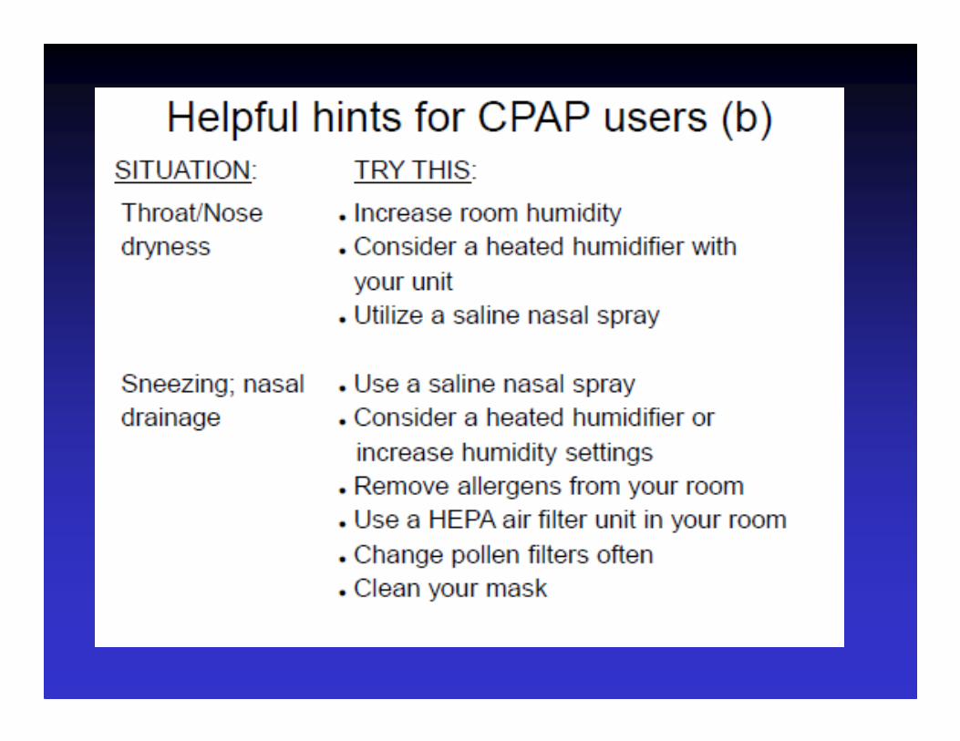
• Nasal congestion• Rhinorrhea• Epistaxis• Sinus discomfort• Oronasal dryness• Skin rash• Abrasion• Conjunctivitis• Chest discomfort• Difficulty exhaling• Aerophagia• Pneumothorax (rare)• Pneumoencephaly (rare)

CPAP ADHERENCE:
PSYCHOLOGICAL BARRIERS• Claustrophobic• Embarrassment• Vanity• Personality Type Disorders• Support System

CPAP ADHERENCE:
EDUCATIONAL BARRIERS
• Comprehension• Patient’s knowledge of obstructive sleep apnea• Patient’s knowledge of interface system• Patient’s knowledge of delivery system

CPAP COMPLIANCE:
PHYSICAL BARRIERS
• Physical Handicaps• Extreme Obesity• Nasal Dryness• Stomach Distention• Retrognathic• Under Treated
• Over Treated• Sinus Problems• Puffing• Ear Discomfort• Eye Irritation• Material Sensitivity

To explain better the pathophysiologyof obstructive sleep apnea and its consequences
To emphasize the magnitude of the clinical problem
To make familiar the practical aspects of the treatmenttechniques
To identify unmet needs
To make familiar the practical aspects of the treatmenttechniques
CPAP and sleep disorders
Workshop
Fulvio Braido Allergy and Respiratory Diseases Department
University of Genoa