CP-673451 inhibits the formation of neutrophil ...
108
From the Department of Infectious Diseases and Microbiology of the University of Lübeck Director: Prof. Dr. med. Jan Rupp CP-673451 inhibits the formation of neutrophil extracellular traps induced by PMA and immobilized immune complexes Dissertation for Fulfillment of Requirements for the Doctoral Degree of the University of Lübeck from the Department of Natural Sciences Submitted by Gigy Varghese From Mahé, India Lübeck 2017
Transcript of CP-673451 inhibits the formation of neutrophil ...

From the Department of Infectious Diseases and Microbiology
of the University of Lübeck
Director: Prof. Dr. med. Jan Rupp
CP-673451 inhibits the formation of neutrophil extracellular
traps induced by PMA and immobilized immune complexes
Dissertation
for Fulfillment of
Requirements
for the Doctoral Degree
of the University of Lübeck
from the Department of Natural Sciences
Submitted by
Gigy Varghese
From Mahé, India
Lübeck 2017

First referee: Prof. Dr. Tamás Laskay
Second referee: Prof. Dr. Hauke Busch
Date of oral examination: 23.11.2017
Approved for printing: Lübeck, 28.11.2017

This work is dedicated to my beloved mother, Alice Varghese
1
Table of Contents
Table of Contents ............................................................................................................................. 1
List of Abbreviations ...................................................................................................................... 5
Summary ............................................................................................................................................ 7
Zusammenfassung .......................................................................................................................... 9
Chapter 1. Introduction............................................................................................................... 11
1.1 Neutrophil granulocytes ................................................................................................................. 11
1.2 Neutrophil activation and extravasation ................................................................................ 11
1.3 Degranulation ..................................................................................................................................... 12
1.4 Phagocytosis ........................................................................................................................................ 13
1.5 Cytokine production ......................................................................................................................... 13
1.6 Reactive oxygen species (ROS) production ............................................................................. 14
1.7 Neutrophil extracellular trap formation (NETosis) ........................................................... 15
1.7.1 NETosis: a unique cell death .............................................................................................. 16
1.7.2 NET inducers ............................................................................................................................ 16
1.7.3 Anti-microbial activity of NETs......................................................................................... 17
1.7.4 NETosis associated- autoimmune diseases ................................................................. 17
1.7.5 ROS-dependent NETosis...................................................................................................... 18
1.7.6 ROS-independent NETosis.................................................................................................. 21
1.8 Platelet-derived growth factor receptors (PDGFRs) .......................................................... 23
1.9 CP-673451: inhibitor molecule targeting PDGFRs .............................................................. 24
Objectives of the Study ................................................................................................................ 26
Chapter 2. Materials and Methods .......................................................................................... 27

2
2.1 Materials ...................................................................................................................................................... 27
2.1.2 Solutions, buffers and media ................................................................................................ 27
2.1.3 Laboratory supplies ................................................................................................................ 28
2.1.4 Chemicals and reagents ......................................................................................................... 29
2.1.5 Other selective inhibitors ...................................................................................................... 31
2.1.6 Ready to use kits ....................................................................................................................... 31
2.1.7 Antibodies ................................................................................................................................... 31
2.1.8 Cytokines ..................................................................................................................................... 32
2.1.9 Cell lines/cell line lysate ........................................................................................................ 32
2.1.10 Instruments .............................................................................................................................. 33
2.1.11 Software .................................................................................................................................... 34
2.2 Methods ........................................................................................................................................................ 35
2.2.1 Neutrophil isolation from human blood .......................................................................... 35
2.2.2 Culturing U87-MG Cell lines ................................................................................................. 35
2.2.3 Cytospin and Diff-Quick staining ........................................................................................ 35
2.2.4 Preparation of immobilized immune complexes ......................................................... 36
2.2.5 Detection of intra- and extracellular ROS ....................................................................... 36
2.2.6 SytoxGreen detection of NETs ............................................................................................. 37
2.2.7 Fluorescent microscopy for NETs ...................................................................................... 37
2.2.8 Neutrophil activation assay .................................................................................................. 38
2.2.9 Neutrophil migration assay .................................................................................................. 39
2.2.10 Neutrophil phagocytosis assay ......................................................................................... 39
2.2.11 Western blot analysis ........................................................................................................... 40
2.2.12 Phospho-enrichment of threonine phosphorylated proteins .............................. 41
2.2.13 Assessment of viability by Annexin V-PI staining ..................................................... 42
2.2.14 LDH-Cytotoxicity assay........................................................................................................ 43
2.2.15 RNA isolation from human neutrophils ........................................................................ 44
2.2.16 RNA quantification ................................................................................................................ 44
2.2.17 RNA Seq ..................................................................................................................................... 45
2.2.18 RNA Seq data analyses ......................................................................................................... 45
2.2.19 Statistical analysis ................................................................................................................. 45
Chapter 3. Results ......................................................................................................................... 46

3
3.1 Identification of inhibitors which do not inhibit ROS release but inhibit NETosis ...... 46
3.2 Dose dependent inhibition of PMA- and iIC-induced NET formation by CP-673451 .. 51
3.3 Effect of Doxorubicin in PMA- and iIC-induced NETosis ......................................................... 56
3.4 Screening for potential targets of CP-673451 involved in NETosis inhibition .............. 61
3.5 Effects of Sunitinib on PMA- and iIC-induced ROS and NETosis .......................................... 63
3.6 Neutrophils do not express PDGF-Receptor ................................................................................. 65
3.7 CP-673451 does not block upstream pathways of PMA-induced ROS production
involved in NETosis......................................................................................................................................... 66
3.8 CP-673451 does not block upstream pathways of iIC-induced ROS production
involved in NETosis......................................................................................................................................... 68
3.9 CP-673451 treatment leads to phosphorylation of Akt, ERK1/2, and P38 MAPK ....... 69
3.10 Screening for changes in tyrosine, serine, and threonine phosphorylated proteins
upon CP-673451 treatment ........................................................................................................................ 70
3.11 CP-673451 does not inhibit Ionomycin induced-NETosis .................................................... 72
3.12 CP-673451 does not induce apoptosis or necrosis in neutrophils .................................... 74
3.13 CP-673451 does not affect neutrophil effector functions other than NETosis ........... 75
3.13.1 CP-673451 does not affect neutrophil phagocytic ability ................................................ 76
3.13.2 The effect of CP-673451 on neutrophil migration............................................................... 77
3.13.3 CP-673451 does not inhibit neutrophil activation .............................................................. 78
3.14 CP-673451 activates neutrophils ................................................................................................... 79
3.15 The neutrophil-activating property of CP-673451 is not due to endotoxin-
contamination .................................................................................................................................................. 79
3.16 CP-673451 induces differential gene expression changes in human neutrophils ..... 80
Chapter 4. Discussion................................................................................................................... 83
4.1 CP-673451 induces neutrophil activation and inhibits an unknown pathway of
NETosis, downstream of ROS production ............................................................................................. 83

4
Bibliography ................................................................................................................................... 93
Supplementary data .................................................................................................................. 101
I. Target-selective inhibitor library from Selleckchem (Houston, USA) ................ 101
Acknowledgements ................................................................................................................... 104
Curriculum Vitae ........................................................................................................................ 105
5
List of Abbreviations
Ab Antibody
AUC Area under the curve
BSA Bovine serum albumin
CD cluster of differentiation
CGD Chronic granulomatous disease
DMSO Dimethyl sulfoxide
DNA Deoxyribonucleic acid
DNA Deoxyribonucleic acid
DOX Doxorubicin
DPI diphenyleneiodonium
ELISA Enzyme linked immuno sorbent assay
Erk1/2 Extracellular signal–regulated kinases 1/2
FCS Fetal calf serum
FITC Fluorescein isothiocyanate
FM Fluorescent microscopy
fMLP N-Formyl methionyl-leucyl-phenylalanine
HSA Human serum albumin
IC Immune complex
IFN-γ Interferon gamma
iIC Immobilized immune complexes
IL Interleukin
IO Ionomycin
LDH Lactate dehydrogenase
LPS Lipopolysaccharide
MAPK Mitogen-activated protein kinases
MPO Myeloperoxidase
NADPH Nicotinamide adenine dinucleotide phosphate
NE Neutrophil elastase
NETs Neutrophil extracellular traps
NOX NADPH oxidase
OD Optical density
6
PBMCs Peripheral blood mononuclear cells
PBS Phosphate buffered saline
pDCs Plasmacytoid dendritic cells
PDGF Platelet derived growth factor
PDGFR Platelet derived growth factor receptor
PE Phycoerythrin
PHOX Phagocytic oxidase.
PI Propidium iodide
PI3K Phosphatidylinositol-4,5-bisphosphate 3-kinase
PKC Protein kinase C
PKCδ Protein kinase C delta
PMA Phorbol 12-myristate 13-acetate
PMB Polymyxin B
RA Rheumatoid arthritis
RIN RNA integrity number
RNA Ribonucleic acid
ROS Reactive oxygen species
RT Room temperature
SDS Sodium dodecyl sulfate
SLE Systemic lupus erythematosus
SOD Superoxide dismutase
TCA Trichloro acetic acid
TLR Toll-like receptor
TNF Tumor necrosis factor
VEGF Vascular endothelial growth factor
VEGFR1/2 Vascular endothelial growth factor receptor-1/2
7
Summary
Neutrophils are essential cells of the innate antimicrobial defense. A recently identified
anti-microbial function is the formation of Neutrophil Extracellular Traps (NETs) that
are composed of decondensed chromatin and antimicrobial granular contents. NETs
can capture and kill invading pathogens. However, the granule proteins on the NETs can
also serve as auto-antigens leading to autoimmunity. NETs are released from
neutrophils undergoing a unique kind of cell death the so called NETosis. The
mechanisms involved in NETosis are poorly understood. In the present study, the
signaling mechanisms involved in NETosis were investigated.
NETosis was induced with PMA and immobilized immune complexes (iIC) which both
induce Reactive Oxygen Species (ROS) dependent NETosis. A library of 141 inhibitors of
known signaling pathways was screened for molecules that inhibit the formation of
NETs but do not inhibit ROS production to identify a NETosis inhibitor, downstream of
ROS production. Through the screening, I identified a potent inhibitor of NETosis, CP-
673451 which inhibited NETosis induced by PMA and iIC. CP-673451 was found to
inhibit the formation of both PMA- and iIC-induced NETs in a dose dependent manner
without inhibiting ROS release. CP-673451 treatment did not inhibit PMA induced
phosphorylation of PKC, p38 MAPK, ERK1/2, and Akt. The iIC-induced phosphorylation
of Src, p38 MAPK, PI3K, Akt, and ERK1/2 was also not affected by CP-673451. These
findings suggest that CP-673451 exerts its inhibitory effect downstream of these
pathways and ROS production. However, the inhibitor did not inhibit ROS-independent
ionomycin-induced NETosis.
CP-673451 is known to target Platelet-derived growth factor receptor (PDGFR).
However, my experimental results suggest that the inhibitory effect of CP-673451 on
NETosis is independent of action on the known target, PDGFR. In addition, by using
western blots, I could confirm the absence of PDGFR in human neutrophils. CP-673451
is not cytotoxic for neutrophils (up to 10µM) and do not inhibit basic neutrophil
functions such as activation, migration, and phagocytosis. However, TNF-α mediated
random migration was affected by the higher concentration of CP-673451 (above 1µM).
Furthermore, the study revealed that CP-673451 activates certain neutrophil functions
such as the upregulation of CD11b and shedding of CD62L. The activation was not
inhibited by polymyxin treatment, confirming that the activation phenotype was not

8
due to LPS contamination. In support to the surface marker expression results, CP-
673451 induced the phosphorylation of p38 MAPK, ERK1/2, and Akt in human
neutrophils. Although, the study could not identify the molecular target of CP-673451
involved in NET-inhibition, the target of CP-673451 is apparently further downstream
of p38 MAPK, ERK1/2, and Akt pathways. The results suggest that CP-673451 is
activating the neutrophils through the same pathways which are involved in ROS
production but downstream to ROS production it leads to NETosis inhibition through an
unknown mechanism. The preliminary gene expression studies also indicated that CP-
673451 is involved in inducing gene expression changes in neutrophils. However, their
role in inhibition of NETosis needs to be further analyzed. Elucidating the molecular
targets of CP-673451 in neutrophils could lead to the identification of novel signaling
events involved in NET formation.

9
Zusammenfassung
Neutrophile Granulozyten sind essenzielle Zellen der angeborenen antimikrobiellen
Abwehr. Eine kürzlich identifizierte antimikrobielle Funktion dieser Zellen ist die
Ausbildung von extrazellulären Gebilden (NETs, engl. Neutrophil Extracellular Traps),
welche aus dekondensiertem Chromatin und antimikrobiellen Granula bestehen. NETs
können eindringende Pathogene fangen und töten. Allerdings können die granulären
Proteine der NETs auch als Auto-Antigene dienen, welche zu Autoimmunität führen
können. Um NETs freisetzen zu können, durchlaufen Neutrophile Granulozyten eine
einzigartige Form des Zelltodes – der sogenannten NETosis. Die darin involvierten
Mechanismen sind bis zum heutigen Zeitpunkt kaum nachvollzogen. In der
vorliegenden Arbeit, sollen diese Mechanismen untersucht werden.
NETosis wurde durch PMA und immobilisierte Immunkomplexe ausgelöst, welche
beide die Produktion von reaktiven Sauerstoffradikalen (ROS, engl. Reactive Oxigene
Species) iniziieren. Ein Satz von 141 Inhibitoren bekannter Signalwege wurde auf
Moleküle durchsucht, welche die Ausbildung von NETs, aber nicht die Produktion von
ROS beeinträchtigen. Dies geschah, um einen Inhibitor zu identifizieren, welcher die
Ausbildung von NETs jenseits der ROS-Produktion hemmt.
Tatsächlich konnte ich einen potenten Inhibitor der NETosis identifizieren: CP-673451,
welcher die durch PMA und iIC induzierten NET Freisetzung dosierungsabhängig
hemmt, ohne die Ausbildung von ROS zu vermindern. CP-673451 Behandlung
verminderte nicht die PMA induzierte Phosphorilierung von PKC, p38 MAPK, ERK1/2
und Akt. Die iIC-induzierte Phosphorilierung von Src, p38 MAPK, PI3K, Akt und ERK1/2
war ebenfalls nicht durch CP-673451 beeinflusst. Diese Ergebnisse zeigen, dass CP-
673451 seine inhibitorischen Effekte nach der obengenannten Signal wegen und der
ROS-Produktion ausübt. Allerdings konnte dieser Inhibitor nicht die von ROS
unabhängige Induktion von NETs durch Ionomycin verhindern.
Es ist bekannt, dass CP-673451 an den PDGF-Rezeptor (Platelet-derived growth factor
receptor) bindet. In meinen Versuchen konnte ich allerdings zeigen, dass der
inhibierende Effekt von CP-673451 auf die NETose von dem bekannten
Bindungsprotein, dem PDGF-Rezeptor, unabhängig ist. Tatsächlich konnte ich die
Abwesenheit dieses Bindungsproteins in Neutrophilen Granulozyten mit Hilfe von

10
Western Blots beweisen. CP-673451 ist in einer Konzentration bis 1µM nicht
zytotoxisch für Neutrophile Granulozyten und inhibiert keine Grundfunktionen der
Zelle, wie zum Beispiel die Möglichkeit der Aktivierung, Migration oder Phagozytose.
Allerdings war die TNF-α vermittelte Migration beeinträchtigt ab einer
Inhibitorkonzentration über 1µM. Außerdem zeigte meine Studie, dass CP-673451
bestimmte Funktionen der Neutrophilen aktiviert, wie beispielsweise die
Hochregulation von CD11b auf der Zelloberfläche oder den Abbau von CD62L. Die
Aktivierung konnte nicht durch Polymyxin inhibiert werden, was darauf schließen lässt,
dass der aktivierte Phänotyp der Zelle nicht durch eine eventuelle LPS kontamination
ausgelöst wurde.
Zusätzlich zur Veränderung der Zelloberflächenmarker konnte gezeigt werden, dass CP-
673451 die Phosphorylierung von p38 MAP, ERK1/2, und Akt in humanen Neutrophilen
induziert. Obwohl diese Studie nicht das molekulare Ziel von CP-673451 identifizieren
konnte, welches in die NET-inhibition involviert ist, liegt der Angriftspunkt von CP-
673451 vermutlich in der Signalkaskade nach p38 MAPK, ERK1/2 und Akt befinden. So
kann davon ausgegangen werden, dass CP-673451 in den Reaktionswegen, welche zur
ROS-Produktion führen, eine aktivierende Rolle einnimmt, jenseits der ROS Produktion
allerdings für eine Inhibierung der NETose durch einen bisher nicht aufgeklärten
Mechanismus sorgt. Ergebnise der vorangegangenen Genexpressionsanalyse zeigen
auch, dass CP-673451 in der Veränderung der Genexpression von Neutrophilen
beteiligt ist. Jedoch muss seine Rolle in der Inhibition von NETose weiter untersucht
werden.
Die Zielproteine für CP-673451 in humanen Neutrophilen Granulozyten zu finden
könnte dazu beitragen neue Signalwege zu identifizieren, welche in der NET Ausbildung
eine entscheidende Rolle spielen.

11
Chapter 1. Introduction
1.1 Neutrophil granulocytes
Neutrophil granulocytes are the most abundant white blood cells in human circulation
system (40% to 75%) [1]. They are formed from stem cells in the bone marrow and are
short lived and replaced continuously throughout life [2]. Neutrophils are the first cells
to be recruited to the area of infection and are part of innate immunity. They are well
known for their anti-microbial functions like release of reactive oxygen species (ROS),
phagocytosis, degranulation, cytokine release and the recently discovered formation
neutrophil extracellular traps (NETs) containing anti-microbial proteins [2, 3].
1.2 Neutrophil activation and extravasation
The recruitment of neutrophils from circulation to the site of infection is a very critical
in controlling infection. Bacterial-derived stimulants such as LPS, fMLP and the tissue
resident leukocyte-derived pro-inflammatory cytokines tumor necrosis factor (TNF)-α,
and interleukin (IL-)-1β can trigger endothelial cells to produce adhesion molecules on
their surfaces such as P-selectin, E-selectin and ICAM [4, 5]. Neutrophils check the
vessel walls and the circulating neutrophils can recognize stimulated endothelial cells.
P-selectin glycoprotein ligand-1 (PSGL-1) and L-selectin expressed on the surface of
neutrophils recognize the endothelial inflammatory signals [4, 6]. These molecules
engage with P- and E- selectins of the endothelial cells which results in selectin
mediated neutrophil tethering. It is followed by neutrophil rolling [7]. During rolling,
neutrophils engage with stimulants which lead to clustering of β2 integrins on the
surface of neutrophils leading to neutrophil activation. The β2 integrin family protein
(LFA-1 and Mac-1) mediates arrest of rolling neutrophils and facilitates firm adhesion.
The β2 integrins then engage with members of the ICAM-1 family and facilitate
neutrophils to transmigrate to the target tissue [4, 8] (Fig. 1).
Neutrophils recognize pathogens via various cell surface and intracellular receptors.
Neutrophils also have numerous receptors that recognize host -derived proteins (such
as IgG and complement) opsonizing the microbe. Pathogen-associated molecular
patterns (PAMPs) such as LPS, peptidoglycan, and bacterial DNA are recognized by

12
neutrophil pattern-recognition receptors (PRRs). Many of these also engage with
damage-associated molecular patterns (DAMPs) (e.g., mitochondrial DNA, released by
necrotic cells). The PRRs C-type lectin receptors (e.g. Dectin-1), recognizes fungal β-
glucan. Another group of PRRs is TLRs, which recognize lipids, carbohydrates, peptides,
DNA, and single- and double-stranded RNA. At the RNA level, neutrophils express TLR1,
−2, −4, −5, −6, −8, and −10 (and, after GM-CSF treatment, TLR9). Other PRRs include the
cytosolic microbial sensors NOD1 and NOD2 (which recognize peptidoglycan-related
molecules of gram-negative and gram-positive bacteria, respectively). The receptor
recognition in turn activates neutrophils by intracellular signaling for various anti-
microbial functions.
Figure 1. Extravasation of neutrophils and its anti-microbial functions. From [4].
1.3 Degranulation
Neutrophils are densely packed with secretory granules containing cytotoxic anti-
microbial mediators. The secretion of cytotoxic mediators via exocytosis is referred to
as degranulation. There are at least four types of granules: primary granules
(azurophilic granules), secondary granules (specific granules), tertiary granules and
secretory vesicles [9]. Azurophil granules are the main storage site of most toxic
mediators like elastase, myeloperoxidase (MPO), cathepsins and defensins. The specific

13
granules and tertiary granules contain lactoferrin and metalloprotease-9 respectively
[10, 11]. The stored proteinases and anti-microbial peptides in granules fuse with the
phagosome during pathogen uptake. Granules fuse with plasma membrane causing the
extracellular release of its contents. Generally, the release of secretory vesicles and
tertiary vesicles occur during neutrophil activation and release of secondary and
primary granules occur during phagosome leakage [9]. Neutrophils mainly contain
cationic peptides which are released and these bind to the negatively charged surface
components of pathogens resulting in membrane permeabilization and bind to
intracellular targets results in disruption of the pathogen [9, 11, 12].
1.4 Phagocytosis
Neutrophils are capable of ingesting microorganisms, so they are called as phagocytes
and the process of ingestion is called phagocytosis. Opsonization of microbes with
immunoglobulins and complement factors enables efficient recognition of the antigens
by the neutrophils [13]. The pathogen is engulfed into into a phagosome (phagocytic
vesicle) [14]. Then the fusion of lysosome with the phagosome takes place, forming a
phagolysosome. Concurrently, a strong oxidative burst is initiated in the phagosome by
NADPH oxidase upon triggering of specific cell surface receptors, leading to the
generation of highly toxic Reactive Oxygen Species (ROS). Together with the toxic
hydrolytic granular contents, ROS play an important role in bacterial killing [13].
1.5 Cytokine production
As one of the first cell types to arrive at sites of infection, neutrophils secrete cytokines
and chemokines that are critical in the inflammatory response and contribute to and
regulate immune responses. The most abundantly produced cytokine in neutrophils is
IL-8, which primarily serves to recruit other neutrophils [15, 16]. Similarly, neutrophils
produce many pro- and anti- inflammatory cytokines. Neutrophil-derived
proinflammatory IL-1β and TNF-α induce other cells to produce neutrophil chemo
attractants. Neutrophils produce also several anti-inflammatory cytokines including
TGF-β, IL-1ra etc [17, 18].

14
1.6 Reactive oxygen species (ROS) production
Reactive oxygen species (ROS) primarily produced via NADPH oxidase during activation
which plays an important role in killing microorganisms by neutrophils [19]. The
assembly of a functional NADPH oxidase (NOX) is formed by translocation of the
cytosolic NADPH oxidase components p47phox, p67phox, and p40phox to the
membrane, where gp91phox (NOX2), gp22phox, and the GTPase Rac2 (or Rac1) reside
[9, 20, 21]. NADPH oxidase transfers an electron of the complex to the oxygen molecule
in the phagosome or in the cytosol, generating superoxide anion (O2-) [20–23] and
hydrogen peroxide (H2O2), which is formed by superoxide dismutase (SOD) activity [22,
23]. Most of the generated hydrogen peroxide is further processed by myeloperoxidase
(MPO). MPO then catalyzes the formation of hypochlorous acid (HOCl) by oxidation of
chloride ions, the primary oxidant bactericidal agent produced by neutrophils [19, 24]
(Fig. 2).
Figure 2. Generation of reactive oxygen species. From: rndsystems.com
Reactive oxygen species are highly toxic and play important role in neutrophil
microbicidal activities [4]. Neutrophils of chronic granulomatous disease (CGD)
patients, cannot form the reactive oxygen compounds (most importantly the superoxide
radical) due to defective phagocyte NADPH oxidase and thus have the poor killing of
ingested pathogens [25]. Diphenyleneiodonium (DPI) is commonly used agent to
inhibit reactive oxygen species (ROS) production [26]. NADPH oxidase inhibition by DPI
is mediated by targeting the flavin-containing subunit, withdrawing an electron from
the oxidase and subsequently inhibiting superoxide formation [26-28].

15
1.7 Neutrophil extracellular trap formation (NETosis)
A novel antimicrobial mechanism of neutrophils has been described in 2004, upon
activation the neutrophils release DNA and their granule contents, forming Neutrophil
Extracellular Traps (NETs) [29]. It has been suggested that NETs are formed during
active cell death, recently named NETosis. NETosis represents a form of cell death
distinct from apoptosis and necrosis [30]. These extracellular chromatin structures,
which contain histones and neutrophil granule proteins, can trap and kill a broad
spectrum of microbes, including bacteria, fungi, protozoa, and viruses [31] (Fig. 3).
There are two main NET release mechanisms proposed. The classical ROS-dependent
NET-formation mechanism and the early/rapid ROS-independent mechanism [32, 33].
NETosis can be induced by various stimuli, most of them involve ROS production in
neutrophils [34]. The oxidative burst triggers the dissociation and activation of
neutrophil elastase (NE) from a membrane-associated complex called azurosome in a
myeloperoxidase (MPO)-dependent process [35]. From the cytoplasm, NE translocates
to the nucleus [36]. Furthermore, NE degrades histones, thereby promoting chromatin
decondensation [37]. Histone deimination by peptidyl arginine deiminase 4 (PAD4) is a
prominent post-translational modification in NETs that is induced by inflammatory
stimuli [38-40]. In neutrophils, inhibition of PAD4 prevents citrullination of histone H3
and significantly reduces NET release induced by a calcium-ionophore or Shigella
flexneri bacteria in differentiated HL60 cells [40]. Furthermore, PAD4 deficient mice
failed to induce NETs [41] indicating citrullination is an important step in certain types
of NETosis. The degradation and disassembly of the actin cytoskeleton take place
towards the end of NETosis which may further facilitate the disruption of the
cytoplasmic membrane, a requirement for NET release [35].
Since reactive oxygen species (ROS) and NET constituents can damage host tissue, it is
important that these pathways are tightly regulated [42]. Recent studies suggest that
many externalized NET components are potential autoantigens and can be involved in
the generation of autoimmune responses [43]. Nevertheless, the mechanism of NET
formation is not completely understood.

16
1.7.1 NETosis: a unique cell death
The term ETosis describes the process of cell death that leads to extracellular traps
formation. When these ETs are produced by neutrophils, the term NETosis is used [44,
45]. In contrast to apoptosis or programmed necrosis, both the nuclear and granular
membranes disintegrate during NETosis, but plasma membrane integrity is maintained
[30]. No morphologic signs of apoptosis are observed, such as membrane blebbing,
nuclear chromatin condensation or phosphatidylserine exposure before plasma
membrane rupture [30]. Caspase activity is detected during apoptosis, but not during
PMA-induced NETosis. Furthermore, inhibition of caspases did not affect PMA-induced
NETosis [46]. The addition of necrostatin-1, an inhibitor of necroptosis also did not
affect PMA-induced NETosis, indicating NETosis is different from necroptosis [46].
Moreover, stimuli that induce NETs did not promote the release of the cytosolic protein
like lactate dehydrogenase (LDH), and activated cells excluded vital dyes for at least two
hours after stimulation, ruling out necrosis as an associated phenomenon [29].
1.7.2 NET inducers
A large variety of microbes can induce NET formation. NET-inducing microbes include
whole bacteria as well as cell surface components of both Gram-positive bacteria and
Gram-negative bacteria. For example, LPS and bacteria-derived peptide formyl-Met-
Leu-Phe (fMLP) stimulation leads to NET release [29, 38, 47]. NET inducing bacteria
include, among others, Staphylococcus aureus, Pseudomonas aeruginosa [48-50]. Not
only bacteria are capable of inducing NETs but also pathogenic fungi such as Candida
albicans and Aspergillus fumigatus [51, 52]. NETs capturing HIV and promoting its
elimination through MPO and α-defensin has been described, but the role of NETs in
fighting viral infections remains unclear. Many parasites like Leishmania amazonensis
and its surface lipophosphoglycan have been also reported to induce NETs. (46–48).
Various other stimulants have been also described to induce NETs such as phorbol
myristate acetate (PMA), IL-8, interferons, as well as activated platelets [53]. The
antigen-antibody complex forming immune complexes (ICs) can activate neutrophils
and form NETs. Both the soluble and immobilized forms of immune complexes have
been also known to induce NETosis [38, 54, 55].
17
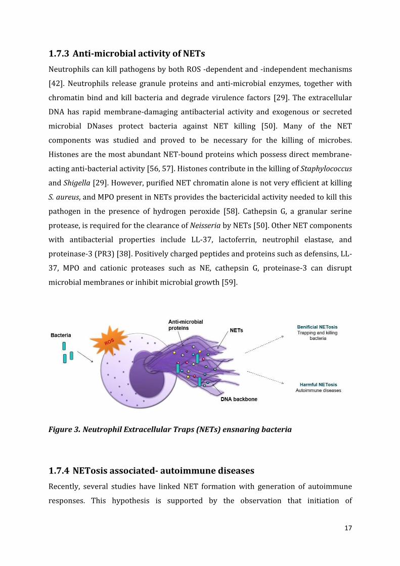
1.7.3 Anti-microbial activity of NETs
Neutrophils can kill pathogens by both ROS -dependent and -independent mechanisms
[42]. Neutrophils release granule proteins and anti-microbial enzymes, together with
chromatin bind and kill bacteria and degrade virulence factors [29]. The extracellular
DNA has rapid membrane-damaging antibacterial activity and exogenous or secreted
microbial DNases protect bacteria against NET killing [50]. Many of the NET
components was studied and proved to be necessary for the killing of microbes.
Histones are the most abundant NET-bound proteins which possess direct membrane-
acting anti-bacterial activity [56, 57]. Histones contribute in the killing of Staphylococcus
and Shigella [29]. However, purified NET chromatin alone is not very efficient at killing
S. aureus, and MPO present in NETs provides the bactericidal activity needed to kill this
pathogen in the presence of hydrogen peroxide [58]. Cathepsin G, a granular serine
protease, is required for the clearance of Neisseria by NETs [50]. Other NET components
with antibacterial properties include LL-37, lactoferrin, neutrophil elastase, and
proteinase-3 (PR3) [38]. Positively charged peptides and proteins such as defensins, LL-
37, MPO and cationic proteases such as NE, cathepsin G, proteinase-3 can disrupt
microbial membranes or inhibit microbial growth [59].
Figure 3. Neutrophil Extracellular Traps (NETs) ensnaring bacteria
1.7.4 NETosis associated- autoimmune diseases
Recently, several studies have linked NET formation with generation of autoimmune
responses. This hypothesis is supported by the observation that initiation of

18
autoimmune responses often occurs following microbial infections [38]. Importantly,
neutrophils from patients with various autoimmune diseases appear more prone to
NETose. For example, neutrophils from rheumatoid arthritis (RA) patients exhibited
increased spontaneous NET formation compared to healthy individuals [60]. NETs
include the targets of most autoantibodies found in RA, systemic lupus erythematosus
(SLE), and vasculitis [61, 62]. The clinical and biological overlaps observed between and
RA, SLE, or SLE and vasculitis suggest that NETosis can be a major triggering event
common to these disorders [63].
Many autoantibodies have so far been described in RA, but only anti-citrullinated
protein antibodies (ACPA) are considered specific disease marker with sufficient
specificity and sensitivity to be used as diagnostic tests of RA [64]. NETs contain
histones which are converted from arginines to citrullines by peptidyl arginine
deiminase IV (PAD4). The deiminated chromatin may function to capture bacterial
pathogens. The complex of bacterial antigens and deiminated chromatin may be
internalized by host phagocytes. The uptake and processing of deiminated chromatin
together with bacterial adjuvants by phagocytes may induce the presentation of
modified histone epitopes and co-stimulation, thus yielding a powerful stimulus to
break tolerance. Autoantibodies against deiminated histones are prevalent in systemic
lupus erythematosus (SLE) and patients with rheumatoid arthritis (RA). These
observations clearly suggest that histone deamination can act as an autoantibody
stimulant [65].
NETs containing antimicrobial proteins including DNA and LL37 combination are
potent stimulus for plasmacytoid dendritic cells (pDCs) to synthesize type I IFNs [66].
Type I IFNs have antimicrobial roles but also have potent immunostimulatory effects in
autoimmune diseases like SLE and psoriasis [38, 66, 67].
1.7.5 ROS-dependent NETosis
NETosis is classified into ROS-dependent and ROS-independent types. However, how
ROS contribute to NETosis is not clear. Requirements for oxidant generation depend on
the stimulus which induces NET formation [68]. The most frequently used compound to
induce NETosis is PMA, a synthetic activator of the protein kinase C (PKC) family of

19
enzymes. PKC is directly responsible for activation of NADPH oxidase and ROS
production [38]. Neutrophils from patients with chronic granulomatous disease (CGD),
with non-functional NADPH-oxidase, failed to induce PMA-induced NET release [30, 69].
The requirement for ROS production was confirmed as NADPH oxidase inhibitor like
diphenylene iodinium (DPI) and ROS-scavengers inhibited NETosis induced by PMA and
S.aureus [30, 68, 70]. However, exogenous hydrogen peroxide, which is membrane
permeable, could induce NETs, as hydrogen peroxide stimulates MPO downstream of
NADPH oxidase. Superoxide itself is not essential, but the conversion of superoxide to
hydrogen peroxide and to perchloric acid is essential for NET release. NETosis pathway
in CGD neutrophils were rescued with the addition of exogenous peroxide [30, 68, 71].
Similarly, Candida albicans also induce ROS mediated NETosis in human neutrophils as
well as in mouse neutrophils. Neutrophils from mice lacking functional NADPH oxidase
(gp91–/–) mice, fail to produce ROS upon stimulation and could not make NETs [72, 73].
Immune complexes (ICs) can stimulate neutrophils to form NETs [74, 75]. Both soluble
and immobilized immune complexes (iIC) are capable of inducing NETs [54, 55]. Pre-
treatment with DPI had no effect on the soluble IC-induced NET formation, suggesting
that ROS is not critical here [54]. However, ROS was found crucial for immobilized
immune complex induced NET formation. The NADPH-oxidase inhibitor (DPI) and MPO
inhibitor aminopyrine as well as ROS-scavengers were shown to abolish iIC-induced
NETosis [55].

20
Figure 4. PMA- and immobilized Immune complex (iIC)- induced NETosis
1.7.5.1 Signaling mechanisms known in PMA-induced NETosis
NET formation induced by PMA is independent of transcription as well as of protein
synthesis. This suggests that neutrophils contain all the factors required for NET
formation when they emerge from the bone marrow as differentiated cells [76].
NETosis induced by PMA is PKC and NADPH oxidase (NOX) dependent. PMA stimulates
conventional (α, βI, βII, γ) and novel (δ, ε, η, θ) PKC isoforms [77]. Conventional PKCs
have a prominent role in NET formation. Furthermore, PKCβ is the major isoform
crucial in NET formation [78]. NADPH subunit p47phox is phosphorylated to acquire a
conformational rearrangement to expose the domains that are important for the
NADPH oxidase function, and this phosphorylation is mediated by PKC [79].
Downstream of PKC and upstream of NADPH oxidase is Raf–MEK–ERK kinase pathway,
which leads to NET signaling [80]. PMA-induced ROS mediated NETosis results in
phosphorylation or activation of ERK1/2 and p38 MAPK pathway [81] (Fig. 4).

21
However, conflicting evidence exists as to whether ERK is activated upstream or
downstream of ROS production [80, 81]. Apparently, an additional contribution of ERK
to this process may not be required as PKC can directly activate NADPH oxidase [82-85].
In addition, Syk is found to be involved in PMA dependent ROS and NET production, as
Syk inhibition reduced the NET formation and almost abolished ROS production [55].
PMA-mediated phosphorylation of Akt has been shown, where Akt induces NETosis
while suppressing apoptosis [86].
1.7.5.2 Signaling mechanisms known in iIC-induced NETosis
Immobilized immune complexes (iICs) stimulate the release of NETs in an NADPH-MPO
associated and ROS-dependent manner. iICs are recognized by FcᵧRIIIB and its signaling
partner macrophage-1 Ag (Mac-1: CD11b/CD18). Mac-1 does not seem to play a role in
iIC-induced ROS production as blocking of Mac-1 had no effect on MPO-dependent ROS
and only slight inhibition of NADPH-oxidase (NOX) dependent super oxide production.
iIC-induced ROS depends on both FcᵧRIIA (CD32) and FcᵧRIIIB (CD16). However, only
FcᵧRIIIB is sufficient for iIC-induced NET release.
FcᵧRIIIB and Mac-1 downstream activates Src/Syk signaling, which activates
neutrophils. Src/Syk further activates ERK1/2, PI3K/ Akt, and p38 MAPK pathways.
Enhanced phosphorylation of, ERK1/2, PI3K/Akt and p38 MAPK is seen in iIC-
stimulated neutrophils. The inhibitor studies on these intracellular molecules confirmed
their importance. ERK1/2 inhibition almost abolished NET formation, whereas
treatment with inhibitors of Akt and p38 MAPK partially inhibited iIC-induced NET
formation. The activation of ERK1/2, PI3K/Akt or p38 MAPK results directly from Src
family kinases/receptor activation and is not ROS mediated, as the ROS inhibition did
not affect their phosphorylation [55] (Fig. 4).
1.7.6 ROS-independent NETosis
NETosis can also occur in certain conditions through ROS-independent pathways [68,
87]. Generation of ROS did not complement the defect in NET formation by neonatal
neutrophils, as it did in adult cells with inactivated NADPH oxidase, demonstrates that
ROS is not sufficient for downstream signaling in neonate neutrophils [87]. NADPH

22
oxidase-independent NETosis that can be induced by stimuli including calcium
ionophores [88]. Furthermore, a unique and very rapid way of NETosis in response to S.
aureus showed that the first 5–60 min of NETosis is ROS-independent [89].
Nevertheless, the mechanisms for the NOX-independent pathway of NETosis are not
well understood.
1.7.6.1 Signaling mechanisms known in Ionomycin-induced NETosis
Ionomycin, a natural Ca2+ Ionophore produced by the Gram-positive bacterium
Streptomyces conglobatus can induce rapid NADPH oxidase (NOX)-independent NETosis
[88, 90, 91]. Ionomycin acts as a motile Ca2+ carrier and enhances Ca2+ influx by direct
stimulation of store-regulated cation entry across biological membranes [92]. In 2015
Douda et al. studied Ionomycin induced- NETosis. They showed that activation of the
calcium-activated potassium channel of small conductance (SK channel) induces NOX-
independent NETosis. In neutrophils with calcium influx, SK channels activate
mitochondrial ROS production and activate potassium current. Mitochondrial ROS is
needed for Ionomycin induced NOX-independent NETosis, but not for NOX-dependent
NETosis. Furthermore, a large amount of mitochondrial ROS is being produced during
NOX-independent NETosis, but not during NOX-dependent NETosis. In contrast to NOX-
dependent NETosis, ERK is not substantially activated in NOX-independent NETosis,
and inhibiting ERK does not inhibit NETosis. p38 and Akt are activated in both NOX-
dependent and NOX-independent types of NETosis. However, the inhibition of p38 did
not inhibit NOX-dependent NETosis [88]. PAD4 is a calcium-dependent enzyme, shown
to be critical in Ionomycin-induced NETosis. Inhibition of PAD4 nearly abolishes
Ionomycin induced NETs [40]. PAD4 mediates citrullination, which is the conversion of
positively charged arginine side chains into polar but uncharged citrulline side chains,
by deimination [93].

23
1.8 Platelet-derived growth factor receptors (PDGFRs)
Platelet-derived growth factor receptors
(PDGFR) are cell surface tyrosine kinase
receptors for members of the platelet-derived
growth factor (PDGF) family [94]. The PDGF
family consists of five members (i.e., disulfide-
bonded dimers of homologous A-, B-, C-, and
D-polypeptide chains, and the AB
heterodimer) [95]. The PDGF-α receptor binds
all PDGF chains except the D chain, whereas
the β receptor binds PDGF-B and -D; thus, the
different PDGF isoforms can induce αα-, αβ-, or
ββ-receptor dimers [96]. PDGFR structure
consists of Ig-like domains in their extracellular part, a single transmembrane domain,
and an intracellular part consisting of a well-conserved juxtamembrane domain, a
tyrosine kinase domain with a characteristic inserted sequence without homology with
kinases, and a less well-conserved carboxy-terminal tail [96] (Fig. 5). Gene knockout
studies in mice indicated that PDGF and PDGF receptors have important roles to
promote proliferation, migration, and differentiation of specific cell types during the
embryonal development [97].
PDGF-A is expressed in most epithelial cells and PDGFR-α is expressed in most
mesenchymal interstitial cells. The PFGF-A is important during organogenesis in
stimulating cell proliferation. PDGF-B is expressed in most endothelial cells and is
responsible for the proliferation of smooth muscle cells of the vessels and pericytes
during angiogenesis [96, 97]. In 1982, Platelet-derived growth factor (PDGF) was shown
to be chemotactic for monocytes and neutrophils [98]. On the contrary, in 1995 a study
showed that neutrophils lack detectable mRNA for PDGF alpha-receptor and beta-
receptors. This indicated that human neutrophils possibly do not possess functional
PDGF receptors [99].
Dimerization is the key event in PDGF receptor activation as it allows for receptor
autophosphorylation on tyrosine residues in the intracellular domain [100].
Autophosphorylation activates the receptor kinase and provides docking sites for
Figure 5. Structure of PDGFR

24
downstream signaling molecules [97, 101]. Both PDGFR-α and PDGFR-β engage several
well-characterized signaling pathways like Ras-MAPK, PI3K, and PLC-γ which are
involved in multiple cellular and developmental responses. PDGFRs connect to Ras-
MAPK mainly through the adaptor proteins. Adapter proteins bind the activated PDGFR
through its SH3 domains. Furthermore, Ras protein is activated, leading to downstream
activation of Raf-1 and the MAPK cascade. MAPK signaling activates gene transcription,
leading to stimulation of cell growth, differentiation, and migration [102, 103].
PDGFRs are mainly linked to certain cancers which are caused due to genetic
aberrations leading to uncontrolled PDGF signaling in tumor cells. PDGFs are known
also to help to recruit different types of stromal cells and promote angiogenesis. This
process supports the invasion of metastatic cells [96, 97, 104]. To control overactivity of
PDGF signaling various pharmacological antagonists have been developed. Several
types of inhibitors are now available, including inhibitory antibodies against the
receptors, and low molecular weight inhibitors of PDGF receptor kinases etc. [105]. The
most efficient ways to block PDGFR signaling is to inhibit the PDGFR kinase activity.
Kinase inhibitors act by binding at or near the ATP-binding pocket of the kinase domain.
Several kinase inhibitors (example; CP-673451) have been developed that block
PDGFRs (Fig. 5), but the inhibitors available so far are not completely specific [97, 106-
108].
1.9 CP-673451: inhibitor molecule targeting PDGFRs
CP-673451 is a pharmacological selective inhibitor of PDGFRα/β and PDGF-BB-
stimulated autophosphorylation of PDGFR-beta with IC50 ranging from 1nM-10nM in
cell-free assays. It exhibits >450-fold selectivity to PDGFRβ over other angiogenic
receptors (e.g., vascular endothelial growth factor receptor 1 and 2 (VEGFR-1, VEGFR-
2)) [106]. The chemical name of CP-673451 is 1-[2-[5-(2-Methoxyethoxy)-1H-
benzimidazol-1-yl]-8-quinolinyl]-4-piperidinamine. Its molecular formula is C24H27N5O2
and it has a molecular weight of 417.52 (Fig. 6) [106].

25
Figure 6. Molecular structure of CP-673451
CP-673451 has shown to have anti-angiogenic and anti-tumor activity. CP-673451
inhibits PDGFR-beta phosphorylation, selectively inhibits PDGF-BB-stimulated
angiogenesis in vivo, and causes significant tumor growth inhibition in multiple human
xenograft models [106]. Furthermore, CP-673451 inhibits the tumor growth in
Colo205, LS174T, H460, and U87MG xenograft models. Inhibition of angiogenesis or
tumor growth is correlated with plasma and tumor concentration and inhibition of
phospho-PDGFR in vivo. U87MG human glioblastoma xenografts express PDGFR on the
tumor cells; thus, inhibition could be due to a direct antitumor effect as well as an
antiangiogenic effect. In kinase assays, CP-673451 does not show substantial potency
against any other kinase tested, including, VEGFRs [106, 108-110].

26
Objectives of the Study
The process of neutrophil extracellular trap formation (NETosis) has been identified in
2004. Since then, several studies have been carried out to investigate the mechanisms
involved in NET formation. These studies identified some molecular pathways involved
in ROS-dependent NETosis which can be induced by PMA and iICs. However, the
molecular pathways identified in ROS-dependent NETosis are upstream of ROS
production. The signaling mechanism of NETosis downstream of ROS production is still
not clear. Therefore, major objective of this study was to identify signaling mechanisms
involved in NETosis downstream of ROS production.
As experimental approach, it was planned that an inhibitory library will be screened for
substances which inhibit NETosis but do not inhibit ROS production. This approach
might possibly give a specific inhibitor of NETosis which targets downstream to ROS
production.
At the second stage of the study, the aim was to identify the target of the NETosis
inhibitor to understand the mechanism of inhibition. The molecular target of the
inhibitor was planned to confirm by other similar target inhibitors and the effect of the
inhibitor on known pathways activated by PMA and iIC. It was hypothesized that the
inhibitor which inhibits NETosis independent of ROS pathways could be used as a
general inhibitor of NETosis. This was planned to be tested with the ROS-independent
NETosis stimulus, Ionomycin.
The third aim of the study was to test whether the inhibitor affects basic neutrophil
functions including activation, migration and phagocytosis. This could clarify if the
pathway of NETosis is also involved in the regulation of other neutrophil functions. To
check if the inhibitor results changes in gene expressions in neutrophils, a high-
throughput RNA-seq approach to profile transcriptional responses was envisaged.

27
Chapter 2. Materials and Methods
2.1 Materials
2.1.2 Solutions, buffers and media
10 x Tris buffered saline (TBS): 200 mM Tris Ultra + 1.4 mM Sodium chloride, pH
to 7.6.
1xRIPA buffer: Dilute 1:10 ready-made 10xRIPA buffer in aqua
dest with 1:10 ratio protease inhibitor (cOmplete
mini)
4 x loading buffer: 0.16 M Tris Ultra pH 6.80 + 30 % glycerol + 2 %
SDS + 0.71 M 2-β-mercaptoethanol + 0.002 %
bromophenol blue.
Acetate buffer
(Substrate buffer for migration
assay);
6.8 g CH3COONa*3H2O in 400ml aqua dest with
HCl to adjust pH to 4 and with aqua dest up to
500 ml
Blocking buffer for immune
complex formation:
TBS + 0.1 % Tween 20 + 1% BSA, filtered.
Blocking buffer for Western blot: TBS + 0.1 % Tween 20 + 5% BSA.
Blotting buffer: 25 mM Tris Ultra + 192 mM glycine + 20%
methanol.
CL-medium RPMI 1640 medium special with 20 mM HEPES
without NaHCO3, pH7.2, without phenol red
Complete medium: RPMI 1640 medium + 200 mM L- glutamine + 20
mM HEPES + 10 % FCS + 100U/100µg/ml
Penicillin/Streptomycin.
DMEM: DMEM (41965-039) +10 % FCS +100 U/100
µg/ml Penicillin/ Streptomycin
Electrophoresis buffer (5x): 125 mM Tris Ultra + 0.960 M glycine + 0.5 %
sodium dodecyl sulfate.
FACS buffer: PBS + 1 % BSA + 0.01 % sodium azide + 1 %
human serum.
28
Glycine buffer (Stop solution for
migration assay):
15 g glycin in 400 aquadest, with NaOH to adjust
the pH to 10.3 with aqua dest fill up to 500 ml
NET-medium: CL-medium (RPMI 1640 medium special
preparation from Biochrome #FZ 1235+ 20 mM
HEPES w/o NaHCO3, pH7.2, w/o phenol red) +
0,5% HSA (Baxter) + 0,5 % Human serum
albumin
TBS-Tween: TBS + 0.1 % Tween 20
2.1.3 Laboratory supplies
Cell culture flask, 250 ml, 75 cm² with filter screw cap
Greiner bio-one, Frickenhausen
Cell culture plates (96, 24, 12, 6 well, flat
bottom)
Greiner bio-one, Frickenhausen
Costar 3472 Corning®-Transwell® cell
culture inserts 24 mm with 3 μm pore
polycarbonate membrane insert
Costar- Corning, New York, USA
Eppendorf tubes (1.5; 2 ml (PP)) Biopure Eppendorf, Hamburg
Extra Thick Blot Filter Paper Biorad, Munich
Microscope slides Menzel, Braunschweig
Microtestplate + lid (96-well, V-bottom) Sarstedt, Nümbrecht
Neubauer chambers BRAND Gmbh + CO KG, Wertheim
Nitrocellulose (NC) membrane Bio-Rad, Munich
Nunclon ELISA PLATES Nunc, Langenselbold
Pipette 2, 5, 10, 25 ml Greiner bio-one Frickenhausen
Pipette filter tips Nerbe plus, Winsen
Plastic tubes (15 ml (PP), 50 ml (PP)) Sarstedt, Nümbrecht
Precast gels Any Kd Bio-Rad, Munich
S-Monovette 9 ml, lithium-heparin Sarstedt, Nümbrecht
Stericup® Filter Units Merck Millipore, Billerica,
Massachusetts, United States
Thermonox cover slides 13mm (174950) Thermo Fisher Scientific, Waltham,

29
MA, USA
Transfer pipette 3.5 ml Sarstedt, Nümbrecht
U-tubes for flow cytometry Sarstedt, Nümbrecht
2.1.4 Chemicals and reagents
2-β-Mercaptoethanol Sigma-Aldrich, Steinheim
Acetone Merck, Darmstadt
Annexin V FLUOS Roche Diagnostics, Mannheim
Bovine serum albumin (BSA) Sigma-Aldrich, Steinheim
Bromophenol blue dye Serva, Heidelberg,
Calcium chloride Sigma-Aldrich, Steinheim
CL-medium (RPMI 1640 medium special
preparation #FZ 1235)
Biochrom, Berlin
cOmplete™, Mini Protease Inhibitor Cocktail Roth, Karlsruhe
Coomassie Thermo Fisher Scientific, Waltham, MA, USA
Crystal violet Sigma-Aldrich, Steinheim
Diff-Quick® fixative solution Medion Diagnostics, Duedingen,
Switzerland
Diff-Quick® staining set Medion Diagnostics, Duedingen,
Switzerland
DMEM Gibco, Karlsruhe
Dmso (Dimethyl sulfoxide) Sigma-Aldrich, Steinheim
DPI (diphenyleneiodinium chloride) Sigma-Aldrich, Steinheim
Fetal calf serum (FCS) Sigma-Aldrich, Steinheim
Glycine Sigma-Aldrich, Steinheim
HEPES Biochrom, Berlin
Histopaque® 1077 Sigma-Aldrich, Steinheim
Histopaque® 1119 Sigma-Aldrich, Steinheim
Human serum albumin (HSA) Apotheke
Immersions oil Carl Zeiss, Jena
Immmobilion™ western Millipore, MA, USA
Ionomycin calcium salt (I0634) Sigma-Aldrich, Steinheim
30
Isopropanol Roth, Karlsruhe
Latex beads (FluoSpheres® Polystyrene
Microspheres, 1.0 µm)
Invitrogen, Eugene, OR, USA
L-Glutamine Biochrom, Berlin
Lipopolysaccharide E. coli 0111: B4 (LPS) Sigma-Aldrich, Steinheim
Luminol Sigma-Aldrich, Steinheim
Methanol Baker, Deventer, The Netherlands
N-Formylmethionyl-leucyl- phenylalanine
(fMLP)
Sigma-Aldrich, Steinheim
PageRuler™ Unstained Protein Ladder Thermo Fisher Scientific, Waltham,
MA, USA
Paraformaldehyde Sigma-Aldrich, Steinheim
PBS (1 x) sterile solution Pharmacy of University of Lübeck,
Lübeck
PBS (10 x) sterile solution Gibco, Karlsruhe
PBS (20 x) sterile solution Cell Signaling, Leiden, The Netherlands
Penicillin/streptomycin Biochrom, Berlin
Percoll® Sigma-Aldrich, Steinheim
PhoStop tablets Roche Diagnostics, Mannheim
Pierce Prestained Protein MW Marker Thermo Fisher Scientific, Waltham,
MA, USA
PMA (phorbol 12-myristate 13-acetate) Sigma-Aldrich, Steinheim
PMB (Polymyxin-B) Biochrom, Berlin
Poly-L-Lysin Sigma-Aldrich, Steinheim
ProLong™ Gold Antifade Mountant Thermo Fisher Scientific, Waltham,
MA, USA
Propidium iodide Sigma-Aldrich, Steinheim
RIPA Buffer (10x) Cell Signaling, Leiden, The Netherlands
RPMI 1640 medium Sigma-Aldrich, Steinheim
Sodium azide Merck, Darmstadt
Sodium chloride Merck, Darmstadt
Sodium dodecylsulfate Sigma-Aldrich, Steinheim

31
Staphylococcus aureus bioparticles®, Alexa
Fluor 488 conjugate
Thermo Fisher Scientific, Waltham,
MA, USA
SytoxGreen (nucleic acid stain) Invitrogen, Eugene, OR, USA
TCA (Trichloro acetic acid) Sigma-Aldrich, Steinheim
Tris Ultra Roth, Karlsruhe
Triton X-100 Merck, Darmstadt
Trypan blue solution 0.4 % Sigma-Aldrich, Steinheim
Trypsin-EDTA (0.25%) Gibco, Karlsruhe
Tween -20 Sigma-Aldrich, Steinheim
β-Glucuronidase (G8420) Sigma-Aldrich, Steinheim
2.1.5 Other selective inhibitors
Etoposide Selleckchem (Houston, USA)
Flumequine Selleckchem (Houston, USA)
Motesanib Diphosphate (AMG-706) Selleckchem (Houston, USA)
Sunitinib Selleckchem (Houston, USA)
Vandetanib (ZD6474) Selleckchem (Houston, USA)
2.1.6 Ready to use kits
Immobilon Western HRP Substrate Merck, Darmstadt
NextSeq 500/550 High Output Kit Illumina, San Diego, California, USA
Pierce™ LDH Cytotoxicity Assay Kit Thermo Fisher Scientific, Waltham, MA,
USA
QIAamp RNA Blood Mini Kit Qiagen, Hilden
RNase-Free DNase Set Qiagen, Hilden
2.1.7 Antibodies
Goat anti-HSA- IgG Cell Signaling, Leiden, The Netherlands
Goat anti-rabbit-HRP-linked Cell Signaling, Leiden, The Netherlands
Horse anti-mouse-HRP-linked Cell Signaling, Leiden, The Netherlands
Mouse anti-human CD11b-FITC BioLegend, San Diego, CA, USA

32
Mouse anti-human IgG New England Biolabs, USA
Rabbit anti-HSA -IgG Sigma-Aldrich, Steinheim
Rabbit anti-human PDGFR Cell Signaling, Leiden, The Netherlands
Rabbit anti-human phospho-Akt (Thr308) Cell Signaling, Leiden, The Netherlands
Rabbit anti-human phospho-Erk1/2
(p44/42)
Cell Signaling, Leiden, The Netherlands
Rabbit anti-human phospho-p38 MAPK
(Thr180/Tyr182)
Cell Signaling, Leiden, The Netherlands
Rabbit anti-human phospho-PI3 Cell Signaling, Leiden, The Netherlands
Rabbit anti-human phospho-PKC α/β Cell Signaling, Leiden, The Netherlands
Rabbit anti-human phospho-PKC δ Cell Signaling, Leiden, The Netherlands
Rabbit anti-human phospho-Serine Merck, Darmstadt
Rabbit anti-human phospho-Src Cell Signaling, Leiden, The Netherlands
Rabbit anti-human phospho-Threonine Cell Signaling, Leiden, The Netherlands
Rabbit anti-human phospho-Threonine
(Sepharose® bead conjugate)
Cell Signaling, Leiden, The Netherlands
Rabbit anti-human phospho-Tyrosin Cell Signaling, Leiden, The Netherlands
Rabbit anti-human β-actin, HRP-linked Cell Signaling, Leiden, The Netherlands
2.1.8 Cytokines
Recombinant human IFN-γ R&D Systems, Wiesbaden-Nordenstadt
Recombinant human IL-8 R&D Systems, Wiesbaden-Nordenstadt
Recombinant human TNF-α PeproTech, Rocky Hill, NJ, USA
2.1.9 Cell lines/cell line lysate
T129 lysate Department of Neurosurgery, University of
Lübeck
U87-MG Department of Neurosurgery, University of
Lübeck

33
2.1.10 Instruments
Analytical balance BP61S Sartorius, Göttingen
Balance Sartorius, Göttingen
Bioanalyzer Agilent, Santa Clara, California, USA
Block thermostat HB Peqlab, Erlangen
Carl Zeiss, Jena Carl Zeiss, Jena
Centrifuge 5417R Eppendorf, Hamburg
Centrifuge Biofuge fresco Kendro Heraeus, Langenselbold
Centrifuge Megafuge 2.0R Kendro Heraeus, Langenselbold
Centrifuge Microfuge R Beckmann, Munich
Centrifuge Mikro 12-24 Hettich, Tuttlingen
Centrifuge Multifuge 3 and SR Kendro Heraeus, Langenselbold
CO2 Incubator IG 150 Jouan, Unterhaching
Cytocentrifuge Cytospin 3 Shandon, Frankfurt
Deep freezers, −20°C, −70°C Liebherr, Ochsenhausen
Flow cytometer Canto II Becton Dickinson, Heidelberg
Fusion Fxt chemiluminescence reader Vilber Loumat, Eberhardzell
Incubator without CO2 Agilent Technologies, Santa Clara, CA, USA
Infinite 200 Pro reader Tecan, Crailsheim
Keyence Microscope Osaka, Osaka Prefecture, Japan
Laminar flow workbench Biohit, Cologne
Magnetic stirrer: Ikamag, Reo IKA Labortechnik, Staufen
Microscope Axiocam HRc Carl Zeiss, Jena
Microscope Axiostar plus Carl Zeiss, Jena
Multichannel pipette Eppendorf, Hamburg
NanoPhotometer Pearl® Impeln, Munich
pH-meter Inolab WTW GmbH, Weilheim
Pipette boy Eppendorf, Hamburg
Ricoh HR-10m camera Ricoh, Tokyo, Japan
Semi-dry protein transfer cell Bio-Rad, Munich
Shaker Vibrofix VF1 Electronic Janke & Kunkel IKA® Labortechnik, Staufen
Water bath Köttermann, Uetze

34
2.1.11 Software
AxioVision release 4.8 software Carl Zeiss, Jena
BD FACSDiva™ software 9 Becton Dickinson, Franklin Lakes, NJ, USA
Bioanalyzer 2100 Expert Software Agilent, Santa Clara, California, USA
BioD1 Vilber Loumat, Eberhardzell
BZ II analyzer software
Keyence, NeuIsenburg
GraphPad Prism 5.0 and 6.0 La Jolla, CA USA
ImageJ software NIH, Bethesda, USA
Tecan i-control 1.7 software Tecan, Crailsheim

35
2.2 Methods
2.2.1 Neutrophil isolation from human blood
Peripheral blood was collected by venipuncture from healthy donors in lithium heparin-
containing tubes. 12.5 ml Histopaque 1119 was layered in a 50-ml falcon tube and 12.5
ml of Histopaque 1077 was layered over it carefully. To the prepared bilayer gradient,
25ml blood was layered. The gradient was centrifuged for 5 min at 300 x g followed by
25 min at 800 x g. After centrifugation, the top layer containing plasma and the
Histopaque 1077 layer rich in lymphocytes and monocytes were discarded and the last
layer containing erythrocytes were discarded. The granulocyte rich layer of Histopaque
1119 layer was collected in 50 ml Falcon tube and washed with PBS for 10 min at 800 x
g. The granulocytes were then resuspended in 2ml of complete medium and layered on
the top of Percoll gradient. The Percolll gradient was prepared in a 15 ml tube by
layering with densities 1.105 g/ml (85 %), 1.100 g/ml (80 %), 1.093 g/ml (75 %), 1.087
g/ml (70 %), and 1.081 g/ml (65 %) from the bottom to the top, respectively. The
gradient column was centrifuged at 800 x g for 25 min. After centrifugation, the
interphase between the 70 % and 85 % Percoll layers was collected and washed with
PBS at 800 x g for 10 min and resuspended in complete medium. All the centrifugation
steps were performed at room temperature. The cell preparations contained >99 %
granulocytes. Neutrophil purity was >98 % as determined by morphological
examination of cytocentrifuged slides stained with Diff Quick.
2.2.2 Culturing U87-MG Cell lines
U87-MG cells were continuously cultured in Dulbecco's Modified Eagle Medium (DMEM
with FCS) at 37°C in humidified atmosphere containing 5 % CO2 until they are confluent.
Cells were exposed to trypsin/EDTA to detach the adherence at 37°C for 5 min and
DMEM containing FCS was added to stop trypsinization. The medium was renewed
every 2-3 days.
2.2.3 Cytospin and Diff-Quick staining
Neutrophils (5x10⁵/ml in 100 μl complete medium) were cytocentrifuged at 400 x g for
5 min. The slides were air dried, fixed in Diff-Quick® fixative solution and,

36
subsequently, stained by Diff-Quick staining set according to the manufacturer’s
protocol.
2.2.4 Preparation of immobilized immune complexes
Immobilized immune complexes (iICs) were prepared by using human serum albumin
(HSA) as antigen and anti–HSA IgG rabbit polyclonal antibody as described previously
[111]. 100 μl of 20 mg/ml HSA in 50 mM bicarbonate buffer (pH 9.6) was coated
overnight at 4˚C in 96 well Lumitrac 600 high-binding plates for chemiluminescence-
based ROS detection and in Flurotrac 600 high-binding plates for fluorescence-based
NET detection. The plates were then washed with 200 μl PBS containing 0.05% Tween
20 (wash buffer) and then blocked with 200 μl 1% biotin-free BSA in PBS (blocking
buffer) for 1 h at room temperature (RT), following 1 h incubation with anti–HSA rabbit
IgG (∼10 mg/ml) diluted 1:400 in blocking buffer. The IC-coated wells were then
washed twice with wash buffer and once with assay- medium. For fluorescence
microscopic experiments, iICs were coated as described above by using 0.5 ml volume
of reagents instead of 200 μl in an 24-well ibiTreat slides.
2.2.5 Detection of intra- and extracellular ROS
The intra- and extracellular ROS produced by neutrophils were measured by using
luminol-based chemiluminescence assay [112, 113]. All chemiluminescent assays were
performed in chemiluminescent medium (CL-medium). For the real-time-ROS-kinetics,
neutrophils (4x10⁵/ml cells/200 µl CL-medium) were added to the iIC-coated or to
uncoated wells containing 0.1% dmso, medium for iIC-mediated ROS detection. For
PMA mediated ROS production, 4x10⁵/ml cells/200 µl CL-medium was treated with
PMA (20nM) or with 0.1% dmso or CL-medium. All the ROS detection assays were
performed in Flurotrac 600 high-binding white plates. Different concentrations of
inhibitors (10 µM, 3 µM, 1 µM, 0.3 µM, 0.1 µM) and 0.06 mM luminol were also added at
the same time. In preliminary screening for inhibitors, a single concentration of 10 µM
was used and cell suspension pre-treated for 30 min with 20nM DPI at 37°C was used as
inhibition control. ROS-dependent chemiluminescence was analyzed using an Infinite
200 reader and Tecan i-control 1.7 software. ROS release was monitored for 1 h every

37
1-2 min at 37˚C. For statistical analysis, the area under the curve (AUC) of each sample
was calculated.
2.2.6 SytoxGreen detection of NETs
The time kinetics of NET release was assessed by using the non-cell permeable DNA dye
SytoxGreen [114-116]. For iIC-mediated ROS detection, neutrophils (2x10⁵ cells/200 µl
NET-medium) were added to the iIC-coated or to uncoated wells. For PMA mediated
ROS production, 2x10⁵ cells/200 µl NET-medium were treated with PMA (20nM) or
with 0.1% dmso or medium. In Ionomycin-induced NET assays, 2x10⁵ cells/200 µl in
complete medium without FCS were tested with 7 µM, 5 µM, and 2.5 µM concentrations
of Ionomycin for inducing NETosis. For the experiments with the inhibitor, 5 µM
concentration of Ionomyin were used. For PMA and iIC NETs, NET-medium was used
and for Ionomycin-induced NET formation, complete medium without FCS was used. All
NET assays are performed in Flurotrac 600 high-binding black plates. Different
concentrations of inhibitors (10 µM, 3 µM, 1 µM, 0.3 µM, 0.1 µM) and 5 µM SytoxGreen
were also added to the wells at the same time. In preliminary screening for inhibitors, a
single concentration of 10 µM was used and cell suspension pre-treated for 30 min with
20 nM DPI at 37°C was used as inhibition control. The NET-bound SytoxGreen
fluorescence (excitation: 488 nm, emission: 510 nm) was analyzed for 4 h for PMA and
7 h for iIC with 5 min interval at 37°C by using Tecan infinite M200 Pro reader and
Tecan i-control 1.7 Software. For statistical analysis, the area under the curve (AUC)
was calculated.
2.2.7 Fluorescent microscopy for NETs
Fluorescent microscopy of NET formation/inhibition slides was prepared in 13 mm µ-
slides. 5x10⁵ cells/500 µl NET medium were treated with different concentrations of
CP-673451, Doxorubicin (10 µM, 3 µM, 1 µM, 0.3 µM, 0.1 µM), 0.1% dmso, DPI (20 nM)
and NET-medium and incubated for 30 min at 37˚C. The cells were then added to iIC-
coated µ-slides. For samples with PMA as stimulant the cells were added to Poly-L-Lysin
coated μ-slides. The cells were then incubated for 4h and 7 h at 37˚C for PMA and iIC
stimulated plates, respectively. Followed by fixation with 4% paraformaldehyde,

38
staining with SytoxGreen was carried out as described previously [117]. Samples were
analyzed with Keyence BZ-9000E using the BZ II Analyzer Software.
2.2.8 Neutrophil activation assay
The surface marker expressions were analyzed by staining CD11b and CD62L markers
on neutrophils. Upregulation of CD11b and shedding of CD62L was associated with
neutrophils activation and this was monitored by flow cytometry. 5x10⁵ neutrophils in
200 µl complete medium were pre-incubated for 30 min at 37°C, 5% CO2 with CP-
673451 or with 0.1% dmso or with Polymyxin-B (PMB) (10 µg/ml), or with medium
and stimulated with different stimulants; LPS (100 ng/ml), LPS (100 ng/ml) together
with IFNᵧ (200 U/ml), TNF-α (100 ng/ml), fMLP (1 µM) and with inhibitor CP-673451
(10 µM, 3 µM, 1 µM, 0.3 µM, 0.1 µM) for 4 h at 37°C. The cells were then stained with
fluorescent-conjugated antibodies against CD11b and CD62L for 30 min at 4°C. The
surface marker expression changes were analyzed using flow cytometer and analyzed
by FACS Diva software (Fig. 7). Percentage of the cells upregulating CD11b and
shedding CD62L are considered as activated cells.
Figure. 7 Assessment of neutrophil activation using flow cytometry
Neutrophils were pre-treated with 0.1% dmso and stimulated with LPS (100ng/ml) (B) or left unstimulated (A) for 4 h at 37°C, 5% CO2. The cells were then stained with fluorescent-conjugated antibodies against CD11b-PE and CD62L-APC for 30 min at 4°C. Percentage of the cells upregulating CD11b and shedding CD62L was assessed by flow cytometry and analyzed by FACS Diva software. Q1: cells upregulating CD11b and shedded CD62L. Q2+Q4: cells not upregulating CD11b and not-shedding CD62L. Q3: cells shedded CD62L, but not upregulating CD11b.

39
2.2.9 Neutrophil migration assay
The neutrophil migration assay was performed in 24 well trans well plates with a 3µm
pore size (costar 3472). 6x10⁵ neutrophils in 100 µl complete medium were seeded in a
24 well plate and treated with different concentrations of the inhibitors (10 µM, 3 µM, 1
µM, 0.3 µM, 0.1 µM), or 0.1 % dmso, or medium. The cell suspension was incubated for
30 min at 37°C, followed by transferring into the upper filter of the trans well. Into the
lower well of the trans well was added with 600µl of IL-8 (25 ng/ml), or TNF-α
(50ng/ml) or medium. The cells were allowed to migrate for 1h at 37°C. The number of
migrated cells was measured by using the beta-glucuronidase assay. The migrated cells
in the lower well and 6x10⁵ untreated fresh neutrophils in 600 µl were lysed with 100 µl
of 1 % Triton X-100 for 10 min. 100µl of the untreated fresh neutrophil lysates were
transferred to a 96 well Nunc transparent plate and serially diluted to get the standard
curve. For the beta-glucuronidase assay, 100µl and 600µl of substrate mix (containing
4-Nitrophenyl β-D-glucuronide) in acetate buffer to the 96 and 24 wells respectively
and incubate overnight at 37°C. Next day, glycine buffer was added 100µl and 600µl to
stop the reaction in both 96 and 24 well respectively. 300µl of the 24 well lysate was
transferred to the 96 well plates and measured the absorbance at 405 nm and reference
620 nm. The migration indexes are calculated by normalizing the OD values of medium-
treated neutrophils migrating in response to IL-8 or TNF-α as migration index of 1. The
migration indices of inhibitor-treated neutrophils migrating in response to IL-8 or TNF-
α are calculated from their OD values in relation to the index of 1.
2.2.10 Neutrophil phagocytosis assay
Neutrophils (5x10⁵ cells/100 μl complete medium) were pre-incubated for 30 min with
different concentrations of CP-673451 or 0.1% dmso or medium. Subsequently, Alexa-
Fluor 488 conjugated non-viable Staphylococcus aureus bioparticles in (2:1 ratio; S.
aureus to neutrophils) or FluoSphere carboxylate-modified latex microspheres with a
diameter of 1 µm were added in 1 to 10 ratios to neutrophils. To certain samples
stimulants LPS (100 ng/ml) and IFNᵧ (200 U/ml) were also added, and incubated for
further 30 min at 37°C with 5 % CO2. Cells were washed in a v-bottom plate at 800 x g
for 5 min to remove extracellular bacteria/beads. Trypan blue was added to quench the
fluorescence of extracellular bacteria/beads sticking on the neutrophil surface. The

40
percentage of cells performed phagocytosis and the quantity of ingested
bioparticles/beads were assessed by flow cytometry and analyzed by FACS Diva
software (Fig. 8).
Figure. 8 Assessment of neutrophil phagocytosis using flow cytometry. Neutrophils were co-incubated with FITC-labelled-FluoSphere carboxylate-modified latex beads added in 1 to 10 ratio to neutrophils and stimulated with LPS (100ng/ml) and IFNᵧ (200 U/ml) or left unstimulated in complete medium for 30 min at 37°C, 5%CO2. Trypan blue was added to quench the fluorescence of extracellular bacteria/beads sticking on the neutrophil surface. Percentage of the cells which ingested beads and the cells which did not ingest beads were obtained by flow cytometry and analyzed by FACS Diva software. P2: non-phagocytosing neutrophils and P3: phagocytosing neutrophils. (A) Unstimulated neutrophils with beads, (B) stimulated neutrophils with beads.
2.2.11 Western blot analysis
Neutrophils (5x10⁶ cells/1 ml NET medium) were unstimulated or stimulated with PMA
or iICs for 15 min at 37˚C. In some cases, neutrophils were pre-incubated for 30 min
with 3µM CP-673451 at 37˚C. For PDGFR- western blots, U87-MG cells (5x10⁶/ml) were
used. Whole cell lysates were prepared using TCA as described [118]. Briefly, after
incubation, the cells were centrifuged 5 min at 400 x g. The pellets were then
resuspended in 500 µl of ice cold 10 % TCA solution and incubated for 10 min on ice
and subsequently centrifuged for 5 min at 14000 x g at 4°C. The pellets were washed
two times with 500 µl 100 % acetone for 5 min at 14000 x g at 4 °C. The pellet was then
re-suspended in 1x sample buffer and boiled for 7 min at 100°C in case of phospho-
protein western blots and the pellet was heated at 98°C for 4 min for the PDGFRβ blots.
Lysates were then centrifuged for 5 min at 14000 x g and supernatants were collected.
Lysates were electrophoresed on 10 % SDS-PAGE gels and transferred to nitrocellulose
membranes. Membranes were blocked for 30 min at room temperature with blocking
buffer. Then membranes were incubated shaking overnight at 4 °C with primary
antibodies against human phospho-Akt (Thr308), phospho-p44/42 MAPK (ERK1/2,

41
Thr202/Tyr204), phospho p38 MAPK (Thr180/Tyr182), phospho-PKC alpha/beta
II(Thr638/641), phospho-PKC-δ (Tyr311), phospho-PI3K p85 (Tyr458)/p55 (Tyr199),
Phospho-Src Family(Tyr416), anti-PDGFR, Phospho-tyrosine pan, Phospho-threonine,
Phospho-serine, beta-actin. The membranes were then treated with HRP-conjugated
anti-rabbit or anti-mouse IgG for 2 h at room temperature. The chemiluminescence
signals were obtained with the help of Immobilon Western Chemiluminescence HRP
substrate and signals were measured at chemiluminescence reader. For quantification,
the ratio of phospho-proteins signals to beta-actin signal was determined using Bio 1D
software.
2.2.12 Phospho-enrichment of threonine phosphorylated proteins
Phospho enrichment of threonine phosphorylated proteins were achieved by
Immunoprecipitation, a technique which permits the purification of specific proteins
from the protein mixture was used to separate phospho-threonine proteins from the
whole cell lysate. Anti-phospho-threonine Sepharose® Bead Conjugated antibody was
used to enrich phospho-threonine proteins according to manufacturer’s protocol.
Fresh human neutrophils were isolated from blood. 40x106 neutrophils/1 ml NET-
medium were pre-incubated for 30 min with 3µM CP-673451, and control samples were
treated with 0.1% dmso at 37˚C. The neutrophils were stimulated with PMA (20nM) for
20 min 37°C. The cells were then centrifuged at 300 x g for 5 min at RT and washed
once with 1xPBS, followed by lysis with ice cold 1xRIPA buffer with a protease inhibitor
for 10 min in ice. The lysed cells were then centrifuged at 14000 x g at 4°C. Anti-
phospho threonine Sepharose bead conjugates were added at 1 mg/ml concentration to
the lysates and incubated overnight at 4°C shaking. The lysates were then washed five
times with lysis buffer and 20 µl 1x sample buffer was added to the pellet and heated for
4 min at 99°C. The lysates are then centrifuged and loaded into 10 % gel for SDS-PAGE
with molecular weight markers. After the proteins have separated according to sizes in
the gel, the gel was stained with 10 ml sterile Coomassie stain for 1 h and background
stains were removed by washing with 100ml distilled water. Bands of the Control (Ctrl)
lanes were compared to the CP-673451 treated (tr) lanes for finding missing bands. The
missing lanes identified in phospho-threonine blots are compared with the same area in

42
the phospho-threonine lanes in the gel. The method was established to cut out the
positive band control at the corresponding missing band from the coomassie stained gel
and to send it for Mass spectrometry analysis and protein identification. Picture of the
gel was taken with the help of a scanner.
2.2.13 Assessment of viability by Annexin V-PI staining
During apoptosis cells translocate phosphatidylserine (PS) to the external surface of the
membrane. Annexin V has a high affinity for this externalized PS and exhibits calcium-
dependent binding to PS. The measurement of Annexin V binding to the cell surface as
indicative for apoptosis was performed in conjugation with a dye exclusion test using
propidium iodide (PI). Apoptosis was determined by Annexin V-FITC and PI staining.
5x10⁵ cells/100 µl complete medium were added with inhibitors of different
concentrations (10 µM, 3 µM, 1 µM, 0.3 µM, 0.1 µM) or with 0.1%dmso, or with medium
and incubated for 1 h, 4 h, and 7 h and 22 h at 37˚C, 5 % CO2 . After incubation, cells
were stained by Annexin V-FITC (1 µl) and PI (1 µl) for 30 min in the dark at 4°C with 1
mM CaCl2, washed and re-suspended in complete medium. The labeled neutrophil
populations were then analyzed by FACS Canto II and FACS Diva software to
differentiate as viable, apoptotic and necrotic populations. Intact/viable cells were both
Annexin V and PI negative, apoptotic cells were Annexin V positive PI negative, and
necrotic cells were Annexin V and PI positive (Fig. 9).
Figure. 9 Assessment of neutrophil viability at different time points using flow cytometry. Neutrophils were incubated for 1 h (A), 4 h (B), 7 h (C), and 22 h (D) in complete medium at 37˚C with 5 % CO2. The cells were then stained with Annexin V-FITC and PI and cell viability was assessed by flow cytometry and FACS Diva software. The percentage of cells in each quadrant was determined. Both Annexin V and PI double negative cells were defined as viable cells (Q3), Annexin V positive and PI negative cells were defined as apoptotic (Q4), and Annexin V and PI double positive cells were defined as necrotic (Q2).

43
2.2.14 LDH-Cytotoxicity assay
Cytotoxicity of cells was tested with LDH kit from Thermo Scientific™ Pierce™, to
specifically check necrosis of the cells after treatment with inhibitors. Lactate
dehydrogenase (LDH) is a cytosolic enzyme present in many different cell types. Plasma
membrane damage releases LDH into the cell culture medium. Extracellular LDH in the
culture supernatant can be quantified by a coupled enzymatic reaction in which LDH
catalyzes the conversion of lactate to pyruvate via NAD+ reduction to NADH.
Diaphorase then uses NADH to reduce a tetrazolium salt (INT) to a red formazan
product that can be measured at 490nm. The level of formazan formation is directly
proportional to the amount of LDH released into the medium, which is indicative of
cytotoxicity (Fig. 10).
Neutrophils (2x10⁵ cells/100 µl NET medium) were incubated for 1 h and 7 h at 37˚C
with 0.1 µM-3 µM CP-673451 or with 0.1% dmso or NET-medium. The cells were
centrifuged 300 x g and the supernatants were transferred to a new plate. Additionally,
LDHmax (LDH maximum) samples, with completely lysed cells, and LDHspont (LDH
spontaneous) samples from untreated cells were also prepared from neutrophils. The
supernatants of the differentially treated cells were mixed with Reaction Mixture (from
the kit). After 30 min of incubation at RT the reaction was stopped by adding Stop
Solution (from the kit). Absorbance at 490 nm and reference 680 nm is measured using
a plate-reading spectrophotometer. LDH activity was determined from the OD values
obtained. OD values obtained for inhibitor treated samples were compared with the
0.1% dmso controls.
Figure 10. LDH detection principle. From www.thermofisher.com

44
2.2.15 RNA isolation from human neutrophils
To perform RNA-Sequencing and gene expression analysis RNA from neutrophils was
isolated. 10x106 cells in 1ml NET-medium were incubated with 3 µM CP-673451 or with
0.1% dmso or with medium for 30 min at 37˚C, followed by stimulation with PMA for 3
h 30 min at 37˚C. The cells were then washed with ice-cold 1x PBS and continued to
isolate RNA by using Qiagen RNA Blood mini kit according to manufacturer’s protocol.
For DNase treatment, Qiagen RNase-Free DNase Set was used, according to
manufacturer’s protocol. The RNA was eluted in 35 µl of RNAase free water and frozen
immediately for analysis with Nanodrop, Bioanalyzer and for RNA Sequencing
procedures.
2.2.16 RNA quantification
RNA concentrations and purity were assessed with nanophotometer, the interpretation
is by correlating absorbance with concentration. Nucleic acids absorb UV light at 260
nm due to nucleic acids and at 280 nm typically proteins and phenolic compounds have
a strong absorbance. For pure RNA samples, A260/280 ratios should be around 2.1 and
a lower ratio indicates protein contamination. In a pure sample, the A260/230 should
be close to 2.0, higher values indicate the presence of organic contaminants, such as
phenol, TRIzol etc. In addition, the concentration of the total RNA was determined by
the nanophotometer [119]. The RNA measurements were performed according to the
manufacturer’s protocol.
The RNA integrity analysis with Bioanalyzer was performed by Ms. Miriam Freitag
(LIED, University of Lübeck). The integrity of RNA is a major concern for gene
expression studies and traditionally has been evaluated using the 28S to 18S rRNA ratio,
as a method that has been shown to be inconsistent [120]. There are several subtypes of
RNA; tRNA (transfer RNA), rRNA (ribosomal RNA), and mRNA (messenger RNA), all
involved in the process of translation. The most prominent species is (~85%) rRNA and
is the most immediately visible species when analyzed by electrophoresis and thus it’s
used for determining RNA quality [121, 122]. The Agilent 2100 Bioanalyzer helped to
perform a quantification and quality control of RNA with the help of Agilent RNA 6000
Nano Kit according to the manufacturer's protocol. The provided RNA samples moved
45
through micro-channels and sample components were electrophoretically separated.
The fluorescent dye molecules intercalated into RNA strands. They are then detected by
their fluorescence and translated into images (showing bands) and electropherograms
(peaks) for 28S rRNA and 18S [121]. The RNA Integrity Number (RIN) is automatically
obtained. The samples with RIN number equal or above 3 are taken for RNA Seq.
2.2.17 RNA Seq
RNA Seq, an RNA profiling based on next-generation sequencing was used to study gene
expression changes. RNA Sequencing was performed with the help of Illumina
Sequencer and Illumina high through put kit by Dr. Sven Kuenzel (MPI, Plön, Germany)
according to manufacturer’s protocol. Briefly, the mRNA is fragmented and copied into
cDNA. The cDNA is sequenced using high-throughput sequencing method. These reads
were then computationally mapped to a reference genome to a transcriptional map,
where the number of reads aligned to each gene gives a measure of its level of
expression [123]. The RNA Seq data was used to examine the differentially expressed
genes in different treatments.
2.2.18 RNA Seq data analyses
The RNA Seq data analyses were performed by Prof. Hauke Busch (LIED, University of
Lübeck). A principal component analysis (PCA) was performed to evaluate the
differences and relatedness of transcription profiles among different conditions. The
PCA was calculated using the R/Bioconductor package labdsv and is based on the
transcript per million (TPM) values of the 10000 most variable genes across samples
according to their interquartile range (IQR).
2.2.19 Statistical analysis
If not stated differently, the presented data were generated from independent
experiments with neutrophils isolated from different blood donors. Statistical analysis
was performed with the GraphPad Prism software 6 using Student’s t-test for using
GraphPad Prism 6 software. A p-value ≤0.05 was considered statistically significant.

46
Chapter 3. Results
3.1 Identification of inhibitors which do not inhibit ROS release but
inhibit NETosis
Ever since the process of Neutrophil Extracellular Trap (NET) formation was first
described in 2014 [29], there is a growing interest to understand the signaling
mechanisms involved in NET formation. NETosis induced by PMA and iIC is dependent
on ROS production. Although several upstream signaling pathways of ROS production
induced by PMA and iIC have been recently identified, the downstream mechanisms of
ROS production which leads to NETosis are poorly understood [55, 78, 80, 81]. To
specifically understand the downstream pathways of ROS production leading to
NETosis, this study used a screening approach with an inhibitor library containing 141
different molecular target inhibitors (List of the inhibitors - supplementary data-1).
In my experiment, human neutrophils were treated with inhibitors and induced ROS
production and NETosis with PMA. NET and ROS release was measured by SytoxGreen
and luminol, respectively, as described previously [55, 93]. Neutrophils pre-treated with
DPI (20nM) and stimulated with PMA served as positive control for inhibition both ROS
production and NETosis inhibition. The study revealed two inhibitors of PMA-induced
NETosis, which do not inhibit the upstream ROS generation (Fig. 11, 12). The two
inhibitors were D14 and D83 among the D1-D141 inhibitors. The inhibitor D14 is called
CP-673451 (an inhibitor of PDGFR) and D83 is called Doxorubicin (an inhibitor of
Topoisomerase II).
Similarly, neutrophils were treated with inhibitors and induced ROS production and
NETosis with iIC. NET and ROS release was measured by SytoxGreen and luminol,
respectively, as described previously [55]. Neutrophils pre-treated with DPI (20nM)
and stimulated with PMA served as positive control for inhibition both ROS production
and NETosis inhibition. The study revealed two inhibitors of iIC-induced NETosis, which
do not inhibit the upstream ROS generation (Fig. 13, 14). The inhibitors were D14 or
CP-673451 (an inhibitor of PDGFR) and D83 or Doxorubicin (an inhibitor of
Topoisomerase II). Interestingly, the same two inhibitor candidates could inhibit both
PMA- and iIC- induced NETosis (Fig. 11, 13).

47
Figure 11. Screening for inhibitors of PMA-induced NETosis. Primary human neutrophils were treated with 141 inhibitors (at a concentration of 10µM) or controls (dmso and medium) and stimulated with PMA (20nM). PMA induced ROS-dependent NET formation was assessed by measuring extracellular DNA with the SytoxGreen assay. Real-time generation of NETs was monitored for 4 h at 37˚C from neutrophils. The normalized area under the curve (AUC) values of real-time kinetics are shown, where dmso treated-PMA stimulated neutrophils were normalized to 100 %. (mean ± SEM). Inhibition is defined as AUC values below 50 % threshold (shown with the dotted lines) of the dmso treated-PMA stimulated neutrophils. n=3 independent experiments.
CP-673451
Doxorubicin

48
Figure 12. Screening for inhibitors of PMA-induced ROS production. Primary human neutrophils were treated with 141 inhibitors (at a concentration of 10µM) or controls (dmso and medium) and stimulated with PMA (20nM). PMA induced intra-/extracellular ROS production was measured by luminol-amplified chemiluminescence assay. Real-time generation of NETs was monitored for 1 h at 37˚C from neutrophils. The normalized area under the curve (AUC) values of real-time kinetics are shown, where dmso treated-PMA stimulated neutrophils were normalized to 100 %. (mean ± SEM). Inhibition is defined as AUC values below 50 % threshold (shown with the dotted lines) of the dmso treated-PMA stimulated neutrophils. n=3 independent experiments.
CP-673451
Doxorubicin
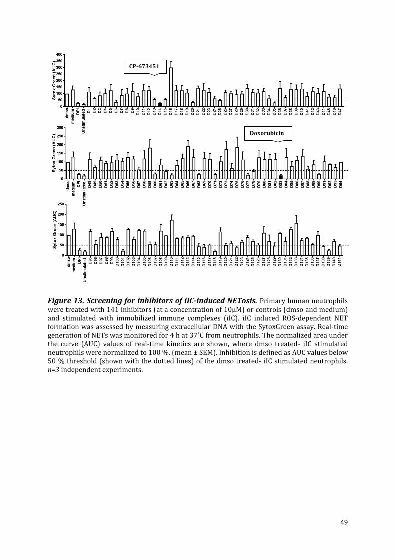
49
Figure 13. Screening for inhibitors of iIC-induced NETosis. Primary human neutrophils were treated with 141 inhibitors (at a concentration of 10µM) or controls (dmso and medium) and stimulated with immobilized immune complexes (iIC). iIC induced ROS-dependent NET formation was assessed by measuring extracellular DNA with the SytoxGreen assay. Real-time generation of NETs was monitored for 4 h at 37˚C from neutrophils. The normalized area under the curve (AUC) values of real-time kinetics are shown, where dmso treated- iIC stimulated neutrophils were normalized to 100 %. (mean ± SEM). Inhibition is defined as AUC values below 50 % threshold (shown with the dotted lines) of the dmso treated- iIC stimulated neutrophils. n=3 independent experiments.
CP-673451
Doxorubicin

50
Figure 14. Screening for inhibitors of iIC-induced ROS production. Primary human neutrophils were treated with 141 inhibitors (at a concentration of 10µM) or controls (dmso and medium) and stimulated with immobilized immune complexes (iIC). iIC induced intra-/extracellular ROS production was measured by luminol-amplified chemiluminescence assay. Real-time generation of NETs was monitored for 1 h at 37˚C from neutrophils. The normalized area under the curve (AUC) values of real-time kinetics are shown, where dmso treated-iIC stimulated neutrophils were normalized to 100 %. (mean ± SEM). Inhibition is defined as AUC values below 50 % threshold (shown with the dotted lines) of the dmso treated-iIC stimulated neutrophils. n=3 independent experiments.
CP-673451
Doxorubicin

51
3.2 Dose dependent inhibition of PMA- and iIC-induced NET formation
by CP-673451
The screening (Fig. 11-14) identified D14 (CP-673451) as a substance which inhibits
both PMA- and iIC-induced NETosis but not PMA- and iIC-induced ROS production.
Next, I investigated the effect of CP-673451 at various concentrations to exclude
potential off-target effects due to high concentrations and to determine the lowest
potential dose needed for NETosis inhibition. PMA and iIC induced ROS production and
NET formation were measured after exposure of neutrophils to different concentrations
of CP-673451. Different concentrations of CP-673451 ranging from 0.1 µM to 3 µM were
tested.
Results from the SytoxGreen and luminol kinetic assay re-confirmed the preliminary
screening results that CP-673451 inhibits PMA-induced NETosis without affecting the
PMA-induced ROS production (Fig. 15). I found that CP-673451 could inhibit PMA-
induced NETosis in a dose dependent manner. CP-673451 inhibited PMA-induced NET
formation at a concentration of as low as 0.3 µM (Fig. 15 (B, D)). However, an
inhibitory effect on ROS production was observed at a concentration of 3 µM but not in
lower concentrations (Fig. 15 (A, C)).
In order to confirm the inhibition of NETosis with CP-673451, a morphological analysis
by fluorescent microscopy (FM) was performed. SytoxGreen staining was used to view
the NET-DNA and the control nuclear DNA with fluorescent microcopy as described
previously [117]. Neutrophils pre-treated with DPI (a common inhibitor of ROS-
dependent NETosis inhibitor) [55] was also included in the staining analyses.
FM analysis confirmed the NET formation with PMA (Fig. 15-E). PMA induced a cloud
like NET structure or sometimes a NETotic cell with decondensed DNA stained with
SytoxGreen was observed (Fig. 15-E). No visible effect of dmso was observed in PMA-
induced NETosis (Fig. 15-E). Moreover, DPI treated neutrophils failed to release NETs
(Fig. 15-E). FM analyses of CP-673451 treated neutrophils confirmed the results from
the kinetic assay and showed a visible dose-dependent inhibition of PMA-induced
NETosis with 1 µM-3 µM concentrations (Fig. 15-E).

52
Similarly, the SytoxGreen and luminol kinetic assay also re-confirmed the preliminary
screening results that CP-673451 inhibits iIC-induced NETosis without affecting the iIC-
induced ROS production. CP-673451 could inhibit iIC-induced NETosis in a dose
dependent manner. CP-673451 inhibited iIC-induced NET formation at a lower
potential concentration of 3 µM (Fig. 16 (B, D)). CP-673451 showed no inhibitory effect
on iIC-induced ROS production (Fig. 16 (A, C)).
FM analysis confirmed the NET formation induced by iIC (Fig. 16-E). iIC induced NETs
were more NET fiber like structures, stained with SytoxGreen (Fig. 16-E). No visible
effect of dmso was observed in iIC-induced NETosis (Fig. 16-E). Moreover, DPI treated
neutrophils failed to release iIC-induced NET formation (Fig. 16-E). FM analyses of CP-
673451 treated neutrophils confirmed the results from the kinetic assay and showed a
visible dose-dependent inhibition of iIC-induced NETosis in the range of 0.3 µM-3 µM
concentrations (Fig. 16-E).

53
Figure 15. Dose dependent inhibition of PMA-induced NETosis by CP-673451 without inhibiting ROS production. Primary human neutrophils were treated with 0.1µM-
54
3µM of CP-673451 or controls (dmso and medium) and stimulated with PMA (20nM) or left unstimulated. PMA induced intra-/extracellular ROS production was measured by luminol-amplified chemiluminescence assay. Real-time generation of ROS was monitored for 1 h at 37˚C from neutrophils. PMA induced ROS-dependent NET formation was assessed by measuring extracellular DNA with the SytoxGreen assay. Real-time release of NETs was monitored for 4 h at 37˚C from neutrophils. Representative real-time kinetics of PMA-induced ROS production (A) and NET release (B) are shown. The normalized areas under the curve (AUC) values of real-time kinetics of ROS production (C) and NET release (D) are shown. (mean ± SEM). n = 6 independent experiments. *p < 0.05, ***p < 0.001 as compared to dmso treated PMA controls. (E) Neutrophils were treated with 3µM-0.1 µM of CP-673451 or 0.1% dmso or medium for 30 min at 37˚C, then stimulated with PMA or left unstimulated. Cells were fixed after 4 h incubation. DNA was stained with SytoxGreen. Images were taken with Keyence BZ-9000E using a 340 Plan Fluor EL NA 0.60 Ph2 objective at 100x and BZ II analyzer software. n = 2 independent experiments

55
Figure 16. Dose dependent inhibition of iIC-induced NETs by CP-673451 without inhibiting ROS production. Primary human neutrophils were treated with 0.1µM-3µM of CP-

56
673451 or controls (dmso and medium) and stimulated with iIC or left unstimulated. iIC induced intra-/extracellular ROS production was measured by luminol-amplified chemiluminescence assay. Real-time generation of ROS was monitored for 1 h at 37˚C from neutrophils. iIC-induced ROS-dependent NET formation was assessed by measuring extracellular DNA with the SytoxGreen assay. Real-time release of NETs was monitored for 7 h at 37˚C from neutrophils. Representative real-time kinetics of iIC-induced ROS production (A) and NET release (B) are shown. The normalized area under the curve (AUC) values of real-time kinetics of ROS production (C) and NET release (D) are shown. (mean ± SEM). n = 6 independent experiments. **p < 0.01 as compared to dmso treated iIC controls. (E) Neutrophils were treated with 3µM-0.1 µM of CP-673451 or 0.1% dmso or medium for 30 min at 37˚C, then stimulated with iIC or left unstimulated. Cells were fixed after 7 h incubation. DNA was stained with SytoxGreen. Images were taken with Keyence BZ-9000E using a 340 Plan Fluor EL NA 0.60 Ph2 objective at 100x and BZ II analyzer software. n = 2 independent experiments
3.3 Effect of Doxorubicin in PMA- and iIC-induced NETosis
The screening (Fig. 11-14) also identified D83 (Doxorubicin) as a substance which
inhibits both PMA- and iIC-induced NETosis but not PMA- and iIC-induced ROS
production. Therefore, I investigated the effect of Doxorubicin at various concentrations
to exclude potential off-target effects due to high concentrations and to determine the
lowest potential dose needed for NETosis inhibition. PMA and iIC induced ROS
production and NET formation were measured after exposure of neutrophils to
different concentrations of Doxorubicin (DOX). Different concentrations of Doxorubicin
ranging from 0.1 µM to 3 µM were tested.
Results from the SytoxGreen and luminol kinetic assay re-confirmed the preliminary
screening results that Doxorubicin inhibits PMA-induced NETosis. I found that
Doxorubicin could inhibit PMA-induced NETosis in a dose dependent manner.
Doxorubicin inhibited PMA-induced NET formation at a lower potential concentration
of 0.1 µM (Fig. 17 (B, D)). However, an inhibitory effect on ROS production was
observed at a concentration of 3 µM but not in lower concentrations (Fig. 17 (A, C)).
To confirm the inhibition of NETosis with Doxorubicin (DOX), a morphological analysis
by fluorescent microscopy (FM) was performed with SytoxGreen. Neutrophils pre-
treated with DPI (a common inhibitor of ROS-dependent NETosis inhibitor) were used
as positive control for NETosis inhibition. PMA induced NET formation and no visible
effect of dmso was observed in PMA-induced NETosis. Moreover, DPI treated
neutrophils failed to release NETs. However, unexpectedly FM analyses of Doxorubicin

57
(DOX) treated neutrophils did not inhibited NET formation as observed in the kinetic
assay (Fig. 17-E).
Similarly, the SytoxGreen and luminol kinetic assay was performed to confirm the
preliminary screening results that Doxorubicin (DOX) inhibits iIC-induced NETosis
without affecting the iIC-induced ROS production (Fig. 18 A-D). Doxorubicin could
inhibit iIC-induced NETosis in a dose dependent manner in the kinetic assay (Fig. 18 (B,
D)). Doxorubicin inhibited iIC-induced NET formation at a lower potential
concentration of 0.1 µM in the kinetic assay (Fig. 18 (B, D)). Doxorubicin (DOX) showed
inhibitory effect on iIC-induced ROS production at a concentration of 3 µM but not at
lower concentrations (Fig. 18 (A, C)).
FM analysis showed NET formation induced by iIC with SytoxGreen staining (Fig. 18-E).
No visible effect of dmso was observed in iIC-induced NETosis (Fig. 18-E). Moreover,
DPI treated neutrophils failed to release iIC-induced NET formation (Fig. 18-E).
However, FM analyses failed to confirm the NETosis inhibition observed in the kinetic
assay (Fig. 18-E). As the FM analyses showed no inhibition of PMA- or iIC-induced
NETosis in morphological analysis, Doxorubicin (DOX) was eliminated from further
studies (Fig. 17-E, 18-E).

58
Figure 17. Effect of Doxorubicin in PMA-induced ROS production and NET formation. Primary human neutrophils were treated with 0.1µM-3µM of Doxorubicin (DOX) or controls
59
(dmso and medium) and stimulated with PMA (20nM) or left unstimulated. PMA induced intra-/extracellular ROS production was measured by luminol-amplified chemiluminescence assay. Real-time generation of ROS was monitored for 1 h at 37˚C from neutrophils. PMA induced ROS-dependent NET formation was assessed by measuring extracellular DNA with the SytoxGreen assay. Real-time release of NET was monitored for 4 h at 37˚C from neutrophils. Representative real-time kinetics of PMA-induced ROS production (A) and NET release (B) are shown. The normalized areas under the curve (AUC) values of real-time kinetics of ROS production (C) and NET release (D) are shown. (mean ± SEM). n = 6 independent experiments*p < 0.05, **p < 0.01, ***p < 0.001 as compared to dmso treated PMA controls. (E) Neutrophils were treated with 3µM-0.1 µM of Doxorubicin (DOX) or 0.1% dmso or medium for 30 min at 37˚C, then stimulated with PMA or left unstimulated. Cells were fixed after 4 h incubation. DNA was stained with SytoxGreen. Images were taken with Keyence BZ-9000E using a 340 Plan Fluor EL NA 0.60 Ph2 objective at 100x and BZ II analyzer software. n = 2 independent experiments.
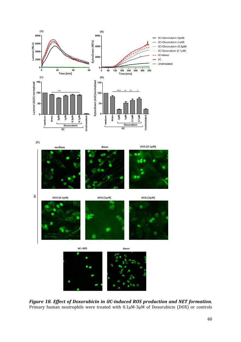
60
Figure 18. Effect of Doxorubicin in iIC-induced ROS production and NET formation. Primary human neutrophils were treated with 0.1µM-3µM of Doxorubicin (DOX) or controls

61
(dmso and media) and stimulated with iIC or left unstimulated. iIC-induced intra-/extracellular ROS production was measured by luminol-amplified chemiluminescence assay. Real-time generation of ROS was monitored for 1 h at 37˚C from neutrophils. iIC-induced ROS-dependent NET formation was assessed by measuring extracellular DNA with the SytoxGreen assay. Real-time release of NET was monitored for 7 h at 37˚C from neutrophils. Representative real-time kinetics of iIC-induced ROS production (A) and NET release (B) are shown. The normalized areas under the curve (AUC) values of real-time kinetics of iIC-induced ROS production (C) and NET release (D) are shown. (mean ± SEM). n = 6 independent experiments. *p < 0.05, **p < 0.01, ***p < 0.001 as compared to dmso treated iIC controls. (E) Neutrophils were treated with 3µM-0.1 µM of Doxorubicin (DOX) or 0.1% dmso or medium for 30 min at 37˚C, then stimulated with iIC or left unstimulated. Cells were fixed after 7 h incubation. DNA was stained with SytoxGreen. Images were taken with Keyence BZ-9000E using a 340 Plan Fluor EL NA 0.60 Ph2 objective at 100x and BZ II analyzer software. n = 2 independent experiments.
3.4 Screening for potential targets of CP-673451 involved in NETosis
inhibition
The small molecule inhibitor CP-673451 has been shown to be target selective and has a
>450-fold selectivity to PDGFRα/β over other angiogenic receptors such as
VEGFR1/2/3 [106]. However, most of the CP-673451’s inhibitory studies were
performed in tumor cells at a dosage of 1 nM-100 nM. As the inhibition of NETosis
needed a higher dosage than the previous studies, the potential off-target effects could
not be excluded. Furthermore, since CP-673451 is known to target for a certain extent
on VEGFR1/2/3, it was necessary to check whether VEGFR1/2/3 can be the target in
NETosis inhibition. The report that neutrophils eventual do not express PDGFR [99]
made a potential off target effect even more likely. Therefore, my first approach to
identify the target was to address potential off-target effects.
To investigate if inhibition of VEGFR1/2/3 is involved in CP-673451-mediated
NETosis inhibition, specific inhibitors of VEGFR 1/2/3 were tested in PMA/iIC induced
NETosis in SytoxGreen-kinetic assay system. Neutrophils were treated with
VEGFR1/2/3 inhibitors: AMG-706 (inhibitor of VEGFR 1/2/3) and ZD6474 (inhibitor of
VEGFR 2/3) and, subsequently NET formation was induced with PMA/iIC. CP-673451
treated neutrophils served as positive control for NETosis inhibition (Fig. 19).
Inhibitors of VEGFR1/2/3 AMG-706 and ZD6474 did not inhibit PMA and iIC induced
NETosis (Fig. 19). CP-673451 as shown previously (Fig. 11, 12) inhibited NET
formation (Fig.19).

62
Figure 19. Inhibitors of VEGFR1/2/3 do not inhibit PMA/iIC-induced NETosis. Neutrophils were treated with 10µM of CP-673451, AMG-706, ZD6474 or dmso or medium and stimulated with PMA or added to iIC coated surfaces or left unstimulated. The release of neutrophil extracellular traps (NETs) was monitored at 37°C, for 4 h for PMA and 7 h for iIC stimulation. Representative real-time kinetics NET release for PMA (A) and iIC (B) was measured by SytoxGreen. Normalized area under the curve (AUC) values for PMA-induced NET formation (C) and iIC-induced NET formation (D) are plotted. (mean ± SEM). n = 3, *p < 0.05 as compared to dmso treated PMA/iIC controls.

63
3.5 Effects of Sunitinib on PMA- and iIC-induced ROS and NETosis
The second approach to clarify the target selectivity of CP-673451 on the NETosis
inhibition was by using another selective inhibitor of PDGFR called Sunitinib in the
PMA- and iIC- induced ROS and NETosis assays. Neutrophils were treated with Sunitinib
and stimulated with PMA and iIC for ROS production and NETosis. Unlike CP-673451,
Sunitinib inhibited PMA and iIC induced ROS production (Fig. 20 (A, C), 21 (A, C)). In
line with the strong inhibitory effect of Sunitinib on iIC-induced ROS production, the iIC-
induced NET release was also markedly inhibited (Fig. 21). However, only a tendency of
inhibition of PMA induced NETosis was observed (Fig. 20 (B, D). These results show
that Sunitinib does not inhibit NETosis without affecting upstream ROS production and
its inhibitory effect is not NETosis specific. Therefore, studying Sunitinib cannot clarify
the effect of CP-673451 on NETosis.
Figure 20. Sunitinib partially inhibits PMA induced ROS production but not PMA-induced NETosis. Neutrophils (1x10⁶/ml in NET medium) were treated with 0.1µM-10µM of Sunitinib or dmso or medium and stimulated with PMA or left unstimulated. Production of intra- and extracellular ROS was monitored for 1 h and release of neutrophil extracellular traps (NETs) induced by PMA was monitored for 4 h at 37°C. Representative real-time kinetics of ROS production was measured by luminol (A). Representative real-time kinetics NET release was measured by SytoxGreen (B). Normalized areas under the curve (AUC) values of ROS production (C) and NET release (D) were plotted. (mean ± SEM). n = 3, *p < 0.05, **p < 0.01 as compared to dmso treated PMA controls.

64
Figure 21. Sunitinib inhibits iIC-induced ROS production and NETosis in a dose dependent manner. Neutrophils (1x10⁶/ml in NET medium) were treated with 0.1µM-10µM of Sunitinib or dmso or medium and incubated on iIC-coated surfaces or left unstimulated. Production of intra- and extracellular ROS was monitored for 1 h and release of neutrophil extracellular traps (NETs) induced by iIC was monitored for 7 h at 37°C. Representative real-time kinetics of ROS production was measured by luminol (A) and NET release was measured by SytoxGreen (B). Normalized area under the curve (AUC) values of ROS production (C) and NET release (D) are plotted (mean ± SEM). n = 3, *p < 0.05, **p < 0.01, ***p < 0.001 as compared to dmso treated iIC controls.

65
3.6 Neutrophils do not express PDGF-Receptor
Platelet-derived growth factor (PDGF) was shown to be chemotactic for neutrophils
[98]. However, later studies showed that neutrophils lack detectable mRNA for and
showed that human neutrophils do not possess functional PDGF receptor [99].
Therefore, the third approach to verify the target selectivity of CP-673451 was to
directly check the protein expression of PDGFR in human neutrophils. Western blot
analysis was performed with neutrophil lysates to check protein expression of PDGFR.
The U-87MG, human glioblastoma cells served as positive control for PDGFR expression
and T129 cell lysate was also used as positive control for PDGFR expression which was
a ready-made lysate. Western blot analysis verified the absence of protein expression of
PDGFR in human neutrophils (Fig. 22). The assay confirmed the expression of PDGFR in
U-87MG and T129 cells (Fig. 22).
Figure 22. Neutrophils do not express PDGFR. Freshly isolated neutrophils form different donors and U-87 MG cell line were lysed and analyzed by western blot with an anti-PDGFR antibody. U-87 MG cells served as positive control for expressing PDGFR. T129 cell line lysates were available ready-made, which served as an additional positive control for the PDGFR expression. Equal loading was shown by reprobing the blots with anti-human β-actin antibody. Neutrophil lysates prepared from 3 donors were analyzed (n = 3).

66
3.7 CP-673451 does not block upstream pathways of PMA-induced
ROS production involved in NETosis
Many studies have shown the activation of PKC and phosphorylation of p38-MAPK,
ERK1/2, PI3K, and Akt occur in response to various NETosis stimuli [55, 77, 80, 86].
PMA induced ROS-dependent NETosis mechanisms were followed by phosphorylation
of PKC, p38-MAPK, ERK1/2, PI3K, Akt. Although these pathways were believed to be
upstream to ROS, they are critical in NETosis [55, 80], therefore it was necessary to
check if CP-673451 treatment inhibits the phosphorylation of these molecules.
Western blot analysis of PMA stimulated neutrophils with or without CP-673451 was
analyzed for phosphorylation of PKC α/βII, p38-MAPK, ERK1/2, PI3K, and Akt.
Phosphorylation of PKC α/βII was enhanced with stimulation of PMA and PKC δ was
phosphorylated after PMA stimulation (Fig. 23). Enhanced phosphorylation of PI3K and
phosphorylation of Akt was seen after PMA stimulation (Fig. 23). The p38 MAPK and
ERK1/2 also phosphorylated after PMA stimulation (Fig. 23). The immunoblotting
results confirmed that the phosphorylation of PKC α/βII, PKC δ, p38-MAPK, ERK1/2,
PI3K, and Akt were not inhibited by CP-673451 treatment (Fig. 23). These findings
indicate that upstream pathways of PMA-induced ROS production, which are involved
in PMA-induced NET formation, are not affected by CP-673451. Interestingly, CP-
673451 alone was tending to activate p38-MAPK, ERK1/2, and Akt pathways (Fig. 23).

67
Figure 23. Signaling events activated after PMA-stimulation in neutrophils are not inhibited by CP-673451. Neutrophils were incubated with medium or dmso or 3µM CP-673451 for 30 min at 37˚C and cells were stimulated with PMA (20nM) for 20 min at 37˚C and whole-cell lysates was prepared and separated by SDS-PAGE. Phosphorylation of PKC α/β, PKC δ, PI3K, Akt, ERK1/2, and p38 MAPK was analyzed by using phospho PKC α/β, Thr638/641) (D40G4), phospho-PKC δ, phospho-PI3K p85 (Tyr458)/p55 (Tyr199), phospho-p44/42 MAPK (ERK1/2, Thr202/Tyr204), phospho-Akt (Thr308), and phospho-p38 MAPK (Thr180/Tyr182) antibodies. Equal loading was shown by reprobing the blots with anti-human β-actin antibody. Blots shown are representative for 2-3 independent experiments. (n=2-3).
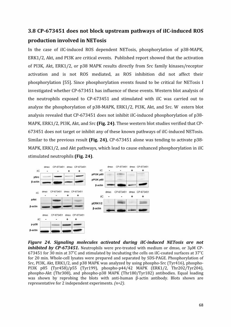
68
3.8 CP-673451 does not block upstream pathways of iIC-induced ROS
production involved in NETosis
In the case of iIC-induced ROS dependent NETosis, phosphorylation of p38-MAPK,
ERK1/2, Akt, and PI3K are critical events. Published report showed that the activation
of PI3K, Akt, ERK1/2, or p38 MAPK results directly from Src family kinases/receptor
activation and is not ROS mediated, as ROS inhibition did not affect their
phosphorylation [55]. Since phosphorylation events found to be critical for NETosis I
investigated whether CP-673451 has influence of these events. Western blot analysis of
the neutrophils exposed to CP-673451 and stimulated with iIC was carried out to
analyze the phosphorylation of p38-MAPK, ERK1/2, PI3K, Akt, and Src. W estern blot
analysis revealed that CP-673451 does not inhibit iIC-induced phosphorylation of p38-
MAPK, ERK1/2, PI3K, Akt, and Src (Fig. 24). These western blot studies verified that CP-
673451 does not target or inhibit any of these known pathways of iIC-induced NETosis.
Similar to the previous result (Fig. 24), CP-673451 alone was tending to activate p38-
MAPK, ERK1/2, and Akt pathways, which lead to cause enhanced phosphorylation in iIC
stimulated neutrophils (Fig. 24).
Figure 24. Signaling molecules activated during iIC-induced NETosis are not inhibited by CP-673451. Neutrophils were pre-treated with medium or dmso, or 3µM CP-673451 for 30 min at 37˚C and stimulated by incubating the cells on iIC-coated surfaces at 37˚C for 20 min. Whole-cell lysates were prepared and separated by SDS-PAGE. Phosphorylation of Src, PI3K, Akt, ERK1/2, and p38 MAPK was analyzed by using phospho-Src (Tyr416), phospho-PI3K p85 (Tyr458)/p55 (Tyr199), phospho-p44/42 MAPK (ERK1/2, Thr202/Tyr204), phospho-Akt (Thr308), and phospho-p38 MAPK (Thr180/Tyr182) antibodies. Equal loading was shown by reprobing the blots with anti-human β-actin antibody. Blots shown are representative for 2 independent experiments. (n=2).

69
3.9 CP-673451 treatment leads to phosphorylation of Akt, ERK1/2,
and P38 MAPK
The activation of p38 MAPK, ERK1/2 pathways are involved in oxidative burst in human
neutrophils [81, 124]. In addition, phosphorylation of p38 MAPK, ERK1/2, Akt are
important in ROS-dependent PMA- and iIC-induced NETosis [55, 80, 86]. Interestingly,
the western blot studies for phospho-proteins after PMA- and iIC- stimulation revealed
an enhanced phosphorylation of p38 MAPK and Akt by CP-673451 alone without any
additional stimulation compared to the dmso treated neutrophils (Fig. 25 (A, B)). In
addition, ERK1/2 also tends to be phosphorylated upon CP-673451treatment (Fig. 25
(C)) compared to the dmso treated neutrophils.
Figure 25. CP-673451 treatment leads to phosphorylation of p38 MAPK, ERK1/2, and Akt in neutrophils. Neutrophils were incubated with dmso or 3µM CP-673451 for 50 min at 37˚C and whole-cell lysates was prepared. dmso treated neutrophils served as solvent control. Whole-cell lysates were separated by SDS-PAGE. Phosphorylation of Akt, ERK1/2, and p38 MAPK was analyzed by using phospho-44/42 ERK1/2 (Thr202/Tyr204), phospho-Akt (Thr308), and phospho-p38 MAPK (Thr180/Tyr182) antibodies. Equal loading was shown by reprobing with anti-human β-actin antibody (A-C). Blots shown are representative of 4-5 independent experiments. For quantification the signals, ratio of pAkt, pERK, and pp38 to β-actin signal were determined using Bo 1D software. Mean ± SEM values of the ratios are plotted in bar graph (A-C). (n=4-5). *p <0.05, **p <0.01, ns=non-significant as compared with solvent control (0.1% dmso).

70
3.10 Screening for changes in tyrosine, serine, and threonine
phosphorylated proteins upon CP-673451 treatment
The eukaryotic protein kinases that directly phosphorylate proteins are divided into
two major classes: those that phosphorylate tyrosine and those that phosphorylate
serine and threonine [125]. Phosphorylation of several proteins are involved in PMA
induced NETosis [80]. To assess if the phosphorylation events induced by PMA is
affected by CP-673451, the tyrosine, threonine, and serine phospho proteins were
screened by immunoblotting.
The phosphorylated tyrosine and serine proteins present in PMA stimulated (Ctrl)
and PMA stimulated and CP-673451 treated (tr) samples had showed similar pattern of
the phospho-protein bands. However, threonine phosphorylated proteins had two clear
bands missing in the CP-673451 treated (tr) neutrophil lysates compared to the control
(Ctrl) (Fig. 26-A). This indicated an inhibitory action by CP-673451 in phosphorylation
of two, so far unknown proteins. According to the molecular weight marker, the bands
were of sizes between 30 kDa and 20 kDa (Fig. 26-B).
To identify the proteins corresponding to the missing bands, all the proteins other than
phospho-threonine proteins needed to be removed so that the purified protein bands
corresponding to missing bands can be analyzed and identified. Therefore, phospho-
protein-enrichment was performed, which purified phospho threonine proteins by
using an anti-phospho threonine antibody immobilized to Sepharose beads. Neutrophils
were treated or untreated with CP-673451 and stimulated with PMA for 20 min. The
lysates were prepared using 1xRIPA buffer containing protease inhibitors. The lysates
were then incubated overnight with the anti-phospho threonine antibody immobilized
to Sepharose beads and washed with the lysis buffer. The proteins were denaturated in
presence of SDS and β-mercapto ethanol and centrifuged to separate the proteins and
beads. The proteins electrophoresed on 10 % SDS-PAGE gels and the gels were then
stained with Coomassie stain to detect proteins. The phospho-threonine bands of sizes
between 30 kDa and 20 kDa were observed. Unfortunately, the bands were not visible
after Coomassie staining. Therefore, I was not able to cut them out for further protein
identification (Fig. 26 (B)).
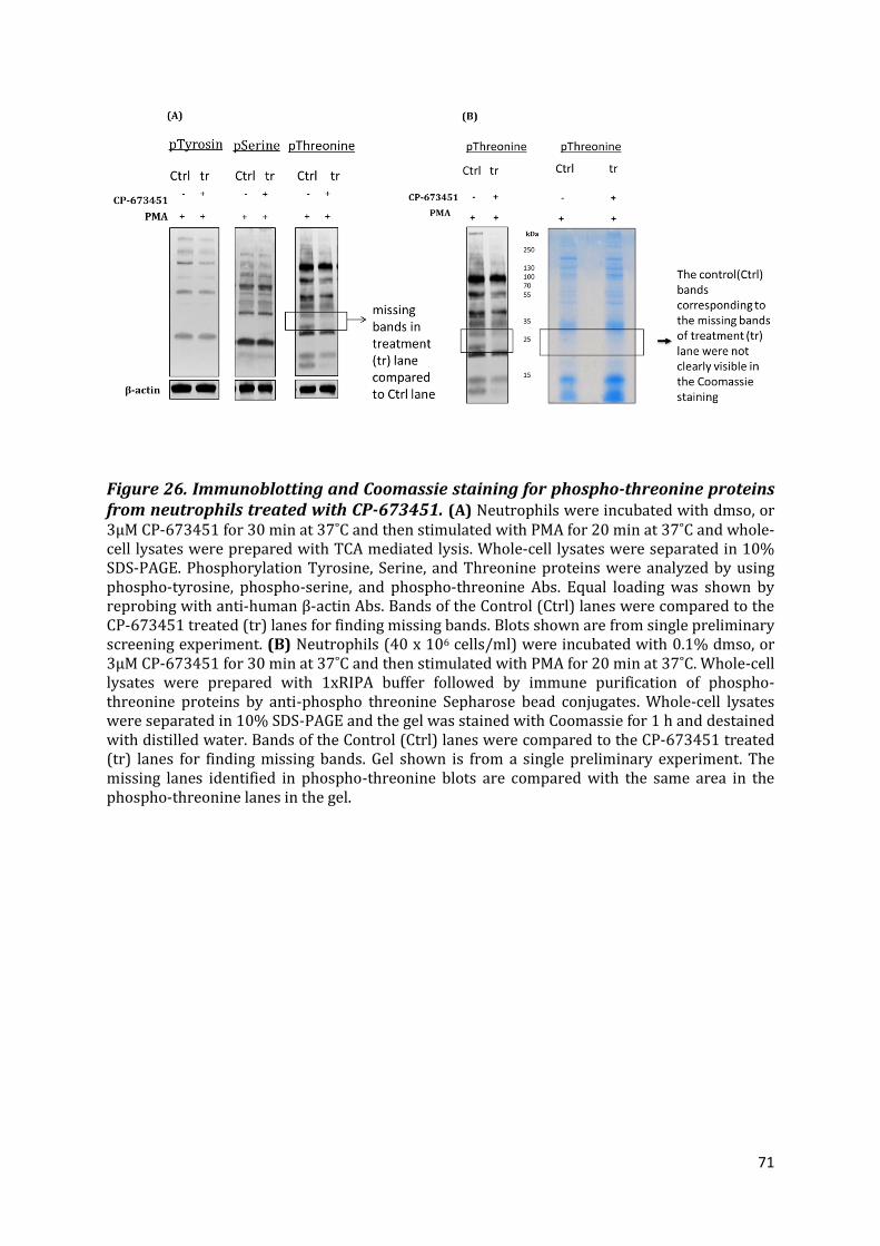
71
Figure 26. Immunoblotting and Coomassie staining for phospho-threonine proteins from neutrophils treated with CP-673451. (A) Neutrophils were incubated with dmso, or 3µM CP-673451 for 30 min at 37˚C and then stimulated with PMA for 20 min at 37˚C and whole-cell lysates were prepared with TCA mediated lysis. Whole-cell lysates were separated in 10% SDS-PAGE. Phosphorylation Tyrosine, Serine, and Threonine proteins were analyzed by using phospho-tyrosine, phospho-serine, and phospho-threonine Abs. Equal loading was shown by reprobing with anti-human β-actin Abs. Bands of the Control (Ctrl) lanes were compared to the CP-673451 treated (tr) lanes for finding missing bands. Blots shown are from single preliminary screening experiment. (B) Neutrophils (40 x 106 cells/ml) were incubated with 0.1% dmso, or 3µM CP-673451 for 30 min at 37˚C and then stimulated with PMA for 20 min at 37˚C. Whole-cell lysates were prepared with 1xRIPA buffer followed by immune purification of phospho-threonine proteins by anti-phospho threonine Sepharose bead conjugates. Whole-cell lysates were separated in 10% SDS-PAGE and the gel was stained with Coomassie for 1 h and destained with distilled water. Bands of the Control (Ctrl) lanes were compared to the CP-673451 treated (tr) lanes for finding missing bands. Gel shown is from a single preliminary experiment. The missing lanes identified in phospho-threonine blots are compared with the same area in the phospho-threonine lanes in the gel.
72
3.11 CP-673451 does not inhibit Ionomycin induced-NETosis
NETosis can be induced by various stimuli and based on the type of stimulus there are
different pathways described [55, 70, 74, 78, 80, 86, 88, 126]. PMA and iIC induce
NETosis in a ROS-dependent manner, where NADPH-oxidase generated ROS plays a
major role in inducing NETosis [55, 80, 127]. However, there is ROS-independent
NETosis such as the NETosis induced by Ionomycin, where inhibition of NADPH oxidase
does not interrupt NETosis [88]. The signaling mechanisms involved in Ionomycin
induced NET formation have been shown to differ from the ones induced by PMA and
iIC [41, 88].
My present study has shown that CP-673451 treatment does not inhibit ROS or
activation of the signaling pathways upstream to ROS is involved in downstream
inhibition of NETosis. In a preliminary experiment, different concentrations (2.57 µM-7
µM) of Ionomycin were tested for inducing NETosis (Fig. 27 (A, C)). An optimal
concentration of 5 µM of Ionomycin found to induce NETosis was decided to use in
further assays. The effect of CP-673451 (0.1 µM -10 µM) on Ionomycin induced NETosis
was tested (Fig. 27 (E-F)), to check if CP-673451 can serve as a general inhibitor of
NETosis. The study showed that, CP-673451 did not inhibit the Ionomycin induced NET
formation even at 10 µM concentrations (Fig. 27 (E, F)). In addition, DPI, an inhibitor of
NADPH-oxidase also did not inhibit Ionomycin-induced NETosis, as it is a NADPH-
oxidase-independent type of NETosis (Fig. 27 (B, D)).

73
Figure 27. CP-673451 does not inhibit Ionomycin- induced NETosis. (A) Neutrophils were stimulated with Ionomycin (IO) (2.5 µM, 5 µM, and 7 µM) or left unstimulated and the release of neutrophil extracellular traps (NETs) was monitored for 4 h at 37°C, measured by SytoxGreen. (B) Neutrophils (1x10⁶/ml) were treated with medium or DPI (20 nM) and then stimulated with IO (5 µM) or PMA (20nM) or left unstimulated. The release of neutrophil extracellular traps (NETs) was monitored for 4 h at 37°C, measured by SytoxGreen. (C, D) Areas under the curve (AUC) values (mean ± SEM) of fluorescence intensities (RFUs) were plotted. (E) Neutrophils (1x10⁶/ml) were treated with 0.1 µM-10 µM of CP-673451 or dmso and stimulated with IO (5 µM) or left unstimulated. The release of neutrophil extracellular traps (NETs) was monitored for 4 h at 37°C, measured by SytoxGreen. (F) Area under the curve (AUC) values (mean ± SEM) of fluorescence intensities (RFUs) was plotted. n = 3, **p <0.01, ns=non-significant as compared with the respective positive controls.
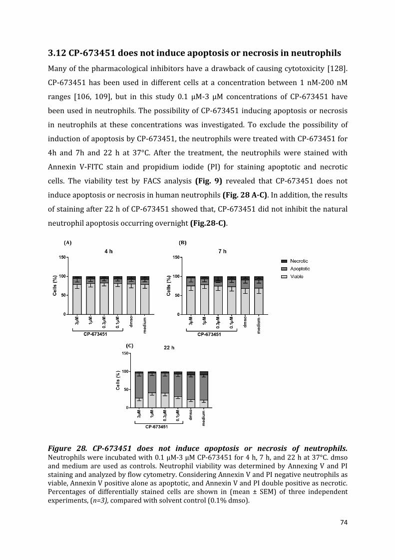
74
3.12 CP-673451 does not induce apoptosis or necrosis in neutrophils
Many of the pharmacological inhibitors have a drawback of causing cytotoxicity [128].
CP-673451 has been used in different cells at a concentration between 1 nM-200 nM
ranges [106, 109], but in this study 0.1 µM-3 µM concentrations of CP-673451 have
been used in neutrophils. The possibility of CP-673451 inducing apoptosis or necrosis
in neutrophils at these concentrations was investigated. To exclude the possibility of
induction of apoptosis by CP-673451, the neutrophils were treated with CP-673451 for
4h and 7h and 22 h at 37°C. After the treatment, the neutrophils were stained with
Annexin V-FITC stain and propidium iodide (PI) for staining apoptotic and necrotic
cells. The viability test by FACS analysis (Fig. 9) revealed that CP-673451 does not
induce apoptosis or necrosis in human neutrophils (Fig. 28 A-C). In addition, the results
of staining after 22 h of CP-673451 showed that, CP-673451 did not inhibit the natural
neutrophil apoptosis occurring overnight (Fig.28-C).
Figure 28. CP-673451 does not induce apoptosis or necrosis of neutrophils. Neutrophils were incubated with 0.1 µM-3 µM CP-673451 for 4 h, 7 h, and 22 h at 37°C. dmso and medium are used as controls. Neutrophil viability was determined by Annexing V and PI staining and analyzed by flow cytometry. Considering Annexin V and PI negative neutrophils as viable, Annexin V positive alone as apoptotic, and Annexin V and PI double positive as necrotic. Percentages of differentially stained cells are shown in (mean ± SEM) of three independent experiments, (n=3), compared with solvent control (0.1% dmso).

75
As necrotic cells disintegrate rapidly, these cells would not be counted by FACS.
Therefore, an LDH release assay was performed after treating neutrophils with CP-
673451 for 4h and 7h at 37°C to confirm its non-toxicity. The amounts of LDH released
after each time points were correlated to the necrosis rate. The LDH assay results were
in agreement with the FACS results, confirming no induction of necrosis due to CP-
673451 treatment (Fig. 29).
Figure 29. CP-673451 does not induce cytotoxicity in neutrophils. Neutrophils were incubated for 1 h and 7 h at 37˚C with 0.1 µM-3 µM CP-673451 or with dmso or NET-medium. The LDH released into the medium was transferred to a new plate. Additionally, LDHmax (LDH maximum) samples, with completely lysed cells, and LDHspont (LDH spontaneous) samples from untreated cells were also prepared. The supernatants of the differentially treated cells were mixed with Reaction Mixture (from the kit) and incubated for 30 min at RT Reactions were stopped by adding Stop Solution (from the kit). LDHmax (LDH maximum) samples served as positive control for lysis/cytotoxicity and LDHspont (LDH spontaneous) served as negative control for lysis/cytotoxicity. Absorbance at 490 nm and reference 680 nm was measured using a spectrophotometer. LDH activity was determined from the OD values obtained whose (mean ± SEM) are shown in of three independent experiments, (n=3), compared with solvent control (dmso).
3.13 CP-673451 does not affect neutrophil effector functions other
than NETosis
To use CP-673451 as NETosis inhibitor in future therapeutic options, it is important to
check whether CP-673451 is affecting or inhibiting neutrophil functions other than
NETosis. At the same time, studying CP-673451 in other functions might provide hint to

76
the pathway it is targeting. Therefore, basic neutrophil functions including; activation,
migration and phagocytosis were assessed after treatment with CP-673451.
3.13.1 CP-673451 does not affect neutrophil phagocytic ability
Phagocytosis is a major anti-microbial function of neutrophils [13, 14]. Inhibiting
NETosis and phagocytosis at the same time could lead to reduced anti-microbial defense
against pathogens. Neutrophils pre-treated with CP-673451 was stimulated with LPS
and IFNᵧ and allowed to phagocytose fluorescent latex beads or S. aureus bioparticles.
The phagocytosis of beads and bioparticles was analyzed by FACS (Fig. 8).
The phagocytosis rate of beads and S. aureus by neutrophils pre-treated with CP-
673451 was not different from the control dmso treated neutrophils after stimulation
(Fig. 30). Hence, no inhibition of the phagocytic function was observed due to CP-
673451 treatment in neutrophils (Fig. 30).
Figure 30. CP-673451 does not inhibit phagocytic function of neutrophils. Neutrophils were incubated with 0.1 µM-3 µM CP-673451 or dmso or medium for 30 min at 37°C. The cells were then co-incubated with FluoSphere carboxylate-modified latex beads added in 1 to 10 ratios to neutrophils (A) or Alexa-Fluor 488 conjugated non-viable Staphylococcus aureus bioparticles in 2:1 ratio; S. aureus to neutrophils (B). Cells were then stimulated with LPS (100ng/ml) and IFNᵧ (200 U/ml) for 30 min at 37°C. Trypan blue was added to quench the fluorescence of extracellular bacteria/beads sticking on the neutrophil surface. Phagocytosis was assessed by flow cytometry and analyzed by FACS Diva software. Percentage of the cells with ingested beads or bioparticles were measured and values were normalized to medium control. Values ± SEM are shown, (n=4) as compared with solvent control (0.1% dmso).

77
3.13.2 The effect of CP-673451 on neutrophil migration
It is important for neutrophils to be able to migrate to the site of infection, mostly by
chemotaxis. Therefore, I tested whether CP-673451 has an influence on the migration of
neutrophils in vitro. CP-673451 treated neutrophils were tested, for chemotaxis, where
the migration is directional towards stimulants like IL-8 [129] and for chemokinesis,
where there is a random migration in response to stimulants like TNF-α [130].
Neutrophils were pre-treated with CP-673451 and allowed to migrate on a trans well
plate with the stimulants IL-8 for chemotaxis and TNF-α for chemokinesis. The migrated
cells were lysed and quantified with the help of β-glucuronidase assay and migration
index values were calculated. Chemotaxis of neutrophils towards IL-8 was not affected
by CP-673451 treatment (Fig. 31). However, TNF-α mediated random migration was
inhibited by 3µM concentrations of CP-673451 (Fig. 31).
Figure 31. CP-673451 does not inhibit neutrophil chemotaxis but affects chemokinesis at higher concentrations. Neutrophils were preincubated with 0.1 µM-3 µM CP-673451 or dmso, or medium for 30 min at 37°C. The neutrophil suspension was transferred to trans well filter in 24 well plate which in its lower well contained chemotactic stimulus, IL-8 (25 ng/ml), or the chemokinesis stimulant, TNF-α (50 ng/ml) or medium. The cells were allowed to migrate for 1h at 37°C. The migrated cells were then lysed with of 1% Triton X-100. parallelly, untreated fresh neutrophil lysates were transferred to a 96 well plate and serially diluted. β-glucuronidase assay was carried out in acetate buffer, containing 4-Nitrophenyl β-D-glucuronide and incubated overnight at 37°C. The reaction was stopped by glycine buffer and the absorbance of the lysates was then measured at 405 nM and reference 620 nM. A standard curve was obtained with serially diluted known concentrations of the cells and OD values were used to determine migration index. The migration index values were normalized to stimulant control. (± SEM). (n=3), *p < 0.05 as compared with solvent control (0.1 % dmso).

78
3.13.3 CP-673451 does not inhibit neutrophil activation
Neutrophil activation plays an important role in the inflammatory response to bacterial
infections. Many stimuli like LPS have been shown to be a major mediator of neutrophil
activation which is accompanied by an early down-regulation of L-selectin (CD62L) and
up-regulation of CD11b on the neutrophil surface [131]. To investigate whether CP-
673451 affects the activation of the neutrophils, neutrophils were pre-treated with CP-
673451 and stimulated with LPS and IFNᵧ, TNF-α, and fMLP. The shedding of CD62L
and upregulation of CD11b was assessed by staining and FACS analysis (Fig. 7). CP-
673451 treatment did not inhibit neutrophil activation in response to any of the stimuli
tested (Fig. 32). Interestingly, with CP-673451 treatment appeared to have a tendency
to enhance the activation phenotype induced by LPS and IFNᵧ, TNF-α, and fMLP (Fig.
32).
Figure 32. CP-673451 does not inhibit LPS+IFNᵧ-, TNF-α-, and fMLP-mediated neutrophil activation. Neutrophils were incubated with 0.1 µM-3 µM CP-673451 or 0.1% dmso or medium for 30 min at 37°C. The cells were then stimulated with LPS (100ng/ml) together with IFNᵧ (200 U/ml), TNF-α (100ng/ml), and fMLP (1µM) for 4 h at 37°C. The cells were then stained with fluorescent-conjugated antibodies against CD11b and CD62L for 30 min at 4°C. Percentage of the cells upregulating CD11b and shedding CD62L was assessed by flow cytometry and analyzed by FACS Diva software. Percentage mean values of neutrophils upregulating CD11b and shedded CD62L are shown, (± SEM), (n=3) as compared with solvent control (dmso).

79
3.14 CP-673451 activates neutrophils
As neutrophils treated with CP-673451 showed a tendency of enhanced activation in
response to stimulants, neutrophils treated with CP-673451 alone were tested for the
activation phenotype by measuring upregulation of CD11b and shedding of CD62L (Fig.
7). Interestingly, CP-673451 could activate neutrophils in a dose dependent manner
which was assessed by upregulation of CD11b and shedding of CD62L by FACS analysis
(Fig. 33). The neutrophil activation by CP-673451 was comparable to LPS-mediated
neutrophil activation (Fig. 33).
Figure 33. CP-673451 activates neutrophils in a dose dependent manner. Neutrophils were incubated for 30 min at 37°C. The cells were then co-incubated with 0.1 µM-10 µM CP-673451 or 0.1% dmso or medium or with LPS (100ng/ml) for 4 h at 37°C. The cells were then stained with fluorescent-conjugated antibodies against CD11b and CD62L for 30 min at 4°C. Percentage of the cells upregulating CD11b and shedding CD62L was assessed by flow cytometry and analyzed by FACS Diva software. Percentage mean values of neutrophils upregulating CD11b and shedded CD62L are shown, (± SEM), (n=3), *p <0.05, **p <0.01, as compared with solvent control (dmso).
3.15 The neutrophil-activating property of CP-673451 is not due to
endotoxin-contamination
Dose dependent activation of neutrophils by CP-673451 was an unexpected result and
the inhibitor molecule has not been reported to exert such property. I therefore
suspected that endotoxin contamination could be responsible for the neutrophil
activating activity of CP-673451. To exclude this possibility, neutrophils were treated
with Polymyxin-B (PMB) and stimulated by CP-673451 or LPS. Polymyxin B (PMB) is
able to neutralize the effect of endotoxin [132]. Activation phenotype was assessed by

80
measuring upregulation of CD11b and shedding of CD62L FACS staining (Fig. 7). PMB
treatment neutralized LPS mediated neutrophil activation, hence no upregulation of
CD11b and shedding of CD62L were observed (Fig. 34). However, CP-673451 mediated
dose-dependent neutrophil activation was not blocked by polymyxin-B, indicating that
the activating property of CP-673451 is not due to LPS contamination (Fig. 34).
Figure 34. Activation property of CP-673451 in neutrophils is not due to endotoxin contamination. Neutrophils were incubated with Polymyxin-B (PMB) (10 µg/ml) or with medium for 30 min at 37°C. The cells were then stimulated with 0.1 µM-10 µM CP-673451 or 0.1% dmso or medium or with LPS (100 ng/ml) for 4 h at 37°C. The cells were then stained with fluorescent-conjugated antibodies against CD11b and CD62L for 30 min at 4°C. Percentage of the cells upregulating CD11b and shedding CD62L was assessed by flow cytometry and analyzed by FACS Diva software. Percentage mean values of neutrophils upregulating CD11b and shedded CD62L are shown, (± SEM), (n=3), *p <0.05, **p <0.01, as compared with solvent control (dmso).
3.16 CP-673451 induces differential gene expression changes in
human neutrophils
The relevance of transcription and translation in NETosis remains unclear. A previous
study argues that PMA-induced NETosis is transcription and translation independent
and neutrophils contain all the factors required for NET formation when they emerge
from the bone marrow as differentiated cells [76]. However, a recent study suggested
that kinases including Erk1/2, cSrc, p38, and Akt activate transcription during PMA-
induced NETosis and inhibition of transcription inhibits PMA-induced NETosis without
affecting the ROS production [133]. As the relevance of transcriptional responses in CP-
673451-mediated signaling in neutrophils were not known and as CP-673451 itself

81
activates Erk1/2, p38, and Akt pathways, its role in modulating gene expression in PMA-
induced NETosis was needed to be clarified. In addition, inhibition of transcription
could inhibit PMA-induced NETosis without inhibiting ROS production. Therefore, the
ability of CP-673451 to suppress transcription was also needed to be analyzed.
Neutrophils were pre-treated with 3µM CP-673451 or dmso and stimulated with PMA
or left unstimulated. Total RNA was extracted and a high throughput RNA Seq was
performed and differential expression of genes was assessed in three independent
experiments. A principal component analysis (PCA) was performed to evaluate the
differences and relatedness of transcription profiles among different conditions. PCA
clustered the multidimensional data into assigned sets of variables and showed
transcription profile patterns in each condition. These analyses showed that
transcriptomes of unstimulated, CP-673451 -treated, PMA-stimulated (NET-forming)
and CP-673451 -treated-PMA-stimulated (NET-inhibiting) neutrophils form distinct
clusters (Fig. 35).
Figure 35. Principle component analysis (PCA) showing differences in transcription profiles among differently treated neutrophils. Neutrophils (10x106 in 1 ml NET-medium) were pre-treated with 3µM CP-673451 or dmso for 30 min at 37°C and stimulated with PMA or left unstimulated 3 h 30 min at 37°C. The total RNA was extracted and a high throughput RNA Seq was performed. The differential transcriptional patterns under each condition were assessed by principle component analysis. The PCA was calculated using the R/Bioconductor package labdsv and based on the transcript per million (TPM) values of the 10000 most variable genes across samples according to their interquartile range (IQR). The three different colors represent different treatments. The green dots; neutrophils treated with diluent control (dmso), black dots; neutrophils treated with CP-673451, red dots; CP-673451 treated neutrophils stimulated with PMA, blue dots; neutrophils stimulated with PMA. (n=3).

82
The principle component analysis (PCA) (Fig. 35) clearly shows the related
transcriptional patterns clustering together with three different donors (showed as
three dots of same color) of same treatment. The data also shows a separated cluster for
unstimulated-dmso treated control (Ctrl) samples from all the other conditions. There
was a clear separation of the samples along the PCA axis (Fig. 35). The CP-673451
treated neutrophil samples also stand out (Fig. 35). Interestingly, there were also
differences between PMA stimulated samples compared to CP-673451 pre-treated-PMA
stimulated samples (Fig. 35). This indicates CP-673451’s involvement in changing
PMA-induced transcriptional pattern.
Comparing the up and down regulated genes of untreated and CP-673451-treated
neutrophils might further provide the involvement of CP-673451 in modulation
neutrophil gene expressions. Furthermore, involvement of CP-673451 in inhibiting
NETosis can be analyzed by comparing the differentially up and down regulated genes
of PMA stimulated neutrophils and CP-673451 treated- PMA stimulated neutrophils.
These data have not been analyzed in detail due to lack of time, but the preliminary
analysis data are provided (in the supplementary CD attached).

83
Chapter 4. Discussion
4.1 CP-673451 induces neutrophil activation and inhibits an
unknown pathway of NETosis, downstream of ROS production
Neutrophils, the first cells to arrive at an infection site exert several potent
antimicrobial mechanisms. NET formation is one of these mechanisms. NETs can
capture and kill the invading pathogen [1, 2]. On the other hand, recent studies show
that many diseases are associated with NETosis including autoimmune diseases [38, 60,
65, 134]. Since NETs expose potential autoantigens such as histones, dsDNA, and
granule enzymes they have been suspected to mediate autoimmune responses due to
immune complexes (ICs) formation [55, 62, 135-137]. However, the molecular
mechanism behind NET formation is still not clear.
There are two main NETosis mechanisms described; the classical, reactive oxygen
species (ROS)-dependent and the early/rapid ROS-independent pathway [32, 33]. Most
of the stimuli described to induce NETosis are dependent on ROS production by the
NADPH- oxidase complex [34]. The importance of ROS production in NETosis has been
shown with the help of several enzymatic inhibitor of NADPH-complex, showing
inhibition of ROS production affects NETosis [30, 70, 87, 127]. Moreover, neutrophils
from CGD patients, who lack a functional NADPH also failed to release NETs [38, 93].
However, it is not yet clear which pathways are involved in NET formation downstream
of ROS production.
In this study, I studied two stimuli; PMA and immobilized immune complex (iIC),
both inducing ROS-dependent NETosis. PMA is a synthetic stimulus. The PMA induced
ROS-dependent NETosis has been studied extensively [138]. Immune complex (iIC) is a
physiological stimulus which has been recently shown to induce NETosis [55]. In this
study, I identified two inhibitors; CP-673451 (targeting PDGFR) and Doxorubicin
(targeting Topoisomerase II) inhibiting PMA and iIC-induced NETosis without affecting
PMA and iIC induced ROS production (Fig. 11-14). Because the inhibitors were
specifically inhibiting NETosis but not the upstream ROS production, the inhibitors
were believed to be targeting downstream of ROS production pathways and upstream
of NETosis.

84
Although Doxorubicin was seen to inhibit PMA- and iIC-induced NETosis in a dose
dependent manner in SytoxGreen kinetic assays, Doxorubicin did not show NETosis
inhibition in morphological analysis (Fig. 17, 18). Moreover, other inhibitors targeting
Topoisomerase II did not inhibit NETosis (data not shown). In addition, Doxorubicin
was not toxic to neutrophils (data not shown). As morphological analysis could not
confirm the kinetic assay results, Doxorubicin was eliminated from the later studies.
The study couldn’t reveal why Doxorubicin showed inhibitory effect on SytoxGreen
signal in the kinetic assay. It was suspected that Doxorubicin might be interfering with
the SytoxGreen signal detection. It has been in discussion in studies that small molecule
libraries used for screening are themselves fluorescent, leading to potential false
positive results through interference [139]. Interestingly, Doxorubicin itself was
fluorescent, which could have led to the false positive inhibitory readout through
interference. Fortunately, a morphological confirmation was applied in this research
which revealed the artifact. A test experiment with DNA together with Doxorubicin by
using the SytoxGreen detection assay could clarify the hypothesis of signal interference
caused by Doxorubicin. As there are several studies based on small molecule libraries
carried out in drug discovery research, it is vital to cut out assay artifacts as early as
possible to avoid spending time and resources pursuing compounds that are not
actually impacting the desired biology but rather are false positives [139].
The small molecule inhibitor, CP-673451 was found inhibiting PMA- and iIC-induced
NETosis in a dose dependent manner in the SytoxGreen-kinetic assay and the results
were in line with the morphological analysis (Fig. 15). In the morphological analyses,
PMA induced NETosis was seen as a cloud like structures or with a very densely packed
DNA in a large NETotic cell stained with SytoxGreen, this phenotype was consistent with
the previous studies [140].
The lowest effective dose of CP-673451 to inhibit PMA-induced NETosis was found to
be 0.3 µM in the SytoxGreen kinetic assay (Fig. 15). To confirm the kinetic assay results,
morphological observations of the neutrophils were performed with SytoxGreen
staining. However, in the morphological observation 1 µM concentration of CP-673451
was needed to inhibit PMA-induced NETosis. The nuclear morphology of neutrophils
was not very clear to examine with the SytoxGreen staining. The CP-673451-treated-
PMA stimulated neutrophils were a slightly larger than the untreated cells and the

85
nuclear lobes were not as clearly seen as in untreated cells (Fig. 15-E). 3µM CP-673451-
treated-PMA stimulated neutrophil’s inhibitory morphology was comparable with DPI-
treated PMA stimulated neutrophils. Similarly, CP-673451 also inhibited iIC-induced
NETosis at a lowest dose of 1µM (Fig. 16). The iIC-induced NETs were thin fiber-like
structures. SytoxGreen staining could clearly show the inhibition of the NET formation
with CP-673451 (Fig. 16-E). However, to visualize the NET-fiber structures properly,
electron microscopic pictures need to be taken.
The most widely used method of NETosis detection is a nuclear staining using a non-
cell permeable dye, SytoxGreen, which is the same technique used in this study. Since
necrotic cells can be stained with SytoxGreen, an assay for neutrophil viability was
conducted. The possibility of CP-673451 inducing apoptosis in neutrophils was
investigated. I found that CP-673451 does not induce neutrophil apoptosis or necrosis
(Fig. 28 (A-C)) and it does not delay or inhibits neutrophil apoptosis (Fig. 28-C). This
indicates that the neutrophil apoptosis pathway has been not affected or inhibited by
CP-673451. The study also excluded that CP-673451 induces necrosis (Fig. 28, 29). The
results of FACS staining with the cell permeable dye propidium iodide and the LDH
release assay excluded the possibility of CP-673451 to cause cytotoxicity (Fig. 28, 29).
These results collectively indicate the specificity of the inhibitor towards specific cell
death, the NETosis. CP-673451 did not induce cytotoxicity (Fig. 29), which makes it
suitable for studies in future therapeutic options for NET-related diseases.
Even though, the study identified CP-673451 as an inhibitor of NETosis, the target of
this inhibitor was needed to be confirmed. However, the pharmacological inhibitors
often show off-target effects which complicate the interpretation of the results and the
understanding of the precise role of the involved receptors and mode of action of the
inhibitors [106]. CP-673451 is a known inhibitor of platelet growth factor receptor
(PDGFR). CP-673451 has shown to be effective against PDGFR at concentrations ranging
from 1 nM-100 nM in several tumor cells. CP-673451 is known to exhibit >450-fold
selectivity toward PDGFR over other angiogenic receptors including vascular
endothelial growth factor receptor- 1, -2, and -3 (VEGFR1/2/3) [106]. In this study, I
have used higher concentrations (up to 3 µM) of CP-673451 for NETosis inhibition (Fig.
15-16), which could lead to off-target effects. Thus, other known targets of CP-673451
such as VEGFR1/2/3 were investigated for playing a role in NETosis. Inhibitors of

86
VEGFR1/2/3 were tested in PMA and iIC-induced NETosis assay, but these inhibitors
could not inhibit NETosis even at a concentration of 10µM (Fig. 19). This showed that
VEGFR1/2/3 were not the target of CP-673451 which is involved in NETosis. However,
this finding does not confirm PDGFR as the target of CP-673451 in neutrophils.
To check if PDGFR is the target involved in NETosis inhibition, another widely available
PDGFR inhibitor, Sunitinib [141] was tested in PMA- and iIC-induced NET assays.
However, Sunitinib did not inhibit PMA-induced NETosis but inhibited iIC-induced
NETosis. However, Sunitinib inhibited also iIC-induced ROS and partially affected PMA-
induced ROS, which indicates that Sunitinib seems to be affecting some other off target
pathways in neutrophils which affect ROS production (Fig. 20, 21). Collectively,
Sunitinib did not act on neutrophils at a similar way as CP-673451, indicating that
inhibition of PDGFR might not be leading to ROS-independent-NETosis-inhibition.
Although in 1988 platelet derived growth factor (PDGF) was shown to be chemotactic
for neutrophils, in 1995 it was showed that human neutrophils lack functional PDGFRs
[98, 99]. As PDGFR is the major target of CP-673451 and the presence of PDGFR in
neutrophils were unclear, I assessed protein expression of PDGFR in human neutrophils
by immunoblotting. I detected no protein expression of PDGFR in neutrophils. This
result was in agreement with another study in 1995 which also showed that neutrophils
lack detectable mRNA for PDGF-receptors [99] (Fig. 22), implying that PDGFR is
apparently not the target of CP-673451 regarding inhibition of NETosis.
PMA is the most frequently used compound to induce ROS-dependent NETosis,
signaling mechanisms involved have been studied broadly [55, 68, 80]. PMA stimulation
leads to direct activation of protein kinase-c (PKC) in neutrophils. NETosis induced by
PMA is PKC and NADPH oxidase (NOX) dependent. PMA stimulates conventional (α, βI,
βII, γ) and novel (δ, ε, η, θ) PKC isoforms [77]. Conventional PKCs have a prominent role
in NET formation. PKCβ is the major isoform crucial in NET formation [78]. PKC
downstream phosphorylates p38-MAPK, ERK1/2, Akt and activates NADPH oxidase [55,
77, 78, 80, 81]. Activation of p38-MAPK and ERK1/2 has been identified to be involved
in ROS generation [78, 80]. However, some studies also found that these pathways are
downstream of ROS production and inhibiting the ROS will also inhibit p38-MAPK,
ERK1/2 activation [81]. PMA-induced NETosis also depends on Akt activation and Akt
activation is downstream of ROS production [86].

87
As it was confirmed from my present study that CP-673451 does not inhibit ROS, it
was also necessary to check if CP-673451 does not inhibit the activation of known
signaling molecules upstream of NETosis. I found that PMA tends to enhance the
induced phosphorylation of conventional isoform, PKC α/β and PMA-induced
phosphorylation of novel PKC isoform, PKC δ (Fig. 23). This results was in agreement
with the activation studies of PKC isoforms induced by PMA [78]. In addition, PMA
induced phosphorylation of p38-MAPK, ERK1/2, and Akt was observed in this study
(Fig. 23). This was in line with the previous results indicating the significance of these
pathways in PMA-induced NETosis [55, 78, 80, 86]. My present study showed that
activation of PKC α/β, PKC δ, p38-MAPK, ERK1/2, and Akt through phosphorylation was
not inhibited by CP-673451 (Fig. 23). This showed that CP-673451 does not inhibit
PMA-induced NETosis by inhibiting any of these molecular targets and indicates that
the target of CP-673451 is probably downstream of PKC, p38-MAPK, ERK1/2, Akt, and
oxidative burst pathways.
Unlike PMA, iIC stimulation in neutrophils requires surface molecule mediated
activation. iIC has been shown to induce ROS production and NET formation in
neutrophils involving activation of FcᵧRIIIB and Mac-1, and further downstream
activating Syk/Src signaling, which further activates ERK1/2, PI3K/Akt, and p38 MAPK
pathways [55]. However, activation of ERK1/2, PI3K/Akt or p38 MAPK results directly
from Src family kinases/receptor activation and is not ROS mediated, as the ROS
inhibition did not affect their phosphorylation [55]. I found that the CP-673451 does not
inhibit phosphorylation of ERK1/2, PI3K/ Akt, and p38 MAPK, indicating that neither
ERK1/2, PI3K/Akt pathways nor the upstream activation of FcᵧRIIIB, Mac-1, Syk/Src
were inhibited by CP-673451 treatment (Fig. 24). This also suggests that the target of
CP-673451 is further downstream of these pathways and ROS production induced by
iIC.
Interestingly, I found that CP-673451 itself activates p38 MAPK, ERK1/2, and Akt
pathways (Fig. 25). Other than in ROS-dependent NETosis, the two major MAPK
pathways extracellular signal-regulated kinases 1 and 2 (ERK 1/ 2) and p38 MAPK are
involved in several cellular functions [142-144]. Similarly, Akt is involved in modulating
neutrophil activation, chemotaxis, and survival other than in ROS-dependent NETosis
[145-148]. As these three signaling molecules are phosphorylated during NETosis, it can

88
also be said that CP-673451 activates the same pathways involved in PMA- and iIC-
induced NETosis (Fig. 25). The significance of CP-673451-mediated activation of p38
MAPK, ERK1/2, and Akt in inhibiting NETosis remains to be clarified. This study
suggests that the target of CP-673451 involved in NETosis inhibition could possibly
downstream of p38 MAPK, ERK1/2, and Akt pathways.
As CP-673451 inhibited both PMA and iIC induced NETosis without affecting the
upstream pathways and ROS production, it was assumed that CP-673451 may affect a
common NETosis pathway-independent of ROS pathway. This hypothesis was
investigated by testing CP-673451 in a ROS-independent-NETosis system, with
Ionomycin. Ionomycin is known to act as a motile Ca2+ carrier which enhances Ca2+
influx and activates PAD4 which induces NETosis in an NADPH-oxidase (NOX)-
independent manner [38, 88, 90, 92]. PAD4, a calcium-dependent enzyme, is shown to
be critical in Ionomycin-induced NETosis. Inhibition of PAD4 nearly abolishes
Ionomycin induced NETs [40]. PAD4 mediates citrullination of histones. Citrullination is
the conversion of positively charged arginine side chains into polar but uncharged
citrulline side chains, by deimination [93]. Ionomycin induced NETosis was not
inhibited by DPI (NOX-inhibitor), which is in line with the results of previous studies
[88, 90]. CP-673451 also did not inhibit Ionomycin-induced NET formation (Fig. 27).
This would mean that Ionomycin-induced NETosis pathway including calcium influx,
PAD4 activation and in turn citrullination of histones is not affected by CP-673451. This
showed that CP-673451 does not inhibit a general NETosis pathway, but might be
exclusively inhibiting NOX-dependent NETosis pathways, downstream of NOX
activation.
As phosphorylation of proteins is a crucial post-translational modification observed
in many described NETosis pathways [55, 80, 86], a preliminary screening for
phosphorylation of all tyrosine, serine, and threonine proteins was conducted to
examine the phosphorylation pattern changes due to CP-673451. I found that CP-
673451 did not inhibit any of the phospho tyrosine or phospho serine proteins, but
inhibited threonine phosphorylation of two proteins (Fig. 26-A). However, when
phospho threonine enriched proteins were examined with coomassie staining to see the
same inhibitory phenotype, the two phospho-proteins were not stained, probably due
to their low abundance (Fig. 26-B). The experiment was not conclusive as it was a

89
single screening. Improvement of phospho threonine enrichment protocol with higher
number of cells might reveal the inhibitory phenotype (the missing bands), which can
be compared with the control samples and the proteins could be cut out and identified
by mass spectrometry.
Most proteins required for neutrophil antimicrobial activity are synthesized during
the granulopoiesis and packed in specialized granules [76]. NETosis, however was not
shown to depend on translation. Nevertheless, there is a bias regarding the relevance of
transcription in Nox-dependent- and Nox-independent-NETosis [76, 133]. PMA- and C.
albicans-induced NETosis were shown to occur independently of de novo gene
expression [76, 149]. As MAPK signaling is involved in NETosis and in activation of a
transcriptional program of neutrophils, two MAPK-mediated pathways were described
in previous studies. One pathway, that relies on transcription to produce chemokines
and to amplify the inflammatory response. The second pathway is transcription-
independent and results in the induction of NET formation and other antimicrobial
effects [76]. A recent study has reported that in PMA-mediated Nox-dependent NETosis,
activation of different transcription factors are driven by various kinase cascades
including Erk, Akt, p38 and cSrc [133].
In this study, I investigated the relevance of gene expression changes in PMA-
induced NETosis inhibition by CP-673451. In the preliminary analysis of the data (Fig.
35), I found that PMA stimulation induced differential gene expression in 4 h compared
to the unstimulated neutrophils. This finding is in agreement with the recent study
showing the transcriptional firing occurring due to PMA-stimulation [133].
Interestingly, I also found that there is differential regulation between PMA stimulated
and CP-673451-treated- PMA stimulated neutrophils. This shows that indeed there
exist gene expression changes between the NETosis and CP-673451-mediated NETosis-
inhibitory processes in neutrophils. This indicated that CP-673451 might be inhibiting
NETosis by changes in gene expressions. Besides, CP-673451 itself results in differential
gene expression compared to untreated neutrophils (Fig. 35). This is possibly related to
the previous results of activation of p38 MAPK, ERK, and Akt (Fig. 25) which
collectively showed the cellular responses by CP-673451 alone. Even though, activation
of p38 MAPK, ERK, and Akt occurs in PMA stimulated neutrophils, the transcriptional
responses between the two responses are distinct (Fig. 35). This indicates the

90
differences in downstream signaling of PMA and CP-673451 after activation of p38
MAPK, ERK, and Akt. A closer look at the up and down regulated genes might reveal the
genes involved in CP-673451-mediated neutrophil activation and CP-673451-mediated
NETosis inhibition. Unfortunately, due to lack of time the analyses of the up and down
regulated gene were not included in this thesis (data available in the supplementary
CD).
CP-673451 has been used in several xenograft models and is suggested as an anti-
tumor drug [106]. NETosis has been shown harmful in several diseases including cancer
and several autoimmune diseases [9, 38, 43, 61-64, 66]. Blocking or controlling NETosis
without affecting other anti-microbial neutrophil functions would be a future treatment
option. In this study, I examined if CP-673451 treatment on neutrophils affect any of
the neutrophil basic anti-microbial functions. I found that CP-673451 does not inhibit
neutrophil activation mediated by pro-inflammatory stimuli; TNF-α, fMLP, LPS and IFNᵧ
(Fig. 32), indicating that CP-673451 does not affect neutrophil activation ability in
response to different stimuli.
Interestingly, CP-673451 itself could activate neutrophils in a dose dependent
manner (Fig. 33). I also confirmed that this activation property of CP-673451 was not
due to endotoxin contamination (Fig. 34). CP-673451 tends to show an additive effect
on activation in the presence of other stimulants. This is in line with the
phosphorylation results of p38 MAPK, ERK1/2, and Akt observed after treatment of
neutrophils with CP-673451 (Fig. 25). These results collectively confirmed the
neutrophil activation by CP-673451. However, the role of the neutrophil activation in
the inhibitory effect of CP-673451 on NETosis remains to be clarified.
Neutrophils migrate towards the sites of infection or inflammation. Chemotaxis or
the directional migration towards the site of infection is triggered by chemokines such
as IL-8. TNF-alpha induces migration of neutrophils is a chemokinetic or random
migration rather than chemotactic response [130]. In this study, I investigated the effect
of CP-673451 on chemotaxis and chemokinesis migrations in trans well assay system. I
showed that chemotaxis mediated by IL-8 was not affected/inhibited by CP-673451
(Fig. 31-A). However, chemokinesis mediated by TNF-α was reduced at a higher dose of
CP-673451 (3µM) (Fig. 31-B). This study couldn’t explain the inhibition of TNF-alpha
mediated migration inhibition with higher concentrations of CP-673451. However, the

91
TNF-alpha mediated neutrophil activation was not affected by CP-673451 (Fig. 32-B).
CP-673451 affected only chemokinesis mediated by TNF-alpha but not chemotaxis
mediated by IL-8 (Fig. 31). However, TNF-alpha mediated migration inhibition by CP-
673451 was not affected by 1µM which is the lowest NETosis inhibitory concentration
(Fig. 31-B).
One of the major anti-microbial neutrophil functions is the phagocytosis [9].
Inhibiting the phagocytic ability of neutrophils would lead to insufficient anti-microbial
defense. In this study, I investigated the effect of CP-673451 in neutrophil phagocytic
ability. The phagocytic ability of the neutrophils was not inhibited by CP-673451 (Fig.
30). This study also showed that ROS production was not affected by CP-673451.
Together, these results suggest that the efficient phagocytosis and probably the killing
the pathogen are not affected by CP-673451. It could be said that even though NETosis
is inhibited by CP-673451 the other important antimicrobial defense functions are
unaffected. These results also indicate that the pathways involved in PMA and iIC-
induced NETosis downstream of ROS generation is unique and different from the
pathways involved in other neutrophil anti-microbial functions and CP-673451 is
selectively inhibit the NETosis pathway. This selectivity of CP-673451 in inhibiting
NETosis makes it considerable for studies on future treatment options for NET-
associated diseases.
This study successfully identified an inhibitor of NETosis, CP-673451. CP-673451
could inhibit PMA- and iIC-induced NETosis without inhibiting PMA- and iIC-induced
ROS production (Fig. 36). Interestingly, it was found that CP-673451 activates
neutrophils by upregulating the surface marker CD11b and by phosphorylating p38
MAPK, Akt and ERK1/2 (Fig. 36). In addition, the study also found that CP-673451
induces gene expression changes in neutrophils. Although the study could not identify
the target of CP-673451 involved in NETosis inhibition, further studies on the identified
activation pathways and the gene expression analyses might help in revealing the target
of CP-673451 involved in NETosis inhibition.
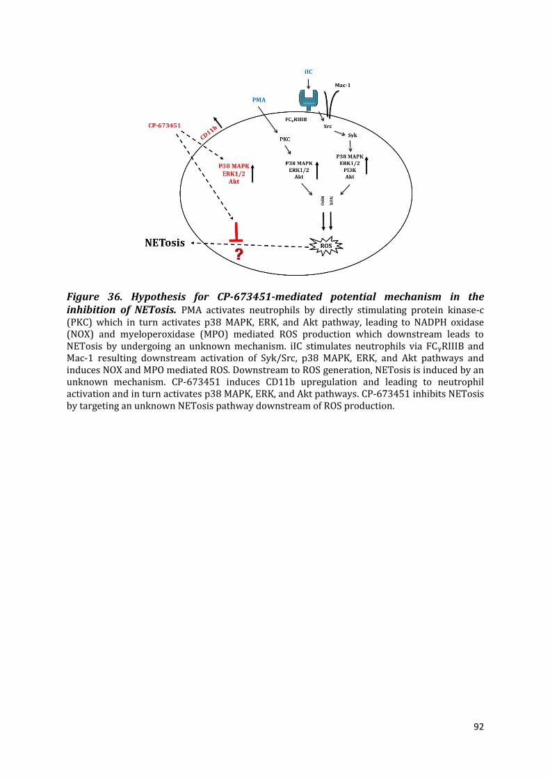
92
Figure 36. Hypothesis for CP-673451-mediated potential mechanism in the inhibition of NETosis. PMA activates neutrophils by directly stimulating protein kinase-c (PKC) which in turn activates p38 MAPK, ERK, and Akt pathway, leading to NADPH oxidase (NOX) and myeloperoxidase (MPO) mediated ROS production which downstream leads to NETosis by undergoing an unknown mechanism. iIC stimulates neutrophils via FCᵧRIIIB and Mac-1 resulting downstream activation of Syk/Src, p38 MAPK, ERK, and Akt pathways and induces NOX and MPO mediated ROS. Downstream to ROS generation, NETosis is induced by an unknown mechanism. CP-673451 induces CD11b upregulation and leading to neutrophil activation and in turn activates p38 MAPK, ERK, and Akt pathways. CP-673451 inhibits NETosis by targeting an unknown NETosis pathway downstream of ROS production.

93
Bibliography
1. Ermert, D., et al., Candida albicans escapes from mouse neutrophils. J Leukoc Biol, 2013. 94(2): p. 223-36.
2. da Silva, F.M., A.M. Massart‐Leën, and C. Burvenich, Development and maturation of neutrophils. VET QUART., 1994. 16(4): p. 220-225.
3. Borregaard, N., Neutrophils, from marrow to microbes. Immunity, 2010. 33(5): p. 657-70.
4. Amulic, B., et al., Neutrophil function: from mechanisms to disease. Annu Rev Immunol, 2012. 30: p. 459-89.
5. Kolaczkowska, E. and P. Kubes, Neutrophil recruitment and function in health and inflammation. Nat Rev Immunol, 2013. 13(3): p. 159-75.
6. McEver, R.P. and R.D. Cummings, Role of PSGL-1 binding to selectins in leukocyte recruitment. J Clin Invest, 1997. 100(11 Suppl): p. S97-103.
7. Kansas, G.S., Selectins and their ligands: current concepts and controversies. Blood, 1996. 88(9): p. 3259-87.
8. Constantin, G., et al., Chemokines trigger immediate beta2 integrin affinity and mobility changes: differential regulation and roles in lymphocyte arrest under flow. Immunity, 2000. 13(6): p. 759-69.
9. Mayadas, T.N., X. Cullere, and C.A. Lowell, The multifaceted functions of neutrophils. Annu Rev Pathol, 2014. 9: p. 181-218.
10. Borregaard, N. and J.B. Cowland, Granules of the human neutrophilic polymorphonuclear leukocyte. Blood, 1997. 89(10): p. 3503-21.
11. Lacy, P., Mechanisms of Degranulation in Neutrophils. Allergy, Asthma & Clinical Immunology, 2006. 2(3): p. 98.
12. Choi, K.Y., L.N. Chow, and N. Mookherjee, Cationic host defence peptides: multifaceted role in immune modulation and inflammation. J Innate Immun, 2012. 4(4): p. 361-70.
13. van Kessel, K.P.M., J. Bestebroer, and J.A.G. van Strijp, Neutrophil-Mediated Phagocytosis of Staphylococcus aureus. Front Immunol, 2014. 5(467).
14. Haas, A., The phagosome: compartment with a license to kill. Traffic, 2007. 8(4): p. 311-30.
15. Wright, H.L., R.J. Moots, and S.W. Edwards, The multifactorial role of neutrophils in rheumatoid arthritis. Nat Rev Rheumatol, 2014. 10(10): p. 593-601.
16. Scapini, P., et al., The neutrophil as a cellular source of chemokines. Immunol Rev, 2000. 177: p. 195-203.
17. Tecchio, C., A. Micheletti, and M.A. Cassatella, Neutrophil-Derived Cytokines: Facts Beyond Expression. Front Immunol, 2014. 5(508).
18. Kasama, T., et al., Neutrophil-derived cytokines: potential therapeutic targets in inflammation. Curr Drug Targets Inflamm Allergy, 2005. 4(3): p. 273-9.
19. Kuwabara, W.M., et al., NADPH oxidase-dependent production of reactive oxygen species induces endoplasmatic reticulum stress in neutrophil-like HL60 cells. PLoS One, 2015. 10(2): p. e0116410.
20. Babior, B.M., J.T. Curnutte, and R.S. Kipnes, Biological defense mechanisms. Evidence for the participation of superoxide in bacterial killing by xanthine oxidase. J Lab Clin Med, 1975. 85(2): p. 235-44.

94
21. Parkos, C.A., et al., Purified cytochrome b from human granulocyte plasma membrane is comprised of two polypeptides with relative molecular weights of 91,000 and 22,000. J Clin Invest, 1987. 80(3): p. 732-42.
22. Nisimoto, Y., et al., The p67(phox) activation domain regulates electron flow from NADPH to flavin in flavocytochrome b(558). J Biol Chem, 1999. 274(33): p. 22999-3005.
23. Han, C.H., et al., Regulation of the neutrophil respiratory burst oxidase. Identification of an activation domain in p67(phox). J Biol Chem, 1998. 273(27): p. 16663-8.
24. Kettle, A.J. and C.C. Winterbourn, Myeloperoxidase: a key regulator of neutrophil oxidant production. Redox Rep, 1997. 3(1): p. 3-15.
25. Segal, B.H., et al., Genetic, biochemical, and clinical features of chronic granulomatous disease. Medicine (Baltimore), 2000. 79(3): p. 170-200.
26. Li, Y. and M.A. Trush, Diphenyleneiodonium, an NAD(P)H oxidase inhibitor, also potently inhibits mitochondrial reactive oxygen species production. Biochem Biophys Res Commun, 1998. 253(2): p. 295-9.
27. Williams, H.C. and K.K. Griendling, NADPH oxidase inhibitors: new antihypertensive agents? J Cardiovasc Pharmacol, 2007. 50(1): p. 9-16.
28. O'Donnell, B.V., et al., Studies on the inhibitory mechanism of iodonium compounds with special reference to neutrophil NADPH oxidase. Biochem J, 1993. 290(Pt 1): p. 41-9.
29. Brinkmann, V., et al., Neutrophil extracellular traps kill bacteria. Science, 2004. 303(5663): p. 1532-5.
30. Fuchs, T.A., et al., Novel cell death program leads to neutrophil extracellular traps. J Cell Biol, 2007. 176(2): p. 231-41.
31. Matoszka, N., et al., [NET and NETosis--new phenomenon in immunology]. Postepy Hig Med Dosw (Online), 2012. 66: p. 437-45.
32. Pilsczek, F.H., et al., A novel mechanism of rapid nuclear neutrophil extracellular trap formation in response to Staphylococcus aureus. J Immunol, 2010. 185(12): p. 7413-25.
33. Yipp, B.G. and P. Kubes, NETosis: how vital is it? Blood, 2013. 122(16): p. 2784-94.
34. Rochael, N.C., et al., Classical ROS-dependent and early/rapid ROS-independent release of Neutrophil Extracellular Traps triggered by Leishmania parasites. Sci. Rep. 2015. 5: p. 18302.
35. Metzler, K.D., et al., Myeloperoxidase is required for neutrophil extracellular trap formation: implications for innate immunity. Blood, 2011. 117(3): p. 953-9.
36. Metzler, K.D., et al., A myeloperoxidase-containing complex regulates neutrophil elastase release and actin dynamics during NETosis. Cell Rep, 2014. 8(3): p. 883-96.
37. Papayannopoulos, V., et al., Neutrophil elastase and myeloperoxidase regulate the formation of neutrophil extracellular traps. J Cell Biol, 2010. 191(3): p. 677-91.
38. Kaplan, M.J. and M. Radic, Neutrophil Extracellular Traps: Double-Edged Swords of Innate Immunity. J Immunol, 2012. 189(6): p. 2689-2695.
39. Neeli, I., S.N. Khan, and M. Radic, Histone deimination as a response to inflammatory stimuli in neutrophils. J Immunol, 2008. 180(3): p. 1895-902.
40. Wang, Y., et al., Histone hypercitrullination mediates chromatin decondensation and neutrophil extracellular trap formation. J Cell Biol, 2009. 184(2): p. 205-13.

95
41. Hemmers, S., et al., PAD4-Mediated Neutrophil Extracellular Trap Formation Is Not Required for Immunity against Influenza Infection. PLOS ONE, 2011. 6(7): p. e22043.
42. Almyroudis, N.G., et al., NETosis and NADPH oxidase: at the intersection of host defense, inflammation, and injury. Front Immunol, 2013. 4: p. 45.
43. Gupta, S. and M.J. Kaplan, The role of neutrophils and NETosis in autoimmune and renal diseases. Nat Rev Nephrol, 2016. 12(7): p. 402-13.
44. Steinberg, B.E. and S. Grinstein, Unconventional roles of the NADPH oxidase: signaling, ion homeostasis, and cell death. Sci STKE, 2007. 2007(379): p. pe11.
45. Wartha, F. and B. Henriques-Normark, ETosis: a novel cell death pathway. Sci Signal, 2008. 1(21): p. pe25.
46. Remijsen, Q., et al., Neutrophil extracellular trap cell death requires both autophagy and superoxide generation. Cell Res, 2011. 21(2): p. 290-304.
47. Itakura, A. and O.J. McCarty, Pivotal role for the mTOR pathway in the formation of neutrophil extracellular traps via regulation of autophagy. Am J Physiol Cell Physiol, 2013. 305(3): p. C348-54.
48. Khan, M.A., et al., JNK Activation Turns on LPS- and Gram-Negative Bacteria-Induced NADPH Oxidase-Dependent Suicidal NETosis. Sci Rep, 2017. 7(1): p. 3409.
49. Yipp, B.G., et al., Infection-induced NETosis is a dynamic process involving neutrophil multitasking in vivo. Nat Med, 2012. 18(9): p. 1386-1393.
50. Halverson, T.W.R., et al., DNA Is an Antimicrobial Component of Neutrophil Extracellular Traps. PLoS Pathog, 2015. 11(1).
51. Lee, M.J., et al., The Fungal Exopolysaccharide Galactosaminogalactan Mediates Virulence by Enhancing Resistance to Neutrophil Extracellular Traps. PLOS Pathogens, 2015. 11(10): p. e1005187.
52. Urban, C.F., et al., Neutrophil extracellular traps capture and kill Candida albicans yeast and hyphal forms. Cellular Microbiology, 2006. 8(4): p. 668-676.
53. Clark, S.R., et al., Platelet TLR4 activates neutrophil extracellular traps to ensnare bacteria in septic blood. Nat Med, 2007. 13(4): p. 463-9.
54. Chen, K., et al., Endocytosis of soluble immune complexes leads to their clearance by FcγRIIIB but induces neutrophil extracellular traps via FcγRIIA in vivo. Blood, 2012. 120(22): p. 4421-31.
55. Behnen, M., et al., Immobilized Immune Complexes Induce Neutrophil Extracellular Trap Release by Human Neutrophil Granulocytes via FcγRIIIB and Mac-1. The Journal of Immunology, 2014. 193(4): p. 1954-1965.
56. Richards, R.C., et al., Histone H1: an antimicrobial protein of Atlantic salmon (Salmo salar). Biochem Biophys Res Commun, 2001. 284(3): p. 549-55.
57. Papayannopoulos, V. and A. Zychlinsky, NETs: a new strategy for using old weapons. Trends Immunol, 2009. 30(11): p. 513-21.
58. Parker, H., et al., Myeloperoxidase associated with neutrophil extracellular traps is active and mediates bacterial killing in the presence of hydrogen peroxide. J Leukoc Biol, 2012. 91(3): p. 369-76.
59. Urban, C.F., et al., Neutrophil extracellular traps contain calprotectin, a cytosolic protein complex involved in host defense against Candida albicans. PLoS Pathog, 2009. 5(10): p. e1000639.
60. Sur Chowdhury, C., et al., Enhanced neutrophil extracellular trap generation in rheumatoid arthritis: analysis of underlying signal transduction pathways and potential diagnostic utility. Arthritis Res Ther, 2014. 16(3): p. R122.

96
61. Garcia-Romo, G.S., et al., Netting neutrophils are major inducers of type I IFN production in pediatric systemic lupus erythematosus. Sci Transl Med, 2011. 3(73): p. 73ra20.
62. Kessenbrock, K., et al., Netting neutrophils in autoimmune small-vessel vasculitis. Nat Med, 2009. 15(6): p. 623-625.
63. Berthelot, J.M., et al., NETosis: At the crossroads of rheumatoid arthritis, lupus, and vasculitis. Joint Bone Spine, 2017. 84(3): p. 255-262.
64. Corsiero, E., et al., NETosis as Source of Autoantigens in Rheumatoid Arthritis. Front Immunol, 2016. 7: p. 485.
65. Dwivedi, N. and M. Radic, Citrullination of autoantigens implicates NETosis in the induction of autoimmunity. Ann Rheum Dis, 2014. 73(3): p. 483-91.
66. Lande, R., et al., Neutrophils activate plasmacytoid dendritic cells by releasing self-DNA-peptide complexes in systemic lupus erythematosus. Sci Transl Med, 2011. 3(73): p. 73ra19.
67. Lin, A.M., et al., Mast cells and neutrophils release IL-17 through extracellular trap formation in psoriasis. J Immunol, 2011. 187(1): p. 490-500.
68. Parker, H., et al., Requirements for NADPH oxidase and myeloperoxidase in neutrophil extracellular trap formation differ depending on the stimulus. J Leukoc Biol, 2012. 92(4): p. 841-9.
69. Stephan, A. and M. Fabri, The NET, the trap and the pathogen: neutrophil extracellular traps in cutaneous immunity. Exp Dermatol, 2015. 24(3): p. 161-6.
70. Lim, M.B., et al., Rac2 is required for the formation of neutrophil extracellular traps. J Leukoc Biol, 2011. 90(4): p. 771-6.
71. Yost, C.C., et al., Impaired neutrophil extracellular trap (NET) formation: a novel innate immune deficiency of human neonates. Blood, 2009. 113(25): p. 6419-6427.
72. Brinkmann, V. and A. Zychlinsky, Beneficial suicide: why neutrophils die to make NETs. Nat Rev Microbiol, 2007. 5(8): p. 577-82.
73. Ermert, D., et al., Mouse neutrophil extracellular traps in microbial infections. J Innate Immun, 2009. 1(3): p. 181-93.
74. Robinson, J.J., et al., Activation of neutrophils by soluble and insoluble immunoglobulin aggregates from synovial fluid of patients with rheumatoid arthritis. Ann Rheum Dis, 1993. 52(5): p. 347-53.
75. Fossati, G., R.C. Bucknall, and S.W. Edwards, Insoluble and soluble immune complexes activate neutrophils by distinct activation mechanisms: changes in functional responses induced by priming with cytokines. Ann Rheum Dis, 2002. 61(1): p. 13-9.
76. Sollberger, G., B. Amulic, and A. Zychlinsky, Neutrophil Extracellular Trap Formation Is Independent of De Novo Gene Expression. PLoS One, 2016. 11(6): p. e0157454.
77. Way, K.J., E. Chou, and G.L. King, Identification of PKC-isoform-specific biological actions using pharmacological approaches. Trends Pharmacol Sci, 2000. 21(5): p. 181-7.
78. Gray, R.D., et al., Activation of conventional protein kinase C (PKC) is critical in the generation of human neutrophil extracellular traps. J Inflamm (Lond), 2013. 10: p. 12.
79. Babior, B.M., J.D. Lambeth, and W. Nauseef, The neutrophil NADPH oxidase. Arch Biochem Biophys, 2002. 397(2): p. 342-4.

97
80. Hakkim, A., et al., Activation of the Raf-MEK-ERK pathway is required for neutrophil extracellular trap formation. Nat Chem Biol, 2011. 7(2): p. 75-77.
81. Keshari, R.S., et al., Reactive oxygen species-induced activation of ERK and p38 MAPK mediates PMA-induced NETs release from human neutrophils. J Cell Biochem, 2013. 114(3): p. 532-40.
82. Kramer, I.M., et al., Purified protein kinase C phosphorylates a 47-kDa protein in control neutrophil cytoplasts but not in neutrophil cytoplasts from patients with the autosomal form of chronic granulomatous disease. J Biol Chem, 1988. 263(5): p. 2352-7.
83. Bouin, A.P., et al., p40(phox) is phosphorylated on threonine 154 and serine 315 during activation of the phagocyte NADPH oxidase. Implication of a protein kinase c-type kinase in the phosphorylation process. J Biol Chem, 1998. 273(46): p. 30097-103.
84. Chessa, T.A., et al., Phosphorylation of threonine 154 in p40phox is an important physiological signal for activation of the neutrophil NADPH oxidase. Blood, 2010. 116(26): p. 6027-36.
85. Konig, M.F. and F. Andrade, A Critical Reappraisal of Neutrophil Extracellular Traps and NETosis Mimics Based on Differential Requirements for Protein Citrullination. Front Immunol, 2016. 7.
86. Douda, D.N., et al., Akt is essential to induce NADPH-dependent NETosis and to switch the neutrophil death to apoptosis. Blood, 2014. 123(4): p. 597-600.
87. Yost, C.C., et al., Impaired neutrophil extracellular trap (NET) formation: a novel innate immune deficiency of human neonates. Blood, 2009. 113(25): p. 6419-27.
88. Douda, D.N., et al., SK3 channel and mitochondrial ROS mediate NADPH oxidase-independent NETosis induced by calcium influx. Proc Natl Acad Sci U S A, 2015. 112(9): p. 2817-22.
89. Pilsczek, F.H., et al., A Novel Mechanism of Rapid Nuclear Neutrophil Extracellular Trap Formation in Response to <em>Staphylococcus aureus</em>. The Journal of Immunology, 2010. 185(12): p. 7413-7425.
90. Kauffman, R.F., R.W. Taylor, and D.R. Pfeiffer, Cation transport and specificity of ionomycin. Comparison with ionophore A23187 in rat liver mitochondria. J Biol Chem, 1980. 255(7): p. 2735-9.
91. Liu, C. and T.E. Hermann, Characterization of ionomycin as a calcium ionophore. J Biol Chem, 1978. 253(17): p. 5892-4.
92. Morgan, A.J. and R. Jacob, Ionomycin enhances Ca2+ influx by stimulating store-regulated cation entry and not by a direct action at the plasma membrane. Biochem J, 1994. 300 ( Pt 3): p. 665-72.
93. Remijsen, Q., et al., Dying for a cause: NETosis, mechanisms behind an antimicrobial cell death modality. Cell Death Differ, 2011. 18(4): p. 581-8.
94. Williams, L.T., Signal transduction by the platelet-derived growth factor receptor. Science, 1989. 243(4898): p. 1564-70.
95. Heldin, C.H. and B. Westermark, Mechanism of action and in vivo role of platelet-derived growth factor. Physiol Rev, 1999. 79(4): p. 1283-316.
96. Heldin, C.H. and J. Lennartsson, Structural and Functional Properties of Platelet-Derived Growth Factor and Stem Cell Factor Receptors. Cold Spring Harb Perspect Biol, 2013. 5(8).
97. Andrae, J., R. Gallini, and C. Betsholtz, Role of platelet-derived growth factors in physiology and medicine. Genes Dev, 2008. 22(10): p. 1276-312.

98
98. Deuel, T.F., et al., Chemotaxis of monocytes and neutrophils to platelet-derived growth factor. J Clin Invest, 1982. 69(4): p. 1046-9.
99. Qu, J., et al., Lack of effect of recombinant platelet-derived growth factor on human neutrophil function. J Immunol, 1995. 154(8): p. 4133-41.
100. Kelly, J.D., et al., Platelet-derived growth factor (PDGF) stimulates PDGF receptor subunit dimerization and intersubunit trans-phosphorylation. J Biol Chem, 1991. 266(14): p. 8987-92.
101. Kazlauskas, A. and J.A. Cooper, Autophosphorylation of the PDGF receptor in the kinase insert region regulates interactions with cell proteins. Cell, 1989. 58(6): p. 1121-33.
102. Bar-Sagi, D. and J.R. Feramisco, Induction of membrane ruffling and fluid-phase pinocytosis in quiescent fibroblasts by ras proteins. Science, 1986. 233(4768): p. 1061-8.
103. Seger, R. and E.G. Krebs, The MAPK signaling cascade. Faseb j, 1995. 9(9): p. 726-35.
104. Hanahan, D. and R.A. Weinberg, The hallmarks of cancer. Cell, 2000. 100(1): p. 57-70.
105. Demetri, G.D., Differential properties of current tyrosine kinase inhibitors in gastrointestinal stromal tumors. Semin Oncol, 2011. 38 Suppl 1: p. S10-9.
106. Roberts, W.G., et al., Antiangiogenic and antitumor activity of a selective PDGFR tyrosine kinase inhibitor, CP-673,451. Cancer Res, 2005. 65(3): p. 957-66.
107. Carroll, M., et al., CGP 57148, a tyrosine kinase inhibitor, inhibits the growth of cells expressing BCR-ABL, TEL-ABL, and TEL-PDGFR fusion proteins. Blood, 1997. 90(12): p. 4947-52.
108. Hojjat-Farsangi, M., Small-molecule inhibitors of the receptor tyrosine kinases: promising tools for targeted cancer therapies. Int J Mol Sci, 2014. 15(8): p. 13768-801.
109. Yu, J.-C., et al., Efficacy of the Novel Selective Platelet-Derived Growth Factor Receptor Antagonist CT52923 on Cellular Proliferation, Migration, and Suppression of Neointima following Vascular Injury. Journal of Pharmacology and Experimental Therapeutics, 2001. 298(3): p. 1172-1178.
110. Zaman, G.J., et al., Tyrosine kinase activity of purified recombinant cytoplasmic domain of platelet-derived growth factor beta-receptor (beta-PDGFR) and discovery of a novel inhibitor of receptor tyrosine kinases. Biochem Pharmacol, 1999. 57(1): p. 57-64.
111. Jakus, Z., et al., Critical but overlapping role of FcgammaRIII and FcgammaRIV in activation of murine neutrophils by immobilized immune complexes. J Immunol, 2008. 180(1): p. 618-29.
112. Briheim, G., O. Stendahl, and C. Dahlgren, Intra- and extracellular events in luminol-dependent chemiluminescence of polymorphonuclear leukocytes. Infect Immun, 1984. 45(1): p. 1-5.
113. Stevens, P. and D. Hong, The role of myeloperoxidase and superoxide anion in the luminol- and lucigenin-dependent chemiluminescence of human neutrophils. Microchemical Journal, 1984. 30(2): p. 135-146.
114. Bianchi, M., et al., Restoration of NET formation by gene therapy in CGD controls aspergillosis. Blood, 2009. 114(13): p. 2619-2622.
115. Brinkmann, V., et al., Neutrophil Extracellular Traps Kill Bacteria. Science, 2004. 303(5663): p. 1532-1535.
99
116. Behnen, M., et al., Extracellular Acidification Inhibits the ROS-Dependent Formation of Neutrophil Extracellular Traps. Front Immunol, 2017. 8: p. 184.
117. Behnen, M., et al., Extracellular Acidification Inhibits the ROS-Dependent Formation of Neutrophil Extracellular Traps. Front Immunol, 2017. 8(184).
118. Sarkar, A., et al., Infection with Anaplasma phagocytophilum activates the phosphatidylinositol 3-Kinase/Akt and NF-kappaB survival pathways in neutrophil granulocytes. Infect Immun, 2012. 80(4): p. 1615-23.
119. Wilfinger, W.W., K. Mackey, and P. Chomczynski, Effect of pH and ionic strength on the spectrophotometric assessment of nucleic acid purity. Biotechniques, 1997. 22(3): p. 474-6, 478-81.
120. Schroeder, A., et al., The RIN: an RNA integrity number for assigning integrity values to RNA measurements. BMC Molecular Biology, 2006. 7: p. 3-3.
121. Khatter, H., et al., Structure of the human 80S ribosome. Nature, 2015. 520(7549): p. 640-645.
122. Imbeaud, S., et al., Towards standardization of RNA quality assessment using user-independent classifiers of microcapillary electrophoresis traces. Nucleic Acids Research, 2005. 33(6): p. e56-e56.
123. Wang, Z., M. Gerstein, and M. Snyder, RNA-Seq: a revolutionary tool for transcriptomics. Nature reviews. Genetics, 2009. 10(1): p. 57-63.
124. Sim, S., et al., NADPH oxidase-derived reactive oxygen species-mediated activation of ERK1/2 is required for apoptosis of human neutrophils induced by Entamoeba histolytica. J Immunol, 2005. 174(7): p. 4279-88.
125. Taylor, S.S., E. Radzio-Andzelm, and T. Hunter, How do protein kinases discriminate between serine/threonine and tyrosine? Structural insights from the insulin receptor protein-tyrosine kinase. Faseb j, 1995. 9(13): p. 1255-66.
126. Kenny, E.F., et al., Diverse stimuli engage different neutrophil extracellular trap pathways. eLife, 2017. 6: p. e24437.
127. The Impact of Various Reactive Oxygen Species on the Formation of Neutrophil Extracellular Traps. Mediators of Inflammation, 2012. 2012.
128. McKim, J.M., Building a Tiered Approach to In Vitro Predictive Toxicity Screening: A Focus on Assays with In Vivo Relevance. Combinatorial Chemistry & High Throughput Screening, 2010. 13(2): p. 188-206.
129. Henkels, K.M., et al., IL-8-INDUCED NEUTROPHIL CHEMOTAXIS IS MEDIATED BY JANUS KINASE 3 (JAK3). FEBS letters, 2011. 585(1): p. 159-166.
130. Smart, S.J. and T.B. Casale, TNF-alpha-induced transendothelial neutrophil migration is IL-8 dependent. Am J Physiol, 1994. 266(3 Pt 1): p. L238-45.
131. Soler-Rodriguez, A.M., et al., Neutrophil activation by bacterial lipoprotein versus lipopolysaccharide: differential requirements for serum and CD14. J Immunol, 2000. 164(5): p. 2674-83.
132. Cardoso, L.S., et al., Polymyxin B as inhibitor of LPS contamination of Schistosoma mansoni recombinant proteins in human cytokine analysis. Microbial Cell Factories, 2007. 6(1): p. 1.
133. Khan, M.A. and N. Palaniyar, Transcriptional firing helps to drive NETosis. Scientific Reports, 2017. 7: p. 41749.
134. Perez-Sanchez, C., et al., Diagnostic potential of NETosis-derived products for disease activity, atherosclerosis and therapeutic effectiveness in Rheumatoid Arthritis patients. J Autoimmun, 2017.

100
135. Hakkim, A., et al., Impairment of neutrophil extracellular trap degradation is associated with lupus nephritis. Proc Natl Acad Sci U S A, 2010. 107(21): p. 9813-8.
136. Radic, M. and T.N. Marion, Neutrophil extracellular chromatin traps connect innate immune response to autoimmunity. Seminars in Immunopathology, 2013. 35(4): p. 465-480.
137. Dwivedi, N., et al., Felty’s Syndrome Autoantibodies Bind to Deiminated Histones and Neutrophil Extracellular Chromatin Traps. Arthritis and rheumatism, 2012. 64(4): p. 982-992.
138. Kamoshida, G., et al., Spontaneous formation of neutrophil extracellular traps in serum‐free culture conditions. FEBS Open Bio, 2017. 7(6): p. 877-886.
139. in Assay Guidance Manual, G.S. Sittampalam, et al., Editors. 2004, Eli Lilly & Company and the National Center for Advancing Translational Sciences: Bethesda (MD).
140. Kawakami, T., et al., Rab27a Is Essential for the Formation of Neutrophil Extracellular Traps (NETs) in Neutrophil-Like Differentiated HL60 Cells. PLoS ONE, 2014. 9(1): p. e84704.
141. Roskoski, R., Jr., Sunitinib: a VEGF and PDGF receptor protein kinase and angiogenesis inhibitor. Biochem Biophys Res Commun, 2007. 356(2): p. 323-8.
142. Cobb, M.H. and E.J. Goldsmith, How MAP kinases are regulated. J Biol Chem, 1995. 270(25): p. 14843-6.
143. Nick, J.A., et al., Selective activation and functional significance of p38alpha mitogen-activated protein kinase in lipopolysaccharide-stimulated neutrophils. J Clin Invest, 1999. 103(6): p. 851-8.
144. Bonner, S., et al., Activation of extracellular signal-related protein kinases 1 and 2 of the mitogen-activated protein kinase family by lipopolysaccharide requires plasma in neutrophils from adults and newborns. Infect Immun, 2001. 69(5): p. 3143-9.
145. Chen, J., et al., Akt isoforms differentially regulate neutrophil functions. Blood, 2010. 115(21): p. 4237.
146. Souza, L.R., et al., G-CSF activation of AKT is not sufficient to prolong neutrophil survival. Journal of Leukocyte Biology, 2013. 93(6): p. 883-893.
147. Zhu, D., et al., Deactivation of phosphatidylinositol 3,4,5-trisphosphate/Akt signaling mediates neutrophil spontaneous death. Proc Natl Acad Sci U S A, 2006. 103(40): p. 14836-41.
148. Zhang, Y., et al., Kinase AKT controls innate immune cell development and function. Immunology, 2013. 140(2): p. 143-152.
149. Borregaard, N., O.E. Sorensen, and K. Theilgaard-Monch, Neutrophil granules: a library of innate immunity proteins. Trends Immunol, 2007. 28(8): p. 340-5.

101
Supplementary data
I. Target-selective inhibitor library from Selleckchem (Houston,
USA)
No. Name of the
Inhibitor
Target No. Name of the
Inhibitor
Target
D1 AZD6244 (Selumetinib)
MEK1 D72 TAME APC
D2 MK-2206 2HCl Akt D73 PHA-665752 c-Met D3 17-AAG
(Tanespimycin) HSP 90 D74 MLN8237 (Alisertib) Aurora A
D4 PFI-1 BRD4 D75 Dutasteride 5-alpha Reductase
D5 Rigosertib (ON-01910)
PLK-1 D76 Doxazosin mesylate α1
D6 BIX 02189 MEK5 D77 MK-1775 Wee1 D7 Candesartan
(Atacand) angiotensin II receptor
D78 BIRB 796 (Doramapimod)
p38α MAPK
D8 Rosuvastatin calcium (Crestor)
HMG-CoA reductase
D79 GSK1292263 GPR119
D9 Bortezomib (Velcade)
20S proteasome
D80 Ketanserin (Vulketan Gel)
HTR2
D10 PD153035 HCl EGFR D81 SB 216763 GSK-3α D11 Barasertib
(AZD1152-HQPA)
Aurora B D82 ADL5859 HCl δ-opioid receptor
D12 Topotecan HCl Topo I D83 Doxorubicin (Adriamycin)
Topo II
D13 AS-605240 p110γ D84 Etomidate GABAA receptor D14 CP 673451 PDGFR D85 Quizartinib (AC220) FLT3 D15 H 89 2HCl PKA D86 Tie2 kinase inhibitor Tie-2 D16 SB 743921 kinesin
spindle protein
D87 PF-4708671 S6K1
D17 Lenalidomide (Revlimid)
TNF-α D88 Clozapine (Clozaril) HTR1
D18 Vismodegib (GDC-0449)
hedgehog D89 Donepezil HCl (Aricept)
AChE
D19 MK-8245 SCD D90 CHIR-124 Chk1 D20 2-
Methoxyestradiol HIF-1α D91 CI994 (Tacedinaline) HDAC1
D21 Tipifarnib (Zarnestra)
FTase D92 Entacapone COMT
D22 EX 527 SIRT1 D93 10058-F4 c-Myc D23 Allopurinol
(Zyloprim) xanthine oxidase
D94 Pancuronium (Pavulon)
nAChR
D24 BGJ398 (NVP-BGJ398)
FGFR D95 ADX-47273 mGlu5

102
D25 Nilotinib (AMN-107)
Bcr-Abl D96 Tideglusib GSK-3β
D26 Iniparib (BSI-201)
PARP1 D97 Sitagliptin phosphate monohydrate
DPP-4
D27 ABT-751 β-tubulin D98 SMI-4a Pim1 D28 Letrozole Aromatase D99 Salbutamol sulfate
(Albuterol) β2
D29 Zibotentan (ZD4054)
ETA D100 KX2-391 Src
D30 AM-1241 CB2 D101 TPCA-1 IKK2 D31 Enalaprilat
dihydrate ACE D102 Mirabegron (YM178) β3
D32 Aliskiren hemifumarate
renin D103 RGD (Arg-Gly-Asp) Peptides
Integrin
D33 Rapamycin (Sirolimus)
mTOR D104 Xylazine HCl α2
D34 Linsitinib (OSI-906)
IGF-1R D105 ZM 336372 c-Raf
D35 TGX-221 p110β D106 Dizocilpine (MK 801) NMDA D36 MDV3100
(Enzalutamide) AR D107 Sodium 4-
Aminosalicylate NF-κB
D37 SP600125 JNK D108 Tariquidar P-gp D38 Dapagliflozin hSGLT2 D109 Tolterodine tartrate
(Detrol LA) mAChR
D39 Ranitidine (Zantac)
Histamine H2-receptor
D110 JTC-801 ORL1
D40 DAPT (GSI-IX) γ-secretase D111 ML130 NOD1 D41 Vandetanib
(Zactima) VEGFR2 D112 PTC124 (Ataluren) CFTR
D42 Varespladib (LY315920)
PLA D113 NSC 23766 Rac
D43 XAV-939 Wnt/β-catenin
D114 Tolvaptan (OPC-41061)
vasopressin receptor 2
D44 Ramelteon (TAK-375)
MT Receptor D115 AZ 3146 Mps1
D45 SB 525334 ALK5 D116 GW441756 TrkA D46 Nebivolol
(Bystolic) β1 D117 Birinapant (TL32711) cIAP1
D47 Clemastine Fumarate
Histamine H1-receptor
D118 PRT062607 (P505-15, PRT2607, BIIB057) HCl
Syk
D48 Mubritinib (TAK 165)
Her2 D119 URB597 FAAH
D49 Y-27632 2HCl ROCK1 D120 PAC-1 Caspase-3 D50 Odanacatib (MK
0822) Cathepsin K D121 WZ 811 CXCR4
D51 BIBR 1532 Telomerase D122 Stattic STAT3 D52 Celecoxib COX-2 D123 UPF 1069 PARP2 D53 Fludarabine
(Fludara) STAT1 D124 BMS-806 (BMS
378806) gp120
D54 Pimobendan (Vetmedin)
PDE3 D125 CH5424802 ALK
D55 PCI-34051 HDAC8 D126 GW9662 PPARγ D56 SB590885 b-Raf D127 SANT-1 Smoothened

103
D57 Enzastaurin (LY317615)
PKCβ D128 GW2580 CSF-1R
D58 Abiraterone (CB-7598)
CYP17 D129 A66 p110α
D59 Fulvestrant (Faslodex)
ER D130 Tofacitinib (CP-690550, Tasocitinib)
JAK3
D60 IC-87114 p110δ D131 Evacetrapib (LY2484595)
CETP
D61 NVP-BEP800 HSP90β D132 IPA-3 Pak1 D62 KU-60019 ATM D133 ABT-199 (GDC-0199) Bcl-2 D63 PF 573228 FAK D134 TAK-875 GPR40 D64 PF 3716556 P-CAB D135 APO866 (FK866) NMPRTase D65 Nutlin-3 MDM2 D136 Pifithrin-α p53 D66 YM155 Survivin D137 NLG919 IDO D67 Decitabine DNA
Methyltransferase
D138 NU7441 (KU-57788) DNA-PK
D68 BX-795 PDK-1 D139 CEP33779 JAK2 D69 Tadalafil (Cialis) PDE5 D140 Rimonabant
(SR141716) CB1
D70 BS-181 HCl CDK5 D141 TCID DUB D71 Roflumilast
(Daxas) PDE4

104
Acknowledgements
First and foremost, I would like to express my sincere gratitude to my supervisor Prof. Dr.
Tamás Laskay for his support in my research. He consistently steered me in the right the
direction whenever I needed help.
I would like to thank Prof. Dr. med. Werner Solbach and Prof. Dr. Jan Rupp, the former and
current directors of Department of Infectious Diseases and Microbiology for giving me the
opportunity to perform my experiments in their department.
I would like to thank Prof. Dr. med Ralf Ludwig from Department of Dermatology, for being my
first mentor and for sharing the chemical library of inhibitors with me. I also thank him for his
positive attitude and vivid discussions.
I would like to thank my second mentor, PD Dr. Norbert Reiling from Research center Borstel,
who took part in extensive discussions regarding my research.
I would like to express my gratitude to Prof. Hauke Busch for analyzing the RNA Seq data in a
very short time and for agreeing to review my PhD thesis.
I would like to thank Prof. Rainer Duden for agreeing to be the Chairman for my PhD thesis.
I also thank PD Dr. Christina Zechel from Department of Neurosurgery, for providing me U87-
MG cell line along with reagents and protocols for its culture and the lysate of T129 cell line.
I would like to thank Ms. Miriam Freitag (from LIED), for performing the RNA-quality analysis
with Bioanalyzer.
I would also like to thank Prof. Dr. med. Detlef Zillikens and the research training group of
Modulation of Autoimmunity (GRK1727), for making me a part of their research group and
giving me opportunities to present my work in various meetings and for funding my project.
I would like to thank the former and current lab members Natallia Salei, Stephanie Lachmann,
Tabea Walther, Martina Behnen-Haerer, and Sonja Möller for their feedback, cooperation and
for the friendship during the last three years.
Last but not the least, I would like to thank my parents, my brother and Vadakkumcherry family
for supporting me spiritually and always standing by my side throughout the PhD period and in
my life in general.

105
Curriculum Vitae
EDUCATION
Research Scholar
Research Training Group GRK 1727
University of Lübeck, Germany
10/2014 - present
Masters in Biological Sciences
Biocentre, University of Cologne, Germany
Department of Plant physiology and biochemistry,
Ecology, Phycology, and Developmental biology
04/2012 – 05/2014
Bachelors in Biotechnology
Administrative Management College, Bangalore, India
Department of Biotechnology, Zoology and Chemistry
08/2007 – 05/2010
PROFESSIONAL EXPERIENCE
Research Assistant
University Clinic of Schleswig-Holstein
Department of Infectious Diseases and Microbiology (KIM)
Research group of Prof. T. Laskay, PhD
10/2014 – 10/2017
Student Research Assistant
Botanical Institute, Biocentre, University of Cologne
Research group of Prof. Dr. U.I. Flügge
05/2013 – 05/2014
Name: Gigy Varghese
Address: Paul-Ehrlich str. 4a
Date and place of birth: 23.05.1990 in Mahe, India