Course of lectures on traumatic injuries in maxillo...
50
1 Mihail Radzichevici Course of lectures on traumatic injuries in maxillo- facial region Chisinau, 2014
Transcript of Course of lectures on traumatic injuries in maxillo...

1
Mihail Radzichevici
Course of lectures on traumatic injuries in maxillo-
facial region
Chisinau, 2014

2
Introduction
In the present study guide for maxillo-facial surgery and dental surgery, briefly, are
described some traumatic injuries in maxillo-facial region, namely etiology,
pathogenesis, diagnostics, of the clinical progression and treatment of the given
diseased peculiarities. Course of study materials on traumatic injuries in maxillo-
facial region will help students of dental departments in the given speciality study.
The present manual is composed in accordance with syllabus approved for students
of dental department, of the State University of Medicine and Pharmacy “Nicolae
Testemitanu” of Republic Of Moldova. The manual contains the lecture material
for students of 4th
year of stomatological department.
Mihail Radzichevici
Teaching assistant of maxillo-facial surgery, implantology and dental therapy
department „Arsenie Guț an” , State University of Medicine and Pharmacy “Nicolae
Testemitanu” of Republic Of Moldova.

3
Topic № 1.
OMF traumatisms, general information. Soft tissues damages.
SOFT TISSUES INJURIES IN THE MAXILLO-FACIAL REGION
All traumatic injuries are divided into occupational (industrial and
agricultural) and non-occupational (household, transport, outdoorsy, sport)
according to the reasons of their production.
Industrial Injury is injury coming from the execution of workers
occupational functions in the production sector or agricultural sector.
Home accident is injury which is not connected with occupational functions
but is produced in the result of household duties performance or home disputes. It
is observed that the frequency of home accidents increases in the spring-summer
period (from April until September). Around 90% of home accidents appear in the
result of stroke and only 10% of home accidents appear in the result of downfall or
due to other reasons. Men prevail women among injured in the ration 4:1. More
often home accidents occur at the age of 20 till 40 years (60%).
Outdoor injury is injury coming from outdoor walking (human downfall
due to disturbance of the general sense of well-being, ice slick, natural disasters
and etc.) which is not connected with transport. About half of injured persons are
persons middle, elderly and old aged. The given injury differs by slight character
of injury (often: hurts, frets, wounds, teeth injury, nasal bones and zygomatic
(malar) complex injuries).
Road accident produces in the result of road traffic incidents. It is
characterized by multiplicity and combined injuries.
Combined injury is the coeval injury of two or more organs which belong
to different anatomico-functional systems. A craniofacial injury is more often type
of combined injuries.
Sport injury produces in the result of fitness and sport. There is defined the
seasonality of sport injury. More often it occurs in winter months (skating, hockey,
skiing) or in summer (football).
Nature of a non - firearm trauma in the maxillo-facial region.
According to nature and degree of injuries, all face soft tissues traumas are
divided into two main groups:

4
1) Isolated injuries of face soft tissues (without skin integument and mouth
mucosa membrane crippling (contusion); with skin integument and mouth mucosa
membrane crippling (racoma or wound));
2) Combined injuries of face soft tissues and viscerocranium bones (without
skin integument and mouth mucosa membrane crippling; with skin integument and
mouth mucosa membrane crippling).
Contusion is a closed mechanic injury of soft tissues without optic
violation of their anatomic crippling. It is produced under the influence of blunt
item with the little force on soft tissues. This is accompanied by the expressed
violation of subjecting tissues (skin structure, muscle) with the preservation of skin
crippling. In the subjecting tissues there are observed injury of small vessels,
hemorrhage and blood tissues imbibitions. There are originated acchymomas (the
hemorrhage in the skin thickness or membrane mucosa), or hematomas (restricted
blood accumulation in the tissues with the cavity formation in it).
Acchymoma is the indicant of viability of tissue injury. The “Flowering” of
acchymoma is a gauge of the trauma limitation. The purple-cyanotic color of
acchymoma is preserved during 2-4 days and a green coloring appears on the 5th -
6th
day after trauma, on the 7th
-8th -10
th day color of the skin is yellow.
Acchymomas disappear in 10 -14 days depending on hemorrhage sizes.
Hematoma is classified depending on:
- their tissue placement (subdermal, submucosal, intraperiosteal,
intermuscular, subfascial);
- localization (buccal, suborbital, periorbital, and other regions);
- the state of effused blood (non-maturated hematoma, infected or
maturated hematoma, organized or encapsulated hematoma);
- the attitude to the blood vessels lumen (non-systaltic, systaltic and
arching).
Frequent contusion of soft tissues could be combined with the injury of
facial skeleton bones. The augment of edema and a non-expressed functional
injury could create a false presentation about only soft tissues damage isolation. X-
ray examination should be executed to specify the diagnoses.
In two first days after trauma, the treatment of soft tissue injuries consists in
overlapping of freeze (the icepack should be overlapped every hour with the
interval 15-20 minutes) on the given area. From the third day after trauma thermal
procedures (UV (ultraviolet) irradiation, ultrasound, phonophoresis with iodine
paraffinotherapy, hot compress and etc.)could be prescribed. Trocsevasin (jelly
2%), heparoid, heparin ointment, Dolgit cream (the cream contains ibuprofen) and
other ointments should be prescribed for the treatment of injury region.

5
In the case of soft tissues acute hematomas (two first days) it is prescribed
the freeze and from the 3rd
-4th
day there are indicated thermal procedures.
Hematomas are opened in the case of their suppuration and encystations
(organized hematoma).
Excoriation (racoma) is the hurt (mechanic trauma) of the superficial skin
stratums (epidermis) or mouth mucosa membrane. More often it produces on the
outpouching body part: nose, chin, and front, superciliary and zygomatic regions.
Often racomas are accompanied by soft tissue injuries and more rarely racomas are
accompanied by face and neck wounds. Such kinds of hurts occupy about 8 % of
all soft tissue hurts (according to our clinic data). The following racoma
cicatrization periods are distinguished:
- from the time of racoma appearance to the time crust appearance (10-12 hours);
- the occlusion of the racoma floor to the level of unaffected skin and then
upward (12-24 hours, and sometimes utill 48 hours);
- the epithelization (4-5 days);
- the falling-off crust (on the 6th -8
th -10
th day); disappearance of racoma trace.
Cicatrization terms can be changed in dependence of racoma sizes. The
cicatrization takes place without any cicatrices formation.
The racoma treatment includes the manipulation by 3% hydrogen dioxide,
chlorhexidine solution, adjustment by dressing of Kureosin solution, adjustment of
collagen pellicle on the wound surface.
Wounds
Wound is a crippling of mucosa membrane along all it thickness (denser and
deeper of lying tissues) caused by mechanic influence.
Wounds are distinguished into:
- superficial and deep,
- nonpenetrating and penetrating (in the mouth and nose cavity, maxillary
antrum, eye socket and etc.)
According to the type and form of injure item, there are distinguished following
wounds:
- compound,
- laceration,
- sword-cut,
- punctured,
- chopped,
- bite,
- crushed,
- degloving wounds.

6
Compound wounds are produced in the case of blunt item stroke with the
simultaneous injury of surrounding tissues. Wounds on the mucosa membrane
could be the result of teeth injury in the case of stroke in check region, upper and
lower lip. Therefore wounds are infected by mouth mucosa microflora. The saliva
which effuses from the wound irritates the skin.
Laceration wound is the wound appeared due to tissues hyperextension. It
is characterized by incorrect edges form, tissue sublation or abruption and by great
zone of their injury. It appears in the case of stroke by abrupt items, fall,
occupational or sport injuries and other.
In dental practice laceration wounds are observed in the case of bur injury,
teeth extraction tongs and other small instruments.
Sword-cut wound is the wound produced by a sharp item. It is
characterized by line or fusiform with equal parallel edges. Sword-cut wounds
dehisce significantly even if they do not penetrate into deep layers of soft tissues of
maxilla-facial region. This occurs because of facial muscles injuries which contract
strong and widen the wound. It creates a false impression about the presence of
tissues defect. In the dental practice sword-cut wounds occurs in the case of
tongue, lip and check injury by the separating disc. The microbial contamination of
these affections is great.
Punctured wound is the wound produced by an acute item with small cross
sections. It is characterized by the narrow and long wound tract. It is observed in
the case of stroke by household piercing objects (knife, awl, screwdriver and et.),
in the dental practice (elevator). The microbial contamination is significant
expressed if injury of soft tissue is produced by an elevator.
Chopped wound is the wound produced because the stroke of a heavy acute
item. It is characterized by a fissured form and high deep. In contrast to sword-cut
wound, chopped wound has more vast soft tissues and wound edges affect. More
often these affects are followed by facial skeleton bones fractures and can
penetrate in cavities (mouth, nose, eye socket, skull, and maxillary antrum). Bones
fractures are usually splintered. Usually the microbal contamination is expressed.
Frequent it is accompanied by wound suppuration, development off posttraumatic
sinusitis and other inflammatory aggravations.
Bite wound is the wound produced by teeth of an animal or a people. It is
characterized by a contamination and by abrupt, crushed edges. If the human was
bit by an animal this wound is contaminated by a pathogenic flora.
Crushed wound is the wound during which production occur the histotripsy
and breakage of tissues. More often than not, there are injured deep placed tissues
and organs (salivary glands, eye-ball, throat, weasand, tongue and teeth), great
vessel and nerves. There are originated vast hemorrhage and asphyxia.

7
Degloving wound is the wound with the complete or almost complete
separation of vast skin flap. Generally there are produced on the bulging parts of
facial skeleton (nose, front, zygomatic area, chin and etc).
Clinical picture peculiarities of soft tissue wounds according to their
localization.
In the case of oral region tissues affection the mucosa membrane is injured
by the acute teeth edges and by broken plastic dentures. More often it is observed
in the lips and check regions. Wounds flood ample and always are infected. If
there are defected the inner and external surface of jaw body alveolar bone and
also hard palate, the mucosa membrane could be not brought together, as it is solid
soldered with periostenium. The injury of the mucosa membrane in the retromolar
region or os, and also of the mouth cavity floor causes a vast hemorrhage and a
speed edema development with the relevant clinical symptoms (pain during the
ingestion, mouth opening, and tongue movement).
Dentist can give a deep wound as by bur (in the time of teeth treatment) as
by a separator disc (during the preparation of teeth to prosthodontic treatable) in
the region of mouth cavity floor soft tissues, tongue and check. In the case of
injury of sublingual tissues region by separating disc, lingual arteria or vein could
be affected which will contribute to the vast hemorrhage. If it is impossible to stop
the hemorrhage of affected vessel by the bandage (in the wound or around it)
should be done along vasoligation of lingual artery of Pirogov’s triangle or
external carotid artery. The tongue injuries occur during human downfall (biting
of the tongue by teeth) or during the cerebral seizure, fishhook injury and other. If
there are affected upper and lower lip it is observed the opening of wound edges.
In the result of affection of orbicular muscle could be the absence of airtight lip-
seal. Compound wounds of periorbital region can violate the eyelids movement,
but oftime eyelid trauma leads to their cicatrices eversion or epicanthus formation
of vertical skin ruga which close the medial triangle of palpebral fissure.
SURGICAL DEBRIDMENT
Surgical debridement is a surgical operation directed to creation of favorable
condition for wound cicatrization, prevention or fight with the wound infection. It
includes the elimination from the wound of nonviable and impure tissues, the final
hemorrhage stop, ablation of necrotizing edges and other arrangements.
Primary surgical debridement is the first wound debridement of a patient.

8
Secondary surgical debridement is the wound debridement which is
leaded according secondary indications, i.e. according to the following changes
subjected by infection development.
Early wound debridement is performed in the first 24 hours after injury.
Primary tardy wound debridement is the primary debridement performed on
the second day after injury, i.e. after 24-48 hours.
Late surgical wound debridement is performed in 48 hours and more.
Surgical debridement peculiarities of maxillo-facial region wounds:
- should be performed in full and at earliest terms;
- it is not allowed to pare (refresh) wound edges, but only nonviable (necrotizing)
tissues should be debrided;
- wounds which penetrate in the mouth cavity should be isolated from the mouth
cavity by the blind saturation of the mucosa membrane with the following
layered closure (muscles, skin);
- as a result of lips injuries from the beginning the red border (Cupid line) should
be put together and needle and then to saturate;
- wound debrides should be obligative amputated. Exception are only debrides
which are in hard-to-reach places (palatal recess) as their search is incidental to
additional traumas;
- in the presence of injury of eyelids or lips red border, the skin and mucosa
membrane are necessary to mobilize to prevent the tissues retraction
(astringent) in order to avoid further intention in the sutures line in some cases.
Sometimes it is necessary to make the displacement of interchanging triangular
flaps;
- in the presence of injury of salivary glands parenchyma it is necessary to suture
the gland capsule and then all subsequent layers. In the presence of canal injury
it is necessary to suture it or to create a false canal;
- wounds are sutured by a blind suture and are drained only if they are infected
(late surgical debridement);
- in the case of expressed edema and a wide edges dihescense are used U-shaped
sutures (for example: on the gauze swab a distance away the wounds borders
1.0-1.5 cm) to prevent sutures cutting out;
- in the presence of soft tissues big penetration defect the surgical debridement is
ended by a suture of a skin with the membrane mucosa mouth cavity to avoid
the jaws cicatrical contraction. It creates favorable conditions for the further
plastic defect closure as well as prevents the formation of gross scar and the
deformation of neighboring tissues;

9
- postoperative wound management is oftener performed by an open method
namely without dressings application on the second and consecutive days of the
treatment;
- to prevent the suture lines disruption, sutures shouldn`t be early removed. Soft
tissues of maxilla-facial region have some characteristic peculiarities by
contrast with other localizations:
- voluminous blood supply;
- good innervations;
- high regeneration abilities;
- expressed local tissues immunity;
- tissues have cells around the face natural ostium which are already partialy
prepared for the cicatrization;
- wounds of mouth cavity mucosa membrane are bounded by saliva and it
contains lysozyme which promotes the regeneration;
- microflora of mouth cavity, nose and maxillary antrum could contribute to
wound contamination;
The surgical wound debridement is made after the cleaning (by antiseptic
means) of skin around the wound. Hairs around the wound should be shaved as
necessary. Once more the wound is debrided by the antiseptic means to eliminate
foreign matters and contaminants. It is performed the local anesthesia and
hemostasis. Devitalized tissues are exsected. The wound is repaired layer-by-layer
by the blind primary saturation. Sutures lines are debrided by the iodine solution or
brilliant green. The antiseptic bandage is applied. The first bandage is made on the
next day after surgery. It is preferably to treat the wound without any bandage by
open method. Only in the presence of contamination and hematomas the bandage
(usual or compressive) should be applied. In the case of development of
inflammatory process in the wound, abscesses must be opened and drained and
then should be prescribed the medicated treatment (antibiotics and other).
The cicatrization of posttraumatic wounds can be as by primary as by
secondary intention.
The cicatrization by the primary intention is the wound cicatrization by the
way of its walls adhesion by fibrin roll with the formation on the surface of crust,
under which happens a quick fibrin substitution by the granulation tissue,
epithelization and formation of tight line cicatrize.
The cicatrization by the secondary intention is the wound cicatrization by
the way of gradual filling of purulent wound cavity by the granulation tissue with
the following epithelization and formation of a cicatrice.

10
The organizational principles of health care delivery:
The first aid to the patient is performed on the incident place (as self-mutual
aid).
The predictor care is performed by persons with the secondary level of
medical education (nurse, parademic).
The primary doctor care foresees the fight with asphyxia, hemorrhage and
collapse. It can be performed by every doctor notwithstanding of specialty.
The secondary doctor care is performed by the doctor, surgeon-dentist in the
dental department or clinics, in the maxilla-facial emergency stations.
The secondary care presupposes:
total pain relief;
the elimination of debrides, blood clots, devitalized tissues and others;
the cleaning of the wound by the antiseptic means;
detailed hemostasis;
the layer-by-layer wound saturation
tetanus prophylaxis (tetanus vaccine);
rabies virus prophylaxis (rabies vaccine) to the patients with the bite wound
(the disease is presented by the motor excitement, convulsions of the
swallowing and breathing muscles, paralysis development in the disease
end-stage);
examination of alcohol intoxication;
the prophylaxis of keloid and hypertrophic scars formation, if they are
mentioned in the anamnesis.

11
Topic № 2
Mandible fractures. Treatment of the mandible fractures (immediate, at
transportation, bone synthesis).
FRACTURES OF LOWER JAWS
Fractures of lower jaw are produces more often than injures of other facial
skeleton bones.
The mandible fractures are usually observed in the typical places (“infirmity
places”): in the region of the central incisors (along the middle line), canine,
premolars, angle of mandible, condylar process cervix (pic. 1).
Pic. 1. Typical places of lower jaw fractures (“infirmity places”)
Classification depending from terms of injury fractures of lower jaw are:
- Current (acute), till 10 days,
- Old, from 11 till 20 days,
- Malunion, more that 20 days.
In everyday practice all fractures of lower jaw are classified according to:
localization and fracture character.
Classification according to the localization:
A) - unilateral; -bilateral;
B) – single; - double; -multiple;
C) –maxilla body fracture (opened, in the limit of tooth line):
а) medial (in the incisors region);
b) mental (in the region of the canine and premolars);

12
c) in the molar region;
d) in the region of maxilla angle (opened and closed).
D fractures in the jaw branch region (closed):
a) condylar process (- floor; - cervix; -head);
b) coronoid process;
c) proper branches (lengthwise or transverse).
According to the fracture character:
a) - Complete; - incomplete (subperiosteal);
b) – undisplaced fragments; - displaced fragments;
c) - linear; - comminuted; - combined;
d) - Isolated; multisystem (with the cerebro-cranial injuries, soft tissues injuries,
other bones injuries).
The lower jaw has an arcual form. The fracture of the lower jaw can be
produced in result of deflection, flexure and compression. The jaw is broken in its
“infirmity” places by the reason of force action (pic.2-3). Deflection of lower jaw
fragments is performed by an action of applied force of fragments own heaviness
and under the action of muscles` draft which are fixed to broken fragments. There
are no fragments displacements in case of intraperiosteal (subperiosteal) fracture.
The movement of the jaw is performed due the action of two muscle groups:
elevating (posterior group) and depressing (anterior group) the lower jaw. The
displacement is the significant the more muscles are fixed to jaw fragments (pic.
4).
Posterior muscles group elevating the lower jaw:
Mastication muscle (m. masseter) begins from the lower border of zygomatic
bone and is fixed to the superficies of the ramus of mandible (tuberositas
masseterica). In case of lateral muscle contraction, the lower jaw is displaced
in direction of the given muscle contraction. In case of bilateral muscle
contraction, the lower jaw is drawn to the upper jaw, in other words the mouth
opens.
Temporal muscle (m. temporalis) occupies the entire space of temporal skull
fossa by its wide beginning and above gets to Гшея temporalis. Muscular
fascicles converge flabellate and are directed from the lower jaw upwards,
dermad (outwards) and some posteriorly. It is formed the firm chorda which
goes under the zygomatic arch and is fixed to the processus coronoideus of
lower jaw. When the temporal muscle contract, the lower jaw raises upwards
and displaces a little posteriorly.

13
Medial pterygoid muscle (m. pterygoideus medialis) begins in the pterygoid
fossa, directs down lateral and fixes on the inner surface of the lower jaw
angle. Muscle fibers are directed upwards, anteriad and inward towards the
lower jaw. In case of bilateral contraction of given muscles, the lower jaw
displaces upwards and moves to the fore. In case of lateral contraction, the jaw
displaced against the contracted muscle.
Lateral pterygoid muscle (m. pterygoideus lateralis) begins on a lower
surface of a greater sphenoid wing and clinoid process. It is fixed to the
condylar process neck and to the bursa and disc of temporomandibular joint.
The muscle comes horizontal almost. The jaw moves to the fore when both
muscles contract simultaneously. If only one muscle contracts, then the lower
jaw displaces laterad, i.e. in direction against the contracted muscle.
Pic. 2. The schematic illustration of production of lower jaw fractures
localized in the place of place of force and in outlands (reflected fracture), in
the case of unilateral force direction.
Pic. 3. The schematic illustration of production of lower jaw fractures in the
case of its compression (the stroke is directed to both parts).
Anterior group of muscle depressing the lower jaw:

14
Mylohyoid muscle (m. mylohyoideus) begins from the linea mylohyoidea on
the interface of lower jaw body and goes inward, down and a little posteriorly.
Along the middle line right and left muscles are connected against each other
and are terminated with a tendon suture, and postoral region are connected to
the body of a sublingual bone. In such a manner contracting the given muscle
depresses the lower jaw and displaces it posteriorly.
Digastric muscle (m. digastricus) consists of two venters connected between
each other by tendon fixed to the body and thyrohyal. Posterior belly of
digastrics muscle begins from the temporal bone mastoid process and goes
downward, anteriad and medial, gradually narrowing to the tendon by the help
of which it is connected with anterior belly of the gastric muscle. The anterior
belly is fixed to the digastrics fossa of lower jaw. The given muscle depresses
the lower jaw and displaces it posteriorly when is contracted.
Geniohyoid muscle (m. geniohyoideus) is placed above the m. mylohyoideus
sidewise from the median palatine suture. It begins from the spina mentalis of
lower jaw and directs to the body of sublingual bone. The lower jaw declines
and displaces posteriorly when is contracted.
Genioglossal muscle (m. genioglossus) begins from the spina mentalis of
lower jaw. It is fixed to the body of sublingual bone divaricating flabellate and
is entwined in the tongue thickness. When it is contracted the lower jaw is
displaced downwards and posteriorly. The character of the fragment
displacement of the lower jaw can be defined taking into consideration the
draft of earlier pointed muscles. Fragments trace slip depends from the
localization, character and direction of fracture fissure.
Pic. 4. The direction of muscle draft fixing to the lower jaw:
1- m.pteryg.lat.,
2- m.pteryg.med.,
3- m.temporalis,
4- m.masseter,

15
5- m.mylohyoideus,
6- m.geniohyoideus,
7- m.digastricus.
General description of the lower jaw fractures
Fractures of maxilla-facial region bones are around 3% from among bones
injures of a human skeleton (Лурье Т. М., 1973, 1986). Fractures of the lower jaw
are from 60 % till 90% among general amount of facial skeleton bones injuries
(Вернадский Ю. И., 1973, 1985; Заусаев В.И., 1981; Кабаков Б.Д., Малышев
В.А., 1981; Робустова Т.Г., Стародубцев В.С., 1990; Тимофеев А.А., 1991,
1997 and other).
According to the Т.М. Лурье data most fractures of lower jaw are accounted
for hard-working age group of population, i.e. at the age of 17 to 40 years (76%),
and in infancy – till 15%.
More or less 80% of fractures are produces in the limits of tooth alignment.
They are opened, namely contaminated. Oftener lower jaw fractures are localized
in the region of angle and submental part but can also be in its frontal part. Nearly
identical there are produces unilateral and bilateral fractures of lower jaw (44% of
unilateral, 49% of bilateral).
Clinical symptomatology.
The complaints are usually different and depend from the place of fracture
and its character. Patients, practically always, worry pains on the define part of
mandible, which sharp increase during its movement, and namely in the case of
pressure to jaw (mastication and nibble). Often patients complaint on the
hemorrhage from the mouth cavity and occlusion violation (joining teeth -
antagonists). Can be violated the sensitivity of the lower lip skin and chin.
Pic.5. Palpation of the lower jaw at patient with the assumption of fracture.
16
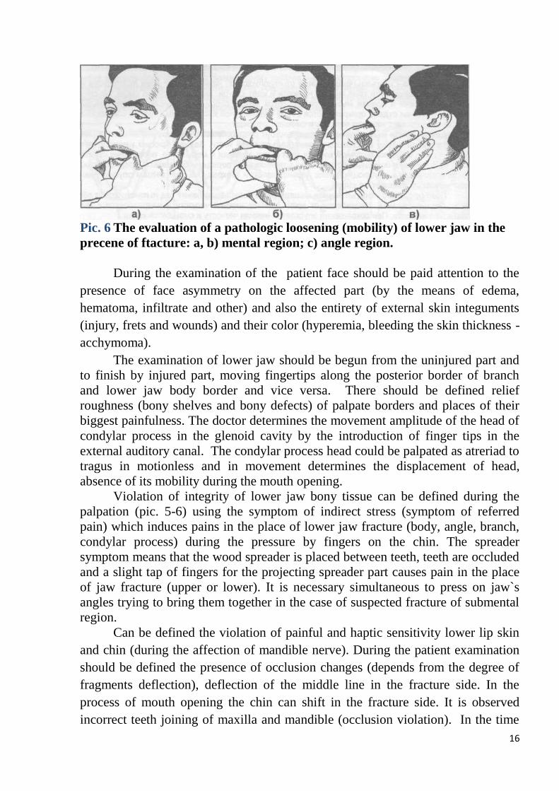
Pic. 6 The evaluation of a pathologic loosening (mobility) of lower jaw in the
precene of ftacture: a, b) mental region; c) angle region.
During the examination of the patient face should be paid attention to the
presence of face asymmetry on the affected part (by the means of edema,
hematoma, infiltrate and other) and also the entirety of external skin integuments
(injury, frets and wounds) and their color (hyperemia, bleeding the skin thickness -
acchymoma).
The examination of lower jaw should be begun from the uninjured part and
to finish by injured part, moving fingertips along the posterior border of branch
and lower jaw body border and vice versa. There should be defined relief
roughness (bony shelves and bony defects) of palpate borders and places of their
biggest painfulness. The doctor determines the movement amplitude of the head of
condylar process in the glenoid cavity by the introduction of finger tips in the
external auditory canal. The condylar process head could be palpated as atreriad to
tragus in motionless and in movement determines the displacement of head,
absence of its mobility during the mouth opening.
Violation of integrity of lower jaw bony tissue can be defined during the
palpation (pic. 5-6) using the symptom of indirect stress (symptom of referred
pain) which induces pains in the place of lower jaw fracture (body, angle, branch,
condylar process) during the pressure by fingers on the chin. The spreader
symptom means that the wood spreader is placed between teeth, teeth are occluded
and a slight tap of fingers for the projecting spreader part causes pain in the place
of jaw fracture (upper or lower). It is necessary simultaneous to press on jaw`s
angles trying to bring them together in the case of suspected fracture of submental
region.
Can be defined the violation of painful and haptic sensitivity lower lip skin
and chin (during the affection of mandible nerve). During the patient examination
should be defined the presence of occlusion changes (depends from the degree of
fragments deflection), deflection of the middle line in the fracture side. In the
process of mouth opening the chin can shift in the fracture side. It is observed
incorrect teeth joining of maxilla and mandible (occlusion violation). In the time

17
of mouth cavity examination there are breakages of the alveolar process mucosa
membrane (bleeding, covered by the fibrin accretion and other), hemorrhage in the
region of transitory fold, sometimes with the bone denudation. Palpatory are
defined acute bony edges under the mucosa membrane and the presence of the
pathological jaw mobility. During the deflection of jaw fragments sometimes can
be seen deducted cervix or teeth root, which is situated in the fracture cleft. On the
X-ray picture is relevant the violation of the bony tissue entirety. The fracture line
comes from the edge of alveolar process till the lower edge of mandible. In the
facture cleft can be a tooth.
TREATMENTS OF THE PATIENTS WITH THE MANDIBLE
FRACTURES
Treatment goals of patients with fractures of lower jaw are to create
conditions for fragments adhesion in right position in recent terms. Herewith a
performed treatment should provide complete recovery of lower jaw functions. To
perform all mentioned above the doctor should:
- Firstly to perform the reposition and fixation of jaw fragments for the
period of fragments union (includes the tooth excision from the fracture
line and initial surgical debridement);
- Secondly to create favorable conditions for the process of reparative
regeneration in bony tissue;
- Thirdly to perform the preventive measures of pyoinflammatory
complications in a bony tissue and surrounding soft tissues.
Liable for excision are:
Broken roots and teeth or completely dislocated from cavity teeth;
Periodontitis teeth with periapical inveterate inflammatory focus;
Teeth with presence of periodontitis or parodontosis of the middle and
severe stage of disease progress;
exposed root is in the fracture fossa or impacted tooth preventing the
right apposition of jaw fragments (penetrating into fracture fossa tooth);
Teeth unresponsive to conservative treatment and supported
inflammatory occurrence.
Temporary immobilization of fragments.
It is performed on the accident place in the ambulance, in any other no
specified medical establishment by paramedical worker or doctors. To the temporal
(transport) immobilization of mandible fragment concern:
• Circular gauze verticomental bandage;
• standart transport bandage (consists from the solid frame – head chin strep of
Entin);

18
• soft chin strep of Pomerantev-Urbansky;
• intermandibular ligature fixation of teeth by wire (according to Ivy)
a)
b)
c)
Pic. 7. Intermandibular Ivy loops: а) ligation; b,c)inter-maxillary fixation.
Permanent fragment immobilization
For immobilization of the mandible fragments are used conservative
(orthopedic) and surgical (operating) methods. More often for permanent fixation
of mandible fragments during its fracture are used wire frame (conservative
method of immobilization). S.S. Tigersted (was a dentist of Russian army, Kiev)
in 1915 year were offered aluminum teeth frames, which are used till present time
in the form of smooth frame – bows, frame with spacer (ripped arcuation) and
double-mandible frame with the anchor split and intermaxillary draft (pic. 8).

19
а) b)
a)
Pic. 8. Variants of the teeth aluminum frames offered by S.S. Tigerstedt:
а) smooth frame - bows; b) frame with spacer (ripped arcuation);
c) double-maxillary frames with the anchor splits and intermaxillary rubber
draft.
Pic. 9. Show of the mouth cavity of the double-maxillary aluminum frame
with the anchor splits and intramaxillary rubber draft.
The frame with the anchor splits are applied on both jaws (pic 9). The
indication of its preparation are fractures of mandible in the limit of teeth line or
beyond its as without the deflection of fragments, as with their deflection, and also
20
with the fractures of maxilla (in the last case it is necessary to apply additionally
verticomental bandage or standard chin strep and cranial cap). On every aluminum
frame are done 5-6 anchor slits, which are placed in the cardinal teeth (second,
fourth and sixth). The size of splits is around 3-4 mm and they are angle wise 35-
40° to the tooth axis. Frames are fixed to teeth by early described method (look the
technique of frame preparation). On the frame fixed on the maxilla, splits are
directed upwards, and on the mandible-downwards. On the anchor splits are put
on rubber rings (they are cutted from the rubber tube 8 mm diameter). To tighten
the ligature wires is necessary every 2-3 days, and also 5-6 days (or as and when
necessary) is necessary to change the rubber draft.
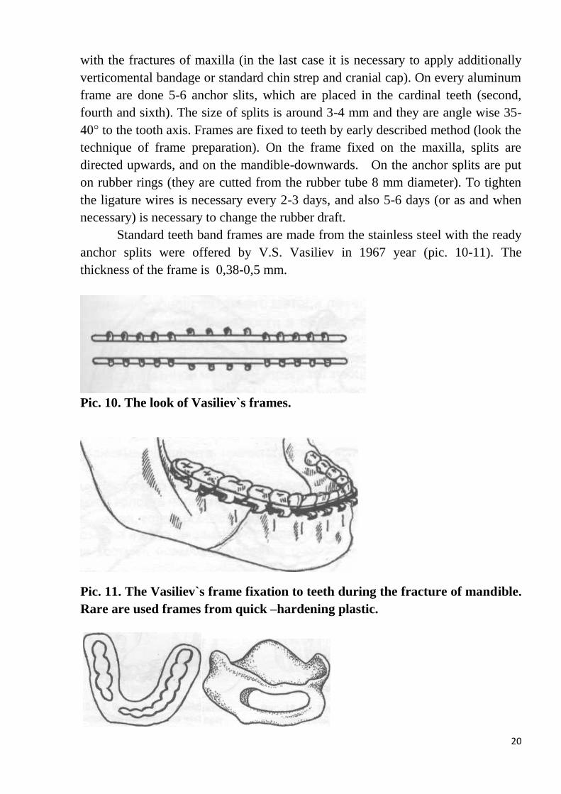
Standard teeth band frames are made from the stainless steel with the ready
anchor splits were offered by V.S. Vasiliev in 1967 year (pic. 10-11). The
thickness of the frame is 0,38-0,5 mm.
Pic. 10. The look of Vasiliev`s frames.
Pic. 11. The Vasiliev`s frame fixation to teeth during the fracture of mandible.
Rare are used frames from quick –hardening plastic.

21
а) b)
Pic. 12. Dentogingival and supragingival frames: а) Veber frame; b) Port
frame;
Osteosynthesis is the surgical method of connection of bony fragments and
elimination of their mobility by the help of fixing means.
Indications to osteosynthesis:
• Insufficient quantity of teeth for frame applying or absence of teeth on the
maxilla and mandible;
• Presence of flexible teeth at the patient with periodont disease, preventing the
usage of conservative method of treatment,
Fractures of lower jaw in the region of condylar process head with the
unreducible fragment in the case of dislocation or incomplete dislocation of jaw
head;
Interposition is the penetration of tissues (muscles, tendons, bony fragments)
between fragments of the broken jaw which impedes the reposition and
fragments consolidation;
Comminuted fractures of the lower jaw, if it is not managed to put together a
bone fragment in right position;
None putted together bone fragments of lower jaw in the result of deflection.
Classification of modern methods of lower jaw fragments osteosynthesis:
1.1. Direct intraosseous osteosynthesis:
1.1.1. simultaneous introduction of fasteners (pins, bolts, nails, screws) in both
fragments.
1.1.2. simultaneous introduction of fasteners in both fragments, but with the usage
of compression devices.
1.1.3. preliminary fixation of fasteners (pins, bolts, nails, screws) in one of
fragments.
1.1.4. preliminary fixation of fasteners) in one of fragments, but with the usage of
compression devices.
1.1.5. Other types of intraosseous osteosynthesis.
1.2. Direct extra-cortical osteosynthesis:

22
1.2.1. Fragments adhesion.
1.2.2. Locking stich.
1.2.3. Other types of direct extra-cortical osteosynthesis.
1.3. Direct intraosseous and extra-cortical osteosynthesis: 1.3.1. Osteosuture is made extra- or intraoral.
1.3.2. Osteosutore in combination with intraosseous pins, bolts, nails, screws and
anchors.
1.3.3. Osteosutore in combination with extra-cortical pins, bolts, nails, plates, mesh
and e.t.c.
1.3.4. Frames, plates, meshes, canals, beams fixing on the bone by screws and
other fixing elements implantable in bone.
1.3.5. Clips of various form input in bone by different apparatus for the mechanic
osteosynthesis other without it.
1.3.6. “Chemical” osteosynthesis with the usage of plastic masses.
1.3.7. Osteosynthesis with the usage of plastic masses in combination with other
materials, for example pins, nails, bolts and other.
1.3.8. Ultrasonic welding of bone.
1.3.9. Other types of direct intraosseous and extra-cortical osteosynthesis.
2.1. Indirect intraosseous osteosynthesis:
2.1.1. Kirshner wire.
2.1.2. bolt, nail and screw devices.
2.1.3. bolt, nail and screw devices, but with the compression – distraction
apparatus.
2.1.4. devices and apparatus using as locating support the head bandage (protector)
bones of facial and cerebral cranium with the input of pins, bolts, nails, and
bone screws in fragments.
2.1.5. Other types of indirect intraosseous osteosynthesis.
2.2. Indirect extra-cortical osteosynthesis:
2.2.1. suspension of lower jaw fragments to the bons of facial or cerebral skull.
2.2.2. Locking stich with gingival splint, prosthesis (according to Black).
2.2.3. apparatus with the usage in capacity of fixative of fragments of elements of
supra bony clamp (bony fastener) with compression – distraction apparatus
and without it.
2.2.4. devices and apparatus using as locating support the head bandage (protector)
bones of facial and cerebral skull, fixation and reposition of fragments which
is performed by the help of fasteners, bolts, nails ad screws.
2.2.5. Other types of indirect extra-cortical osteosynthesis.
2.3. Indirect intraosseous and extra-cortical dental osteosynthesis: 2.3.1. Nails, pins, screws and anchors.

23
2.3.2. Nails, pins, screws and anchors input in one of the fragments and fixating ny
the intermediate redisung – clamp band. The surgical intervention can be
performed by an extraoral or intraoral access. The surgery is made under the
general or local anesthesia.
Pic. 13. Schedules of various usages of titanium
mini-plates for the osteosynthesis of facial skeleton bones.
X-ray symptomatology of the bony tissue healing after fracture.
The healing of the bony tissue after fracture is a complicate biological
process, which has some stages. In first days after trauma effusive blood from the
injured vessels (bone fracture) gets together and reasorbs. Necrotizing small bone
fragments reabsorb and mesenchymal tissue expands which gives a rise for bone
tissue cells. In the following 10-45 days it is observed the formation of primary
callus due to sealed band of loose connective tissue and formation of the
osteogenic tissue which produces bone. In the given period the cell growth of
periostenium, endosteum and paraosseous tissues takes place. It is formed the
osteoid. It is the bony tissue at the stage of formation which precedes the
mineralization of its intercellular substance. In the following, osteoid tissue
calcifies and turn into bone tissue. At the expense of the periosteal and intraosteal
reparative processes the fracture lines do not differentiate at X-ray film in 4-6 and
sometimes more months (this depends on localization and character of fracture,
degree of fragments deflection and etc.).
In the given period the reabsorption of excess bone amount happens and jaw
bony tissue is formed definitive. In the presence of fragments gaping the duration
of lower jaw fracture healing significantly rises due to chondral stage.
Posttraumatic osteomyelitis of lower jaw is identified due to the
appearance of bone tissue loss in the region of mini fracture. In the case of
comminuted fracture, sometimes it is very problematic to differentiate separate
bony fragments from sequester.

24
Topic № 3
Maxilla fractures. Treatment of the maxilla fractures. Dental-
paradontal traumas.
In maxillo-facial region traumatology is distinguished such region of face as
“middle region”. Middle face region from above is bounded by the upper orbital
line and from below by the line of tooth alignment occlusion. This region should
include following bones: nose, orbital, zygomatic complex, maxilla.
Predominantly bones of the middle face zone have a vertical type of
formation of trabecules corpus and the presence of contrefort.
Contrefort (French countre-force means an opposed force) is an
accumulation of plates (thickening) of the upper jaw compact tissue which is
situated in such a way that the tensions appeared during the nibble and food
mastication are shared by the jaw and then is projected to other bones which are
connected with it.
Азенштейн И.М. and Худайбердыев Р. И. (1962) distinguish frontonasal,
zygomatic, pterygopalatine and palatinalcontrefort (abutment).
Frontonasal contrefort is connected with the jugal abutment in the region
of upper and lower eye socket borders.
Palatinal contrefort is connected with frontonasal abutment in the region of
nasal incisure.
Zygomatic, pterygopalatine and palatinal contreforts are connected with
the lower jaw alveolar process.
The given structure of the middle face zone provides their steadiness to the
tension during mastication and is able to stand against the mechanical action.
Fractures appear in the cases when the force of mechanical action exceeds the
endurance of bones structure.
In the middle face area except the zones of increase fastness there are also
the places of feeble impedance. Among these are all walls of maxillary antrum,
lacrimal bone, sieve bone paper plate and alar bone pterygoid bone.
Abutments of upper jaw bones maintain the significant resistance if the
stroke direction acts parallel to contreforts. Upper jaw fractures appears in the
result of force action perpendicular to contreforts. Often there are produced
multiple and different fractures of middle face zone which not frequently are
combined with the brain injury and cerebral cranial bones.
Concomitant injury is the simultaneous injury of two and more anatomy
regions by one or more affecting factors.

25
Combined injury is the injury which appears in the influence result of
different traumatic factors (physical, chemical or biological).
Peculiarities of the facial skeleton architectonics not only create condition
for the brain protection from the traumatic influence but also play very important
role in the delivery of mechanical energy to cerebral structures.
In the presence of facial trauma such severe complications as subdural
hematoma, subarachnoidal hemorrhage, cerebral vessels thrombosis, traumatic
aneurysm, cervical vertebra fractures, basal skull fracture and other could be
determined by close topographic-anatomical relationships of facial and cerebral
skulls.
Clinical symptomatology of concomitant injury depends from the severity
and character of the cerebro-cranial and maxilla-facial trauma. In the presence of
concomitant trauma with the severe cerebro-cranial injuries neurological
symptoms prevail in the clinical presentation which considerable complicate the
diagnostic of maxilla-facial region injures. The X-ray examination performance
not always is managed in necessary projections. Therefore in the presence of
facial skeleton bones injury the main diagnostic method is a clinical method. The
given method requires from the doctor the relevant background and practice in
work with such type of patients.
All cranio-cerebral traumas are divided into 3 types:
• brain concussion;
• cerebral contusion:
а) mild case; b) middle case; c) severe case;
• brain compression:
а) against its concussion; b) without associated concussion.
MAXILLA FRACTURES
It is used Le Fort classification to determine types of maxilla body fractures.
It is established three main types of maxilla body fracture.
The first type of the fracture is characterized in that the fracture line
undergoes under the alveolar bone and under the hard palate (almost parallel to it),
through the lower edge of piriform aperture and through ends of alar bone
pterygoid bones and along the of maxillary antrum floor.
The given fracture is associated with the fracture of Guerrin (described by
him earlier) therefore in the scientific literature the given type of fracture is named
“fracture of Guerrin-Le Fort”. More often such fracture is produced by the stroke
of blunt item in upper lip.

26
The second type of fractures (suborbital, middle).The line of fracture
comes through the nose root (the place of connection of brow tine maxillary and
frontal bone nasal process), then goes over the inner wall of eye socket to the lower
palpebral fissure, gets through it and directs frontwards along the lower wall of
arcula to the place of connection of maxillary malar process with the jugal. Behind,
the fracture line comes through alar bone pterygoid processes.
Oftener such fractures are produced due to stroke of the blunt item in the
region of nasal bridge.
The third type of fractures (subbasal, upper). The fracture line comes in
the region of nasal root (the place of connection of maxillary bones brow tines with
frontal bone nasal process) along the medial eye socket wall to the lower palpebral
fissure, through the alar bone pterygoid processes, then directs frontwards to the
lower arcula wall, through the frontal-zygomatic joint (the place of connection of
brown tine with frontal bone malar process and big alar bone wing) and jugal
bridge, which is formed by a malar process of temporal bone and temporal process
of zygomatic bone.
It can appear due to the stroke of a blunt item in the eye-socket region or
nose floor as well as due to lateral blow in the zygomatic bone region.
Fractures of upper jaw are followed by injures of maxillary antrum walls and
hemorrhage in them. The presence of the blood in the antrum does not means that
the post-traumatic sinusitis will develop yet therefore it is not the indicant to
obligative maxillary sinus surgery.
Other variety of upper jaw fractures are so called sagittal (unilateral)
fractures, when only one upper jaw bone is twisted off. As if the jaw breaks
anteroposteriorly. Outside the fracture line comes in typical place but inside
(medial) along the middle line (along the palatine suture which connects both
upper jaw bones in one upper jaw). Such fractures are the result of blunt item
action and the obliguity of blow power from up downwards in the upper lip region
(upper jaw posterior area).
Mentioned above tree types of upper jaw fractures, according to Le Fort, can
combine against each other. From one side can be present one type of fracture form
other side can be present other fracture type. More often it is present the
combination of the send and the third type of fracture. Atypical fractures of upper
jaw which do not stay with earlier described schemas could be presented. There are
distinguished fractures of processes of upper jaw bone alveolar (it is broken a part
of alveolar with some teeth – pic.14), frontal (oftener it is unilateral) and hard
palate (it appears in the result of the fall on the outstanding object). Comminuted
fracture of the upper jaw bone anterior wall can be present.

27
Pic. 14. Fracture of upper jaw alveolar process
In such a manner I propose the following classification to divide non-
ballistic fractures of upper jaw.
THE CLASSIFICATION OF THE NON-BALLISTIC FRACTURES
OF UUPER JAW AND THEIR COMPLICATIONS (А.А. Тимофеев, 1998)
I. ISOLATED FRACTURES OF UPPER JAW.
1. Fractures of upper jaw body:
- Unilateral (sagittal);
- Typical (according to classification of Le Fort, Vassmund);
- Combined;
- Atypical.
2. Fractures of upper jaws processes:
- Alveolar;
- Frontal;
- Palatal.
3. Comminuted fractures (body and proceses).
II. COMBINED FRACTURES OF UUPER JAW:
- With cerebro-cranial injuries;
- With other bones injuries;
- With the injury of softtissues.
III. COMPLICATIONS OF UPPER JAW FRACTURES:

28
A – early complications (the injury and displacement of the eyeglobe, injury of
vessels and nerves, facial pneumoderma, meningitis and other);
B – late complications (paresis and paralysis of face mimic muscle, ptosis,
osteomyelitis, maxillary sinusitis, face deformation and other).
CLINICAL PICTURE
During the patient examination, the attention should be paid on the face form
defect and the occlusion state (it is connected with the fragments dislocations,
presence of ecchymoma (hemorrhage in the full –thickness skin and membrane
mucosa) or hemorrhages, the character and localization of the soft tissues. It is
observed the elongation and flattening of the middle face region, which is
connected with the infraplacement of maxilla as self consistent alike with
zygomatic bones. There is so called spectacles symptom what means the
hemorrhage in the palpebra pacefollower. The same symptom is presented in the
case of basis crania bones fracture. The difference is in the time of its appearance
and prevalence. In the case of maxilla fracture the spectacles symptom appears at
once after trauma, has a prevalence character and in case of the isolated fracture of
the basis crania bones the symptom appears not earlier than 12 hours (more often
in 24-48 hours) after trauma and does not outstep orbicular muscle of the eye.
In the case of basal skin fractures can be found out the liquorrhea. It is the
escape of cerebrospinal fluid through the defect of hard brain tunic.
Nasal liquorrhea is a liquorrhea in the nasal cavity through the defect of
hard brain tunic in the region of cribliform bone plate or in the place of alar bone
fracture.
Cerebrospinal fluid otorrhea is a liquorrhea from the external auditory
canal in case of the fracture of periodic bone. Visually this symptom is hard to
define due to accompanied hemorrhage. For diagnostic of the liquorrhea presence
it is used the probe of “double spot”. The effused blood forms on the wad gauze
the reddish spot in the centre and the yellow aureolla of cerebrospinal fluid along
the periphery. The symptom of handkerchief means that a clear handkerchief
dampened by neurolymph remains soft, but if dampened by nasal secretion
remains hard (“starch”).
In the case second or third type of maxilla fracture cab be presented the
syndrome of upper orbital fissure (ophthalmoplegia -paralysis of eye muscles),
ptosis (omission of upper eyelid), sensibility absence of upper eyelid and front
skin, widening and fixing of eye apple location (Zachariades N. et al.,1985). In the
case of blood effusion into the orbit it is observed the exophthalmos and diplopy.
In case of zygomatic bones affection appears the zygomatic syndrome which
means the reduction of sensibility in the innervation zone of zygomatic-facial and

29
zygomatic-temporal branches of the IInd branch of trigeminal nerve, paralysis of
separate facial muscle.
During the skin palpation can be defined the crepitation. The sense of
crackle or rattle which appeares in the result of air penetration from the aeriferous
ways in the hypoderm. In the suborbital region is presented the step syndrome
(according the second type of fracture to Le Fort) owing to the bone injury in the
place of adjustment of maxilla bone malar process with the later surface of
zygomatic bone.
There are presented occlusal disturbences as central teeth on the mandible
and maxilla do not occlude against each other therefore the open occlusion
appears. More often it is observed in the case of second type maxilla fracture and it
is connected with the fact that all maxilla outs off the surrounding bones
connection. The maxilla descends down, revolves about its transverse axes and tips
posteriorly (under the influence of involution of medial pterygoid muscles, which
by the one end are connected to the pterygoid process of alar bone, and by the
other end to the medial surface of the mandible corner).
During the intraoral examination can be identified the effusion of blood
under the mucosa membrane and violation of bony tissue entirety (step symptom)
in the region of zygomatic-maxillary suture (the place of connection of maxillary
and zygomatic bones).
The positive symptom of Malevich is the sound of creaked pot which
appears in the result of teeth percussion on the injured part (in case of walls
fractures of upper jaw processes).
Positive symptom of Geoen means pains along the fracture fissure during the
pressure by the index finger on lifters (from the bottom upwards) of the pterygoid
processes of alar bone. The mobility of fragments can be defined if we capture by
hand fingers upper teeth and careful move of the maxilla in the AP dimensions but
other hand fingers we place on the face skin respectively to supposed fracture.
Treatment. In the case of maxilla fracture, Temporary (transport) means
of fragments immobilization are: submental pariental bandage, submental sling,
standard transport bandage, elastic rubber and reticulate bandage. The aim of the
temporary immobilization is to press the mandible to maxilla and to hold them in
such position till the permanent fixation of fragment that which refers to the
secondary care.
Methods of maxilla fragments fixation can be defined:
- Orthopedic (conservative) method of treatment presupposes the fixation of
double-jawed standard or aluminum anchor splints to patients’ maxilla and
mandible teeth. It is applied an intermaxillary rubber draft. The capping rubber
tube is laid between big root teeth for more exact apposition of fragments of

30
upper jaw bone. The given treatment method presupposes the future
immobilization of mandible by the gypsum submental sling and by the skull cap
with rubber draft. The last one can be corrected in the course of the treatment
dynamics.
- Surgical-orthopedic method of treatment presupposes the fixation of the dental
splint to the cranial strong bandage or to unaffected facial bones.
Surgical treatment of upper jaw injuries
R.E. Shands (1956) applied the “transmaxillary bolt” for the strengthening of
the separated upper teeth, which was walked through both upper jaw bones in cross
direction and through the cheek skin with the further strengthening of the given
bolt to the head cap or arch if there were present injuries of skull skin integument.
М.А. Макиенко (1962) proposed to use Kirschner wires which are
introduced at different angles through the broken off upper jaw into uninjured
cranial bones (zygomatic bone or arch, upper jaw process of frontal bone). Wires
are introduced by special equipment. Wires are chipped in such a way as not to
stand proud soft tissues. Additional the author advised to use the Померанцевой -
Урбанской sling or circular dressing bandage.
In the 1995 year М.М. Збарж had a shot to join broken off upper jaw bone
along the frontozygomatic suture by the help of catgut. The result was negative. In
the 1957year the same author retried by the help of steel wire and the result was
positive. In recent years steel plates are used to this effect.
In the presence of frontal wall fracture, В.Г. Центило (1996) proposed the
trepanise of the medial wall of the upper jaw cavity through the lower nasal
passage and by the way of consequent introduction of an antiseptic tampon (for 14
days). There is performed the resorbtion and the fixation of the bone fragment in
right position to the solid achievement.
The most popular surgical method of upper jaw fractures strengthening is
different variants of bones` sutures which join flexible and immobile facial
skeleton bones (the osteosynthesis of wire suture) or the fixation of fractures by the
titanic mini-plates.
TEETH AFFECTION
Clasification:
1. Incomplete teeth fracture (without pulpa opening): dentin and enamel fissure;
dental crown marginal fracture and enamel abruption; dental crown marginal
fracture and dentin and enamel abruption.
2. Complete teeth fracture (with pulpa opening):

31
b) Opened (in the mouth cavity) are fractures with the partial dental crown and
root deformation;
c) Closed (if the entirety of dental crown) is the root fracture.
3. Teeth dislocation:
a) incomplete (partial) tooth dislocation;
b) tooth dislocation (abruption) and abruption of the alveolar process edge.
4. Dental impaction.
In case of tooth injury, the hemorrhage and then necrosis appear in the pulp
which is the reason of the development of inflammatory processes in the periapical
region.
This demands the performance of case follow-up for the pulp viability by
electro-odontometry method. In the presence of pulp necrosis, it should be
extirpated with the following canal filling.
Tooth dislocation is the deflection of the tooth in the socket in any of the
side (in different ways) or in the maxilla sponge tissue, which is accompanied by
breakage of tissue which surrounds the tooth.
Should be defined incomplete, complete and impacted tooth dislocations.
More often are observed the dislocation of frontal teeth on the upper and lower
maxilla. In the case of incomplete dislocation there is the deflection of the teeth in
the lingual (palatal) or buccal part, but tooth did not lose its connection with
socket. Patients` complaints come to pain in the tooth, increasing during the touch
to it, its mobility and deflection in relation to the neighboring teeth. The tooth root
deflects to the opposite to the crownwork side. The mucosa membrane of the
gingival can be broken. On the X-ray picture the tooth root is shortened because of
its slanting position, it is defined the widening of periodontal fissure not only in the
lateral, but also in cacuminal parts of tooth root. In the case of incomplete
dislocation should be tended to tooth preservation. After local anesthesia
performance, it is made the manual reduction of the tooth, its immobilization by
the help of ligature fixation or arch bar on the time around 2-weeks.
In the case of complete dislocation the tooth is completely dislocates from
the socket and lose the connection with it.
Impacted dislocation is the variety of complete dislocation when the last
tooth perforate compact plate of alveola and invade on different depth in the
spongy substance of the maxilla or into the soft tissues, but on the upper maxilla -
and in the cavity (nasal or maxillary). On the X-ray picture the line of periodont is
absent along the whole extent. Reimplantation is performed with the preservation
of alveola walls.
Tooth fracture.

32
Fractures of the tooth can be defined as incomplete (without pulpa opening)
and complete (with pulpa opening). The last one can be open (with the affection of
crownwork) and closed (root fracture), and also transverse, slanting and
lengthwise. The root fractures can occur in the upper, middle and lower its third.
In the case of fishneck of the crownwork with the pulpa opening, the
patient’s complaint on the self-existing pains, which sharp increase during the
influence of any irritant (food, cold air or water). In the place of chipped part of
tooth crownwork is seen the place of denuded pulpa, which can bleed, edema of
the soft tissue of alveolar process. During the root fracture it becomes mobility, the
percussion is painfulness. In the process of palpation it can be discovered that only
a broken part of the tooth is shifted. According the X-ray picture is seen the line of
tooth root facture.
Treatment.
In the case of fishneck of crownwork without pulpa opening is performed
the slicing acute edges and reconstructs the defect of the tooth by the help of filling
or inlay. In the case of crownwork fracture is observed the pulpa opening, then it is
necessary to remove the tooth, the canal is filled and the defect is reconstructed by
the way of inlay preparation. During the root fracture in the edge area, it is
necessary to extract by operative way the fishnecked its part with the obliged
preliminary filling of root canal. The tooth is subjected to the extraction in the case
of fracture of the root lower dental neck and during its lengthwise fracture.
FRACTURES OF THE ALVEOLAR PROCESSES
CLASIFICATION:
Partial- the line of fracture comes through external compact plate and sponge
substance;
• complete – the line of fracture comes through all thickness of alveolar process;
• abruption of alveolar process;
• fracture of the alveolar process, combined with the dislocation or teeth fractures;
comminuted fracture.
The line of fracture comes above the edges of root teeth (on maxilla) or
below them (on mandible) and has arcuated form. The patient complains are self-
existing pains in the region of the injured jaw, increased during the joining of teeth
or during the biting on the hard food. There is the violation of the teeth joining, the
patients neither can nor close the mouth. It is observed hemorrhage from the mouth
cavity. There are complaints for same speech violation.
During the examination is defined edema of the soft tissue of the oral region,
there are bleeding on the skin, frets and wounds. The viscous saliva with the

33
bloody tap springs from the mouth. On the lips and check mucosa membrane there
are hemorrhage, but on the alveolar process can be its breakages and denudation of
the bone or are seen the denudated teeth edges. The occlusion is usually violated.
Can be violated the form of teeth arch. During the palpation of the alveolar process
is observed its pathological mobility along some teeth. The chipped part of alveolar
process is flexible together with teeth. On the X-ray picture it is obvious the line of
maxilla alveolar process fracture and character of violation of teeth root edges
incoming in the fragment.
Treatment is performed under local anesthesia. It is leaded the finger
reduction of slipped fragment of the alveolar process. During the sufficient
quantity of the stable teeth on the affected and non - affected maxilla area is
necessary to apply the smooth frame.

34
Topic № 4
Fractures of the zygomatic-orbital complex. Fractures of the nasal
bones. Complications of OMF traumatisms.
FRACTURES OF ZYGOMATIC BONE AND ARCH
The zygomatic bone is the solidest from facial bones. The zygomatic arch is
formed from the zygomatic bone temporal process and the temporal bone
zygomatic process. More often reasons of zygomatic bone and arch injuries are
household, athletic, transport or industrial injury. Fractures of the zygomatic bone
and arch can be opened or closed, linear or comminuted, without a displacement or
with a displacement of fragments, ballistic or non-ballistic (pic. 15).
The typical places of the zygomatic bone fracture are:
- from the suborbital suture till the zygomatic-alveolar crest (it can be feeled
outside and from the side of mouth cavity in the “step” type),
- in the region of frontal-zygomatic and zygomatic-temporal suture.
In case of zygomatic bone injury the zygomatic bone body shifts inward and
posteriorly which leads to the violation of entirety of the arcular outer wall, and in
the case of turn of the fragment along its axis leads to injury of maxillary cavity
with the breakage of mucosa membrane and appearance of nose bleed.
Pic. 15. Fractures of zygomatic complex: 1-zygomatic bone; 2- zygomatic
arch.
Subjected to the trauma prescription, fractures of zygomatic complex are
considered: fresh – till 10 days, inveterate – from 11 till 30 days, incorrect adherent
and not adherent more than 30 days.
Clinical picture.

35
- The face deformation due to depression (flattening) of the zygomatic region
soft tissues in the result of the deflection of zygomatic bone);
- The presence of “step” symptom in the middle part of the eye socket lower edge
and in the region of the zygomatic-alveolar crest;
- The hemorrhage can appear in the transitory fold in the region of upper
premolars and the first or the second molar;
- The numbness of skin of suborbital region and lower eyelid, the lateral nose
part, upper lip and upper teeth gingival due to suborbital nerve injury;
- The hemorrhage in the orbital cellulose and in the eye sclera;
- The chemosis due to injury of eye socket lateral side;
- The nose hemorrhage in the result of the maxillary cavity injury.
There are complaints for the mouth opening restriction. In the case of
deflections of zygomatic bone frontal process in the eye socket cavity there are
observed pains and difficulties of eyeglobe movement. The diplopy can appear in
the case of significant down deflections of zygomatic bone. On the general X-ray
picture of facial skeleton bones (naso-submental setup) there are entirety violation
of the lower and external edge of eye socket, continuities in the zygomatic-alveolar
crest region and the zygomatic bone temporal process, the reduction of maxillary
cavity transparence due to hemosinus).
In the case of isolated zygomatic arch fracture take place the impaction of
soft tissue due to the deflection of fragments inwards and down. The impaction of
the soft tissues is masked owning to the quick appeared edema, the restriction and
painfulness during mouth opening as well as the difficulty of lateral movements of
lower jaw at the affected part. In the axial X-ray film are visible a deformation of
the zygomatic arch and the violence of continuity.
TREATMENT.
Fractures of the zygomatic bone and arch without expressed deflection of
fragments can be treated by the conservative method which includes the
indications of cold (ice cap or bulla with cold water) in first two days after trauma.
Surgical treatment is applied to all patients who have fractures of
zygomatic bone and arch with fragments deflection. The diaplasis of zygomatic
bone and arch fragments can be performed surgical and non-surgical.
Non-surgical (noninvasive) fragments resorption is performed in the case
of easy reducible acute fractures of zygomatic bone and arch without any
significant deflection of fragments. The doctor introduces a hand index finger or
the metal tongue depressor wrapped by the gauze (can be used the Buyalsky

36
wound scoop) in the hinder region of upper fornix of the buccal cavity and then by
the movement towards to the opposite side of deflection sets the fragments.
Surgical resorption can be divided into extraoral and intraoral. The usage
of dental anchor with the transverse placed handle is more common (pic.16). The
skin discission of a centimeter long is made on the transsection of mutually
perpendicular lines: first line comes along the lower edge and the second line
descends down along the outside edge of orbital cavity. The one teeth anchor is
introduced under the mixed fragment, then catch it from the inside and by the
movement opposite to the deflection reduce the bone (arch) in the right position.
The characteristic flick sounds when fragments are put together in the right
position. The absence of the bony prominence (“step”) along the posterior orbital
edge, the reconstruction of the face symmetry, a free mouth opening and the
accomplishment of lateral movements by upper jaw points to the right fragments
reduction.
Pic. 16. The reduction of the zygomatic arch by means of tenaculum with the
transverse placed handle (Limberg anchor).
Extraoral methods of zygomatic bone resorption are surgically intervention
with the usage of bony suture overlapping or the osteosynthesis of fragments by
miniplates (titanic or stainless steel).

37
FRACTURES OF NASAL BONES
Pic. 17. Nose:
a) Schematic illustration of osteochondral region of external nose:
1. Nasal bone,
2. Small alar cartilages;
3. Greater alar cartilage;
4. Epactile cartilage;
5. Lateral cartilage;
b) Schematic illustration of the osteochondral nasal septum :
1. Frontal sinus;
2. Sphenoidal sinus;
3. Vomer;
4. Nasal crest;
5. Hard palate;
6. Incisive canal;
7. Greater alar cartilage crus;
8. Vomeronasal cartilage;
9. Perpendicular plate of ethmoid bone;
10. Nasal bone.
For the clinician it is more convenient to use the Ю.Н. Волков classification
of nose bone fractures, proposed in 1958 year. According to the given
classification all nose fractures` injures are divided in 3 groups:
1. Nose bones` fractures without fragments displacement and without the
external nose deformation (opened and closed);
2. Nose bones` fractures with fragments displacement and the external nose
deformation (opened and closed);
3. Nasal septum injury.

38
Classification of all nasal bones injures
Pic. 18. The scheme of nasal bones fractures (frontal section):
1:1- nasal septum; 2- nasal bones; 3-frontal bones;
II- fracture in the form of the nasal fornix flattening in consequence of sutures
detachment between the nasal bones, between the frontal processes and nasal
bones.
III- the nasal fracture with the suture detachment between the nasal bone and
frontal process on the side of stroke and the fracture of the frontal process on
the opposite side;
VI- the fracture with the lateral deflection of the nasal arch and inward
impaction of nasal slope fragments.
Clinical picture.
The patients` complaints are:
- the deformation of the nasal arch,
- the nasal hemorrhage,
- the soft tissues edema,
- the hemorrhage in the nasal skin and eyelids,
- pains,
- the violation of nasal breathing and osmesis.
Fractures of the nasal bone can be accompanied by the cerebral contusion
(nausea, faintness and other symptoms).

39
During the examination and palpation is defined the sharp painfulness
edema of soft tissues in the nose region which is developed on lower eyelids. The
bulge is preserved during some days. The hemorrhage can be observed not only in
the skin structure, but also in the region of palpebral conjunctiva. The nasal arch
deformation points to the nasal bones fracture. During the palpation there are
defined bony prominences on the nasal arch and slope. There is present the
mobility of bony fragments (depending on trauma terms). The significant trauma
can result the breaking of nasal bones. The displacement of the nose at the bottom
signifies the frontal processes fracture of the upper jaw and nose bones. The
subdermal crepitation indicates the fracture of cribriform bone with the abruption
of the mucosa membrane and the appearance of emphysema because air penetrates
from the nose through injured tissue under the face skin during the nose blowing.
A straight and lateral X-ray film of nasal bones provides the data about the
localization and character of the fracture.
Pic. 19. The reduction of nasal bones: a) instrumental reposition; b) finger
reposition.
Treatment.
The reduction of nasal fragments is performed under the local or general
anesthesia. The reduction of nasal bone fragments (lateral arch deflection) is
performed by the big finger of a right hand in the case of arcuation to the left and
accordingly by the left hand during the arcuation to the right. The characteristic
flick sounds when fragments are deflected in the normal position. Retroposed
fragments (to the side of nose cavity) are set by the help of nasal narrow elevator,
on which preliminary was put on the steril rubber tube, which guarantees the
atraumatisms (pic.19). The upper and middle nasal ducts are tamponed by the

40
iodoform turunda(pic.20), damped in the liquid paraffin to prevent the repeated
deflection and retention of them in the right position. In the lower nasal duct there
are introduced wrapped buy the iodoform turunda rubber tubes to provide the
respiration. Endonasal fixation is withheld during six –seven days. The collodion
bandage should be applied during the multicomminuted nasal arch fracture. In the
presence of nasal hemorrhage is used the posterior nasal cavity packing (pic.21).
Pic.20. Schematic illustration of anterior nasal cavity packing
In cases when fractures of nasal bones are combined with brain commotion
is required a neurologist consultation, profound rest and strict bed confinement.
When the fractures of nasal bones is combined with fractures of basal skull or
cerebrospinal rhinorrhea, the nasal reposition is temporary contraindicative as there
is a real danger of meningitis development in coming days after trauma. The
reposition deadline is variable and depends on a set of conditions: a fracture
character and complications, patient age, recovery time after basal skull fracture. In
the presence of nasal bones injury connected with upper jaw fractures (Лефор IInd
or IIIrd), the reposition of nasal bones should be made after countertraction and
fixation of upper jaw bones.

41
Pic. 21. Posterior nasal cavity packing : а) catheter introduction; b) passage of
tampon; c) fixation of tampon.
Injures in nasal cavity can result to the future formation of adhesion between
its separate parts or to the tissues replacement (mucosa membrane, concha,
septum) by massive scars (commissures) – synechia, which interrupt the
respiratory and osmetic nasal function. In case of nasal bony malunion it is
developed the expressed deformation of nose in bony or osteocartilaginous parts
what is followed by the violation of nasal breathing. This is the indicant to perform
plastics to eliminate given deformations. Early and adequate treatment provides
good functional and cosmetic outcome.
ORBITAL FRACTURES
Orbital cavity (pic. 22) is the conjugated vallecula in the skull where is
placed eyeglobe with its assist device.

42
Pic. 22. Orbital cavity.
1 – frontal process of upper jaw bone; 2 – lacrimal bone; 3 – orbital plate of
ethmoid bone; 4 – alar bone; 5 – orbital surface of the zygomatic bone; 6 –
orbital part of frontal bone; 7 – palatine bone; 8 –intraorbital foramen of
upper jaw bone; 9 – lower orbital fissure; 10 – superior orbital fissure; 11 –
optic canal.
Medial wall of the orbital cavity is formed by the frontal process of upper
jaw bone, lacrimal bone, orbital plate of ethmoid bone and body of the sphenoid
bone anterior from the optic canal.
Lateral wall is formed by the zygomatic bone orbital surface and greater
wings of sphenoid bones.
Upper wall is formed by the orbital part of the frontal bone and lesser wings
of sphenoid bones.
Lower wall (floor) is formed by the zygomatic bone and upper jaw, and back
part is formed by the orbital surface of a cognominal palatal bone process.
In the upper external orbital cavity angle there is the vallecula for lacrimal
gland, but at the inner third of its upper border there is an incisure for the
cognominal vessels and nerves. Through the upper orbital fissure in the orbital
cavity enter the first ramus of a trifacial nerve, oculomotor, abducent and trochlear
nerve and it is emerged upper orbital vein.
The given zone injures symptomatology is named – orbital apex syndrome.
The optic nerve and ophthalmic artery pass along the orbital canal. Through
the lower orbital fissure infraorbital and zygomatic nerves penetrate in the orbital
cavity, butlower orbital vein emerges. Orbital walls are covered by periostenium
which is closely intergrown with bony frame along its border and the region of
optic canal where periostenium wind into optic nerve membrane.

43
Posterior tooth anlage present close to the lower orbital border in children.
Form and size of orbital cavity by 8-10 ages are in much the same in adults. Four
direct and two oblique muscles provide an essential mobility at all directions:
dermad (abduction) due to lateral rectus, upper and lower oblique muscles; inward
(adduction) due to medial rectus, upper and lower direct muscles; upward doe to
upper direct and lower oblique muscles; down due to lower direct and upper
oblique muscles.
Orbital cavity fractures are different depending on trauma`s mechanism, but
more often there are injures of lower wall owing to fractures of zygomatic bone
and upper jaw per IInd and IIIrd type. Also the inner wall of orbital cavity is
injuredin case of upper jaw fractures per IInd and IIIrd type. In the retrobulbar
space there are developedophthalmocele or orbital apex syndrome -
ophthalimoplegia (ocular paralysis), ptosis (superior eyeliddescent), nonsensibility
of superior eyelid and frontal skin, dilatation and fixed position of pupil due to
hematoma formation. There are present diplopy and visual impairment. There are
fractures of upper and lower orbital border.
Treatment.
The treatment of orbital cavity fractures includes the reconstruction of the
anatomical entirety ofzygomatic and upper jaw bony fragments. In the presence of
the upper or lower orbital border isolated fracture there are made the skin
discussion along a relevant orbital border, exploration of fracture fissure, release of
interponing soft tissues which are reduced and fixed to the uninjured orbital bony
regions by titanium mini plates or chromic catgut.
Comminuted fractures of the lower orbital wall are treated by tight iodoform
tamponade of the maxillary antrum and the reconsctruction of lower orbital wall.
The tampon end is output through the preliminary made fistula in the lower nasal
passage (rhinostoma). The post-surgical wound is sutured. The iodoform tampon
remains in maxillary antrum somewhere about 14 days.
In the presence of lower orbital wallbony defects it is necessary to perform
the plastics by auto bone or by alloplastics (titanium, teflon, silicon and e.t.c.). To
be sensible to the fact that all injures of orbital cavity are followed by
eyeglobeclosed injures (contusion, commotion, impaction) patients should be
treated in cooperation with oculist.

44
COMPLICATIONS OF SOFT TISSUE INJURES
All complications can be divided into immediate (in the incident place),
early (in the stage of medical evacuation and in the medical treatment facility in
the course of the first week after trauma) and late (appear not earlier than a week
after trauma), which can appear during the soft tissue affection.
To the immediate complications refer:
asphyxia,
acute respiratory failure,
hemorrhage,
collapse and shock.
Early complications are:
asphyxia,
respiratory failure,
early hemorrhage,
syndrome of acute disorder of water-electrolytic balance in patient
organism.
Late complications are:
secondary hemorrhage,
bronchopulmonary disorders,
purulence of the wound,
keloids and hypertrophic scars,
contracture of mastication muscles,
posttraumatic ossificans myositis of masticatory muscles,
paresis and paralyses,
parasthesia and hyperesthesia,
salivary fistula and other.
Asphyxia is a pathological state which is determined by an acute and
subacute proceeded hypoxia and hypercapnia manifested by heavy respiratory
failure, blood circulation and nervous function.
Иващенко Г.М. (1951) classifies 5 types of asphyxia according to the
origin:
1) Dislocation asphyxia appears in the result of tongue falling back at
patients with the bilateral mandible fracture (mainly in the
submental region).
2) Obturative asphyxia develops due to closing of upper respiratory tract
by foreign matter, vomiting matters or blood clot.

45
3) Stenotic asphyxia appears due to edema of throat, vocal cords and
subligamentous space tissues in the result of the impaction of
throat posterior spaces by hematoma.
4) Valve asphyxia develops in the case of closing of the laryngeal inlet
by the lacerated tissues flap from the back wall of the palate
(during the entrance the drop flap is stuck and in the valve form
covers air access through the true glottis in the weasand and
bronchi).
5) Aspiration asphyxia is observed in the case of hit (aspiration) in the
weasand and bronchi of vomiting matters, blood clots and mouth
cavity content.
In the presence of dislocation asphyxia an acute management consists in
putting of the patient on one side (on the side of injury) or facedown. In
appropriate case the tongue should be underran (in the horizontal area) and fiber
ends should be fixed around the neck or to the firm applied dressing. Injured
persons in the state of insensibility should be evacuated in the recovery position,
but persons in conscious state should be evacuated in the semirecumbent or
semisitting position. In the case of obturative asphyxia it is necessary to remove
from guttur all blood clots and foreign matters by the finger (wrapped by gauze or
bandage). As far as possible the suction should be used to clear the guttur cavity by
suction unit, which will assure the free air access. It is prohibited to underran the
tongue in the case of obturation asphyxia, as this promote the lift of foreign matters
in the lower area of upper respiratory tract. In the presence of aspiration asphyxia,
it is required to sanitate the trancheobronchial tree by the help of
tracheobronchoscopy (medical endoscope for the visual examination of weasand
and bronchi) though the tracheostoma. It is performed the bronchoscopy namely
the examination of the lower respiratory tracts, based upon the exanimation of the
inner surface of weasand and bronchi. If the given instrument is absent the sanation
of respiratory tracts is performed by the vacuum suction.
If it is impossible to eliminate cause of obturative and aspiration asphyxia as
well as stenotic and valve asphyxia it is performed the surgical intervention
directed to the normalization of aspiration function.
Cricotomy is the prosection of the throat by the way of degloving and
discission of annular cartilage.
Cricotracheotomy is the degloving and discission of the annular cartilage
and upper tracheal rings.
Conicotomy is the discission of throat in the interval between annular and
thyroid cartilage (in the region of elastic cone).

46
Conicocentesis is the puncture by thick needles (3 or 4 pc. according to the
lumen width) of the throat part and interval between annular and thyroid cartilage.
Trachycentesis is the puncture of the weasand by thick needles.
Hemorrhage
The hemorrhage namely bleeding from the blood vessels can be primary
and secondary. Primary hemorrhage appears at once after vessels affection.
Secondary – appears later, in some time, after vessel affection. The secondary
hemorrhage can be early, late and anticipate. Early secondary hemorrhage appears
in 1-3 days after trauma because of expulsion of clot from the injured vessel in the
result of blood pressure rise (for example, during the edema liquidation, in the case
of insufficient immobilization). Late secondary hemorrhage appears in 5-6 days
after trauma and later. It is conditional upon purulent fusion of a clot or wall which
forms the traumatic aneurysm. Secondary anticipate hemorrhage (multiple
appeared) is observed in the presence of purulent-necrotic processes.
The body weight consists of 6-8 % of human blood or 4.5 – 6 liters. Status
of patient is determined by extend of blood loss. Moderate blood loss is 20%
from the volume of blood circulation. Profuse blood loss is accompanied by shock
of a moderate severity and is 20-35% from the volume of blood circulation. Fatal
blood loss is 35-50 % from the volume of blood circulation and is followed by
preagonal and agonal state.
In the result of blood loss can develop a hemorrhagic collapse and then a
hemorrhagic shock.
If the level of hemoglobin in blood is lower than 80g/l and the hematicrit is
lower than 30% ( hematocrit norm: child – 36-44%, women 36-47%, men 40-50
%), the packed red cell or blood transfusion is indicated. It should be remembered
that hematocrit indicators begin to correspond to the real blood loss only after 8-10
hours of blood loss.
Collapse is an acute developed circulatory inefficiency, which is
characterized by the falling of vascular tone and decrease of circulating blood
mass. It is manifested by the sharp lowering of the arterial and venous pressure, by
brain hypoxia signs and arrest of the body vital functions depression.
Shok is an acute developed and life threatening pathological process, which
is characterized by the heavy violations of the central nervous system activity,
blood circulation, respiration and metabolism. The shock is determined by
following clinical features: cold, weeping, pale-cyanotic or marble coloring of the
skin; sharp delayed blood circulation in the nail bed region; trouble and sometimes
confused mental state; dyspnea (labored breathing).

47
In the presence of primary hemorrhage first care consists in overlapping of
compression band on the bleeding wound. In the case of profuse hemorrhage it is
indicated the digital occlusion of the vessel which supply the given anatomic
region. The facial artery is pressed a few anteriad from the place of interception
of mastication muscle frontal edge with the lower edge of mandible. Subsurface
temporal artery is pressed on 1 cm anteriad and upward deviating from the ear
antilobluit. Common carotid artery is pressed by the finger to the vertebra spine
of the sixth neck-bone (this is the place of interception of clavisternomastoid
muscle and horizontal line on the level of upper annular cartilage edge).
RESPIRATORY DISTRESS
Respiratory distress can occur as well as in early and late periods after
trauma. It is conventional to distinguish the respiratory distress according to
central, peripheral and combined type.
In the presence of peripheral type respiratory distress there are obturation of
breathing passages by the vomit masses, blood or mucus which fall in the
breathing passages due to violation of tone of lower jaw muscles, tongue and throat
as well as in the result of pharyngeal reflex lowering. This can be presented as in
the case of maxilla-facial trauma in their pure form as in the case of its connection
with the cerebro-cranial trauma or thorax trauma.
The central type of breathing distress occurs at patients with maxilla facial
trauma which is combined with the cerebro-cranial injuries. This type of
respiratory distress is characterized by full patency of breathing passages against
the expressed cyanosis and dyspnea which appears in the result of violation in the
central nervous system. The patient care consist in the introduction of air syringes
by mouth, nose or by mask with the usage of lungmotor (artificial respirator).
In the presence of combined type breathing disorders main doctor measures
should be oriented to the elimination of obstruction (occlusion) and restoration of
adequate ventilation of tracheobroncheal tree. The treatment of the patient should
take place in the Intensive Care, Anesthesia and Resuscitation Unit.
KELOID SCARS.
In the face wound treatment the optimal cosmetic result can be achieved of
the extent if sutures lines penetrate perpendicular to main masculation directions
along so called “force lines”. If lines of suture run out the direction of force lines
than appear hypertrophic scars namely scars which visible projects above the skin
surface.
Михельсон Н.М. (1938) distinguishes 4 stages of scar formation:
Ist stage – epithelisation (2-2.5 weeks);

48
IInd stage – swelling (3-4 weeks);
IIIrd stage – contraction (2-3 weeks);
IVth stage – malaxation (3-4 weeks).
The excessive skin scar enlargements are divided into 3 groups:
1. hypertrophic scar;
2. false (cicatrical) keloid;
3. true keloid.
True (spontaneous) keloid develops self-existing namely without anterior skin
injury. False (cicactrical) keloid develops in the place of former (burns, ulcers and
others).
MYOSITIS OSSIFICANS (CAVALRY BONE).
The myositis ossificans of the masticatory and temporal muscle can be present
after trauma (the stroke in the region of ramus mandible, traffic traumas and other).
The myositi assificans appears in 1.5-2 months after trauma. The patient`s
compliances are restrictions of mouth opening, the presence of immobile solid and
rather tender swelling with exact borders in the region of the masticatory or rare
temporal muscle. X-ray examination shows the presence of focuses of assification
in the region of the injured muscle. The treatment includes the excision of the post-
traumatic appearances together with the section of changed muscle.

49
Bibliography
1) Burlibaşa C. Chirurgie orală şi maxilofacială. Editura Medicina – Bucureşti:
Editura medicală, 2003.
2) Безрукова В.М., Робустова Т.Г Руководство по хирургической
стоматологии и челюстно-лицевой хирургии, том 1, Москва, 2000.
3) Poбустова Т.Г. Хирургическая стоматология, Издание второе,
переработанное и дополненное, Москва, 2000.
4) Тимофеев А.А. Руководство по челюстно-лицевой хирургии и
хирургической стоматологии. Киев, 2002.
5) Шаргородский А. Г. Клиника, диагностика, лечение и профилактика
воспалительных заболеваний лица и шеи. Москва, 2002.

50
Content
Introduction………………………………………………………………………...2
Topic № 1. OMF traumatisms, general information. Soft tissues damages……….3
Soft tissues injuries in the maxillo-facial region…………………………………...3
Nature of a non - firearm trauma in the maxillo-facial region……………………..3
Wounds……………………………………………………………………………..5
Surgical debridment………………………………………………………………...7
Surgical debridement peculiarities of maxillo-facial region wounds………………8
The organizational principles of health care………………………………………10
Topic № 2. Mandible fractures. Treatment of the mandible fractures (immediate,
at transportation, bone synthesis)…………………………………………………11
Fractures of lower jaws…………………………………………………………...11
Topic № 3.Maxilla fractures. Treatment of the maxilla fractures. Dental-
paradontal traumas………………………………………………………………...24
Maxilla fractures…………………………………………………………………..25
Teeth affection (classification, clinical picture, treatment)……………………….30
Fractures of the alveolar processes………………………………………………..32
Topic № 4 Fractures of the zygomatic-orbital complex. Fractures of the nasal
bones. Complications of OMF traumatisms………………………………………34
Fractures of zygomatic bone and arch…………………………………………….34
Fractures of nasal bones…………………………………………………………..37
Orbital fractures…………………………………………………………………...41
Complications of soft tissue injures (asphyxia, hemorrhage, respiratory distress,
keloid scars, myositis ossificans)…………………………………………………44
Bibliography………………………………………………………………………49