Course 2016 MANDIBLE MUSCLES OF MASTICATIONANDTEMPOROMANDIBULAR JOINT new microsoft power point...
30
MANDIBLE MUSCLES OF MASTICATION AND TEMPOROMANDIBULAR JOINT ESSAM ELDIN ABDELHADY SALAMA 03/24/2022 1
-
Upload
essameahady -
Category
Health & Medicine
-
view
15 -
download
2
Transcript of Course 2016 MANDIBLE MUSCLES OF MASTICATIONANDTEMPOROMANDIBULAR JOINT new microsoft power point...

05/02/2023 1
MANDIBLE MUSCLES OF MASTICATION
ANDTEMPOROMANDIBULAR JOINT
ESSAM ELDIN ABDELHADY SALAMA

05/02/2023 2
Mandible
It consists of a horseshoe-shaped body and a pair of rami The body meets the ramus on each side of the angle of the mandible

05/02/2023 3
BODY Horizontally placed, is composed of two halves fused at the middle On its external surface; Has the symphysis menti on the midline indicates fusion of its halves during its development.Mental foramen is seen below the 2nd premolar tooth , transmits terminal branches of inferior alveolar nerve and vessels.

05/02/2023 4
On the internal surface Mental spines are found in the midline giving attachment to genioglossus above and geniohyoid belowThe mylohyoid line; is seen as an oblique ridge runs backward and laterally from the area of mental spines to the area below and behind the 3rd molar tooth

05/02/2023 5
The submandibular fossa; for the superficial part of the submandibular salivary gland Sublingual fossa; for Sublingual salivary gland Lies in relation to the mylohyoid line;

05/02/2023 6
The upper border of the body is called the alveolar border; contains sockets of teeth. The lower border is called the base it contains;The digastric fossa on either sides of the symphysis menti for the anterior bellis of digastric muscles.

05/02/2023 7
THE RAMUS Is vertically placed; it has an anterior coronoid process and posterior condyloid process (head) the two processes are separated by the mandibular notch

05/02/2023 8
THE RAMUS The lateral surface; Contains rough area for the attachment of masseter muscle

05/02/2023 9
THE RAMUS The medial surface; lies the mandibular foramen for inferior alveolar nerve and vessels. Anterior to the foramen is a projection of bone the lingual for attachment of the sphenomandibular ligament

05/02/2023 10
The mandibular foramen leads into the mandibular canal that connected to the mental foramenThe incisive canal is a continuation forward of the mandibular canal below the incisor teeth

05/02/2023 11
THE RAMUS The medial surface; Lies the attachment of medial pterygoid muscle near the angle The coronoid process receives on its medial surface the attachment of the temporalis muscle Below the head is a short neck; anteriorly gives attachment to lateral pterygoid muscle.

05/02/2023 12
MUSCLES OF MASTICATION

05/02/2023 13
Muscles of mastication1. Temporalis.2. Masseter.3. Lateral pterygoid.4. Medial pterygoid.

05/02/2023 14
TemporalisOrigin: Floor of temporal fossa
and deep surface of temporal fascia.
Insertion: Coronoid process and anterior border of ramus of mandible.
Nerve supply: Deep temporal nerves (from and the anterior division of mandibular nerve).
Action: Elevation and retraction of the mandible.

05/02/2023 15
Masseter
Origin: Lower border and inner surface of the zygomatic arch.
Insertion: Lateral aspect of the ramus of the mandible.
Nerve supply: anterior division of the mandibular nerve.
Action: Elevation and protraction of the mandible.

05/02/2023 16
Lateral pterygoidOrigin:Upper head: from the infratemporal
surface of the greater wing of sphenoid bone.
Lower head: from the lateral surface of the lateral pterygoid plate.
Insertion: anterior aspect of the neck of the
mandible and the articular disc of the temporo-mandibular joint.

05/02/2023 17
Lateral pterygoid
Nerve supply: anterior division of the mandibular nerve.
Action: pulls the head of mandible forward during opening of the mouth (helps in depression of mandible), protracts the mandible and side to side moves it (chewing).

05/02/2023 18
Medial pterygoidOrigin: Superficial head: from the
maxillary tuberosity. Deep head: from the medial
surface of the lateral pterygoid plate.
Insertion: medial surface of the angle of
the mandible.
05/02/2023 19
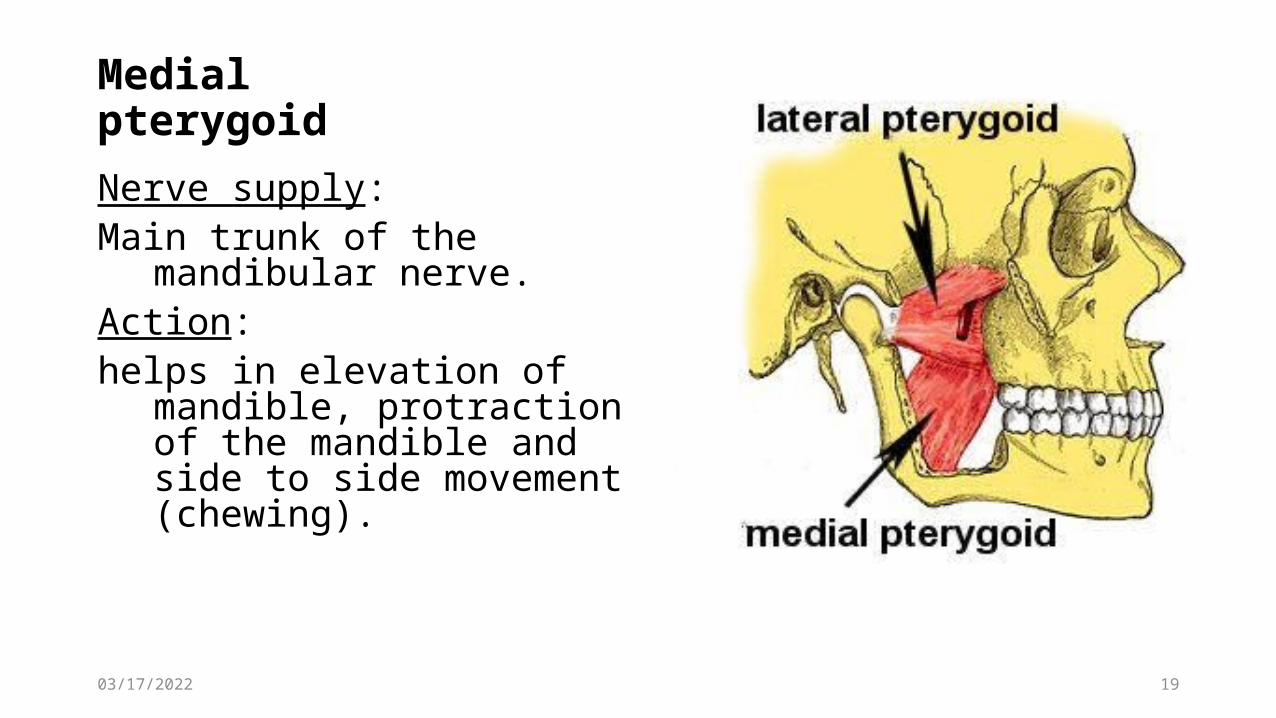
Medial pterygoidNerve supply: Main trunk of the mandibular nerve.Action: helps in elevation of mandible,
protraction of the mandible and side to side movement (chewing).

05/02/2023 20
TEMPOROMANDIBULAR JOINT

05/02/2023 21
TEMPOROMANDIBULAR JOINT It is a condylar synovial joint ArticulationArticular surface and tubercle of the temporal bone Head of the mandibleThe joint cavity is divided into upper and lower parts by an articular disc

05/02/2023 22
TEMPOROMANDIBULAR JOINT CapsuleGives attachment to the margins of the articular disc and lined by synovial membrane Articular disc Its upper surface is concavoconvex to fit into the articular tubercle and fossa

05/02/2023 23
Accessory ligaments
Lateral ligament:between the tubercle of the root of zygoma and lateral surface of the neck of mandible.

05/02/2023 24
Accessory ligaments
Sphenomandibular ligament’From the spine of sphenoid to the lingual of mandibleRelated laterally to the lateral pterygoid, auriculotemporal nerve, maxillary artery, inferior alveolar nerve, and inferior alveolar vessels. Related medially to the chorda tympani and medial pterygoid It is derivative of the first pharangyial arch

05/02/2023 25
Accessory ligaments
Stylomandibular ligamentFrom the tip of the styloid process to posterior border of ramus and angle of mandible

05/02/2023 26
Arterial supplyTwigs from superficial temporal and maxillary arteriesNerve supply Auriculotemporal nerve and nerve to masseter

05/02/2023 27
MOVEMENTSDepression; to open the mouth ;Lateral pterygoid on both sides , helped by digastric, mylohyoid and geniohyoidElevation; to close the mouth;Masseter, temporalis, and medial pterygoid on both sides

05/02/2023 28
MOVEMENTSProtrusion;Lateral and medial pterygoid on both sides.RetractionPosterior fibers of temporalis on both sides.Side to side (chewing);Lateral and medial pterygoid on both sides acting alternately

05/02/2023 29
On opening the mouth the head of the mandible first rotates on the articular disc at the same time forward displacement of the disc on the articular tubercle On closing the mouth the reverse occurs.

05/02/2023 30