Training pK a and logP prediction Jozsef Szegezdi Solutions for Cheminformatics.

1
Could LogP be a principal determinant of biological activity in 18-crown-6 ethers?
Synthesis of biologically active adamantane-substituted diaza-crowns.
Fran Supek[a]
, Tatjana Šumanovac Ramljak[b]
, Marko Marjanović[c]
, Maja Buljubašić[d]
,
Goran Kragol[b]
, Nataša Ilić[c]
, Tomislav Šmuc[a]
, Davor Zahradka[d]
, Kata Mlinarić-
Majerski[b]*
, Marijeta Kralj[c]
*
[a] Division of Electronics,
[b] Division of Organic Chemistry and Biochemistry
[c] Division of
Molecular Medicine, [d]
Division of Molecular Biology, Ruđer Bošković Institute, Bijenička
cesta 54, P. O. Box 180, HR-10002 Zagreb, Croatia;
* Corresponding authors: Dr. Marijeta Kralj, Division of Molecular Medicine, Ruđer
Bošković Institute, Bijenička cesta 54, P. O. Box 180, HR-10002 Zagreb, Croatia, e-mail:
[email protected], tel: +385 1 4571 235; fax: +385 1 456 1 010; Prof. Dr. Kata Mlinarić-
Majerski, Division of Organic Chemistry and Biochemistry, Ruđer Bošković Institute,
Bijenička cesta 54, P. O. Box 180, HR-10002 Zagreb, Croatia; e-mail: [email protected], tel:
+385 1 4680 196; fax: +385 1 4680 195.

2
Abstract
18-crown-6 ethers are known to exert their biological activity by transporting K+ ions across
cell membranes. Using non-linear Support Vector Machines regression, we searched for
structural features that influence antiproliferative activity in a diverse set of 19 known oxa-,
monoaza- and diaza-18-crown-6 ethers. Here, we show that the logP of the molecule is the
most important molecular descriptor, among ~1300 tested descriptors, in determining
biological potency (R2
cv=0.704). The optimal logP was at 5.5 (Ghose-Crippen ALOGP
estimate) while both higher and lower values were detrimental to biological potency. After
controlling for logP, we found that the antiproliferative activity of the molecule was generally
not affected by side chain length, molecular symmetry, or presence of side chain amide links.
To validate this QSAR model, we synthesized six novel, highly lipophilic diaza-18-crown-6
derivatives with adamantane moieties attached to the side arms. These compounds have near-
optimal logP values and consequently exhibit strong growth inhibition in various human
cancer cell lines and a bacterial system. The bioactivities of different diaza-18-crown-6
analogs in Bacillus subtilis and cancer cells were correlated, suggesting conserved molecular
features may be mediating the cytotoxic response. We conclude that relying primarily on the
logP is a sensible strategy in preparing future 18-crown-6 analogs with optimized biological
activity.
Supporting Information Available: Table S1 contains data related to QSAR modeling of
cytostatic activity of the 18-crown-6 compounds.
Supplementary data also at http://dx.doi.org/10.6084/m9.figshare.1279414
Keywords: adamantane-substituted diaza-crowns; biological activity; crown ethers;
ionophores; support vector machine

Graphical Abstract
Among a variety of molecular descriptors tested, the logP was found to have the prominent role in
determining biological activity of oxa-, monoaza- and diaza-18-crown-6 ether derivatives. Six
novel adamantane-substituted compounds had logP values close to the optimum of 5.5 and were
accordingly highly active against cancer cell lines.

Research Highlights
for
Could LogP be a principal determinant of biological activity in 18-crown-6 ethers?
Synthesis of biologically active adamantane-substituted diaza-crowns.
by
Fran Supek, Tatjana Šumanovac Ramljak, Marko Marjanović, Maja Buljubašić, Goran
Kragol, Nataša Ilić, Tomislav Šmuc, Davor Zahradka, Kata Mlinarić-Majerski*, Marijeta
Kralj *
structural features that influence antiproliferative activity in a set of 19 known oxa-,
monoaza- and diaza-18-crown-6 ethers were studied
the logP of the molecule is the most important molecular descriptor, among ~1300
tested descriptors, in determining biological potency (R2
cv=0.704)
to validate this QSAR model six novel highly lypophilic diaza-18-crown-6 derivatives
with adamantane moieties attached to the side arms were synthesized
novel molecules exhibit strong growth inhibition in various human cancer cell lines
and a bacterial system
the bioactivities of different diaza-18-crown-6 analogs in Bacillus subtilis and cancer
cells were correlated
*Research Highlights

3
1. Introduction
Since Pedersen in 1967 reported the synthesis of crown ethers, a new class of
compounds with unusually powerful cationic non-covalent binding properties,[1] they have
attracted extensive and continuous attention. Due to their ability in selective binding of metal
cations, anions, and neutral molecules, crown ethers have important roles in a variety of
applications [1 − 4]. Many different modifications of the crown ethers, such as changing the
ring size, the kind of substituents, and the type of donor atoms, have been synthesized to
enhance their complexation properties [5, 6].
Because of their structural characteristics, crown ethers exhibit ionophoric properties
in membranes, behaving very similarly to the natural ionophores (such as gramicidin,
valinomycin, nonactin, etc.), which makes crown ethers particularly interesting and useful in
chemical and biological research and their pharmaceutical potential remains large [4, 7].
Naturally occurring ionophores, such as metabolites of microorganisms (e.g. Streptomyces
sp.) disrupt the flow of ions either into or out of the cell, which dissipates the cellular ion
gradients leading to physiological and osmotic stress. Bacteria (particularly Gram-positive
bacteria) are very sensitive to this effect [8]. Since cyclic polyethers discriminate among
different ions, they can serve as convenient synthetic model compounds for their biological
counterparts and they also have similar functions [4]. Indeed, crown ethers were found to be
toxic in prokaryotic and eukaryotic cellular systems which led to further studies on their
potential for being developed as pharmacological agents [9]. For example, various approaches
have been developed to prepare crown-based antimicrobial agents [9-12].
Although the cytotoxic effects of crown ethers on mammalian cells (including tumor
cells) were soon recognized, the potential antitumor ability of the compounds has not been
scrutinized. The exceptions are functionalized crown ethers that have been synthetically
designed to alkylate and/or cleave DNA in order to achieve antitumor activity [7, 13]. In a
recent study, we investigated the antiproliferative/antitumor ability of conventional crown
ethers of various ring sizes and their derivatives in vitro and compared their activity to
valinomycin [14]. The results revealed that crown ethers possess marked tumor-cell growth
inhibitory activity and that this activity strongly correlates with both the type of hydrophilic
cavity (the size and the nature of donor atoms) and the characteristics of hydrophobic ring
surrounding, which enhanced the bioactivity by increasing hydrophobicity, possibly due to
requirements for membrane insertion. In a different study, a relationship between molecular
structure of alkyl-substituted diaza-18-crown-6 derivates and cytostatic activity in bacteria

4
and yeast was examined [12]. The authors measured that cytotoxic activity in three systems
peaks with C10 and C12 alkyl side chains, and decreases with either shorter or longer side
chains. This discontinuity in toxicity with increasing side chain length was interpreted to
result from the interplay of two factors: (i) an increase in hydrophobicity that would be
beneficial to activity as it promotes insertion into membranes, and (ii) long chains might
interfere with the 'flip-flop' activity (change of orientation in the membrane) that would be
essential for K+ transport and therefore biological activity. Additionally, a presence of a C=O
group adjacent to the macroring nitrogen atom in the diaza-crown reduced activity beyond
what would be expected by the change in hydrophobicity introduced by the keto group itself.
Our motivation for this work was to further investigate the interplay of these
molecular features, in particular: hydrophobicity, side chain length, and the side-chain amide
linkage, on biological activity of 18-crown-6 ethers against cancerous cells and bacteria. The
studied compounds include oxa-, monoaza- and diaza-18-crown-6 and are shown on Scheme
1. Because this set of compounds is diverse with respect to the side substituents, side-chain
length and, to some degree, the binding properties of the central ring caused by the type of
donor atoms, the rules derived from these compounds should be valid for 18-crown-6 in
general. We have measured the cytostatic activity on several human cell lines, as well as on E.
coli and B. subtilis for the series of previously synthesized 18-crown-6 derivatives, oxa-crown
ethers 7-9 [15,16], monoaza-crown ethers 10-15 [17], and diaza-crowns ethers 16 and 17.
Furthermore, we have derived a computational structure-activity relationship model that
indicated an overarching influence of molecular logP on biological activity, which led us to
synthesize six novel lariat diaza-18-crown-6, 20-25, that contain adamantane moieties in side
arms [18]. Finally, we have experimentally verified that the novel compounds are indeed
active against cancer cells in vitro, as the computational model had predicted.

5
O
O
O
OO
O
O O
HN
O
O
O
O O
HN
O
NH
O
O
O
O
OO
O
O
O
O
OO
O
O
O
O
OO
O
O
O
O
OO
O
O
O
O
OO
O
N
O O
N
O O
N
O O
N
O O
O
HNO
O
NH
O
O
O
N
O O
N
O O
OMe
O
O
Ph
O
O
MeO
Ph
O
O
O
OO
O
14
14
O O
O
O
O
O
O
O
O O
O
O
O
O O
N
O O OO
N
O
O
O
OO
N
O
O
O
OO
N
O
O
O
OO
N
O
O
OO
O
OO
N
O
N
O
OO
N
O
N
O
OO
N
O
N
O
OO
N
O
N
OO O
OO
1 432 5 6
7 8 9
10 11 12 13 14 15
16 17 18 19
OO
N
O
N
OO O
OO
N
O
N
O
20 21 22 23
24 25
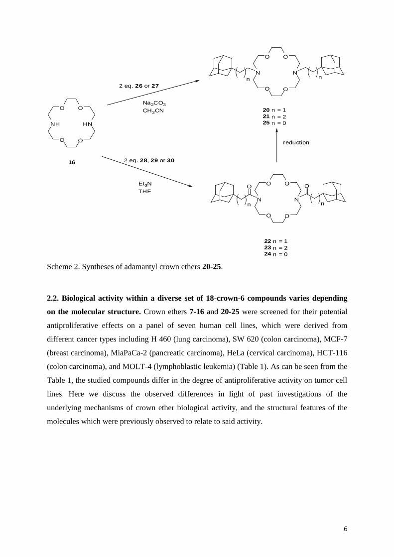
Scheme 1. Structures of crown ethers investigated in this study.
2. Results and Discussion
2.1. Synthesis. The 2-oxadamantane containing crown ethers 8 and 9 were prepared from 1,3-
bis(hydroxymethyl)-2-oxaadamantane by using an approach based on the literature procedure
[15]. The synthetic strategy to prepare monoaza-18-crown-6 12–15 was based on the coupling
reactions of the aza-crown ether 10 with corresponding adamantane derivatives [i.e 1-(2-
tosyloxyethyl)adamantane (26) [19], 1-(3-tosyloxypropyl)adamantane (27) [17] 1-
(chloroethanoyl)adamantane (28) [20], 1-(chloropropanoyl)adamantane (29)] [21]. The same
strategy was applied for the preparation of adamantane derived diaza-crown ethers 20-23. The
coupling reactions of the diaza-18-crown-6 (16) with corresponding adamantane derivatives
26 and 27 (method a) or the corresponding adamantane acyl chlorides 28 or 29 (method b),
followed by reduction of the obtained products with hydride type reagents, in particular
NaBH4, B2H6, or BH3 THF complex, afforded 20, 21, 22, and 23 (Scheme 2) in 54, 59, 56,
and 64 % yield, respectively. However, the crown ether 24 was prepared applying the method
b from diaza-18-crown-6 (16) and 1-adamantanecarbonyl chloride (30) in the yield of 65 %.
The subsequent reduction of 24 using LiAlH4 to afforded 25 in the yield of 49 %.
6
O O
HN
O
NH
O
Et3N
THF
O O
N
O
N
O
O O
O O
N
O
N
O
Na2CO3
CH3CN
n
nn
n
2 eq. 26 or 27
2 eq. 28, 29 or 3016
20 n = 1
21 n = 225 n = 0
reduction
22 n = 1
23 n = 224 n = 0
Scheme 2. Syntheses of adamantyl crown ethers 20-25.
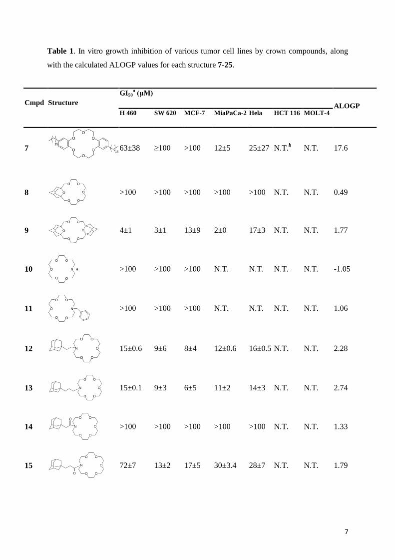
2.2. Biological activity within a diverse set of 18-crown-6 compounds varies depending
on the molecular structure. Crown ethers 7-16 and 20-25 were screened for their potential
antiproliferative effects on a panel of seven human cell lines, which were derived from
different cancer types including H 460 (lung carcinoma), SW 620 (colon carcinoma), MCF-7
(breast carcinoma), MiaPaCa-2 (pancreatic carcinoma), HeLa (cervical carcinoma), HCT-116
(colon carcinoma), and MOLT-4 (lymphoblastic leukemia) (Table 1). As can be seen from the
Table 1, the studied compounds differ in the degree of antiproliferative activity on tumor cell
lines. Here we discuss the observed differences in light of past investigations of the
underlying mechanisms of crown ether biological activity, and the structural features of the
molecules which were previously observed to relate to said activity.
7
Table 1. In vitro growth inhibition of various tumor cell lines by crown compounds, along
with the calculated ALOGP values for each structure 7-25.
Cmpd Structure
GI50a (μM)
ALOGP H 460 SW 620 MCF-7 MiaPaCa-2 Hela HCT 116 MOLT-4
7
O
O
O
OO
O15
15
63±38 ≥100 >100 12±5 25±27 N.T.b N.T. 17.6
8 O
O O
O
O O
>100 >100 >100 >100 >100 N.T. N.T. 0.49
9 O
O O
O O
O
4±1 3±1 13±9 2±0 17±3 N.T. N.T. 1.77
10
O O
N
O
O
O
H
>100 >100 >100 N.T. N.T. N.T. N.T. -1.05
11
O O
N
O
O
O
>100 >100 >100 N.T. N.T. N.T. N.T. 1.06
12
O
O
OO
O
N
15±0.6 9±6 8±4 12±0.6 16±0.5 N.T. N.T. 2.28
13
O
O
OO
O
N
15±0.1 9±3 6±5 11±2 14±3 N.T. N.T. 2.74
14
O
O
OO
O
N
O
>100 >100 >100 >100 >100 N.T. N.T. 1.33
15
O
O
OO
O
N
O
72±7 13±2 17±5 30±3.4 28±7 N.T. N.T. 1.79

8
16
O O
HN
O
NH
O
>100 >100 >100 >100 >100 >100 >100 -1.32
20c
O
N
OO
O
N
4±3 2.6±1.9 4±0.7 1.7±0.6 2.3±1.
4 N.T. N.T. 5.35
21 c
O O
N
O
N
O
1±0.4 1±0.4 2±1 N.T. N.T. 0.4±0.4 1±1 6.25
22 c
O O
N
O
N
O
O O
14±1 12±5 5±2 N.T. N.T. 6±0 6±6 3.45
23 c
O O
N
O
N
O
O O
11±5 19±16 2±0.4 N.T. N.T. 6±4 3±0.5 4.36
24 c
O O
N
O
N
OO O
12±0.3 21±7 9±1 N.T. N.T. N.T. N.T. 3.384
25 c
O O
N
O
N
O 18±3 19±2 21±5 N.T. N.T. N.T. N.T. 4.703
a GI50 - the concentration that causes 50% growth inhibition
b N.T. not tested
c Novel compounds synthesized
according to computational prediction
Oxa-adamantane crown ethers 8 and 9 were chosen because the incorporation of a
rigid polycylic moiety into crown ethers should affect their conformational mobility and
complexation abilities, while the lipophilic moiety (adamantane) should increase the solubility
of the crown ethers in nonpolar media (membrane) and thereby increase their ability for ion
transport via their complexation with metal ions [15]. As demonstrated, only bis(2-
oxaadamantano)-18-crown-6 (9) showed significant antiproliferative activity. Bis(4-hexadecyl
benzo)-18-crown-6 (7) showed very modest antiproliferative activity, presumably because the
presence of long alkyl side-chains attached to benzene ring makes this molecule highly
lipophilic and thus very slightly soluble in aqueous medium. Unsubstituted mono- and diaza-
18-crown-6 (10 and 16, respectively) did not show any antiproliferative activity. On the other

9
hand, substituted aza-18-crown-6 12-15 showed higher activity, although aza-crown ethers 14
and 15 in which the linkage of the adamantane molecule to the aza-18-crown-6 is achieved
through an amide bond showed significantly lower activity compared to 12 and 13, in which
the adamantane is attached by a methylene group. As shown previously, this phenomenon
could be related to their extraction capabilities [17]. Namely, the aza-crown ethers 14 and 15
(that have adamantane molecule linked through an amide bond) have very low affinity for
alkali cations (especially for potassium ion) and poor extraction capabilities, while 12 and 13
displayed significantly higher selectivity toward the extraction of K+. The lipophilicity is
necessary for the potassium extraction [17]. Besides, the potassium complexing abilities were
shown to be influenced also by the side chain length between adamantane moiety and the
macrocyclic ring in similar compounds [17], whereby the compound with two carbon atoms
in the linkage has a better affinity toward alkali cations than the one with three C-atoms.
However, the antiproliferative activity was very similar between 12 and 13, pointing to
unclear importance of length of methylene-linkage between adamantane moiety and crown
ether ring.
2.3. Computational structure-activity relationship analysis of 18-crown-6 reveals logP as
the main determinant of bioactivity. We gathered human tumor cell line cytostatic activity
data from our previous investigation of crown ether antitumor activity [14], (Scheme 1, crown
ethers 1-6, 17-19) and by testing other previously synthesized crown ethers, in particular oxa-
and aza-18-crown-6: 7, 8, 9, 12-15 [15,16,17,22] (Table 1). Using this data, we trained a
regression model that would predict antitumor activity from a large number of molecular
descriptors (see Experimental Section and Supporting Data). The algorithm employed for this
QSAR effort was the Support Vector Machines (SVM) regression. SVMs have desirable
properties of robustness to noise and high generalization power in difficult situations with few
training examples and many descriptors, and have consequently become widely popular in
other sciences, such as computational biology [23] and proteomics [24]. Examples of previous
applications of SVMs in QSAR studies include a classification problem involving
dihydrofolate reductase inhibition by pyrimidines [25], large-scale virtual screening of
molecular databases for inhibitors of multiple enzymes [26] and prediction of anticancer
activity of enkephalin-like peptides containing an unnatural amino acid [27] (for a review, see
reference [28]).

10
The non-linear SVM regression model trained on our 19-compound training set (1-19)
using all available molecular descriptors was estimated to predict activity of unknown
molecules with some accuracy; the root mean square error (RMSE) was 0.76 log GI50 units,
while squared Pearson’s correlation coefficient between measured and predicted values (in
crossvalidation) was R2
cv=0.518. We were able to considerably improve this result by
regressing activity against one molecular descriptor at a time. The highest performing group
of descriptors included two computational logP estimates: the Ghose-Crippen ALOGP
hydrophobicity [29], with RMSE=0.63 logs and R2
cv=0.683 and the Moriguchi MLOGP [30]
with RMSE=0.65 logs and R2
cv=0.645 (Supporting Data, Table S1). Combining the ALOGP
with the MLOGP (squared value) further improved the SVM regression model slightly:
RMSE=0.55 logs, R2
cv=0.753, possibly because the combination of two different
computational logP estimates may yield a closer approximation of the compounds' true logP
values. On the contrary, combining the ALOGP with each of the remaining descriptors did
not lead to an appreciable improvement in accuracy of regression over the ALOGP/MLOGP
combination (Supporting Data, Table S1), and neither did our attempt to summarize the
general trends in molecular descriptors using principal component analysis (Supporting Data,
Table S1) as we have done in a previous study [14] of a more structurally diverse set of crown
ether molecules.
Thus, in this set of compounds logP is a primary determinant of biological activity,
and the relationship of the two variables is non-linear: the predicted activity peaks at about
Ghose-Crippen logP=5.2, and is reduced towards lower or higher logP values (Fig. 1).
Molecules with high logP values are poorly soluble in aqueous media thus hindering the
activity because the compounds cannot be dissolved in the cell culture medium. Also, the
series of alkyl-substituted diaza-18-crown-6 compounds from a previous study by Leevy et al.
[12] have been shown to have a similar relationship of hydrophobicity to activity measured in
a different biological system - yeast and bacteria. It is tempting to speculate how the same
molecular features may be relevant for toxicity of diaza-18-crown-6 compounds throughout
the living world, which is perhaps not unexpected given the similarity of the biological target
- the cellular membrane - and given the universal importance of potassium ion homeostasis in
cells [31].


12
lower activity than the logP-based model predicts. Therefore, it is likely that an additional
variable such as e.g. the short side chain length may reduce the activity of 25 (vs. 20 and 21).
The cell cycle influence-experiments (Figure 2) showed that the compound having two
C-atoms in the linkage (20) has a slightly less pronounced effect comparing to 21 (three C-
atoms) on the cell cycle of SW 620 cells. Both compounds induced strong G1 phase delay,
accompanied with marked S phase reduction, at 5 and 10 μM concentration. Also, both
compounds induced accumulation of subG1 cells (apoptotic cells) at 10 μM concentration.
Apoptosis induction was additionally assessed by annexin V binding assay (Figure 3). These
results are in accordance with our previously published results on the crown ether influence
on the cell cycle, which is strong G1 phase delay, along with the activation of apoptosis [14].
0
20
40
60
80
100
SubG1 G0/G1 S G2/M
Pe
rce
nta
ge
of
ce
lls
(%
)
SW620, 24h
control 20 5 µM 20 10 µM 21 5 µM 21 10 µM
0
20
40
60
80
100
SubG1 G0/G1 S G2/M
Pe
rce
nta
ge
of
ce
lls
(%
)SW620, 48h
control 20 5 µM 20 10 µM 21 5 µM 21 10 µM
Figure 2. Cell cycle analysis of SW 620 cells treated with 5 and 10 μM compounds 20 and
21, 24 h, or 48 h. The histograms show percentages of live cells in G0/G1, S or G2/M phase,
along with the number of dead (subG1) cells, where subG1 population is expressed as a
percentage of total number of measured cells/counts.

13
Figure 3. The results of the annexin-V test performed with SW 620 cells after 48 h with
crown ether 20. The results are shown as percentages of viable cells (lower left quadrant),
percentages of early apoptotic cells (lower right quadrant) and percentages of late apoptotic
cells (upper right quadrant).
To further investigate the relationship of logP to molecular activity and the possible
contribution of other structural features of molecules, we combine the initial training set (1-
19) with the six novel compounds 20-25 and their experimentally measured activity against
cancer cells. The results of SVM regression remained essentially unchanged, with
computational logP estimates again having a prominent place among the relevant molecular
descriptors (Supporting Information, Table S1). Here, the Ghose-Crippen ALOGP emerges as
the most relevant single descriptor (RMS error=0.58 logs, R2
cv=0.704) and is closely followed
by the Moriguchi MLOGP (RMSE=0.65) and by the dataset with all 1318 descriptors
combined (RMSE=0.65, full data in Supporting Information, Table S1). The optimal value of
ALOGP on this expanded set of 25 molecules was found to be 5.5 (Fig. 1). There is a caveat
about how precise is this estimate of the optimal logP: computational logP estimates may not
be very accurate, especially with larger molecules – the error of the ALOGP algorithm itself
was previously found to range between 0.7 and 1.1 logs [32]. In our 25-molecule dataset, the
Ghose-Crippen ALOGP could be complemented by several descriptors that quantify
periodicity in distribution of polarizable, voluminous or massive atoms in the molecular
structure. Most promininently this included the Moran autocorrelation [33] of lag 4, weighted
by atomic polarizability or atomic volume (descriptor names "MATS4p" and "MATS4v"), but
also to a similar degree the GETAWAY descriptors [34]: leverage-weighted autocorrelations

14
of lag 5 and 7, weighted by mass ("HATS5m" and "HATS7m"); full data in Supporting
Information, Table S1). By including these descriptors alongside the ALOGP, some further
reduction of regression error was possible (best RMS error=0.47, crossvalidation R2
cv=0.803
for ALOGP+MATS4p). Judging by the magnitude of the error reduction, however, these
descriptors are of lesser relevance to biological activity in comparison to the ALOGP.
2.5. Symmetry, side chain length, amide side chain linkage, and adamantane moieties:
are they relevant for cytostatic activity of 18-crown-6? Some specific structural features of
crown ether molecules were previously discussed as related to biological activity, and we turn
to re-examine these effects using our structurally diverse set of 18-crown-6 ethers while
controlling for the overarching influence of the compounds' logP. For instance, Gokel and
collaborators [12] stated that the increasing length of the alkyl side chains in the examined
series of diaza-18-crown-6 compounds is a factor that limits biological activity, and
speculated how this might be related to the long side chains interfering with membrane 'flip-
flop' activity necessary for ion transport. In our set of 25 derivatives of 18-crown-6 that
contains both diaza-, monoaza- and all-oxa crowns and that also has more diverse
substituents, we do not find that the descriptors quantifying substituent length produce highly
predictive models either alone or when complemented by Ghose-Crippen ALOGP
(Supporting Information, Table S1). The examined descriptors included molecular span
("SPAN", R2
cv=0.410), mass-weighted radius of gyration ("RGyr", R2
cv=0.118), length-to-
breadth ratio ("L/Bw", R2
cv=0.408) and asphericity ("ASP", R2
cv=0.292). Let us examine an
example from our data: the compounds 5 and 6, where 6 is much more hydrophobic
(ALOGP=5.7 vs. 8.6), leading to a lower activity predicted by our model as the logP is further
from the optimal value of 5.5. In this case the substituent length cannot be applied as an
explanation for almost a degree of magnitude difference in biological activity between 5 and
6. However, the case of compound 25 illustrates a different trend: the logP-based model
overestimates its activity (Fig. 1), indicating the very short length of the side chains may have
reduced the activity of 25 (compared to 20 or 21) beyond what would be expected from the
logP. Therefore, in addition to the logP, it is possible that the substituent length might be
relevant for bioactivity in some specific cases, and it would take a carefully designed
experiment to disentangle the two influences.
Our previous work involved finding relevant molecular descriptors that relate to
anticancer activity in a set of 15 crown ether-related molecules [14]. The employed set of
molecules encompassed crown ethers of varying central ring sizes, their open-ring derivatives
15
and crown ether fragments. We interpreted the results as the biological activity being dictated
by the orientation and asymmetry of hydrophobic groups and the distribution of polarizable
elements. Here, we attempt to refine these rather general findings by focusing on a more
strictly defined set of molecules: oxa-, monoaza- and diaza-18-crown-6. While the structural
features of the central ring ("distribution of polarizable elements", see above) are kept
constant, the properties of the substituents and side chains important for membrane insertion
can be examined more thoroughly. Here, the logP has emerged as the most relevant molecular
feature. In our previous work [14] we had only found a weak (linear) correlation of crown
ether logP to biological activity; the difference may be due to the scarcity of the data points in
[14], and also to a less structurally consistent set of compounds.
Previously [14] we were not able to clearly determine to what degree the molecular
symmetry is relevant to crown ether biological activity, therefore we take the opportunity to
investigate this factor in this new set of the 18-crown-6. Five asymmetrical crown ethers 11,
12, 13, 14, and 15 were matched with symmetrical crown ethers 19, 18, 2, 9, and 17
respectively, having approximately equal Ghose-Crippen ALOGP values, and their biological
activities were compared. We did not find consistently higher activities for the symmetrical
molecules (Figure 4a). Thus, within the examined 18-crown-6 set, molecular symmetry does
not appear to play a role in determining cytostatic activity on human cancer cell lines. Next,
we have also examined if biological activity is influenced by the type of linkage (amide vs.
amine) between the ring of aza-18-crown-6 ether and the side chain, the structural feature
discussed previously [12]. Using five ALOGP-matched pairs of compounds, we found no
clear regularities of difference in activity of molecules with amine-linked vs. amide-linked
side-chains (Figure 4b). The result obtained by Leevy et al. [12], where the amide group
abolished biological activity of diaza-18-crown-6, may be specific to long alkyl side chains
(C10 and C12) that Leevy et al. [12] used to show this effect. Finally, we employed the same
logP-matching approach to test whether the presence of adamantane moiety affects cytostatic
activity of crown ethers, and found that adamantane does not add to biological activity of 18-
crown-6 beyond what would be expected by its contribution to logP (Figure 4c).
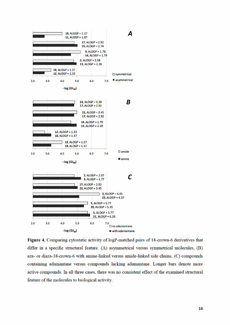

17
Our approach of matching crown ethers by their logP would necessarily result in some
of the logP-matched pairs of molecules differing in structural features other than the feature
currently examined. For instance, we may examine a logP-matched pair to see if symmetry of
molecule affects activity, where one of the two molecules may be an oxa-crown ether while
the other is a diaza-crown ether. In other words, when examining symmetry, type of bond or
presence of particular substituent, the confounding variable we control for is the logP, while
we do not control other structural features. Even so, the logP-matched compounds are (with a
few notable exceptions, see e.g. compound 3 in Figure 4c) well matched in activity regardless
of other possible differences in structure, meaning that the logP does have a prominent role in
determining a compound's activity. This rule was derived from a diverse set of 18-crown-6,
meaning that (i) it may or may not be applicable for crown ethers of other ring sizes, and that
(ii) while it shows a trend valid across various 18-crown-6, this does not rule out that
additional structure-activity relationship rules may exist for some specific compounds.
2.6. Cytostatic activity of 18-crown-6 derivatives towards bacteria is correlated to
activity against human cancer cells. In order to examine whether the same molecular
properties are indeed relevant for the biological activity of the 18-crown-6 derivatives in cells
of different organisms, we have conducted additional growth inhibition experiments on two
common bacteria: Escherichia coli and Bacillus subtilis. E. coli was almost completely
resistant to any of the compounds we subjected it to (2-6, 9, 14, 15, 17, 18 and 20-25). The
highest growth inhibition was recorded for compound 21, only 26% at the highest tested
concentration of 10-4
M, corresponding to a GI50 of approx. 10-3
M. All other compounds
caused E. coli growth inhibition <15% at 10-4
M. E. coli is a Gram-negative bacterium,
meaning it is enclosed by two layers of membrane; we anticipate that the outer membrane
would have to become saturated with the crown ether before the compound enters the inner
membrane, and both membranes would need to have some crown ether dissolved in them for
the ion transport to take place into the bacterium, or in the other direction. Leevy et al.[12]
have also noted that E. coli was less susceptible to alkyl-substituted diaza-18-crown-6
compounds than B. subtilis or yeast. In our experiments, B. subtilis (having a single cell
membrane) has indeed proven to be much more sensitive to a structurally more varied set of
18-crown-6 compounds (data in Supporting Table S1). In the sixteen compounds where
cytostatic activity to B. subtilis was above detection threshold, we found a roughly linear
correlation (Figure 5) between the log GI50 values on human cell lines, and on B. subtilis. A

18
more complex relationship, as captured by a second degree polynomial, fits the data slightly
better (Fig. 5, grey line) but the interpretation does not change: the correlation implies that the
same molecular features of 18-crown-6 ethers that enable cell killing of human cells are also
highly relevant for toxicity to bacterial cells. In designing a hypothetical antimicrobial 18-
crown-6 derivative, an insight into the compounds' structural features that cause an increase in
the relative bacterial vs. human toxicity would be useful. However, when we regressed the
residuals from Figure 5 against the full set of molecular descriptors using non-linear SVM, we
found no relationship (not shown). Thus, the differences in activity against Bacillus vs. human
cell lines that we did observe are mostly due to measurement noise and not to a genuine
difference of biological effect that we could relate to molecular structure. Also note that the
linear function in Figure 5 has a slope <1, indicating that the toxicity to human cells is
predicted to increase faster than the toxicity to bacteria. Taken together with the fact that we
found no structural determinants that would relate to increased selectivity against bacteria vs.
human cells (see above), one cannot escape the conclusion that 18-crown-6 as a group do not
appear promising as antibacterial therapeutics. This is not to say that a few of the 18-crown-6
derivatives are not interesting for future research. In particular, the compound 5 and the novel
compound 25 have a favorable ratio of antibacterial vs. human cell cytostatic activity (Fig. 5):
they have approx. equal GI50 values in both biological systems (this is actually an encouraging
finding; one should keep in mind that bacteria are generally unaffected by antitumor drugs at
the concentrations used to treat tumors [35, 36]). Regarding the biological mechanism-of-
action that leads to bacterial cell death, the 18-crown-6 are analogous to other ionophores
which have been recently emerged as medically relevant antimicrobials. For instance, the
peptide antibiotic daptomycin that rapidly kills bacteria through K+ efflux and membrane
depolarization [37] has been approved for human use. Daptomycin is effective against Gram-
positive bacteria, including difficult-to-treat infectious agents such as methicillin-resistant
Staphylococcus aureus (MRSA), or vancomycin-resistant enterococci (VRE) [37].

19
3п
21мо1222
232
207
1718р
1
24
25
y = 0.734x - 0.607R² = 0.63
R² = 0.68
-6.0
-5.0
-4.0
-3.0
-7.0 -6.0 -5.0 -4.0 -3.0
log(
GI 5
0) o
n B
aci
llu
s su
bti
lis
log(GI50) on human cell lines
Figure 5. Cytostatic activity of 18-crown-6 ethers on the bacterium Bacillus subtilis correlates
with cytostatic activity on human cancer cell lines. The 16 compounds with activity on B.
subtilis above detection threshold are shown on the chart (1-5, 7, 12, 15, 17, 18, 20-25). The
full black line is a linear function fit to the data points; the gray curve is a quadratic curve fit.
3. Conclusions
In summary, we found that the major contribution to biological activity of oxa-,
monoaza-, and diaza-18-crown-6 derivatives comes from the logP of the molecule which is
determined by hydrophobicity of the attached substituents and side-chains. The optimum logP
is approximately at 5.5, while both higher and lower values of the logP are detrimental to
activity. Structural features of the tested 18-crown-6 ethers, such as the symmetry of the
molecule, type of the substituent, type of the linkage of side-chain or side-chain length, did
not appear to affect biological activity beyond their contribution to the logP. In addition, we
have prepared six new diaza-18-crown-6, 20-25 and experimentally verified the activity
predicted by the logP-based model.

20
4. Experimental
4.1. Chemistry.
IR spectra were recorded on a FT-IR-ABB Bomem MB 102 spectrophotometer in
KBr. 1H and
13C NMR spectra were recorded on a Bruker AV- 300 or 600 MHz. The NMR
spectra were taken in CDCl3 using TMS as a reference and chemical shifts are reported in
ppm. HRMS were obtained on an Applied Biosystems 4800 Plus MALDI TOF/TOF
instrument (AB, Foster City, CA). Elemental analyses for carbon, hydrogen, and nitrogen
were performed on a Perkin-Elmer 2400 elemental analyzer and a Perkin-Elmer series II
CHNS analyzer 2400. The purity of compounds is >95%. Solvents for chromatography were
of HPLC purity. All compounds were routinely checked by TLC with Fluka aluminium oxide
on TLC-PET foils. 1,3-Bis(hydroxymethyl)-2-oxaadamantane [38], 1-(2-
tosyloxyethyl)adamantane (26) [19], 1-(3-tosyloxypropyl)adamantane (27) [17], 1-
(chloroethanoyl)adamantane (28) [20], 1-(chloropropanoyl)adamantane (29) [21], crown
ethers 10 [37], 11 [17], 16 [40], 17 [17], were prepared by literature procedure.
4.1.1. General procedure for the preparation of adamantane derived diaza-18-
crown-6 ethers 20-25.
Method a: In a reaction vessel under a stream of N2, two equivalents of corresponding
tosylate and one equivalent of diaza-18-crown-6 were dissolved in acetonitrile. To the stirred
solution, four equivalents of Na2CO3 were added. The reaction mixture was heated at 80 °C,
for five days, after which the reaction mixture was cooled to the ambient temperature and
concentrated in vacuo. The solid residue was suspended in CH2Cl2, and filtered through a
plug of celite. The combined filtrates were concentrated under reduced pressure to afford oily
product. If needed, product was additionally purified by column chromatography.
Method b: In a reaction vessel under a stream of N2, one equivalent of diaza-18-
crown-6 was dissolved in THF, and 2.5 equivalents of triethylamine were added. The
resulting mixture was stirred at ambient temperature for 10 min, and a solution of
corresponding acyl chloride (two equivalents) was added. The reaction mixture was stirred at
ambient temperature for additional 24 hours, filtered and the filtrate was evaporated under
reduced pressure. The solid residue was suspended in CH2Cl2, washed with saturated solution
of NaCl and dried over anhydrous MgSO4. After removal of the solvent, under reduced

21
pressure, the oily product was obtained. The product is additionally purified by column
chromatography.
4.1.2. N,N’-Bis[2-(1-adamantyl)ethyl]-4,13-diaza-18-crown-6 (20). By following
the general procedure (Method a), crown ether 20 was obtained via reaction of 1-(2-
tosyloxyethyl)adamantane (26, 0.668 g, 0.002 mol) and diaza-18-crown-6 (0.262 g, 0.001
mol). The crude product was purified by column chromatography on Al2O3 (act. II-III) using
02% MeOH in CH2Cl2 as an eluent, thereby affording 0.317 g (54%) of 20 as a colorless
oil. Analytically pure sample was obtained by re-chromatography on a small column of Al2O3
(act. II-III) using 02% MeOH in CH2Cl2 as an eluent.
1H NMR (CDCl3) /ppm: 1.20–1.30 (m, 4H), 1.47 (br. s, 12H), 1.55–1.75 (m, 12H),
1.92 (br. s, 6H), 2.45–2.60 (m, 4H), 2.70–2.85 (m, 8H), 3.55–3.70 (m, 16H); 13
C NMR
(CDCl3) /ppm: 28.55 (d, 6C), 31.65 (s, 2C), 37.07 (t, 6C), 40.69 (t, 2C), 42.43 (t, 6C), 49.66
(t, 2C), 53.75 (t, 4C), 69.80 (t, 4C), 70.60 (t, 4C); IR (KBr) /cm-1
: 2903 (s), 2852 (s), 1628
(s), 1462 (m), 1133 (m), 1110 (m), 1096 (m); HRMS calculated for [C36H62N2O4+H]+
587.4782, found 587.4758; Anal. Calcd. for C36H62N2O4: C 73.68, H 10.65, N 4.77, found: C
73.12, H 10.23, N 4.46.
4.1.3. N,N'-Bis[3-(1-adamantyl)propyl]-4,13-diaza-18-crown-6 (21). By following
the general procedure (Method a), crown ether 21 was obtained by reaction of 1-(2-
tosyloxypropylyl)adamantane (27, 1.50 g, 0.0043 mol) and diaza-18-crown-6 (0.563 g, 0.0022
mol). The crude product was purified via column chromatography on Al2O3 (act. II-III) using
02% MeOH in CH2Cl2 as an eluent, thereby affording 0.783 g (59%) of product as a
colorless oil. Analytically pure sample was obtained by re-chromatography on a small column
of Al2O3 (act. II-III) using 02% MeOH in CH2Cl2 as an eluent.
1H NMR (CDCl3) δ/ppm: 0.95–1.05 (m, 4H), 1.35–1.40 (m, 4H), 1.42 (br. s.12H),
1.60–1.75 (m, 12 H), 1.93 (br. s, 6H), 2.40–2.50 (m, 4H), 2.75–2.80 (m, 8H), 3.50–3.70 (m,
16H); 13
C NMR (CDCl3) δ/ppm: 20.09 (t, 2C), 28.66 (d, 6C), 32.06 (s, 2C), 37.16 (t, 6C),
42.12 (t, 2C), 42.43 (t, 6C), 53.81 (t, 4C), 56.92 (t, 2C), 69.91 (t, 2C), 70.65 (t, 6C); IR (KBr)
/cm-1
: 2901 (s), 2845 (s), 1645 (s), 1450 (m), 1119 (m); HRMS calculated for
[C38H66N2O4+H]+ 615.5095, found 615.5085; Anal. Calcd. for C38H66N2O4: C 74.22, H 10.82,
N 4.56, found: C 73.99, H 10.44, N 4.26.
4.1.4. N,N'-Bis[1-oxo-2-(1-adamantyl)ethyl]-4,13-diaza-18-crown-6 (22). By
following the general procedure, (Method b) crown ether 22 was obtained by reaction of
22
diaza-18-crown-6 (0.68g, 0.0026 mol) with 1-(chloroethanoyl)adamantane (28, 1.10 g, 0.0052
mol). The crude product was purified via column chromatography on Al2O3 (act. II-III) using
02 % MeOH in CH2Cl2 as an eluent, thereby affording 0.861 g (56%) of product as a
colorless oil. Analytically pure sample was obtained by re-chromatography on a small column
of Al2O3 (act. II-III) using 02% MeOH in CH2Cl2 as an eluent.
1H NMR (CDCl3) δ/ppm: 1.62–1.72 (m, 24 H), 1.97 (br. s, 6H), 2.12 (s, 4H), 3.50–
3.70 (m, 24 H); 13
C NMR (CDCl3) δ/ppm: 28.38 (d, 6C), 33.37 (s, 2C), 36.54 (t, 6C), 42.42 (t,
6C), 45.48 (t, 1C), 45.56 (t, 1C), 46.33 (t, 1C), 46.57 (t, 1C), 48.97 (t, 1C), 49.18 (t, 1C),
68.97 (t, 1C), 69.63 (t, 1C), 69.81 (t, 1C), 70.08 (t, 1C), 70.22 (t, 1C), 70.39 (t, 1C), 70.44 (t,
1C), 70.59 (t, 1C), 171.17 (s, 1C), 171.24 (s, 1C); IR (KBr) /cm-1
: 2905 (s), 2845 (s), 1449
(m), 1351 (m), 1126 (s); HRMS calculated for [C36H58N2O6+H]+ 615.4368, found 615. 4379;
Anal. Calcd. for C36H58N2O6: C 70.32, H 9.51, N 4.56, found: C 69.99, H 9.26, N 4.64.
4.1.5. N,N'-Bis[1-oxo-3-(1-adamantyl)propyl]-4,13-diaza-18-crown-6 (23). By
following the general procedure (Method b), crown ether 23 was obtained by reaction of
diaza-18-crown-6 (0.63g, 0.0024 mol) with 1-(chloropropanoyl)adamantane (29, 1.09 g,
0.0048 mol). The crude product was purified via column chromatography on Al2O3 (act. II-
III) using 02% MeOH in CH2Cl2 as an eluent, thereby affording 1.014 g (64%) of product
as a colorless oil. Analytically pure sample was obtained by re-chromatography on a small
column of Al2O3 (act. II-III) using 02% MeOH in CH2Cl2 as an eluent.
1H NMR (CDCl3) δ/ppm: 1.35-1.45 (m, 4H), 1.47 (br. s, 12H), 1.60–1.75 (m, 12H),
1.95 (br. s, 6H), 2.25–2.35 (m, 4H), 3.55–3.70 (m, 24H); 13
C NMR (CDCl3) δ/ppm: 26.18 (t,
1C), 26.30 (t, 1C), 28.29 (d, 6C), 31.65 (s, 2C), 36.80 (t, 6C), 39.19 (t, 1C), 39.22 (t, 1C),
41.88 (t, 6C), 46.45 (t, 1C), 46.77 (t, 1C), 48.44 (t, 1C), 48.62 (t, 1C), 69.09 (t, 1C), 69.67 (t,
1C), 69.78 (t, 1C), 70.07 (t, 2C), 70.20 (t, 1C), 70.41 (t, 1C), 70.59 (t, 1C), 173.86 (s, 1C),
173.91 (s, 1C); IR (KBr) /cm-1
: 2899 (s), 2845 (s), 1450 (m), 1350 (m), 1125 (s), 1071 (m);
HRMS calculated for [C38H62N2O6+H]+ 643.4681, found 643.4706; Anal. Calcd. for
C38H62N2O6: C 70.99, H 9.72, N 4.36, found: C 70.37, H 9.41, N 4.34.
4.1.6. N,N'-Bis(1-adamantanoyl)-4,13-diaza-18-crown-6 (24). By following the general
procedure (Method b) crown ether 24 was obtained by reaction of diaza-18-crown-6 (0.73 g,
0.0028 mol) with 1-adamantanecarbonyl chloride (30, 1.14 g, 0.056). The crude product was
purified via column chromatography on Al2O3 (act. II-III) using 02% MeOH in CH2Cl2 as
an eluent, thereby affording 1.074 g (65%) of product as a colorless oil. Analytically pure

23
sample was obtained by re-chromatography on a small column of Al2O3 (act. II-III) using
02% MeOH in CH2Cl2 as an eluent.
1H NMR (CDCl3) δ/ppm: 1.72 (br. s, 12H), 1.95 – 2.05 (m, 18H), 3.50 – 3.70 (m, 24H).
13C
NMR (CDCl3) δ/ppm: 28.39 (d, 6C), 36.47 (t, 6C), 39.08 (t, 6C), 41.95 (s, 2C), 48.53 (t, 4C),
70.14 (t, 4C), 70.62 (t, 4C), 176.87 (s, 2C). IR (KBr) /cm-1
: 2905 (s), 2850 (s), 1615 (s),
1461 (m), 1408 (m), 1228 (m), 1123 (s), 1102 (s), 1067 (s). HRMS calculated for
[C34H54N2O6+H]+
587.4055, found: 587.4060.
4.1.7. N,N'-Bis[(1-adamantylmethyl)]-4,13-diaza-18-crown-6 (25). Compound 25
was obtained by reduction of 18-crown-6 24 with LiAlH4. The solution of crown ether 24
(0.50 g, 0.00085 mol) in Et2O (30 ml) was added dropwise to the suspension of LiAlH4 (0.213
g, 0.056 mol) in Et2O (120 ml). The reaction mixture was heated at the reflux temperature for
7 hours. After cooling to the ambient temperature the excess of LiAlH4 was destroyed by
careful dropwise addition of water. The Et2O solution was decanted, washed with saturated
solution of NaCl and dried over MgSO4. After removal of the solvent under reduced pressure
the oily product was obtained. The crude product was purified via column chromatography on
Al2O3 (act. II-III) using 02% MeOH in CH2Cl2 as an eluent, thereby affording 0.234 g
(49%) of product as a colorless oil. Analytically pure sample was obtained by re-
chromatography on a small column of Al2O3 (act. II-III) using 02% MeOH in CH2Cl2 as an
eluent.
1H NMR (CDCl3) δ/ppm: 1.49 (br. s, 12H), 1.55 – 1.75 (m, 12H), 1.93 (br. s, 6H), 2.11 (s,
4H), 2.76 (t, 8H, J = 6.10 Hz), 3.55 – 3.65 (m, 16H). 13
C NMR (CDCl3) δ/ppm: 28.41 (d, 6C),
34.90 (s, 2C), 37.15 (t, 6C), 41.00 (t, 26C), 56.97 (t, 4C), 70.07 (t, 2C), 70.48 (t, 4C), 70.62 (t,
4C). IR (KBr) /cm-1
: 2899 (s), 2845 (s), 1453 (w), 1292 (w), 1125 (m), 1074 (w), 991 (w).
HRMS calculated for [C34H58N2O4+H]+ 559.4469, found: 559.4482.
4.2. Biology
4.2.1. Antiproliferative Activity on tumor cells. The HeLa (cervical carcinoma), MCF-7
(breast carcinoma), SW 620 (colon carcinoma), HCT 116 (colon carcinoma) MiaPaCa-2
(pancreatic carcinoma) and H 460 (lung carcinoma) cells were cultured as monolayers and
maintained in Dulbecco's modified Eagle's medium (DMEM), while MOLT-4 cells (acute
lymphoblastic leukemia) were cultured in suspension in RPMI medium, both supplemented
with 10% fetal bovine serum (FBS), 2 mM L-glutamine, 100 U/mL penicillin and 100 μg/mL
streptomycin in a humidified atmosphere with 5% CO2 at 37 °C.

24
The growth inhibition activity was assessed as described previously, according to the
slightly modified procedure of the National Cancer Institute, Developmental Therapeutics
Program.[14,41] Briefly, the cells were inoculated onto standard 96-well microtiter plates on
day 0. Test agents were then added in five consecutive 10-fold dilutions (10-8
to 10-4
mol/L)
and incubated for further 72 hours. Working dilutions were freshly prepared on the day of
testing. The solvent (DMSO) was also tested for eventual inhibitory activity by adjusting its
concentration to be the same as in working concentrations (maximal concentration of DMSO
was 0.25%). After 72 hours of incubation, the cell growth rate was evaluated by performing
the MTT assay which detects dehydrogenase activity in viable cells. The absorbency (OD,
optical density) was measured on a microplate reader at 570 nm. Each test point was
performed in quadruplicate in at least two individual experiments. The results are expressed
as GI50, which is the concentration necessary for 50% of inhibition. The GI50 values for each
compound are calculated from dose-response curves using linear regression analysis by fitting
the test concentrations that give PG (percentage of growth) values above and below the
reference value (i.e. 50%). Each result is a mean value from three separate experiments.
4.2.2. Cell Cycle Analysis. The 2×10⁵
cells were seeded per well, in a 6-well plate. After 24
hours, the tested compounds were added at various concentrations. After the desired length of
time the attached cells were trypsinized, combined with floating cells, washed with phosphate
buffer saline (PBS) and fixed with 70% ethanol. Immediately before the analysis, the cells
were washed with PBS and stained with 1µg/ml of propidium iodide (PI) with the addition of
0.2 µg/µl of RNAse A. The stained cells were then analyzed with Becton Dickinson
FACScalibur flow cytometer (20000 counts were measured). The percentage of the cells in
each cell cycle phase was determined using ModFit LT™ software (Verity Software House)
based on the DNA histograms.
4.2.3. Annexin V test. Detection and quantification of apoptotic cells at single cell level, was
performed using Annexin V-Alexa 488 conjugate (Molecular Bioprobes, Invitrogen). The
cells were seeded in 6-well plates (2×10⁵ cells/well) and treated according the required
schedule. After the desired length of time, both floating and attached cells were collected. The
cells were then washed with PBS with the addition of 0.5% BSA, pelleted and resuspended in
the Annexin binding buffer (10 mM HEPES, 140 mM NaCl, 2.5 mM CaCl2, pH 7.4)
containing Annexin V-Alexa 488 conjugate, 7-actinomicyn D (7-AAD, Molecular Bioprobes,
Invitrogen) according to the manufacturer’s recommendations. Camptothecin (10 M, Sigma)
was used as the control apoptosis-inducing agent. The cells were then analyzed on Becton

25
Dickinson FACScalibur flow cytometer (15000 counts were measured). Fluorescence
compensation and analysis was performed with FlowJo (TreeStar Inc.). Annexin V positive
cells were determined to be early apoptotic and both Annexin V and 7-AAD positive cells
were determined to be late apoptotic/necrotic cells. Each test point was performed in duplicate
in two individual experiments
4.2.4. Antiproliferative effect on bacteria Bacillus subtilis and Escherichia coli. Cultures
of the bacteria B. subtilis and E. coli were grown in the Luria-Bertani (LB) medium [43] at 37
°C with shaking, until they reached optical density at 600 nm of approximately 0.1 (OD600
0.1). To determine the colony forming ability before adding the tested compounds, we made
series of decimal dilutions in the phosphate buffer (Na2HPO4×2H2O 0.0333 mol/L, KH2PO4
0.0333 mol/L), and plated the 10-6
dilution onto the LB-plates.
At that point (t=0) we divided the starting culture into five aliquots; one of them served as
untreated control, whereas in other four we added the tested compounds in the following
concentrations: 10-4
M, 10-5
M, 10-6
M and 10-7
M. Cultures were grown for approximately
three mass doubling times (70 minutes for B. subtilis and 105 minutes for E. coli) at 37 °C.
Afterwards we made a series of dilutions in the phosphate buffer (described above) and plated
the diluted cultures (10-6
and/or 10-7
dilutions) onto LB-plates to determine the colony
forming ability for the control culture and the percentage of growth inhibition in treated
cultures. The plates were incubated overnight at 37 °C and the colonies were counted the next
day. The number of colonies formed multiplied by the 1/dilution yields the number of colony
forming units (CFU) per ml. Percent of growth inhibition at a certain concentration of the test
compound, and the GI50 values, were calculated as for the tumor cell essay. Each test point
was performed in quadriplicate in at least two individual experiments.
4.3. Computational Analysis.
4.3.1. Preparation of data set. Human tumor cell line cytostatic activity data from nine 18-
crown-6 derivatives 1-6, 17-19 was collected from our previous work on anticancer activity of
crown ethers in general [14]. Although the GI50 values were obtained for the five cell lines,
only the log GI50 values obtained for three cell lines (SW 620, MCF-7 and H460) that are
represented in previously published measurements (see below) were averaged to obtain an
overall estimate of biological activity. The differences in sensitivity of individual cell lines
were rather small (see Table 1) and therefore we assumed no relevant information would be

26
lost by the averaging of log GI50 values over cell lines. Results on antiproliferative activity of
compounds 1-6 and 17 were previously published [14] and are additionally provided in
Supporting data. We expanded this set of biological activity with novel bioactivity
measurements of other ten crown ethers, here numbered as 7-16. The overall cytostatic
activity was again described as an average GI50 value over the same three human cell lines,
grown in identical conditions and measured using the same experimental protocol. These 19
crown ethers with known biological activity constitute our initial training set. In addition to
these molecules, the dataset contains six novel diaza-18-crown-6 derivatives 20-25 whose
activity was first predicted computationally, then they were synthesized and their biological
activity was measured experimentally. The SMILES computational notations of the 25
compounds in our dataset were submitted to the E-Dragon 5.4 web service [43] which
predicts a probable three-dimensional structure of the molecules using CORINA [44] and then
computes 1666 molecular descriptors per compound. After removal of descriptors invariant
within our data, 1318 descriptors remained; to promote re-use by other researchers, this
dataset is provided in full in Supporting Information Table S1 and on the authors' website at
http://anticancer.irb.hr/. We used Marvin 5.1.1 (ChemAxon, http://www.chemaxon.com) to
visualize, browse, and otherwise handle the molecular structures.
4.3.2. Computational modeling of cytostatic activity from structure. The Support Vector
Machines (SVM) algorithm [45] for regression (ε-SVR variety) was applied to this data, as
implemented in the LibSVM software [46] adapted for the Weka 3.5.8 data mining
environment [47]. Per recommendation by the LibSVM authors [48], we utilized a radial basis
function kernel, and optimized the SVM's meta-parameters „c“ and „gamma“ in an exhaustive
search for a combination yielding the best root-mean-square error (RMSE) estimate on
crossvalidation. Gamma was varied exponentially from 2-15
to 23 in steps of 2
1, and c was
varied from 2-5
to 215
in steps of 21. Five runs of four-fold cross-validation were used to
estimate the RMSE, and also the Pearson's correlation coefficient between the predicted and
actual values (here denoted as Rcv). In order to find a single molecular descriptor most
informative for prediction of biological activity, the SVM meta-parameter optimization run
was repeated for each descriptor separately. The meta-parameters optimal for the whole 1318
descriptor dataset are likely to differ from the meta-parameters optimal for each descriptor,
and are also likely to differ between the individual descriptors. The optimal combination of
meta-parameters for each descriptor, and the crossvalidation error estimates, are given in
Supporting Table S1 and on the authors' website at http://anticancer.irb.hr/. In addition to the

27
mean RMSE and Rcv across the five cross-validation runs, the standard deviation of the
RMSE and Rcv were also recorded. A low mean RMSE was the primary criterion for
determining if a descriptor is informative, but a lower standard deviation is also desirable
since it signifies that the model is robust to the specific choice of molecules in the training set.
ACKNOWLEDGMENTS
This work was supported by the Croatian Ministry of Science, Education and Sports (Grants
no. 098-0982464-2514 to MK; 098-0982933-2911 to KMM; 098-0000000-3168 to TŠ; 098-
0982913-2862 to DZ). The authors are thankful to Dr. Ljerka Tušek Božić for a sample of
compound 7.

28
References
[1] C. Pedersen, J. Am. Chem. Soc. 89 (1967) 2495-2496.
[2] J. S. Bradshaw, R. M. Izatt, A. V. Bordunov, C. Y. Zhu, J. K. Hathaway, in
Comprehensive Supramolecular Chemistry, Pergamon, New York, 1996, p. 35.
[3] G. W. Gokel, O. F. Schall, in Comprehensive Supramolecular Chemistry, Pergamon,
New York, 1996, p. 97.
[4] G. W. Gokel, W. M. Leevy, M. E. Weber, Chem. Rev. 104 (2004) 2723-2750.
[5] R. M. Izatt, K. Pawlak, J. S. Bradshaw, R. L. Bruening, Chem. Rev. 91 (1991) 1721-
2085.
[6] F. Vögtle, Ed. , Host Guest Complex Chemistry, Springer-verlag, (Berlin, New York),
1981.
[7] M. Kralj, L. Tusek-Bozić, L. Frkanec, ChemMedChem 3 (2008) 1478-1492.
[8] A. J. Houlihan, J. B. Russell, J. Antimicrob. Chemother. 52 (2003) 623-628.
[9] W. Tso, W. Fung, M. W. Tso, J. Inorg. Biochem. 14 (1981) 237-244.
[10] W. Tso, W. Fung, Inorg. Chim. Acta 46 (1980) L33-L34.
[11] W. Tso, W. Fung, Inorg. Chim. Acta 55 (1981) 129-134.
[12] W. M. Leevy, M. E. Weber, M. R. Gokel, G. B. Hughes-Strange, D. D. Daranciang, R.
Ferdani, G. W. Gokel, Org. Biomol. Chem 3 (2005) 1647-1652.
[13] S. Vogel, K. Rohr, O. Dahl, J. Wengel, Chem. Commun. (Camb.) (2003) 1006-1007.
[14] M. Marjanović, M. Kralj, F. Supek, L. Frkanec, I. Piantanida, T. Smuc, L. Tusek-Bozić,
J. Med. Chem. 50 (2007) 1007-1018.
[15] K. Mlinarić-Majerski, G. Kragol, Tetrahedron 57 (2001) 449-457.
[16] K. Mlinarić-Majerski, A. Višnjevac, G. Kragol, B. Kojić-Prodić, J. Mol. Struct. 554
(2000) 279-287.
[17] K. Mlinarić-Majerski, T. Šumanovac Ramljak, Tetrahedron 58 (2002) 4893-4898.
[18] M. Kralj, K. Majerski, T. Šumanovac Ramljak, M. Marjanović, Adamantane Derivatives
of Aza-Crown Ethers and Their Use in Treatment of Tumor (patent Application,
#P20090355A).
[19] J. G. Henkel, J. T. Hane, G. Gianutsos, J. Med. Chem. 25 (1982) 51-56.
[20] W. Lunn, W. Podmore, S. Szinai, J. Chem. Soc. C (1968) 1657-1660.
[21] W. Oppolzer, R. Moretti, Tetrahedron 44 (1988) 5541-5552.
[22] L. Tušek, Comitato Nazionale Energia Nucleare RT/CHI (1975) 1-24.
[23] A. Ben-Hur, C. S. Ong, S. Sonnenburg, B. Schölkopf, G. Rätsch, PLoS Computational
Biology 4 (2008) e1000173.
[24] F. Supek, P. Peharec, M. Krsnik-Rasol, T. Smuc, Proteomics 8 (2008) 28-31.
[25] R. Burbidge, M. Trotter, B. Buxton, S. Holden, Comput. Chem. 26 (2001) 5-14.
[26] R. N. Jorissen, M. K. Gilson, J. Chem. Inf. Model. 45 (2005) 549-561.
[27] M. Gredičak, F. Supek, M. Kralj, Z. Majer, M. Hollósi, T. Šmuc, K. Mlinarić-Majerski,

29
Š. Horvat, Amino Acids 38 (2010) 1185-1191.
[28] O. Ivanciuc, in Reviews in Computational Chemistry (Eds.: K. Lipkowitz, T. Cundari, D.
Boyd), J. Wiley & Sons Inc., Hoboken, NJ., 2007, pp. 291-400.
[29] A. K. Ghose, G. M. Crippen, J. Comput. Chem. 7 (1986) 565-577.
[30] I. Moriguchi, S. Hirono, Q. Liu, I. Nakagome, Y. Matsushita, Chem. Pharm. Bull. 40
(1992) 127–130.
[31] C. D. Bortner, J. A. Cidlowski, Arch. Biochem. Biophys. 462 (2007) 176-188.
[32] R. Mannhold, G. I. Poda, C. Ostermann, I. V. Tetko, J. Pharm. Sci. 98 (2009) 861-893.
[33] P.A.P. Moran, Biometrika. 37 (1950) 17-23.
[34] V. Consonni, R. Todeschini, M. Pavan, J. Chem. Inf. Comp. Sci. 42 (2002) 682-692.
[35] C. A. Bodet, J. H. Jorgensen, D. J. Drutz, J. Antimicrob. Chemother. 28 (1985) 437-439.
[36] R. Henriksson, S. Holm, B. Littbrand, Acta Oncologica. 29 (1990) 43-46.
[37] J. N. Steenbergen, J. Alder, G. M. Thorne, F. P. Tally, J. Antimicrob. Chemother. 55
(2005) 283-288.
[38] K. Mlinarić-Majerski, G. Kragol, T. Ramljak, Synlett 2008 (2008) 405-409.
[39] R. A. Schultz, B. D. White, D. M. Dishong, K. A. Arnold, G. W. Gokel, J. Am. Chem.
Soc. 107 (1985) 6659-6668.
[40] V. J. Gatto, K. A. Arnold, A. M. Viscariello, S. R. Miller, C. R. Morgan, G. W. Gokel, J.
Org. Chem. 51 (1986) 5373–5384.
[41] M. Boyd, K. Paull, Drug Develop. Res. 34 (1995) 91-109.
[42] J. Sambrook, D. W. Russell, Molecular Cloning: A Laboratory Manual, Cshl Press,
2001.
[43] I. V. Tetko, J. Gasteiger, R. Todeschini, A. Mauri, D. Livingstone, P. Ertl, V. A.
Palyulin, E. V. Radchenko, N. S. Zefirov, A. S. Makarenko, et al., J. Comput. Aided
Mol. Des. 19 (2005) 453-463.
[44] J. Sadowski, J. Gasteiger, G. Klebe, J. Chem. Inf. Comp. Sci. 34 (1994) 1000-1008.
[45] C. J. Burges, Data Mining and Knowledge Discovery 2 (1998) 121-167.
[46] C.-C. Chang, C.-J. Lin LIBSVM: a library for support vector machines. (2001) Software
available at http://www.csie.ntu.edu.tw/~cjlin/libsvm
[47] Y. EL-Manzalawy, V. Honavar. WLSVM: Integrating LibSVM into Weka Environment.
(2005) Software available at http://www.cs.iastate.edu/˜yasser/wlsvm.
[48] C.-W. Hsu, C.-C. Chang, C.-J. Lin. A practical guide to support vector classification.
Technical report, Department of Computer Science, National Taiwan University. (2003)
http://www.csie.ntu.edu.tw/~cjlin