Theories of Literary Criticism English 113 Literature-Based Research Suzann Ledford.

Copyright © 2015 Association of Technical Personnel in Ophthalmology® (ATPO®). All rights reserved
COT Certification
Written Exam
Review, Part 1February 24, 2018
Presenter: Laura Lee Hopkins, COMT, OCT – C, ROUB
Criteria for Certification & Recertification, JCAHPO
Book only:
The Ophthalmic Assistant, 9th Edition
by Stein, Stein & Freeman
Preparing for the
COT Exam: A Study Guide
Includes: descriptions of the
exam, screen captures, sample
exam questions, and study tips.
Ophthalmic Medical Assisting: An Independent Study Course, 5th Edition, AAO

Fundamentals for Ophthalmic Technical Personnel,Barbara Cassin
Certified Ophthalmic Technician Exam Review Manual,2nd Edition,
Janice Ledford
COMPUTER-BASED EXAM
▪ 200 Questions Scored.
▪ There are10-25 questions which are not scored. You will not be able to identify these questions.
▪ Three hours to complete the exam.▪ Four distracters for each question.
▪ Each question has “ONE BEST” response.▪ While taking the exam, you can:
- Answer the question. - Mark the question for review at the end of
the exam.
All patient’s history recorded with accuracy, in the patient’s own words, if possible
This history is part of the entire record which provides valuable information
Additions and corrections must be made properly.
Chief Complaint
History of Present Illness (HPI)
Past Ocular History
Medical and Surgical History
Medications
Allergies
Social History
Family History
Name (nickname) and Title
Address
Telephone numbers
Social Security number
Date of birth (age)
Employer/Occupation
Spouse or parent (guardian)
Power of attorney
Chief complaint asks the question what is the reason for your visit? “Why is the patient here today?”
This is the subjective problem in the patient’s own words
Identify the problem with accuracy and efficiency
Listen first, then document it
Location (OD vs. OS)
Quality (Sharp or dull pain)
Severity (mild, moderate or severe)
Duration (How long does it last?)
Timing (When did it start?)
Context (Were you doing anything when it happened?)
Modifying factors (Has the patient tried any treatments?, Does anything make it better or worse?)
Associated signs and symptoms (flashes/floaters)

How many HPI elements can you count in the following Hx:
“Cataracts OU; c/o glare; especially with PM driving; c/o very blurred vision x 6 months.”
Surgeries and laser procedures
Diagnosis of all eye diseases or conditions Eye injuries
Contact lens and/or spectacle wear historyAge at first eye glass prescription?Were eyes equal then?
Eye therapies, patching, prisms, etc.Past IOL information is important◦ All should include date of surgery, which eye,
surgeon’s name, and specific IOL info
Complete ROS,(review of systems) past and present
The ROS should include 14 body systemsand must be signed and dated by the
examining physician
Illnesses◦ Includes any therapies, such as
chemotherapy, radiation, etc.◦ DM, HTN, heart/kidney disease, headaches
Surgeries
Injuries
Is it really a negative finding, or an omission?
Allergic/ Immunologic
Cardiovascular/ Cardiac
Constitutional Symptoms
Ears, Nose, Mouth, and Throat
Endocrine
Eyes
Gastrointestinal
Genitourinary
Hematologic
Integumentary
Musculoskeletal
Neurological
Psychiatric
Respiratory
Prescribed medicines◦ Name, dosage, (strength if applicable),
purpose
OTC medications, Vitamins
Herbs and supplement
Watch spelling
Match medication listed to a medical condition
All prescribed drops and ointments
All the OTC drops and ointments “Lid scrubs” or similar therapies◦ Dosage, strength, and specifics all should
be included ◦ Documentation of last dosage, if
indicated, i.e. History of glaucoma◦ Compliance may also be noted
Drug
Systemic
Ophthalmic
Environmental
Allergy vs. Adverse Reaction◦ Allergy- can be dangerous (i.e. itching,
constricting airway)
◦ Adverse reaction- uncomfortable (i.e. nausea, dizzy, bad taste in mouth, etc.)
Usage of ◦ Tobacco
◦ Alcohol
◦ Drugs
Sexual History (STDs)
Occupation
Does anyone else in the family have a condition like yours?
Pertinent family history: Diabetes, hypertension, cancer, retinal disease, glaucoma, cataracts
May uncover vital information
More specific questions may be required
Absence of data should not be construed as negative family history, negatives must be documented

Dilator Muscle: Dilates; Innervated by sympathetic branch of autonomic nervous system
Sphincter Muscle: Constricts; Innervated by the parasympathetic branch of ANS
science20.com
Retina Optic Nerve Chiasm
Midbrain (3rd nerve nucleus)
3rd nerve Ciliary Ganglion Iris Sphincter & Ciliary Body
Efferent Anatomy Afferent Anatomy
Size (in mm)◦ In each eye in Dim and Bright light and
◦ Note comparison between size of two eyes
Equal vs. Aniscoria
Shape- round, irregular
Light reaction
◦ Direct response in each eye and
◦ Direct response relative to each other
◦ Direct response Swinging Flashlight Test
RAPD
◦ Consensual response
◦ Hippus
◦ Fixed
Accommodative reaction
Recording pupil evaluation results◦ Normal: PERL (or PERRLA) and no APD
Lighting conditions
Accommodation
Medication
Hormones
~3mm ~7mm
Anisocoria
Marcus Gunn/ APD Horner’s Syndrome Third Nerve Palsy Argyll Robertson Adies Pupil Iritis Anisocoria Pharmacolgic

Hard lenses
Soft lenses
Toric lenses
Astigmatism
Bifocal
Aphakic
Extended wear
Gas permeable
Truncated
Bandage lenses
Oxygen permeability
Lens characteristics
Rigid lenses
Do you understand the difference between spherical and toric CL’s?◦ Know when to use and when to avoid.
◦ Front, back, & bi-toric◦ Lens markings…
Rotation compensation: LARS (left add, right subtract)
Bifocal/multi-focal CL’s Aphakic CL’s Piggyback lenses Treating keratoconus patients with CL’s Truncated
Base Curve
Power
Diameter
Dk Value
https://www.myalcon.com/products/contact-lenses/
Curvature of the back of a contact lens
Typical base curves of soft contacts 8.4, 8.6, 8.8
Base curve is crucial for a good fit.◦ Too tight
◦ Too loose
Amount of refractive correction in the lens
Measured in diopters
May be spherical or contain astigmatic correction
May also contain presbyopic correction
Measured in millimeters (mm)
Soft CLs larger that corneal diameter
RGPs much smaller diameter
Dk refers to the oxygen permeability of the lens
The higher the Dk value, the greater the amount of oxygen that passes through the material of the lens.
Dry Eyes
Keratoconus
Astigmatic Correction
Presbyopic Correction
Soft Lenses vs. Gas Permeable
Fitting CL’s What are the advantages & disadvantages of “hard” vs. soft CL’s?
Hard (PMMA) vs. RGP (silicone acrylic, silicone vs. flouropolymers)
Soft lenses◦ Most are HEMA material◦ Now fluoropolymers◦ Spincast vs. cast molded vs. lathe-cut
Know general differences in materials

Goals:◦ Centration
◦ Stable vision
◦ Good movement
Lens considerations:◦ Keratometry/ base curve
◦ Corneal diameter/ lens diameter
◦ Pupil diameter
◦ Dry eyes?
◦ Lid tightness (may need bigger lens if lid laxity)
Keratometry
Corneal diameter
Pupil diameter
Tear secretion
Eyelid tightness and fissure size
Fluorescein pattern
Spectacle prescription conversion
Over-refraction
Pediatric
Contraindications
Keratometry:◦ Used to determine Base Curve
◦ Considered normal when between 42.00-45.00D
◦ Considered flat when less than 42.00D
◦ Considered steep when greater than 45.00D
Note mire quality:◦ Clear vs. 1+ to 4+ distorted
Corneal warpage from CL’s
Topography
Needed to determine the power of the contact lens.
The amount of astigmatism in the refraction determines if a spherical or an astigmatic (Toric) lens will be used.
Typically, more than a diopter of astigmatism indicates the need for a Toric lens.
If less than one diopter of astigmatism, then the spherical equivalent of the refraction [in minus (-) cylinder] is used to determine the power of the contact.
Equivalent of a glasses prescription expressed only as a sphere
Used to calculate spherical contact lenses
Used to reduce the amount of cylinder in a glasses prescription
Step 1) Take half of the cylinder.
Step 2) Add this to the sphere algebraically.
Step 3) Drop the original cylinder & axis.
Ex: -4.00 -2.00 x 90Half of -2.00 = -1.00
(-1.00) +(-4.00) = -5.00 sph
Answer = -5.00 sph
+2.00 -1.00 x 75
Half of -1.00 = -0.50
-0.50 + (+)2.00 = +1.50
Answer = +1.50 sph
www.bausch.com.za www.bausch.com.za
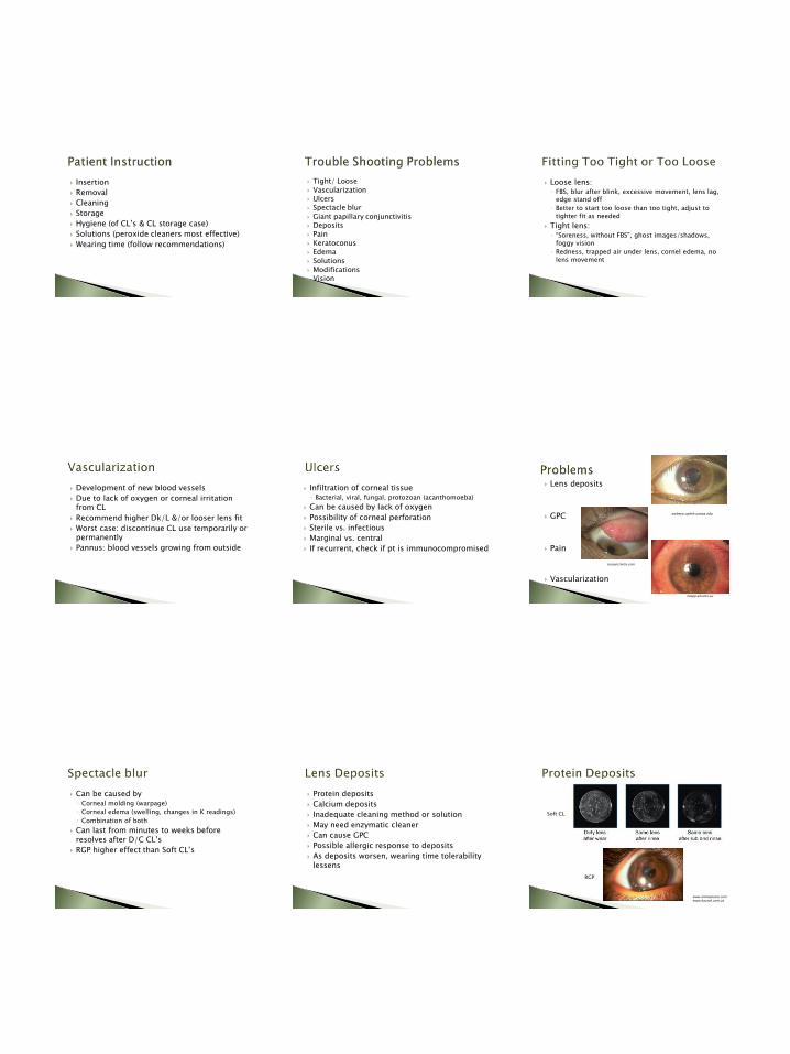
Insertion
Removal
Cleaning
Storage
Hygiene (of CL’s & CL storage case)
Solutions (peroxide cleaners most effective)
Wearing time (follow recommendations)
Tight/ Loose
Vascularization Ulcers Spectacle blur
Giant papillary conjunctivitis Deposits Pain
Keratoconus Edema
Solutions Modifications Vision
Loose lens:◦ FBS, blur after blink, excessive movement, lens lag,
edge stand off
◦ Better to start too loose than too tight, adjust to tighter fit as needed
Tight lens:◦ “Soreness, without FBS”, ghost images/shadows,
foggy vision
◦ Redness, trapped air under lens, cornel edema, no lens movement
Development of new blood vessels
Due to lack of oxygen or corneal irritation from CL
Recommend higher Dk/L &/or looser lens fit
Worst case: discontinue CL use temporarily or permanently
Pannus: blood vessels growing from outside
Infiltration of corneal tissue◦ Bacterial, viral, fungal, protozoan (acanthomoeba)
Can be caused by lack of oxygen
Possibility of corneal perforation
Sterile vs. infectious
Marginal vs. central
If recurrent, check if pt is immunocompromised
Lens deposits
GPC
Pain
Vascularization
webeye.ophth.uiowa.edu
iloapp.akrams.eu
conjunctivitis.com
Can be caused by ◦ Corneal molding (warpage)
◦ Corneal edema (swelling, changes in K readings)
◦ Combination of both
Can last from minutes to weeks before resolves after D/C CL’s
RGP higher effect than Soft CL’s
Protein deposits
Calcium deposits
Inadequate cleaning method or solution
May need enzymatic cleaner
Can cause GPC
Possible allergic response to deposits
As deposits worsen, wearing time tolerability lessens
RGP
Soft CL
www.amoeyecare.comwww.bausch.com.za

Thinning of cornea, bulging corneal tissue
Irregular astigmatism
Progressive steepening as ectasia worsens
RGP can correct for early to mid-stage KCN
RGP acts as new front surface for refraction
Munson’s sign and bulge , as pt looks down, examiner can see the bulge
Power
Base curve
Diameter
Central thickness
Edge profile
Instruments: radiuscope (b.c. of lens), V-groove, Loupe, Shadowgraph, Lensometer (power of hard or RGP lens)
Acuity Projectors
Ophthalmoscopes
Retinoscopes
Lensometers
Perimeters
Tangent Screen
Phoropters
Slit Lamps
Ultrasound biometry
Keratometers
Lenses
Tonometers
Muscle Light
Special Instruments (Equipment)
Surgical Instruments
Used to measure visual acuity
Electrical connections
Projector-blow off dust
Slides-lint-free cloth, photo-paper, replace darkened slides
Bulb-avoid touching with your fingers (oils lessen the life of bulb)
Screens-
Mirror-use only canned air or specified cleaning cloths
Other
Used to visualize/examine inside of the eye, usually the fundus◦ Direct
◦ Indirect
Electrical connections or battery
Bulb
Loose lenses
(20D, 90D, others)
Direct
Indirect
Used to conduct retinoscopy (objective observance of refractive error)
Front mirror-blow off dust
Battery- Lithium ion, rechargeable
Bulb-store instrument upright, filaments bend/distort light if stored horizontally
Used to measure the refractive power of the lenses in glasses (or RGP/hard CLs)
Manual lensometer◦ Cleaning lenses (blow off dust), dials (soft cloth),
stands (do not lubricate/call professional)
Auto-lensometer◦ Electrical connections
◦ Cleaning lenses, stands
◦ Utilizing correct modes
Used to measure field of vision◦ Humphrey visual field
◦ Goldmann visual field
◦ Other
Calibrating lighting conditions
Cleaning
Maintenance
Trial lens maintenance

Used to measure field of vision
Maintenance of tangent screen
The 1-meter black felt screen is mounted on a spring roller with mounting brackets. It also comes with a 1-1/2mm white test object, 18" wand, black
marking pins and 50 double chart sheets.
Used for the measurement of refractive error including sphere, cylinder, axis, distance and near, with/without prism, IPD, level, etc.
Cleaning lenses-blow off dust, lens tissue
Face shields- clean &/or exchange as needed
Other maintenance-always let a professional service inside the phoropter ~Q2yrs
Do not use alcohol of phoropter
(except face shields)
Used to examine the eye at various magnifications; also used for tonometry
Electrical connections
Bulb
Cleaning the: ◦ lenses/mirrors (dust
brush/cloth/photo-glass cleaner & cotton balls)
◦ stand (clean the pad, sewing machine oil at ball joint)
Maintenance of joystick, dials, etc.
Calibration of tonometry unit
Haag-striet-usa.com
A-scans and/or B-scans used to measure axial length and/or orbital abnormalities
Electrical connections
Cleaning the probe
Maintenance of probe, foot pedal, unit, printer (changing paper)
Calibrating the unit
www.sonomedescalon.com
Used to measure the corneal curvature, determine amount/location of astigmatism, identify corneal irregularities
Electrical connections
Bulb-check for deposits/
darkening, & replace as needed
Occluder
Check calibration with silver calibration spheres; call service professional if needed
Used to magnify a view of the eye for examination
Cleaning, maintenance, and storage◦ Loose lenses
Non-contact: Hruby lens, Condensing lenses, funduscopic lenses
Contact: Gonio, fundus contact lens, Koeppe lens
◦ Trial lenses
◦ Gonio lenses
◦ RGP/hard contact lenses
◦ Soft contact lenses (Bandage CLs)
◦ Other
Used to measure intraocular pressure (IOP)
Cleaning, maintenance, and storage◦ Goldmann
◦ Tonopen
◦ Non-Contact tonometer
◦ Perkins tonometer
◦ Schiötz tonometer
◦ Other
Used to illuminate external and some internal structures of the eye; used as a fixation focal point
Battery
Bulb- store instrument upright, filaments bend/distort light if stored horizontally
Used to measure various elements of structure and performance of the eye
Cleaning, maintenance, and storage◦ Auto-refractor
◦ Pachymeter
◦ Endothelial Cell Count/Specular Microscopy
◦ Topography units
◦ OCTs, HRTs
◦ Cameras
◦ Other
http://www.ophthalmic-instrument.com/store/p25/Tomey_RT-7000_Auto_Ref_%2F_Ker_%2F_Topography.html

Used to perform various surgical procedures
Cleaning/ Sterilizing of instruments
Clean, maintenance, and storage ◦ Sharps
◦ Disposables
◦ Reusable instruments
◦ Microscopes
◦ Loupes
Used to neutralize or “read” a prescription from a pair of glasses, loose lenses or rigid contact lenses.
Used to locate optical centers of lenses.
Used to “read” prism in lenses.
Used to “read” bifocals, trifocals (adds)
Sphere Cylinder power/axis Prism Multifocal power Multifocal induced prism Base curve Lensometer Lens “clock” Estimation with loose lenses Aphakic lenses Recording prescription Transposition
Several brands on the market
Measures quickly and accurately the sphere, cylinder, axis and prism of a lens.
Digital display can be printed on paper tape.
No focusing of eyepiece or target is required.
Eliminates need for mathematical determination of cylinder or add power.
Focus Eyepiece
Place spectacles on the platform so that both lenses are resting on the platform.
Center the target
Focus target using the power wheel and axis wheel.
Manual Lensmeter Target
➢All target lines come into focus at the same time. Thin target lines and wide target lines come
into focus at different times. (Here the thin lines are in focus, but wide lines are not.)
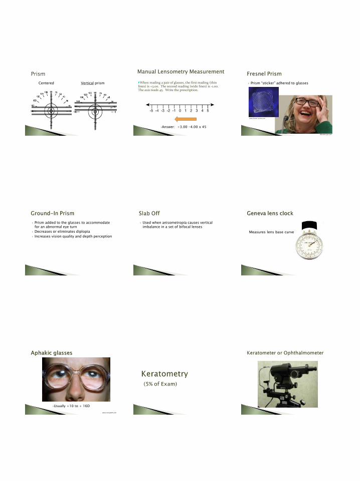
Centered Vertical prism When reading a pair of glasses, the first reading (thin lines) is +3.00. The second reading (wide lines) is -1.00. The axis reads 45. Write the prescription.
Answer: +3.00 -4.00 x 45
Manual Lensometry Measurement
Prism “sticker” adhered to glasses
Abcnews.go.com
www.fresnel-prism.com
Prism added to the glasses to accommodate for an abnormal eye turn
Decreases or eliminates diplopia
Increases vision quality and depth perception
Used when anisometropia causes vertical imbalance in a set of bifocal lenses
Measures lens base curve
Usually +10 to + 16D
www.mrcophth.com

Measures the central curvature of the anterior cornea
Readings are called K-readings
Measures in two meridians
Measured in diopters
Average cornea has a power of 42-44 D
Contact lens fitting
IOL calculations
Keratoconus detection
Irregular cornea detection
Focus the eye piece
Instruct the patient
Position the patient
Position the keratometer
Focus the mires
Locate the axis by rotating the drum
Align the plus signs and minus signs
Read/Record the measurement
Basic understanding of chart documentation to meet coding requirements
Basic understanding of coding levels
E&M codes
HIPAA regulations
Research related guidelines
The patient’s right to privacy.

Protecting patient privacy
Trained, skilled competent staff members
Equipment is calibrated/cleaned/good working order
Professional conduct
Code of Ethics
Respect and Sensitivity
A patient’s chart is a legal document and should be treated as such.
Health Insurance Portability and Accountability Act of 1996
➢Restricts use and disclosure of health information
➢Confidential communications concerning patient’s condition and treatment
➢Printed copy of the privacy practice of the provider
Patients have the right to make the decision about what they will and will not allow.
Patient is educated and asked to help in the decision making process.
Patient must be informed of common and uncommon risks.
These steps must be documented in the patient’s chart.
The technique of preventing infection and the growth of microorganisms.✓Never touch the eye with a dropper bottle or
ointment tube during instillation of meds.
✓Clean tonometer tips properly.
✓Use tonopen covers.
✓Wipe down equipment, occluders, chin rests, forehead straps, chairs, etc. between patients.
Personal Protective Equipment
How to handle a blood spill
What to do if a needle-stick occurs
Sharps containers
BioHazard containers
Universal Precautions:◦ Treat every patient as though he or she has an
infectious disease.
Sanitation◦ Hand hygiene- VERY important
◦ Personal Protective Equipment (PPE)
Contamination◦ Safe handling & disposal of sharps/waste
Ocular Fluid or tissue samples collected for evaluation of abnormalities◦ Lid
◦ Corneal
◦ Conjunctival
Collected in Surgery or Clinic

Scrapings or smears of ocular tissues collected for evaluation of infection or disease
Chocolate
Blood
Sabouraud
Mannitol Salt
Topical Administration◦ Drops (Solution vs. Suspension vs. Emulsion)
Advantage: Easy, quick absorption into eye with little systemic absorption
Disadvantage: May not penetrate cornea, may not extend beyond anterior segment
◦ Ointments
Advantage: longer contact time, more difficult for kids to flush away with tears
Disadvantage: blurred vision, longer contact time may irritate cornea
◦ Sustained release
Advantage: longer contact time, dose less often
Disadvantage: uncomfortable, can dislodge
Injections ◦ IV (intravenous)- rapid absorption
◦ IM (intramuscular)- for slower absorption of higher doses
◦ Periocular- inject around the eye
◦ Retrobulbar- behind the eye
◦ Subconjunctival- under the conjunctiva
◦ Intracameral- into the anterior chamber
◦ Intravitreous- into vitreous
Systemic (oral)
Complications of each
•Systemic (oral)
• Advantage: increased compliance, less hassle than eye drops/ointments, longer lasting effects
• Disadvantage: systemic absorption, possible adverse reaction, longer lasting effects
www.drugs.com
Describe proper instillation
Identify classes of drops, color of caps, generic names
Hygiene
Proper instillation
Proper Storage
Dosing
Compliance
Expiration and Usage
Allergies
Side Effects
Contraindications
Diagnostics:◦ Topical Anesthesia
◦ Mydriatics and Cycloplegics
◦ Ophthalmic Dyes
Therapeutics:◦ Antibiotics (bacteriostatic,
bacteriocidal)
◦ Antivirals
◦ Antifungals
◦ Glaucoma (reduce aqueous production/increase outflow)
◦ Dry Eye/Mast Cell Stabilizers
◦ Coriticosteriods
◦ NSAIDS
◦ Combination Drugs
◦ Allergy/ Antihistamines

Local
Eye Drops
Ointment
Gel
Periocular (sub-conj, sub-tenons, peribulbar, retrobulbar)
Intraocular (intracameral, intravitreal)
Systemic
Oral
IV
Intramuscular
Therapeutic Ophthalmic Drugs:
◦ A drug used to treat an ocular disease Understanding the Color Code:◦ TAN: anti-infectives or anti-microbials
◦ PINK: anti-inflammatories or steroids
◦ GRAY: NSAIDs
◦ RED: mydriatics or cycloplegics
◦ GREEN: miotics
◦ YELLOW/BLUE: beta-blockers
◦ PURPLE: adrenergic antagonist
◦ ORANGE: carbonic anhydrase inhibitors
◦ TURQUOISE: prostaglandin analogues
DEFINITIONS: Fixation: maintaining the gaze in a constant direction
Vergence: simultaneous movement of both eyes in opposite directions to obtain/maintain single binocular vision◦ Convergence: simultaneous inward movement of eyes
Divergence: simultaneous outward movement of eyes
Ductions: an eye movement involving one eye◦ Abduction: Horizontal lateral eye movement ◦ Adduction: Horizontal medial eye movement◦ Supraduction: Vertical upward eye movement◦ Infraduction: Vertical downward eye movement
Versions: an eye movement involving both eyes moving synchronously and symmetrically in the same direction
DEFINITIONS: CONT’D Saccade: fast eye movements that move the eye
from one target to another Pursuit: slow, smooth eye movements that track
a target Nystagmus: a form of involuntary eye movement
characterized by alternating smooth pursuit in one direction and saccadic movement in the other direction
Accommodation: the process by which the eye increases optical power to maintain a clear image (focus) on an object as it draws near
Stereopsis: the ability of the eyes and brain to interpret a presented image as three dimensional
DEFINITIONS: CONT’D Tropia: misalignment of one eye, relative to the
other, during binocular viewing Phoria: Similar to strabismus, except the deviation
only occurs after binocular vision is interrupted Amblyopia: the lack of development of normal
sight in one eye during childhood, because the unaffected eye is favored by the brain
Pseudostrabismus: the false appearance of crossed eyes
Anisometropia: unequal refractive error between the two eyes
DEFINITIONS: CONT’D Sherrington’s Law:◦ Every unit of innervation to the agonist is
accompanied by a reciprocal amount of relaxation to the antagonist muscle.
◦ Example: If the medial rectus contracts, the lateral rectus of the same eye must relax
Herring’s Law:◦ The fixing eye determines how much innervation
goes to the agonist of that eye. An equal and simultaneous amount of innervation then goes to its yoke (helper) in the other eye.
◦ Example, the medial rectus of the right eye works with the lateral rectus of the left eye to turn both eyes to the left
Used to detect weakness in the extraocular muscles
Test the horizontal, vertical and oblique meridians.
Primary and the eight cardinal positions of gaze
Strabismus◦ Eso-, Exo-, Hyper-, Hypo-
◦ Tropias
◦ Phorias
Amblyopia
Suppression
Diplopia

Muscle Primary Function Secondary Function
Tertiary Function
Medial rectus Nasal(adduction)
None None
Lateral rectus Temporal (abduction)
None None
Superior rectus Upward(elevation)
Incyclotorsion Adduction
Inferior rectus Downward (depression)
Excyclotorsion Adduction
Superior oblique Incyclotorsion Depression Abduction
Inferior oblique Excyclotorsion Elevation Abduction
Muscle Innervating Nerve
Superior oblique CN IV (Trochlear Nerve)
Lateral rectus CN VI (Abducens Nerve)
Superior rectus CN III (Oculomotor Nerve)
Inferior rectus CN III (Oculomotor Nerve)
Medial rectus CN III (Oculomotor Nerve)
Inferior oblique CN III (Oculomotor Nerve)
Tropia:
A misalignment of one eye, relative to the other, during binocular viewing
It typically involves a lack of coordination between the EOM’s which prevents bringing the gaze of each eye to the same point in space and preventing proper binocular vision
Phoria:
Deviation of the eyes occurring after binocular vision is interrupted
If you remove the sensory information about the eye's position in the orbit (alternating cover test), there is no stimulus to binocular fusion, and the eye will move to a position of "rest"
The difference between this position, and where it would be were the eye uncovered, is the phoria
Usually asymptomatic unless it is not overcome by fusional vergence, and then signs and symptoms appear (decompensated phoria)
Pseudostrabismus: The false appearance of crossed eyes
Generally occurs in infants and toddlers whose facial features are not fully developed◦ the bridge of their nose is wide and flat.
Refractive or Anisometropic Amblyopia:
Refractive amblyopia may result from anisometropia
The eye which provides the brain with a clearer image typically becomes the dominant eye
The image in the other eye is blurred, which results in abnormal development of one half of the visual system
Form-Deprivation Amblyopia: Results when the ocular media become
opaque◦ ex: cataracts or corneal scarring
Opacities prevent adequate visual input from reaching the eye, and therefore disrupt development
If not treated in a timely fashion, amblyopia may persist even after the cause of the opacity is removed
Ptosis or some other problem causing the upper eyelid to physically occlude a child's vision, may also cause amblyopia

Strabismic Amblyopia:
Strabismus usually results in normal vision in the preferred sighting eye, but may cause abnormal vision in the deviating eye
Children's brains can suppress images from one of the eyes, eliminating the double vision
This interrupts the brain's normal development, resulting in the amblyopia
Cover Test:◦ Determine the presence, magnitude, direction and
frequency of tropias and phorias.
Unilateral Cover (Cover-Uncover) Test
Alternating Cover Test
Unilateral Cover (Cover-Uncover) Test: Direct the patient to look at the fixation target ◦ test distance 1st, then near
For testing of the right eye, place the occluder over the patients left eye while closely observing the patients right eye for movement after the left eye is covered
Repeat for left eye
If no movement of either the right or left eye is detected, the patient does not have a tropia◦ proceed to alternating cover test to detect the presence of a
phoria
If movement of the fellow eye is detected when testing either the right or left eye, the patient has a tropia
Unilateral Cover (Cover-Uncover) Test: Note direction –◦ Exotropia – deviating eye moves inward after the fellow
eye is covered◦ Esotropia – deviating eye moves outward after the fellow
eye is covered◦ Hypertropia – deviating eye moves down after the fellow
eye is covered◦ Hypotropia – deviating eye moves up after the fellow eye
is covered
Note laterality – R, L, or alternating Note frequency – Constant or intermittent Proceed to alternating cover test to measure the
magnitude of deviation
Alternating Cover Test: The occluder is introduced and held in front the eye
for 1-2 sec and then moved quickly to the other eye and held in place for 1-2 sec ◦ repeat at least 3 times – not allowing binocular
fixation to occur If no movement, the patient does not have a phoria or
tropia If a phoria is detected, note the direction –◦ Exophoria – eye moves inward after the fellow eye is covered◦ Esophoria – eye moves outward after the fellow eye is covered◦ Hyperphoria – eye moves down after the fellow eye is covered◦ Hypophoria – eye moves up after the fellow eye is covered
Neutralize devotion using prism
Hirshberg’s test: To detect the difference between
strabismus and pseudostrabismus
Direct a penlight into the child's eyes
When the child is looking at the light a reflection can be seen
If the eyes are aligned with one another then the reflection from the light will be in the same spot of each eye
If strabismus is present then the reflection from the light will not be in the same spot of each eye
Krimsky Prism Test:
Neutralize the Hirschberg reflex with prism
Place prism before the fixating eye until the light reflexes are symmetrical
Diagnostic Positions of Gaze:
This test evaluates the functioning of the six extraocular muscles and cranial nerves III, IV, and VI
NOSEMR/CN3
IO/CN3
SO/CN4
LR/CN 6
SR/CN3
IR/CN3
OD
Maddox Rod: Used to objectively measure a
heterophoria by placing maddox rod in front of one eye of a subject and viewing a spot of light binocularly
The Maddox rod and eye together form a long streak of light perpendicular to the axis of the grooves and this retinal image is so unlike the image formed in the other eye that the fusion reflex is not stimulated

Maddox Rod: cont’d If there is a phoria the streak of light will
not intersect the spot of light For horizontal phorias the rod axis is placed
horizontally and for vertical phorias, vertically
The amount and type of the phoria can be quantified by placing a prism of appropriate power and direction in front of either eye such that the streak appears superimposed on the spot of light
Maddox Rod: cont’d Worth 4-Dot Test: A clinical test for suppression Patient wears glasses with a red lens over RIGHT and
a green lens over LEFT eye The patient is shown a modified flashlight with four
holes ◦ Traditionally, the holes are arranged with the top hole
showing only red light, the left and right showing only green light, and the bottom showing white light
Because the red filter blocks the green light and the green filter blocks the red light, it is possible to determine if the patient is using both eyes simultaneously
Worth 4-Dot Test: cont’d Normal binocular vision = four lights
Eye behind red lens (right) is suppressed = three lights
Eye behind green lens (left) is suppressed = two lights
Patient has diplopia = five lights
Stereopsis (Titmus Fly Stereotest):
Consists of various vectograms◦ including one with a stereoscopic pattern
representing a housefly
approximately 3000 seconds of arc of retinal disparity at 40 cm
The other vectograms of the test provide finer tests for stereoscopic acuity
Near Point of Convergence (NPC): This test measures the distance from
your eyes to where both eyes can focus without double vision
Hold a small target in front of patient and slowly move it closer until the patient either experiences double vision, or you notices a break in fusion
◦ or it touches the patients nose
Record in mm (or to the nose)
Normal = < 7mm
Near Point of Accommodation “Push Up Test”:
Slowly move relatively small letters closer to the eye until they become blurry
Measure the distance the letters became blurry
This is the near point of accommodation
Patching/Dilation of the “good eye”
Muscle Therapy
Strabismus Surgery
Congenital Cataract Surgery
Corneal Surgery
Strengthens patient’s ability to converge

Involuntary rapid movement of the eyeball
May be lateral, vertical, rotary or mixed.
Occlusion of one eye may make nystagmus greater.
Visual acuity is usually better if vision is tested with both eyes.
Bibliography Ophthalmic Medical Assisting, An Independent Study Course,
(E. Newmark & M.A. O’Hara, 5th Edition, 2012, AAO)
The Ophthalmic Assistant, (H.A. Stein, R.M. Stein and M.I. Freeman, Eighth Edition, 9th Edition, 2013, Elsevier Inc.)
Fundamentals for Ophthalmic Technical Personnel, (B. Cassin, 1995, W.B. Sanders Company)
Certified Ophthalmic Technician Exam Review Manual, J. K. Ledford, 2nd Edition, Slack.
Certified Ophthalmic Assistant Exam Review Manual, J. K. Ledford, 3rd Edition, Slack.
Atlas of Strabismus, (G. VonNoorden & E. Maumenee, 4th
Edition, 1983, C.V. Mosby Co.)
Study the areas you feel weakest
Form a study group
Borrow/purchase additional resources
Create your own study aides
Attend in-services
Ask for assistance
Get Certified!