Cosmetic benefits of astaxanthin on humans subjects*
5
Regular paper Cosmetic benefits of astaxanthin on humans subjects* Kumi Tominaga, Nobuko Hongo, Mariko Karato and Eiji Yamashita * Fuji Chemical Industry Co. Ltd., Kamiichi, Toyama, Japan Two human clinical studies were performed. One was an open-label non-controlled study involving 30 healthy female subjects for 8 weeks. Significant im- provements were observed by combining 6 mg per day oral supplementation and 2 ml (78.9 μM solution) per day topical application of astaxanthin. Astaxanthin derived from the microalgae, Haematococcus pluvia- lis showed improvements in skin wrinkle (crow’s feet at week-8), age spot size (cheek at week-8), elasticity (crow’s feet at week-8), skin texture (cheek at week- 4), moisture content of corneocyte layer (cheek in 10 dry skin subjects at week-8) and corneocyte condition (cheek at week-8). It may suggest that astaxanthin derived from H. pluvialis can improve skin condition in all layers such as corneocyte layer, epidermis, ba- sal layer and dermis by combining oral supplementa- tion and topical treatment. Another was a randomized double-blind placebo controlled study involving 36 healthy male subjects for 6 weeks. Crow’s feet wrinkle and elasticity; and transepidermal water loss (TEWL) were improved after 6 mg of astaxanthin (the same as former study) daily supplementation. Moisture con- tent and sebum oil level at the cheek zone showed strong tendencies for improvement. These results sug- gest that astaxanthin derived from Haematococcus pluvialis may improve the skin condition in not only in women but also in men. Key words: astaxanthin, healthy human subjects, Haematococcus pluvialis, skin condition Received: 17 October, 2011; accepted: 01 March, 2012; available on-line: 17 March, 2012 INTRODUCTION Astaxanthin, is widely and naturally distributed in marine organisms, including crustaceans such as shrimps and crabs; and fish such as salmon and sea bream. In fact, it is one of the oldest carotenoids isolated and iden- tified from lobster, Astacus gammarus (Kuhn et al., 1938). Astaxanthin was first commercially used for pigmen- tation only in the aquaculture industry. Later in 1991, when the biological activity from potent antioxidative properties and the physiological function as a vitamin A precursor in fish and mammals (rats) were reported, asta- xanthin as a food supplement started gaining acceptance (Miki, 1991; Matsuno, 1991). Further reports suggest that astaxanthin does not have any pro-oxidative nature like β-carotene and lycopene (Martin et al., 1999) and its po- tent anti-oxidative property is exhibited on the cell mem- brane (Goto et al., 2001). Among various health-promot- ing effects of astaxanthin have been reported (Yuan et al., 2010) including anti-inflammatory effects (Lee et al., 2003). There are few studies on the skin. In terms of dermatological actions, suppression of hyper-pigmenta- tion (Yamashita, 1995) and inhibitions of melanin syn- thesis and photoaging (Arakane, 2002) have been report- ed. We have previously reported three clinical studies to evaluate either topical or oral supplementation effects of astaxanthin derived from the microalgae H. pluvialis. The first as a small pilot study was the repeated topical ap- plication test of a cream containing not only astaxanthin but other active ingredients and effective base materi- als (Seki et al., 2001). A double blind placebo controlled study using a dietary supplement containing astaxanthin and tocotrienol from palm oil was the second (Yamashi- ta, 2002). In the third study, we reported the effects of a dietary supplement containing only astaxanthin in a sin- gle blind placebo controlled study (Yamashita, 2006). All these studies were either oral supplementation or topical application trials using female subjects. Here we report an open-labeled non-controlled clinical study by combin- ing both oral supplementation and topical treatment of astaxanthin involving healthy female subjects and a ran- domized double-blind placebo controlled study by asta- xanthin oral supplementation involving 36 healthy male subjects. METHOD Materials. The material for oral supplementation con- tained AstaREAL ® Oil 50F (Fuji Chemical Industry Co., Ltd., Toyama, Japan), 5% w/w astaxanthin H. pluvialis extract and canola oil as soft gel capsules. Each capsule contained 3 mg of astaxanthin. Identical placebo cap- sules for control were prepared with only canola oil in soft capsules. The product for external use had 0.094% AstaTROL™-Hp (5% w/w astaxanthin H. pluvialis ex- tract, Fuji Chemical Industry Co., Ltd., Toyama, Japan) resulting in 78.9 µM astaxanthin solution without any other active ingredient and effective base materials. Subjects and study design. As far as the first study (Study-1) is concerned, thirty (30) healthy wom- en in Japan, aged 20 to 55 years old, participated after obtaining their informed consent. Astaxanthin wash- out period was eight weeks before start. One capsule as internal supplementation was administered to each subject twice daily, after breakfast and dinner respec- tively. 1ml of the topical application was applied onto the whole face of each subject twice daily every morn- ing and evening after washing. Test duration was eight weeks starting from October, 2008. Measurements Vol. 59, No 1/2012 43–47 on-line at: www.actabp.pl * e-mail: [email protected] *Presented at the 16th International Symposium on Carotenoids, 17–22 July, 2011, Kraków, Poland Abbreviations: TWEL, transepidermal water loss.
-
Upload
truongtuong -
Category
Documents
-
view
218 -
download
0
Transcript of Cosmetic benefits of astaxanthin on humans subjects*
Regular paper
Cosmetic benefits of astaxanthin on humans subjects*Kumi Tominaga, Nobuko Hongo, Mariko Karato and Eiji Yamashita*
Fuji Chemical Industry Co. Ltd., Kamiichi, Toyama, Japan
Two human clinical studies were performed. One was an open-label non-controlled study involving 30 healthy female subjects for 8 weeks. Significant im-provements were observed by combining 6 mg per day oral supplementation and 2 ml (78.9 μM solution) per day topical application of astaxanthin. Astaxanthin derived from the microalgae, Haematococcus pluvia-lis showed improvements in skin wrinkle (crow’s feet at week-8), age spot size (cheek at week-8), elasticity (crow’s feet at week-8), skin texture (cheek at week-4), moisture content of corneocyte layer (cheek in 10 dry skin subjects at week-8) and corneocyte condition (cheek at week-8). It may suggest that astaxanthin derived from H. pluvialis can improve skin condition in all layers such as corneocyte layer, epidermis, ba-sal layer and dermis by combining oral supplementa-tion and topical treatment. Another was a randomized double-blind placebo controlled study involving 36 healthy male subjects for 6 weeks. Crow’s feet wrinkle and elasticity; and transepidermal water loss (TEWL) were improved after 6 mg of astaxanthin (the same as former study) daily supplementation. Moisture con-tent and sebum oil level at the cheek zone showed strong tendencies for improvement. These results sug-gest that astaxanthin derived from Haematococcus pluvialis may improve the skin condition in not only in women but also in men.
Key words: astaxanthin, healthy human subjects, Haematococcus pluvialis, skin condition
Received: 17 October, 2011; accepted: 01 March, 2012; available on-line: 17 March, 2012
InTROduCTIOn
Astaxanthin, is widely and naturally distributed in marine organisms, including crustaceans such as shrimps and crabs; and fish such as salmon and sea bream. In fact, it is one of the oldest carotenoids isolated and iden-tified from lobster, Astacus gammarus (Kuhn et al., 1938). Astaxanthin was first commercially used for pigmen-tation only in the aquaculture industry. Later in 1991, when the biological activity from potent antioxidative properties and the physiological function as a vitamin A precursor in fish and mammals (rats) were reported, asta-xanthin as a food supplement started gaining acceptance (Miki, 1991; Matsuno, 1991). Further reports suggest that astaxanthin does not have any pro-oxidative nature like β-carotene and lycopene (Martin et al., 1999) and its po-tent anti-oxidative property is exhibited on the cell mem-brane (Goto et al., 2001). Among various health-promot-ing effects of astaxanthin have been reported (Yuan et al., 2010) including anti-inflammatory effects (Lee et al., 2003). There are few studies on the skin. In terms of
dermatological actions, suppression of hyper-pigmenta-tion (Yamashita, 1995) and inhibitions of melanin syn-thesis and photoaging (Arakane, 2002) have been report-ed. We have previously reported three clinical studies to evaluate either topical or oral supplementation effects of astaxanthin derived from the microalgae H. pluvialis. The first as a small pilot study was the repeated topical ap-plication test of a cream containing not only astaxanthin but other active ingredients and effective base materi-als (Seki et al., 2001). A double blind placebo controlled study using a dietary supplement containing astaxanthin and tocotrienol from palm oil was the second (Yamashi-ta, 2002). In the third study, we reported the effects of a dietary supplement containing only astaxanthin in a sin-gle blind placebo controlled study (Yamashita, 2006). All these studies were either oral supplementation or topical application trials using female subjects. Here we report an open-labeled non-controlled clinical study by combin-ing both oral supplementation and topical treatment of astaxanthin involving healthy female subjects and a ran-domized double-blind placebo controlled study by asta-xanthin oral supplementation involving 36 healthy male subjects.
METhOd
Materials. The material for oral supplementation con-tained AstaREAL® Oil 50F (Fuji Chemical Industry Co., Ltd., Toyama, Japan), 5% w/w astaxanthin H. pluvialis extract and canola oil as soft gel capsules. Each capsule contained 3 mg of astaxanthin. Identical placebo cap-sules for control were prepared with only canola oil in soft capsules.
The product for external use had 0.094% AstaTROL™-Hp (5% w/w astaxanthin H. pluvialis ex-tract, Fuji Chemical Industry Co., Ltd., Toyama, Japan) resulting in 78.9 µM astaxanthin solution without any other active ingredient and effective base materials.
Subjects and study design. As far as the first study (Study-1) is concerned, thirty (30) healthy wom-en in Japan, aged 20 to 55 years old, participated after obtaining their informed consent. Astaxanthin wash-out period was eight weeks before start. One capsule as internal supplementation was administered to each subject twice daily, after breakfast and dinner respec-tively. 1ml of the topical application was applied onto the whole face of each subject twice daily every morn-ing and evening after washing. Test duration was eight weeks starting from October, 2008. Measurements
Vol. 59, No 1/201243–47
on-line at: www.actabp.pl
*e-mail: [email protected]*Presented at the 16th International Symposium on Carotenoids, 17–22 July, 2011, Kraków, PolandAbbreviations: TWEL, transepidermal water loss.
44 2012K. Tominaga and others
of each test item were performed at three points, at the beginning of the study, after four weeks and af-ter eight weeks. The study was open-label and non-controlled.
On another study (Study-2), thirty-six (36) healthy men in Japan, aged 20 to 60 years old, participated af-ter obtaining their informed consent. The subjects were divided into the two groups, astaxanthin supplemented (n=18) and placebo (n=18). After an eight week wash-out period one capsule was orally administered to each subject twice daily, after breakfast and dinner respec-tively. The test period was six weeks starting from October, 2008. Measurements of each test item were performed at the beginning of the study and after six weeks. The study was performed under a randomized double-blind and placebo controlled manner.
Conditions of measurement. The measurements were performed 15 minutes after the subjects were al-lowed to rest in a seated position after washing their fac-es in an environmental test room conditioned to 20±2°C (or 68±3.6°F) room temperature and 45±10% relative humidity in the both studies.
Measurement parameters. Wrinkle. Skin surface photographs for crow’s feet condition evaluation were recorded using the Facial Stage (Moritex Co., Tokyo, Japan). Wrinkle topography measurements were made from negative skin replicas of the left crow’s feet and calculated by the ASA-03RXD (Asahibiomed Co., Ltd., Yokohama, Japan) image analysis based on six parame-ters — deepest point of the deepest wrinkle, mean depth of the deepest wrinkle, maximum width of the deep-est wrinkle, area ratio of all wrinkles, mean depth of all wrinkles and volume ratio of all wrinkles.
Elasticity. Skin elasticity of the left crow’s feet area was measured by ASA-GP1 (Asahibiomed Co., Ltd.).
Age spot. Skin surface photographs for cheek condi-tion evaluation were also recorded using the Facial Stage with normal and UV lamps. Comparison of the most outstanding age spot in the left cheek between 0 and
8 weeks were determined by image analysis using Im-ageJ (NIH, USA).
Skin texture. Skin topography for cheek (left side) condition evaluation were determined with replicas by the ASA-03RXD image analysis based on four param-eters — number of texture, mean depth of texture, volume ratio of texture/volume ratio of all texture and projection number of texture. Corneocyte of the left cheek was collected by Scotch tape stripping and was applied to hematoxylin-eosin stain. The area was calcu-lated by ImageJ analysis on a prepared slide at a magni-fication of 200 times.
Moisture content. Skin moisture contents of the left crow’s feet for wrinkle evaluation and cheek for skin texture evaluation, respectively, were recorded using the ASA-M2 (Asahibiomed Co., Ltd.).
Sebum oil: Skin sebum oil content at the left cheek was measured by the SEBU sheet around the nose.
Transepidermal Water Loss (TEWL): TEWL at the left cheek was measured by the ASA-CT1 (Asahibiomed Co., Ltd.).
Wrinkle, elasticity, age spots, skin texture and mois-ture content were measured for Study-1. And wrinkle,
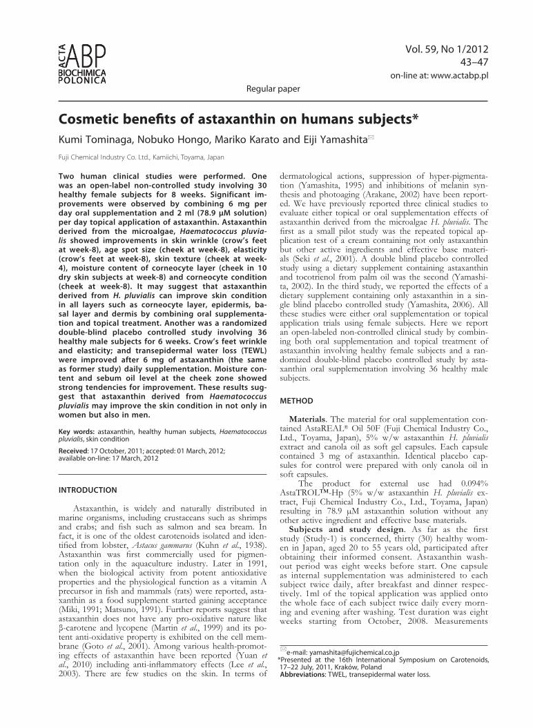
Figure 1. Skin surface photographs and replica images of crow’s feet.
Figure 2. Wrinkle parameters from replica image analysis.By paired t-test: *p<0.05 **p<0.01.

Vol. 59 45Cosmetic effects of astaxanthin on human subjects
elasticity, moisture content, sebum oil and TWEL were measured for Study-2.
RESuLTS
Study-1
Wrinkle, moisture content & elasticity. Figure 1 shows skin surface photographs and replica images of the crow’s feet area in two subjects at week-0 and -8. Visual wrinkle reductions were observed respectively. Significant improvements on four parameters were ob-served. Deepest point of the deepest wrinkle (at week-8 with p<0.01), mean depth of the deepest wrinkle (at week-4 and -8 with p<0.01), maximum width of the deepest wrinkle (at week-8 with p<0.01) and mean depth of all wrinkles (at week-4 and -8 with p<0.05) out of six as shown in Fig. 2. Moisture content of corneocyte layer in the left crow’s feet did not show any significant differences before and after the treatment (not shown). Elasticity of crow’s feet area significantly improved at both week-4 and -8 (Fig. 3).
Age spot. Figure 4 shows skin surface photographs with both normal and UV lamps of the cheek area in two subjects at week-0 and -8. Visual age spot reduc-tions were observed in the both subjects. The age spot area was significantly treated at week-8 as shown in Fig. 5.
Skin texture & moisture content. Figure 6 shows skin topographic replica images of the cheek in two sub-jects at week-8. Visual rough skin improvements were observed in the both subjects. There was a significant improvement on the parameter, mean depth of texture (at week-8 with p<0.01) out of four as shown in Fig. 7. Total area of the corneocyte at week-8 significantly im-proved from the start period (Fig. 8). Moisture content
of corneocyte layer in the cheek among all subjects did not show any significant differences. However, in ten dry skin subjects out of thirty showed a significant in-crease with p<0.05 at week-8 (not shown).
Study-2
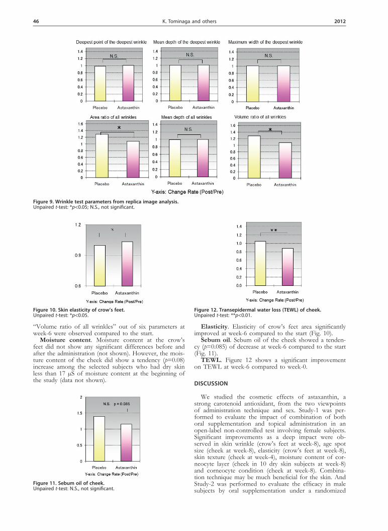
Wrinkle. As shown in Fig. 9, significant improve-ments in two parameters “Area ratio of all wrinkles” and
Figure 3. Skin elasticity of crow’s feet.By paired t-test: *p<0.05 **p<0.01.
Figure 4. Skin surface photographs with both normal and uV lamps of cheek.
Figure 5. Age spot area of cheek.By paired t-test: **p<0.01
Figure 6. Skin topographic replica images of cheek.
Figure 7. Mean depth of texture of cheek.By paired t-test: *p<0.05 **p<0.01.
Figure 8. Size of total area of corneocyte.By paired t-test: *p<0.05.
46 2012K. Tominaga and others
“Volume ratio of all wrinkles” out of six parameters at week-6 were observed compared to the start.
Moisture content. Moisture content at the crow’s feet did not show any significant differences before and after the administration (not shown). However, the mois-ture content of the cheek did show a tendency (p=0.08) increase among the selected subjects who had dry skin less than 17 µS of moisture content at the beginning of the study (data not shown).
Elasticity. Elasticity of crow’s feet area significantly improved at week-6 compared to the start (Fig. 10).
Sebum oil. Sebum oil of the cheek showed a tenden-cy (p=0.085) of decrease at week-6 compared to the start (Fig. 11).
TEWL. Figure 12 shows a significant improvement on TEWL at week-6 compared to week-0.
dISCuSSIOn
We studied the cosmetic effects of astaxanthin, a strong carotenoid antioxidant, from the two viewpoints of administration technique and sex. Study-1 was per-formed to evaluate the impact of combination of both oral supplementation and topical administration in an open-label non-controlled test involving female subjects. Significant improvements as a deep impact were ob-served in skin wrinkle (crow’s feet at week-8), age spot size (cheek at week-8), elasticity (crow’s feet at week-8), skin texture (cheek at week-4), moisture content of cor-neocyte layer (cheek in 10 dry skin subjects at week-8) and corneocyte condition (cheek at week-8). Combina-tion technique may be much beneficial for the skin. And Study-2 was performed to evaluate the efficacy in male subjects by oral supplementation under a randomized
Figure 9. Wrinkle test parameters from replica image analysis.Unpaired t-test: *p<0.05; N.S., not significant.
Figure 10. Skin elasticity of crow’s feet.Unpaired t-test: *p<0.05.
Figure 11. Sebum oil of cheek.Unpaired t-test: N.S., not significant.
Figure 12. Transepidermal water loss (TEWL) of cheek.Unpaired t-test: **p<0.01.

Vol. 59 47Cosmetic effects of astaxanthin on human subjects
double-blind placebo controlled condition. Significant improvements were observed in wrinkle and elasticity of crow’s feet and TWEL at cheek at week-6 compared to start. Tendencies of improvement in moisture content and sebum oil at cheek were also observed. Astaxanthin supplementation exhibited cosmetic benefits in not only female but male subjects.
The wrinkle parameters used in the present studies has been authorized by the Japanese Cosmetic Science Society (Task Force Committee for Evaluation of Anti-aging Function, 2007) as a guideline for the functional assessment of anti-wrinkle product. The Committee pre-scribes that a product can be effective in wrinkle reduc-tion in the case of significant improvement from at least one parameter. It is also provided that any parameters are equivalent. Significant improvements from four pa-rameters in Study-1 and from two parameters in Study-2 were observed respectively. The female subjects in Study-1 were unrestrained in any cosmetic behavior such as skin care or dietary supplement. Bedside, the male subjects in Study-2 were absent from any cosmetic be-havior. It seems that a double administration by combin-ing oral supplementation and topical treatment should be recommended for wrinkle reduction and oral supplemen-tation might be more potent than topical treatment. The mechanism of action of wrinkle reduction by astaxanthin could be explained as a dermis condition improvement through collagen fiber recovery. Astaxanthin promotes collagen fiber recovery by protecting the dermal layer from singlet oxygen damage which has been substanti-ated by an in vitro study using human dermal fibroblasts (Tominaga et al., 2009). There are the reasons why the moisture content of corneocyte layer in crow’s feet was not significantly changed before and after the test and the ASA-GP is available to a measurement of dermic elasticity. Elasticity was also improved as a result of col-lagen fiber recovery both in Study-1 & -2. Significant in-hibition of melanogenesis in age spots were observed in the same manner as the other reports (Yamashita, 1995; Arakane, 2002) by suppressing the oxidative polymeriza-tion in melanocytes and inflammation in epidermis in Study-1. Regarding improvement of rough skin by asta-xanthin treatment on the mean depth of texture and the size of total area of the corneocyte in Study-1, it’s the first finding. It seems that the improvements resulted in the moisture content increase in the cheek among dry skin subjects. The mean moisture content in the all sub-jects at start was approximately 20 µS in Study-1. In gen-eral, the range of moisture content of dry skin is 12–15 µS. A significant increase was observed in ten subjects whose moisture content was less than 17 µS at the start. In Study-2 moisture content of the cheek show a strong tendency increase among the selected subjects less than 17 µS at the start. Topical treatment might be more deeply involved in the improvement of rough skin than oral supplementation. Corneocyte consists of the dead epidermal cells. Astaxanthin treatment might normalize
the corneocyte conditions protecting the keratinocyte differentiation and cornification from oxidative dam-ages such as inflammation in epidermis. Excess oxidized sebum oil causes rough skin and aging odor. It’s well-known that men have more sebum oil production than women. Astaxanthin supplementation may help to re-duce rough skin and aging odor protecting the sebum oil from peroxidation. TEWL is a marker for the bar-rier functions in corneocyte layer. It also seems that the significant TWEL improvements resulted in normalizing the corneocyte condition. Atopic skin patients who have high TEWL may be treated by astaxanthin supplementa-tion.
In conclusion these results may suggest that astaxan-thin derived from H. pluvialis can improve skin condition in all layers such as corneocyte layer, epidermis, basal layer and dermis by combining both oral supplementa-tion and topical treatment and oral supplementation of astaxanthin can improve the skin condition in not only women but also men.
REFEREnCES
Arakane K (2002) Superior skin protection via astaxanthin. Carotenoid Science 5: 21–24.
Goto S, Kogure K, Abe K, Kimata K, Kitahama K, Yamashita E, Terada H (2001) Efficient radical trapping at the surface and inside the phospholipid membrane is responsible for highly potent anti-oxidative activity of the carotenoid astaxanthin. Biochim Biophys Acta 1515: 251258.
Kuhn R, Sorensen NA (1938) The coloring matters of the lobster (Astacus gammarus L.). Z Angew Chem 51: 465–466.
Lee SJ, Bai SK, Lee KS, Namkoong S, Na HJ, Ha KS, Han JA, Yim SV, Chang K, Kwon YG, Lee SK, Kim YM (2003) Astaxanthin inhibits nitric oxide production and inflammatory gene expression by suppressing IκB kinase-dependent NF-κB activation. Mol Cells 16: 97–105.
Martin HD, Ruck C, Schmidt M, Sell S, Beutner S, Mayer B, Walsh R (1999) Chemistry of carotenoid oxidation and free radical reactions. Pure Appl Chem 71: 2253–2262.
Matsuno T (1991) Xanthophylls as precursors of retinoids. Pure Appl Chem 63: 81–88.
Miki W (1991) Biological functions and activities of animal carotenoids. Pure Appl Chem 63: 141–146.
Seki T, Sueki H, Kono H, Suganuma K, Yamashita E (2001) Effects of astaxanthin from Haematococcus pluvialis on human skin-patch test; skin repeated application test; effect on wrinkle reduction. Fragrance J 12: 98–103.
Task Force Committee for Evaluation of Anti-aging Function (2007) Guideline for evaluation of anti-wrinkle products in “Guidelines for evaluation of cosmetic functions”. J Jpn Cosmet Sci Soc 31: 411–431.
Tominaga K, Hongo N, Karato M, Yamashita E (2009) Protective ef-fects of astaxanthin against singlet oxygen induced damage in hu-man dermal fibroblasts in vitro. Food Style 21 13: 84–86.
Yamashita E (1995) Suppression of post-UVB hyperpigmentation by topical astaxanthin from krill. Fragrance J 14: 180–185.
Yamashita E (2002) Cosmetic benefit of dietary supplements includ-ing astaxanthin and tocotrienol on human skin. Food Style 21 6: 112–117.
Yamashita E (2006) The effects of a dietary supplement containing astaxanthin on skin condition. Carotenoid Science 10: 91–95.
Yuan JP, Peng J, Yin K, Wang JH (2010) Potentialhealth-promoting effects of astaxanthin: a high-value carotenoid mostly from microal-gae. Mol Nutr Food Res 54: 1–16.