Corynebacterium & Bacillus - Microscopic appearance - Colonial morphology.
33
Corynebacterium & Bacillus - Microscopic appearance - Colonial morphology
-
Upload
nikolas-roylance -
Category
Documents
-
view
235 -
download
0
Transcript of Corynebacterium & Bacillus - Microscopic appearance - Colonial morphology.

Corynebacterium & Bacillus
- Microscopic appearance
- Colonial morphology


Corynebacterium diphtheriae
- Nasal, nasopharyngeal and tonsillar
diphtheria.


Corynebacterium diphtheriae
- Cutaneous diphtheria.

Corynebacterium diphtheriae
- Throat, nasopharyngeal swabs.
- Skin swab.

Corynebacterium diphtheriae
- Gram positive pleomorphic, long, thin, and curved forms can be seen and also short rods and rods enlarged at one end (clubshaped).

Corynebacterium diphtheriae
- They often appear in clusters, joined at angles like Chinese letters.

- C. diphtheriae often appears beaded due to the
presence
of dark staining granules in the rods.
-These granules, known as volutin or metachromatic
granules, are energy-storing inorganic
polyphosphate
units. In some strains the granules form at the ends
of
the rods.
- The granules are most numerous after the organism
has
been cultured on a protein-rich medium such as
Dorset
egg or Loeffler serum.
Albert Staining of volutin granules

Albert Staining of volutin granules
1- Fix the dried smear using alcohol.
2- Cover the smear with the toluidine blue
malachite green stain for 3–5 minutes.
3-Wash off the stain with clean water.
4- Tip off all the water.

Albert Staining of volutin granules
5- Cover the smear with Albert’s iodine for
1 minute. Wash off with water.
6- Wipe the back of the slide clean, and place it
in
a draining rack for the smear to air-dry.
7- Examine the smear microscopically to look
for
bacteria containing metachromatic granules

Albert Staining of volutin granules
Bacteria cells . . . . . . . . . . . . . . . . . . . . . . Pale green
Metachromatic granules . . . . . . . . . . . . Green-black

Corynebacterium diphtheriae
- They often appear in clusters, joined at angles like Chinese letters.

Tinsdal medium:- grey-black raised colonies surrounded by a dark brown area.

-Brown colour due to formation of H2S which result from interaction between cystine and tellurite.

C. diphtheriae grows rapidly on these media, producing significant growth in 4–6 hours.

Bacillus

Bacillus anthracis

Bacillus anthracis
- Cutaneous ANTHRAX.
Bacillus anthracis
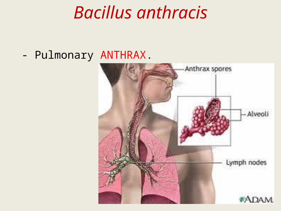
- Pulmonary ANTHRAX.

Bacillus anthracis
- Enteric ANTHRAX.
- Meningoencephalitis.

B. anthracis is a high risk infectious
pathogen, therefore handle specimens
and infected material with care, wearing
protective gloves and face mask, and
following recommended safety
procedures.

Bacillus anthracis
- Fluid aspirated from cutaneous lesions.
- Sputum.
- Blood for culture.
- CSF.

large, 5–8 X 1.5 μm, Gram positive, non-motile bacillus, often appearing joined end to end in chains
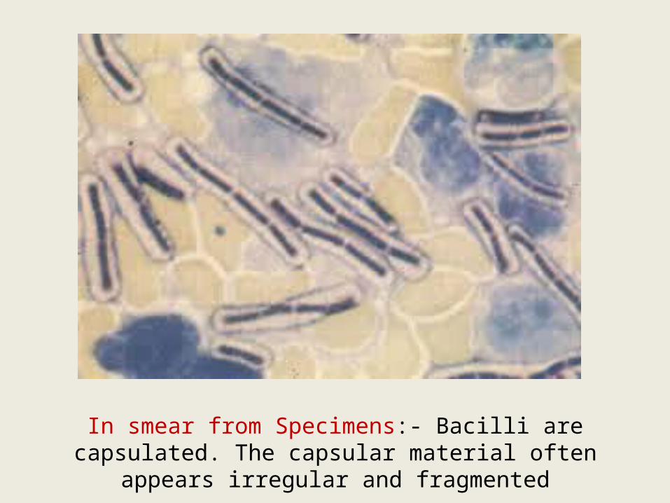
In smear from Specimens:- Bacilli are capsulated. The capsular material often appears irregular and
fragmented

In smears from aerobic cultures: Bacilli are non-capsulated
but contain oval spores (same diameter as the bacilli), giving the
organisms a beaded appearance.
They occur in chains.

Fixation of smears:-
B. anthracis is not killed by heat-fixation.
Smears should be chemically fixed by
immersing the dry smears in a container
of potassium permanganate solution for
10–15 minutes.

Bacillus anthracis
Blood agar:- large grey-white 2-5 mm in diameter
irregular with wavy edges colonies (non or slightly
haemolytic).

Bacillus cereus
- Food poisoning.
- Opportunistic infections in
immunocompromised
persons (bacteraemia, pnumonia and
wound
infection).
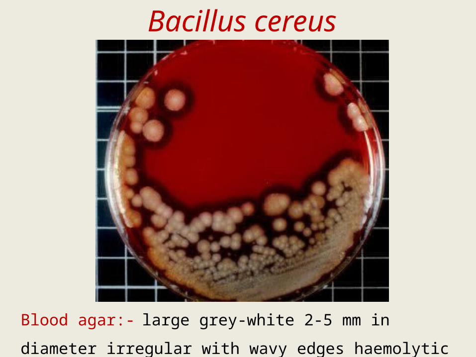
Bacillus cereus
Blood agar:- large grey-white 2-5 mm in diameter
irregular with wavy edges haemolytic colonies.

B.cereus rapidly liquefies the gelatin along and out from
the line of inoculation.