Corresponding Author: Sang Soo Eun, MD, PhD. Spine …jmisst.org/upload/pdf/JMISST-16-005.pdf ·...
20
Simultaneous L4-5 transforaminal and L5-S1 interlaminar percutaneous endoscopic lumbar discectomy for L4-5 down migrated disc: a technical case report Sang Soo Eun, MD, PhD , Sang-Ho Lee, MD, PhD* Department of Orthopaedic surgery, *Neurosurgery, Spine Health Wooridul Hospital(SHWH) Gang Nam, Seoul, Korea Corresponding Author: Sang Soo Eun, MD, PhD. Spine Health Wooridul Hospital(Gangnam) 445, Hakdong-ro, Gangnam-gu Seoul, 135-951, Republic of Korea Phone: +82-2-513-8947 Fax: +82-2-513-8154 Email: [email protected] Running head: Simultaneous L4-5 transforaminal and L5-S1 interlaminar PELD for L4-5 down migrated disc Acknowledgement: This study was supported by a grant from Wooridul Spine Foundation.
Transcript of Corresponding Author: Sang Soo Eun, MD, PhD. Spine …jmisst.org/upload/pdf/JMISST-16-005.pdf ·...

Simultaneous L4-5 transforaminal and L5-S1 interlaminar percutaneous endoscopic
lumbar discectomy for L4-5 down migrated disc: a technical case report
Sang Soo Eun, MD, PhD , Sang-Ho Lee, MD, PhD*
Department of Orthopaedic surgery, *Neurosurgery, Spine Health Wooridul Hospital(SHWH)
Gang Nam, Seoul, Korea
Corresponding Author: Sang Soo Eun, MD, PhD.
Spine Health Wooridul Hospital(Gangnam)
445, Hakdong-ro, Gangnam-gu
Seoul, 135-951, Republic of Korea
Phone: +82-2-513-8947
Fax: +82-2-513-8154
Email: [email protected]
Running head: Simultaneous L4-5 transforaminal and L5-S1 interlaminar PELD for L4-5
down migrated disc
Acknowledgement: This study was supported by a grant from Wooridul Spine Foundation.

1
Simultaneous L4-5 transforaminal and L5-S1 interlaminar percutaneous endoscopic
lumbar discectomy for L4-5 down migrated disc: a technical case report

2
Abstract
A 44 years-old woman was diagnosed as L4-5 down migrated disc herniation and decided
for percutaneous endoscopic lumbar discectomy(PELD). Since significant amount of disc
material is present in both subannular lesion and down migrated portion, transforaminal
approach for L4-5 and interlaminar approach for L5-S1 were planned. Herniated disc was
successfully removed. Simultaneous transforaminal and interlaminar PELD can be an
minimal invasive option for treatment of migrated disc herniation in selected cases.
Keywords: PELD, transforaminal, interlaminar, simultaneous, migrated disc

3
Introduction
Choi et al.2 reported L5S1 interlaminar approach to treat L4-5 down migrated disc
herniation. Developed from this previous technique, authors present a technical case report
for simultaneous L4-5 transforaminal and L5-S1 interlaminar PELD in L4/5 down migrated
disc herniation.
Case Report
A 44 years-old woman presented with severe left lower extremity radiating pain and back
pain, and ambulation was not possible. MRI showed left sided large amount of disc
herniation with down migration at L4-5(Fig. 1). The patient did not want open surgery nor
general anesthesia. We decided for percutaneous endoscopic lumbar discectomy(PELD).
Since significant amount of disc material is present in both subannular lesion and down
migrated portion, transforaminal approach for L4-5 and interlaminar approach for L5-S1
were planned. Left side transforaminal approach at L4-5 was first performed to decompress
subannular disc herniation(Fig.2). Down migrated disc was also to be removed at
transforaminal approach but forceps could not reach to grasp it. Rigid working channel was
introduced through L5-S1 left interlaminar window. Down migrated herniated disc was seen
just after splitting ligamentum flavum (Fig.3). The multi-fragmented disc was removed
completely (Fig. 4 and 5). Two 0.8 cm sized post-operative scars are observed. The pain

4
disappeared and patient was discharged next day.
Discussion
PELD is recognized as a minimal invasive surgical option for treatment of disc herniation.
One of the biggest advantages of PELD is preservation of the soft and bony tissues that are
usually resected in standard discectomy.4 Other advantages include minimal anesthesia,
shorter hospital stay, faster return to activities of daily living, lower estimated blood loss
during surgery, lower cost of postoperative care, and lower infection rates. 6
For treatment of migrated disc herniation, numerous methods has been proposed. Lee et al.
5 treated migrated disc herniations by “half and half technique” and “epiduroscopic
technique”. Choi et al. 1
reported transforaminal foraminoplasty to approach for highly down
migrated disc herniation. Kim et al. 3
reported transforaminal contralateral apparoch in highly
down migrated disc to decrease chance of exiting nerve root damage. Choi et al. 2
reported
treatment L4-5 down migrated disc herniation via L5-S1 interlaminar approach.
Authors thought that it would be difficult to remove whole disc from only transforaminal
or interlaminar approach. There would be insufficient decompression if only one approach
was performed. Reason for not removing transforaminal working channel(Fig.2) was that
authors wanted to use triangulation technique as arthroscopic surgery which uses one portal
as a viewing portal and the other as a working portal. However, disc fragments were well
5
removed with each approach, and biportal manipulation was not necessary.
Indication of this technique is limited to L4-5 down migrated disc with wide L5-S1
interlaminar window.
Conclusion
Simultaneous transforaminal and interlaminar PELD can be an minimal invasive option for
treatment of migrated disc herniation in selected cases.

6
References
1. Choi G, Lee SH, Lokhande P, Kong BJ, Shim CS, Jung B, et al: Percutaneous
endoscopic approach for highly migrated intracanal disc herniations by
foraminoplastic technique using rigid working channel endoscope. Spine 33:508-515,
2008
2. Choi G, Prada N, Modi H N, Vasavada N B, Kim J-S, Lee S-H: Percutaneous
endoscopic lumbar herniectomy for high-grade down-migrated L4-L5 disc through an
L5-S1 interlaminar approach: a technical note. Minim Invasive Neurosurg 53:147-
152, 2010
3. Kim JS, Choi G, Lee SH: Percutaneous endoscopic lumbar discectomy via
contralateral approach: a technical case report. Spine 36:1173-1178 2011
4. Kim SS, Michelsen CB: Revision Surgery for failed back surgery syndrome. Spine
17:957-960 1992
5. Lee S, Kim SK, Lee SH, Kim WJ, Choi WC, Choi G, et al: Percutaneous endoscopic
lumbar discectomy for migrated disc herniation: classification of disc migration and
surgical approaches. Eur Spine J 16:431-437 2007
6. Rasouli MR, Rahimi-Movaghar V, Shokraneh F, Moradi-Lakeh M, Chou R:
Minimally invasive discectomy versus microdiscectomy/open discectomy for
symptomatic lumbar disc herniation. Cochrane Database of Systematic Reviews Issue

7
9 2014

8
Figure Legends
Figure 1. (A) Sagittal MRI shows high-grade down-migrated L4-5 disc herniation. (B) Axial
MRI at L4-5 disc level shows significant amount subannular disc herniation. (C) Axial MRI
at L5 lower pedicle level shows large amount of down migrated disc.
Figure 2. (A) Antero-posterior and (B) Lateral view of intraoperative fluoroscopy showing
placement of L4-5 transforaminal and L5-S1 interlaminar rigid working channels. (C) Note
caudal to cranial direction of working channel and how far up the focep can reach.
Figure 3. Intraoperative endoscopic pictures of interlaminar approach (A) Down migrated
disc was seen just after splitting ligamentum flavum. (B) Blue stained herniated disc is seen
next to left side of S1 root. (C) S1 root is observed after removal of disc.
Figure 4. Picture shows removed multiple fragments of herniated disc. Disc fragments
removed from transforaminal approach are inside the circle. Note large sized disc materials
from interlaminar approach.
Figure 5. Postoperative sagittal MRI shows complete removal of disc.

Fig-1A

Fig-1B

Fig-1C
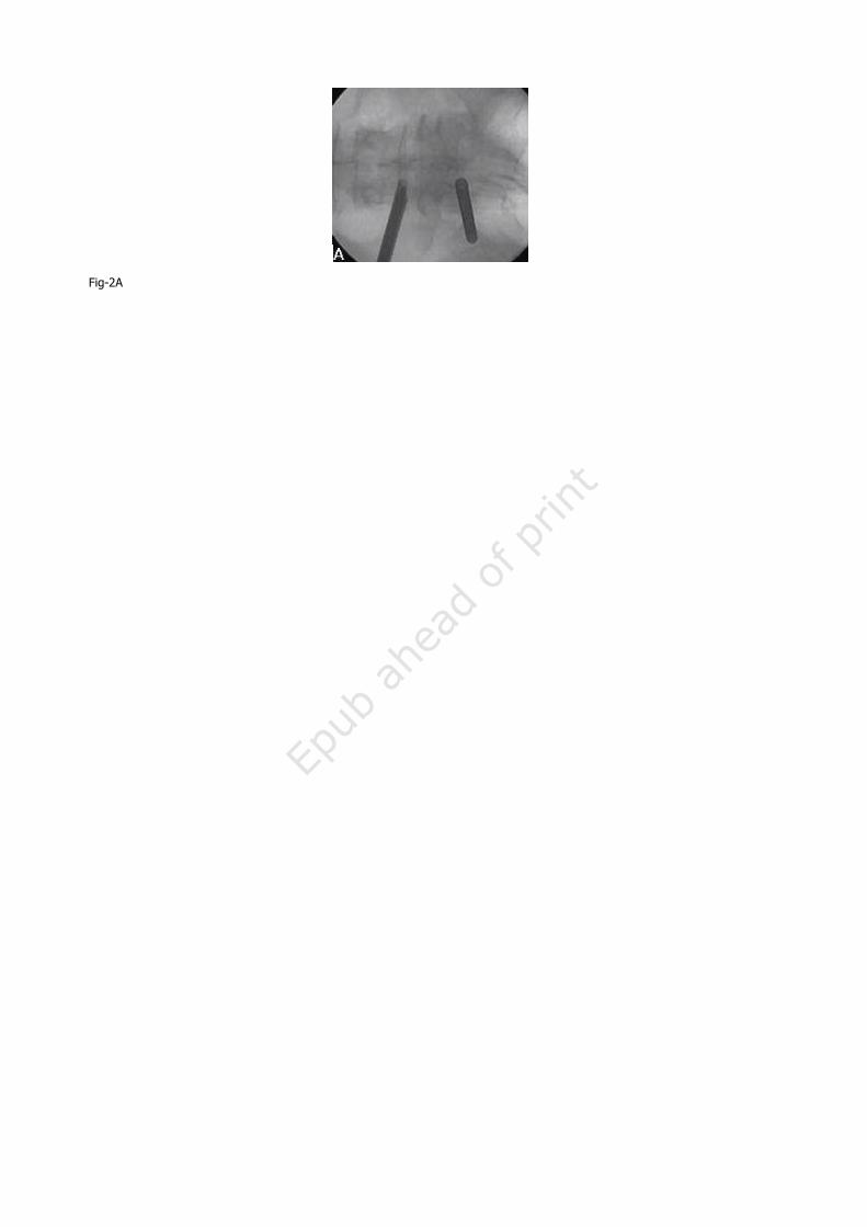
Fig-2A

Fig-2B

Fig-2C

Fig-3A
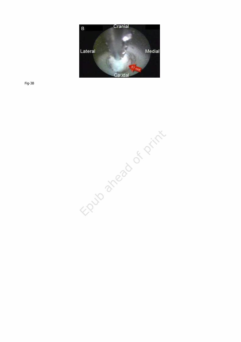
Fig-3B

Fig-3C

Fig-4

Fig-5