Correlation of SPECT imaging, biochemical parameters · PDF fileCorrelation of SPECT imaging,...
14
©FUNPEC-RP www.funpecrp.com.br Genetics and Molecular Research 12 (4): 5964-5977 (2013) Correlation of SPECT imaging, biochemical parameters and mutation with systolic dysfunction D. Alenizi 1 , N.A. Kizilbash 2 , O. Gill 3 , A. Abukanna 1 , S. Malik 4 and A. Badawy 5 1 Department of Medicine, Faculty of Medicine, Northern Border University, Arar, Saudi Arabia 2 Department of Biochemistry, Faculty of Medicine, Northern Border University, Arar, Saudi Arabia 3 Punjab Institute of Nuclear Medicine, Faisalabad, Pakistan 4 Department of Animal Sciences, Faculty of Biological Sciences, Quaid-i-Azam University, Islamabad, Pakistan 5 Department of Medical Biochemistry, Faculty of Medicine, Mansoura University, Mansoura, Egypt Corresponding author: N. Kizilbash E-mail: [email protected] Genet. Mol. Res. 12 (4): 5964-5977 (2013) Received November 21, 2012 Accepted August 8, 2013 Published November 26, 2013 DOI http://dx.doi.org/10.4238/2013.November.26.6 ABSTRACT. We investigated systolic dysfunction by the use of biochemical laboratory tests and perfusion single-photon emission computed tomography imaging in 32 Pakistani subjects exhibiting symptoms of this disorder. To investigate underlying genetic causes, such as familial hypercholesterolemia, DNA samples from these subjects were screened by PCR-SSCP and DNA sequencing to detect changes in the low density lipoprotein receptor gene (LDLR). A novel mutation (1171G>A) in exon 8 and two polymorphisms (1167G>A and 1413 A>G) in exons 8 and 10 of the LDLR gene were found. In silico tools such as SIFT, PolyPhen-2, KD4v, and Project HOPE were used
Transcript of Correlation of SPECT imaging, biochemical parameters · PDF fileCorrelation of SPECT imaging,...
©FUNPEC-RP www.funpecrp.com.brGenetics and Molecular Research 12 (4): 5964-5977 (2013)
Correlation of SPECT imaging, biochemical parameters and mutation with systolic dysfunction
D. Alenizi1, N.A. Kizilbash2, O. Gill3, A. Abukanna1, S. Malik4 andA. Badawy5
1Department of Medicine, Faculty of Medicine, Northern Border University, Arar, Saudi Arabia2Department of Biochemistry, Faculty of Medicine, Northern Border University, Arar, Saudi Arabia3Punjab Institute of Nuclear Medicine, Faisalabad, Pakistan4Department of Animal Sciences, Faculty of Biological Sciences,Quaid-i-Azam University, Islamabad, Pakistan 5Department of Medical Biochemistry, Faculty of Medicine,Mansoura University, Mansoura, Egypt
Corresponding author: N. Kizilbash E-mail: [email protected]
Genet. Mol. Res. 12 (4): 5964-5977 (2013)Received November 21, 2012Accepted August 8, 2013Published November 26, 2013DOI http://dx.doi.org/10.4238/2013.November.26.6
ABSTRACT. We investigated systolic dysfunction by the use of biochemical laboratory tests and perfusion single-photon emission computed tomography imaging in 32 Pakistani subjects exhibiting symptoms of this disorder. To investigate underlying genetic causes, such as familial hypercholesterolemia, DNA samples from these subjects were screened by PCR-SSCP and DNA sequencing to detect changes in the low density lipoprotein receptor gene (LDLR). A novel mutation (1171G>A) in exon 8 and two polymorphisms (1167G>A and 1413 A>G) in exons 8 and 10 of the LDLR gene were found. In silico tools such as SIFT, PolyPhen-2, KD4v, and Project HOPE were used
5965
©FUNPEC-RP www.funpecrp.com.brGenetics and Molecular Research 12 (4): 5964-5977 (2013)
Coronary artery disease: SPECT, biochemical tests and mutations
to predict the effect of this mutation on protein structure and function.
Key words: LDLR gene polymorphism; Systolic dysfunction;LDL receptor; Familial hypercholesterolemia; SPECT
INTRODUCTION
The death rate due to coronary heart disease is 0.41% in Pakistan (Samad et al., 1992). According to the National Health Survey of Pakistan (NHSP), the rate of occurrence of hy-pertension among Pakistanis is 17.9%, while it is 10% for diabetes and 13% for high blood cholesterol (Khan and Mallick, 1992). One of the factors required for development of coronary heart disease is atherosclerosis (Davignon and Ganz, 2004). Atherosclerosis is a condition in which an artery wall thickens as a result of the accumulation of fatty materials such as choles-terol. It is caused largely by the accumulation of macrophage white blood cells and promoted by low-density lipoproteins (LDL). Atherosclerosis is commonly referred to as hardening of the arteries and is caused by the formation of multiple plaques in arteries (Maton et al., 1993).
Normal functioning of low-density lipoprotein receptor (LDLR) is important for mainte-nance of normal levels of plasma LDL cholesterol (LDL-C). LDLR regulates the concentration of plasma LDL-C by receptor-mediated endocytosis. Mutations in LDLR gene produce familial hy-percholesterolemia (FH). The estimated prevalence of heterozygous FH is 0.2% (1:500) in most populations of the world. Worldwide, an estimated ten million people are affected by FH. Patients with FH are characterized by a high plasma LDL-C starting in early childhood, tendon xanthomas and an increased risk of premature coronary artery disease (Austin et al., 2004).
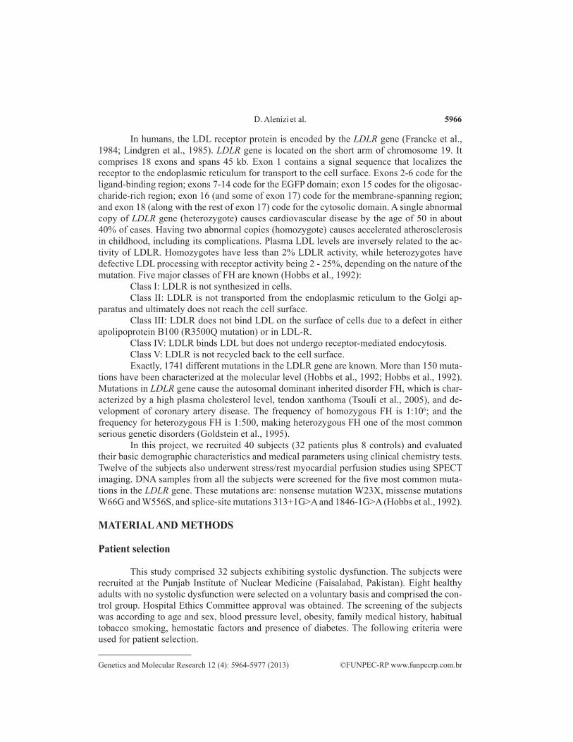
LDLR is a cell-surface receptor that recognizes apoB100 protein in the phospholipid outer layer of LDL particles (Figure 1). It mediates the endocytosis of cholesterol-rich LDL. The recep-tor also recognizes apoE protein found in chylomicron remnants and very low density lipoprotein remnants (intermediate density lipoprotein). The can be regarded as a chimeric protein. It is made up of a number of domains that can function independently of each other (Yamamoto et al., 1984).
Figure 1. X-ray crystal structure of low density lipoprotein receptor in complex with PCSK9 (PDB-file: 3m0c).
5966
©FUNPEC-RP www.funpecrp.com.brGenetics and Molecular Research 12 (4): 5964-5977 (2013)
D. Alenizi et al.
In humans, the LDL receptor protein is encoded by the LDLR gene (Francke et al., 1984; Lindgren et al., 1985). LDLR gene is located on the short arm of chromosome 19. It comprises 18 exons and spans 45 kb. Exon 1 contains a signal sequence that localizes the receptor to the endoplasmic reticulum for transport to the cell surface. Exons 2-6 code for the ligand-binding region; exons 7-14 code for the EGFP domain; exon 15 codes for the oligosac-charide-rich region; exon 16 (and some of exon 17) code for the membrane-spanning region; and exon 18 (along with the rest of exon 17) code for the cytosolic domain. A single abnormal copy of LDLR gene (heterozygote) causes cardiovascular disease by the age of 50 in about 40% of cases. Having two abnormal copies (homozygote) causes accelerated atherosclerosis in childhood, including its complications. Plasma LDL levels are inversely related to the ac-tivity of LDLR. Homozygotes have less than 2% LDLR activity, while heterozygotes have defective LDL processing with receptor activity being 2 - 25%, depending on the nature of the mutation. Five major classes of FH are known (Hobbs et al., 1992):
Class I: LDLR is not synthesized in cells.Class II: LDLR is not transported from the endoplasmic reticulum to the Golgi ap-
paratus and ultimately does not reach the cell surface.Class III: LDLR does not bind LDL on the surface of cells due to a defect in either
apolipoprotein B100 (R3500Q mutation) or in LDL-R.Class IV: LDLR binds LDL but does not undergo receptor-mediated endocytosis.Class V: LDLR is not recycled back to the cell surface.Exactly, 1741 different mutations in the LDLR gene are known. More than 150 muta-
tions have been characterized at the molecular level (Hobbs et al., 1992; Hobbs et al., 1992). Mutations in LDLR gene cause the autosomal dominant inherited disorder FH, which is char-acterized by a high plasma cholesterol level, tendon xanthoma (Tsouli et al., 2005), and de-velopment of coronary artery disease. The frequency of homozygous FH is 1:106; and the frequency for heterozygous FH is 1:500, making heterozygous FH one of the most common serious genetic disorders (Goldstein et al., 1995).
In this project, we recruited 40 subjects (32 patients plus 8 controls) and evaluated their basic demographic characteristics and medical parameters using clinical chemistry tests. Twelve of the subjects also underwent stress/rest myocardial perfusion studies using SPECT imaging. DNA samples from all the subjects were screened for the five most common muta-tions in the LDLR gene. These mutations are: nonsense mutation W23X, missense mutations W66G and W556S, and splice-site mutations 313+1G>A and 1846-1G>A (Hobbs et al., 1992).
MATERIAL AND METHODS
Patient selection
This study comprised 32 subjects exhibiting systolic dysfunction. The subjects were recruited at the Punjab Institute of Nuclear Medicine (Faisalabad, Pakistan). Eight healthy adults with no systolic dysfunction were selected on a voluntary basis and comprised the con-trol group. Hospital Ethics Committee approval was obtained. The screening of the subjects was according to age and sex, blood pressure level, obesity, family medical history, habitual tobacco smoking, hemostatic factors and presence of diabetes. The following criteria were used for patient selection.
5967
©FUNPEC-RP www.funpecrp.com.brGenetics and Molecular Research 12 (4): 5964-5977 (2013)
Coronary artery disease: SPECT, biochemical tests and mutations
All the experimental subjects selected belonged to the age group 15-35 years. A 500-mL blood sample was obtained from both the subjects and controls and stored in EDTA-coated vacu-tainers. All subjects were divided into three groups on the basis of serum LDL-cholesterol value.
Group-I (control group): 8 subjects with normal serum cholesterol and LDL-CGroup-II: 20 subjects with high serum cholesterol or LDL-C but not in the range of FHGroup-III: 12 subjects selected on the basis of high serum cholesterol (>7.5 mM) or
LDL-C (>4.9 mM)
Measurement of total cholesterol
The method for cholesterol measurement used hexane extraction after hydrolysis with alcoholic KOH followed by reaction with the Liebermann-Burchard color reagent, which con-tains sulfuric acid, acetic acid and acetic anhydride (Abel et al., 1952).
Measurement of LDL-C
The method for measuring LDL-C was the beta quantification method in which the beta designation refers to the electrophoretic term for LDL. Beta-quantification combines ul-tracentrifugation and chemical precipitation (Bachorik, 2000).
Myocardial perfusion scintigraphy
In SPECT imaging, a radioactive pharmaceutical product (a radionuclide) such as 99mTc-MIBI, which decays with the emission of gamma rays, is injected in the patient. The principle of SPECT imaging is that under conditions of stress, diseased myocardium receives less blood than normal myocardium. A healthy tissue takes up a known amount of the radio-active pharmaceutical product and it appears as a bright area in the SPECT image. If a tissue is abnormal, the uptake of the radiopharmaceutical can be increased or decreased and it can appear as a more intense spot or a dark area, respectively, on the image. Such an image can be interpreted by a cardiologist as a suspected diseased state. Different processing algorithms al-low a reproducible and reliable assessment of left ventricular (LV) function, which is validated against various references (Michelena and VanDecker, 2005).
SPECT imaging was performed on the 12 group-III subjects after induction of phar-macological stress. It revealed the distribution of the radiopharmaceutical, and therefore the
Criterion Description of patients: a) Total cholesterol concentration above 7.5 mM in adults or above 6.7 mM in children under 16 years, or low density lipoprotein cholesterol concentration above 4.9 mM in adults or above 4.0 mM in children; b) Tendon xanthoma in the patient or a 2nd degree relative; c) DNA-based evidence of mutation in the LDLR gene; d) Family history of myocardial infarction before age 50 years in a 3rd de-gree relative or before age 60 years in a 1st degree relative; e) Family history of high total cholesterol concentration above 7.5 mM in a 1st or 2nd degree relative.
5968
©FUNPEC-RP www.funpecrp.com.brGenetics and Molecular Research 12 (4): 5964-5977 (2013)
D. Alenizi et al.
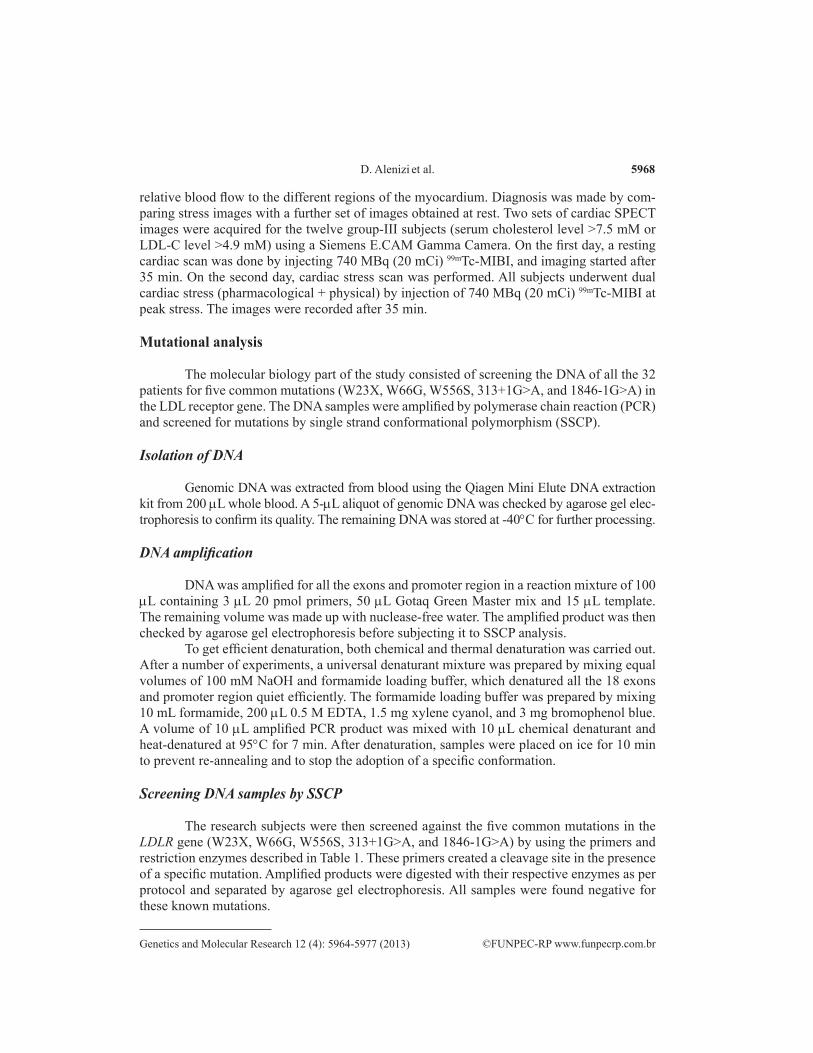
relative blood flow to the different regions of the myocardium. Diagnosis was made by com-paring stress images with a further set of images obtained at rest. Two sets of cardiac SPECT images were acquired for the twelve group-III subjects (serum cholesterol level >7.5 mM or LDL-C level >4.9 mM) using a Siemens E.CAM Gamma Camera. On the first day, a resting cardiac scan was done by injecting 740 MBq (20 mCi) 99mTc-MIBI, and imaging started after 35 min. On the second day, cardiac stress scan was performed. All subjects underwent dual cardiac stress (pharmacological + physical) by injection of 740 MBq (20 mCi) 99mTc-MIBI at peak stress. The images were recorded after 35 min.
Mutational analysis
The molecular biology part of the study consisted of screening the DNA of all the 32 patients for five common mutations (W23X, W66G, W556S, 313+1G>A, and 1846-1G>A) in the LDL receptor gene. The DNA samples were amplified by polymerase chain reaction (PCR) and screened for mutations by single strand conformational polymorphism (SSCP).
Isolation of DNA
Genomic DNA was extracted from blood using the Qiagen Mini Elute DNA extraction kit from 200 mL whole blood. A 5-mL aliquot of genomic DNA was checked by agarose gel elec-trophoresis to confirm its quality. The remaining DNA was stored at -40°C for further processing.
DNA amplification
DNA was amplified for all the exons and promoter region in a reaction mixture of 100 mL containing 3 mL 20 pmol primers, 50 mL Gotaq Green Master mix and 15 mL template. The remaining volume was made up with nuclease-free water. The amplified product was then checked by agarose gel electrophoresis before subjecting it to SSCP analysis.
To get efficient denaturation, both chemical and thermal denaturation was carried out. After a number of experiments, a universal denaturant mixture was prepared by mixing equal volumes of 100 mM NaOH and formamide loading buffer, which denatured all the 18 exons and promoter region quiet efficiently. The formamide loading buffer was prepared by mixing 10 mL formamide, 200 mL 0.5 M EDTA, 1.5 mg xylene cyanol, and 3 mg bromophenol blue. A volume of 10 mL amplified PCR product was mixed with 10 mL chemical denaturant and heat-denatured at 95°C for 7 min. After denaturation, samples were placed on ice for 10 min to prevent re-annealing and to stop the adoption of a specific conformation.
Screening DNA samples by SSCP
The research subjects were then screened against the five common mutations in the LDLR gene (W23X, W66G, W556S, 313+1G>A, and 1846-1G>A) by using the primers and restriction enzymes described in Table 1. These primers created a cleavage site in the presence of a specific mutation. Amplified products were digested with their respective enzymes as per protocol and separated by agarose gel electrophoresis. All samples were found negative for these known mutations.
5969
©FUNPEC-RP www.funpecrp.com.brGenetics and Molecular Research 12 (4): 5964-5977 (2013)
Coronary artery disease: SPECT, biochemical tests and mutations
Gel analysis by SSCP
The composition of the acrylamide gel is a very important parameter for SSCP analy-sis. Glycerol added gels of various compositions were prepared. A series of experiments were carried out to adjust the composition of the gel for each exon. The gels were run in ice cold 1X TBE buffer at 16°C for various run times in a Bio-Rad Mini Protean Terta Cell.
DNA sequencing
A total of 12 reaction mixtures were sent for sequencing. DNA was amplified in 100 mL reaction mixture, purified and cleaved from the oligonucleotide with the help of the Qiagen PCR purification kit. The cleaved PCR products were sent to Eurofines (Canada) for sequencing.
RESULTS
Laboratory tests
All subjects and controls were required to provide a complete medical history and were given a thorough clinical examination. Mean systolic and diastolic blood pressure were measured according to a standardized protocol after at least 10 min rest using a validated auto-mated machine and appropriate cuff. Blood samples from the subjects and controls were used for the estimation of total cholesterol and LDL-C.
Table 2 shows the characteristics of the subjects; age of patients ranged from 15 to 35 years. Serum levels of total cholesterol and LDL-C were not significantly higher in patients as compared to controls (P > 0.05). There was also no significant difference in the presence of diabetes between the subjects and the controls (P > 0.05). However, significant differences were found for presence of hypertension and smoking habit (P < 0.001) between subjects and controls.
Myocardial perfusion scintigraphy
Single photon emission computed tomography scans were done on 12 patients to check for evidence of coronary artery disease and to assess left ventricular function. Seven patients were found to have extensive ischemia affecting the apex, inferior wall, septum and anterior wall.
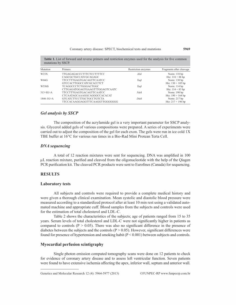
Mutation Primers Restriction enzymes Fragments after cleavage
W23X TTGAGAGACCCTTTCTCCTTTTCC Alul Norm: 118 bp CAGCGCTGCCATCGCAGAGC Htz: 118 + 88 bpW66G TTCCTTTGAGTGACAGTTCAATCC Taql Norm: 130 bp GTCCACTTGGCCATCGCACCTCT Htz: 130 + 105 bpW556S TCAGGCCCTCTGGGACTGGC Taql Norm: 114 bp CTTGAGATGGAGTGAAGTTTGGAGTCAATC Htz: 114 + 83 bp313+IG>A TTCCTTTGAGTGACAGTTCAATCC Ndel Norm: 190 bp CTCAATAGCAAAGGCAGGGCCACACAT Htz: 190 + 164 bp1846-1G>A GTCATCTTCCTTGCTGCCTGTCTA Ddel Norm: 217 bp TTCCACAAGGAGGTTTCAAGGTTGGGGGGG Htz: 217 + 196 bp
Table 1. List of forward and reverse primers and restriction enzymes used for the analysis for five common mutations by SSCP.
5970
©FUNPEC-RP www.funpecrp.com.brGenetics and Molecular Research 12 (4): 5964-5977 (2013)
D. Alenizi et al.
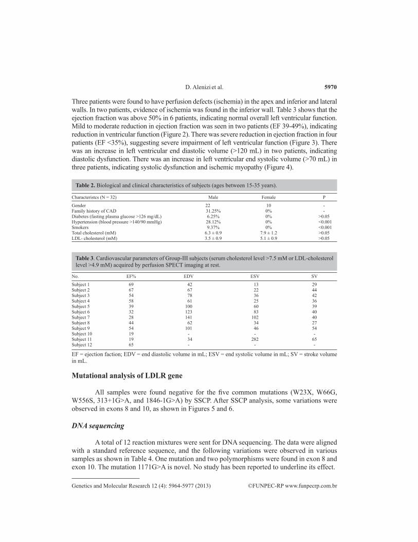
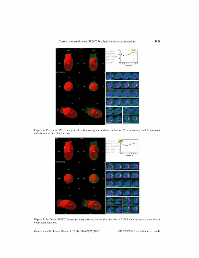
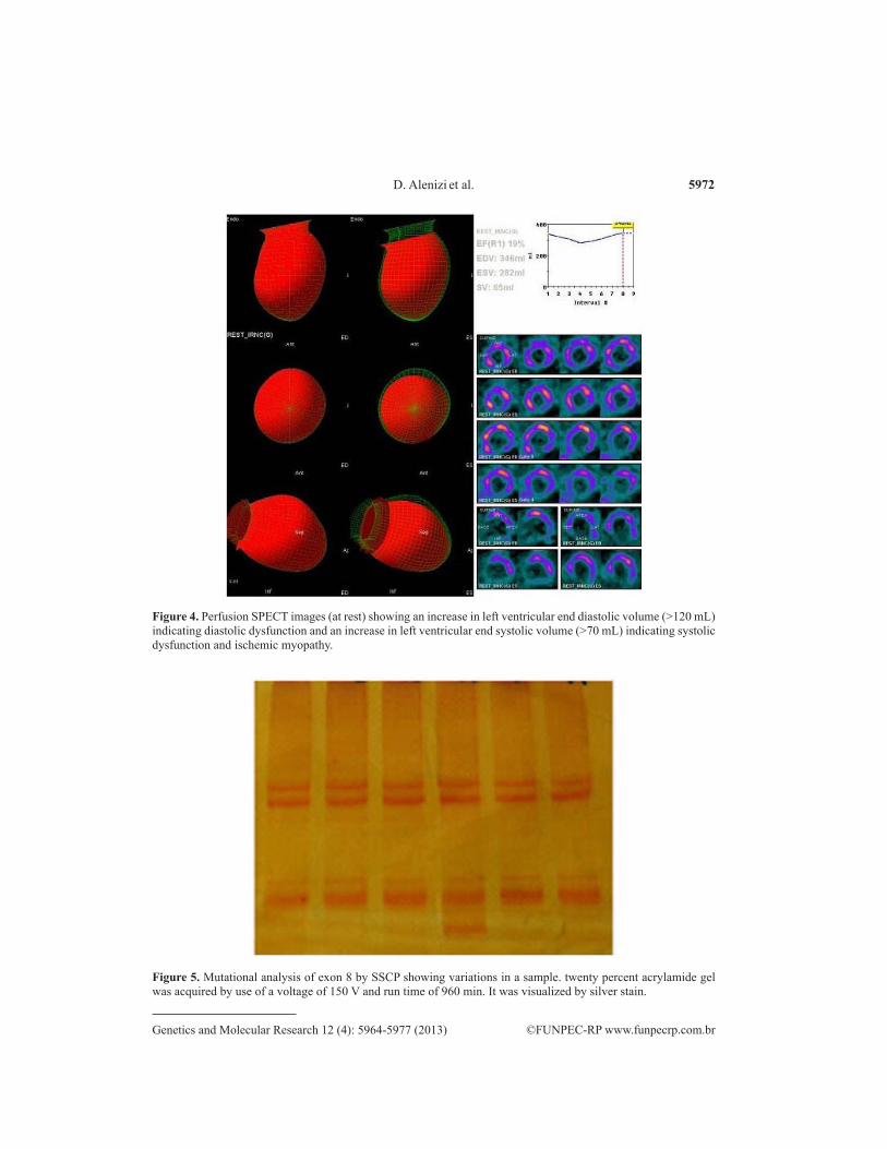
Three patients were found to have perfusion defects (ischemia) in the apex and inferior and lateral walls. In two patients, evidence of ischemia was found in the inferior wall. Table 3 shows that the ejection fraction was above 50% in 6 patients, indicating normal overall left ventricular function. Mild to moderate reduction in ejection fraction was seen in two patients (EF 39-49%), indicating reduction in ventricular function (Figure 2). There was severe reduction in ejection fraction in four patients (EF <35%), suggesting severe impairment of left ventricular function (Figure 3). There was an increase in left ventricular end diastolic volume (>120 mL) in two patients, indicating diastolic dysfunction. There was an increase in left ventricular end systolic volume (>70 mL) in three patients, indicating systolic dysfunction and ischemic myopathy (Figure 4).
Characteristcs (N = 32) Male Female P
Gender 22 10 -Family history of CAD 31.25% 0% -Diabetes (fasting plasma glucose >126 mg/dL) 6.25% 0% >0.05Hypertension (blood pressure >140/90 mmHg) 28.12% 0% <0.001Smokers 9.37% 0% <0.001Total cholesterol (mM) 6.3 ± 0.9 7.9 ± 1.2 >0.05LDL- cholesterol (mM) 3.5 ± 0.9 5.1 ± 0.9 >0.05
Table 2. Biological and clinical characteristics of subjects (ages between 15-35 years).
No. EF% EDV ESV SV
Subject 1 69 42 13 29Subject 2 67 67 22 44Subject 3 54 78 36 42Subject 4 58 61 25 36Subject 5 39 100 60 39Subject 6 32 123 83 40Subject 7 28 141 102 40Subject 8 44 62 34 27Subject 9 54 101 46 54Subject 10 19 - - -Subject 11 19 34 282 65Subject 12 65 - - -
Table 3. Cardiovascular parameters of Group-III subjects (serum cholesterol level >7.5 mM or LDL-cholesterol level >4.9 mM) acquired by perfusion SPECT imaging at rest.
Mutational analysis of LDLR gene
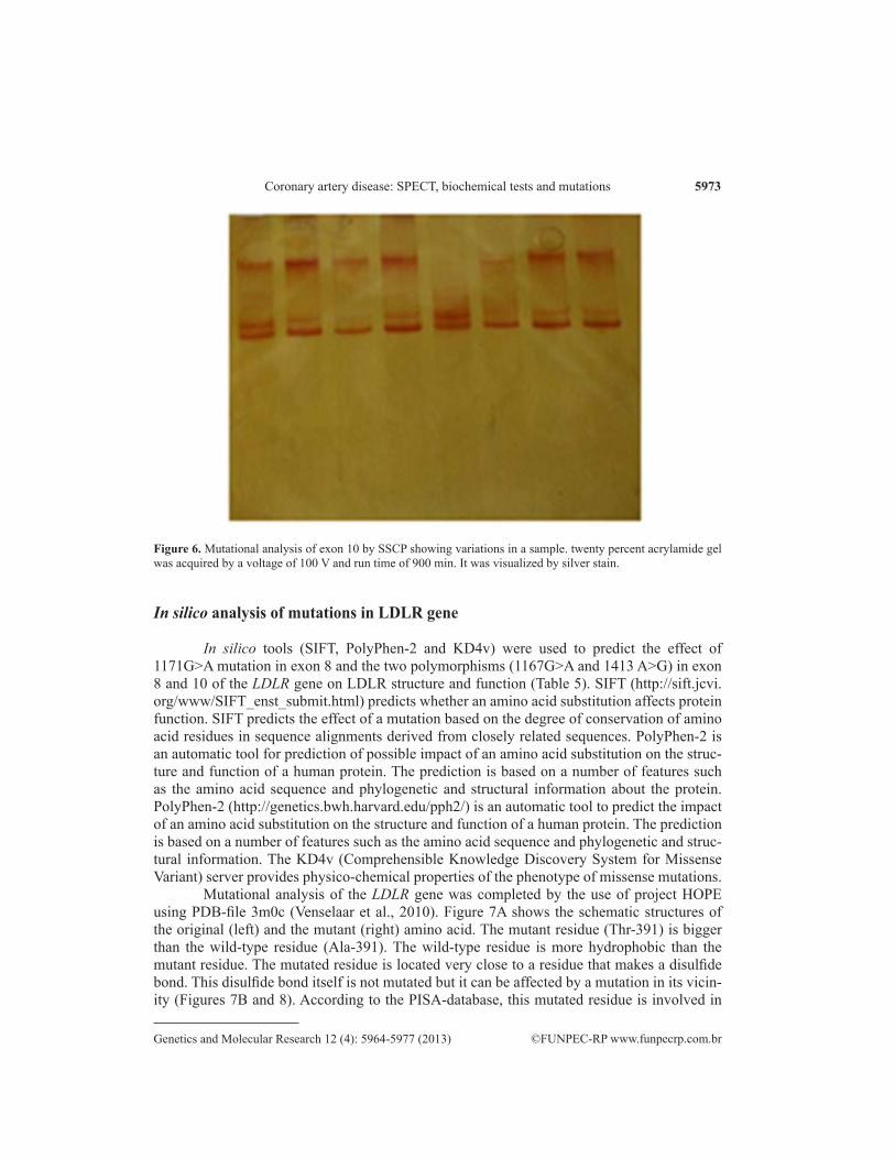
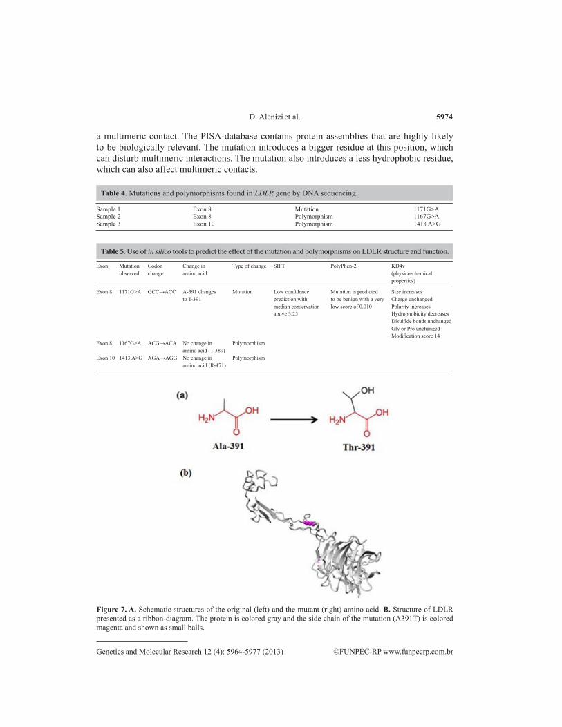
All samples were found negative for the five common mutations (W23X, W66G, W556S, 313+1G>A, and 1846-1G>A) by SSCP. After SSCP analysis, some variations were observed in exons 8 and 10, as shown in Figures 5 and 6.
DNA sequencing
A total of 12 reaction mixtures were sent for DNA sequencing. The data were aligned with a standard reference sequence, and the following variations were observed in various samples as shown in Table 4. One mutation and two polymorphisms were found in exon 8 and exon 10. The mutation 1171G>A is novel. No study has been reported to underline its effect.
EF = ejection faction; EDV = end diastolic volume in mL; ESV = end systolic volume in mL; SV = stroke volume in mL.
5971
©FUNPEC-RP www.funpecrp.com.brGenetics and Molecular Research 12 (4): 5964-5977 (2013)
Coronary artery disease: SPECT, biochemical tests and mutations
Figure 2. Perfusion SPECT images (at rest) showing an ejection fraction of 39% indicating mild to moderate reduction in ventricular function.
Figure 3. Perfusion SPECT images (at rest) showing an ejection fraction of 32% indicating severe reduction in ventricular function.
5972
©FUNPEC-RP www.funpecrp.com.brGenetics and Molecular Research 12 (4): 5964-5977 (2013)
D. Alenizi et al.
Figure 4. Perfusion SPECT images (at rest) showing an increase in left ventricular end diastolic volume (>120 mL) indicating diastolic dysfunction and an increase in left ventricular end systolic volume (>70 mL) indicating systolic dysfunction and ischemic myopathy.
Figure 5. Mutational analysis of exon 8 by SSCP showing variations in a sample. twenty percent acrylamide gel was acquired by use of a voltage of 150 V and run time of 960 min. It was visualized by silver stain.
5973
©FUNPEC-RP www.funpecrp.com.brGenetics and Molecular Research 12 (4): 5964-5977 (2013)
Coronary artery disease: SPECT, biochemical tests and mutations
In silico analysis of mutations in LDLR gene
In silico tools (SIFT, PolyPhen-2 and KD4v) were used to predict the effect of 1171G>A mutation in exon 8 and the two polymorphisms (1167G>A and 1413 A>G) in exon 8 and 10 of the LDLR gene on LDLR structure and function (Table 5). SIFT (http://sift.jcvi.org/www/SIFT_enst_submit.html) predicts whether an amino acid substitution affects protein function. SIFT predicts the effect of a mutation based on the degree of conservation of amino acid residues in sequence alignments derived from closely related sequences. PolyPhen-2 is an automatic tool for prediction of possible impact of an amino acid substitution on the struc-ture and function of a human protein. The prediction is based on a number of features such as the amino acid sequence and phylogenetic and structural information about the protein. PolyPhen-2 (http://genetics.bwh.harvard.edu/pph2/) is an automatic tool to predict the impact of an amino acid substitution on the structure and function of a human protein. The prediction is based on a number of features such as the amino acid sequence and phylogenetic and struc-tural information. The KD4v (Comprehensible Knowledge Discovery System for Missense Variant) server provides physico-chemical properties of the phenotype of missense mutations.
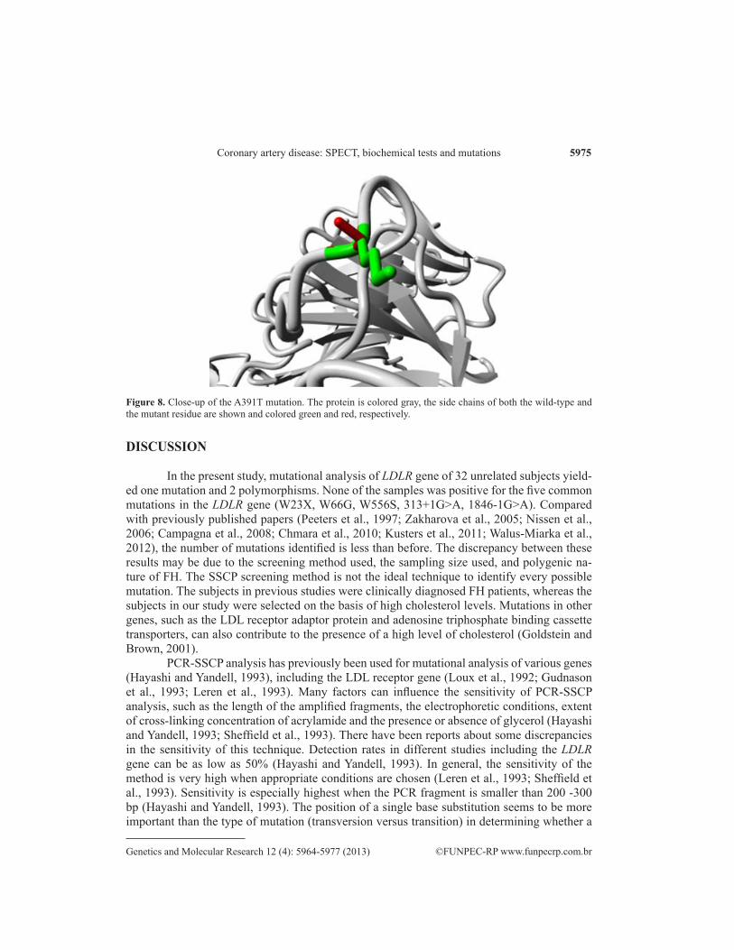
Mutational analysis of the LDLR gene was completed by the use of project HOPE using PDB-file 3m0c (Venselaar et al., 2010). Figure 7A shows the schematic structures of the original (left) and the mutant (right) amino acid. The mutant residue (Thr-391) is bigger than the wild-type residue (Ala-391). The wild-type residue is more hydrophobic than the mutant residue. The mutated residue is located very close to a residue that makes a disulfide bond. This disulfide bond itself is not mutated but it can be affected by a mutation in its vicin-ity (Figures 7B and 8). According to the PISA-database, this mutated residue is involved in
Figure 6. Mutational analysis of exon 10 by SSCP showing variations in a sample. twenty percent acrylamide gel was acquired by a voltage of 100 V and run time of 900 min. It was visualized by silver stain.
5974
©FUNPEC-RP www.funpecrp.com.brGenetics and Molecular Research 12 (4): 5964-5977 (2013)
D. Alenizi et al.
a multimeric contact. The PISA-database contains protein assemblies that are highly likely to be biologically relevant. The mutation introduces a bigger residue at this position, which can disturb multimeric interactions. The mutation also introduces a less hydrophobic residue, which can also affect multimeric contacts.
Sample 1 Exon 8 Mutation 1171G>ASample 2 Exon 8 Polymorphism 1167G>ASample 3 Exon 10 Polymorphism 1413 A>G
Table 4. Mutations and polymorphisms found in LDLR gene by DNA sequencing.
Exon Mutation Codon Change in Type of change SIFT PolyPhen-2 KD4v observed change amino acid (physico-chemical properties)
Exon 8 1171G>A GCC→ACC A-391 changes Mutation Low confidence Mutation is predicted Size increases to T-391 prediction with to be benign with a very Charge unchanged median conservation low score of 0.010 Polarity increases above 3.25 Hydrophobicity decreases Disulfide bonds unchanged Gly or Pro unchanged Modification score 14Exon 8 1167G>A ACG→ACA No change in Polymorphism amino acid (T-389)Exon 10 1413 A>G AGA→AGG No change in Polymorphism amino acid (R-471)
Table 5. Use of in silico tools to predict the effect of the mutation and polymorphisms on LDLR structure and function.
Figure 7. A. Schematic structures of the original (left) and the mutant (right) amino acid. B. Structure of LDLR presented as a ribbon-diagram. The protein is colored gray and the side chain of the mutation (A391T) is colored magenta and shown as small balls.
5975
©FUNPEC-RP www.funpecrp.com.brGenetics and Molecular Research 12 (4): 5964-5977 (2013)
Coronary artery disease: SPECT, biochemical tests and mutations
DISCUSSION
In the present study, mutational analysis of LDLR gene of 32 unrelated subjects yield-ed one mutation and 2 polymorphisms. None of the samples was positive for the five common mutations in the LDLR gene (W23X, W66G, W556S, 313+1G>A, 1846-1G>A). Compared with previously published papers (Peeters et al., 1997; Zakharova et al., 2005; Nissen et al., 2006; Campagna et al., 2008; Chmara et al., 2010; Kusters et al., 2011; Walus-Miarka et al., 2012), the number of mutations identified is less than before. The discrepancy between these results may be due to the screening method used, the sampling size used, and polygenic na-ture of FH. The SSCP screening method is not the ideal technique to identify every possible mutation. The subjects in previous studies were clinically diagnosed FH patients, whereas the subjects in our study were selected on the basis of high cholesterol levels. Mutations in other genes, such as the LDL receptor adaptor protein and adenosine triphosphate binding cassette transporters, can also contribute to the presence of a high level of cholesterol (Goldstein and Brown, 2001).
PCR-SSCP analysis has previously been used for mutational analysis of various genes (Hayashi and Yandell, 1993), including the LDL receptor gene (Loux et al., 1992; Gudnason et al., 1993; Leren et al., 1993). Many factors can influence the sensitivity of PCR-SSCP analysis, such as the length of the amplified fragments, the electrophoretic conditions, extent of cross-linking concentration of acrylamide and the presence or absence of glycerol (Hayashi and Yandell, 1993; Sheffield et al., 1993). There have been reports about some discrepancies in the sensitivity of this technique. Detection rates in different studies including the LDLR gene can be as low as 50% (Hayashi and Yandell, 1993). In general, the sensitivity of the method is very high when appropriate conditions are chosen (Leren et al., 1993; Sheffield et al., 1993). Sensitivity is especially highest when the PCR fragment is smaller than 200 -300 bp (Hayashi and Yandell, 1993). The position of a single base substitution seems to be more important than the type of mutation (transversion versus transition) in determining whether a
Figure 8. Close-up of the A391T mutation. The protein is colored gray, the side chains of both the wild-type and the mutant residue are shown and colored green and red, respectively.
5976
©FUNPEC-RP www.funpecrp.com.brGenetics and Molecular Research 12 (4): 5964-5977 (2013)
D. Alenizi et al.
mutation is detected (Sheffield et al., 1993). The sensitivity of a mutation detection method has usually been evaluated by the rate of detection of already known mutations. However, a more definitive evaluation of the sensitivity of such a mutation screening method is to compare the rate of detected sequence variations with the results of DNA sequencing of the same amplified fragments, assuming of course that the sequencing method used actually is capable of detect-ing all sequence variations.
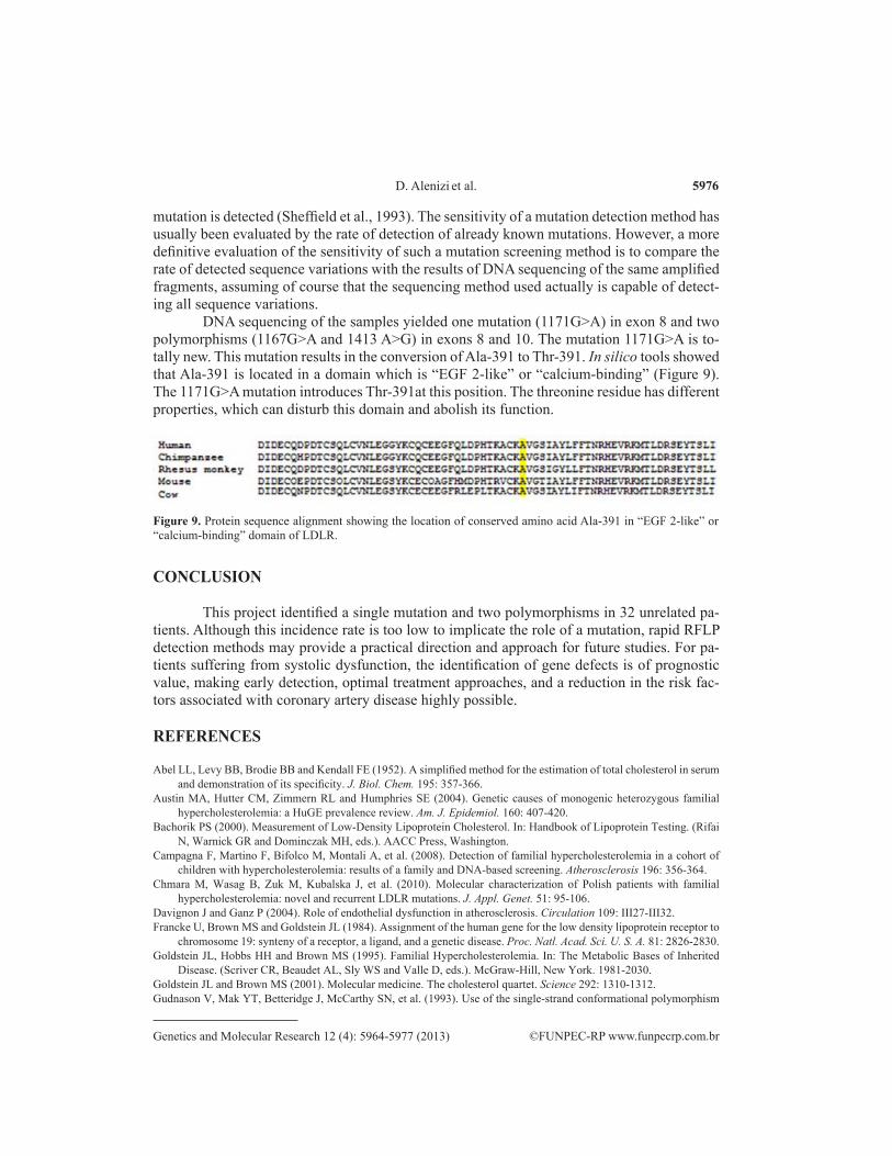
DNA sequencing of the samples yielded one mutation (1171G>A) in exon 8 and two polymorphisms (1167G>A and 1413 A>G) in exons 8 and 10. The mutation 1171G>A is to-tally new. This mutation results in the conversion of Ala-391 to Thr-391. In silico tools showed that Ala-391 is located in a domain which is “EGF 2-like” or “calcium-binding” (Figure 9). The 1171G>A mutation introduces Thr-391at this position. The threonine residue has different properties, which can disturb this domain and abolish its function.
Figure 9. Protein sequence alignment showing the location of conserved amino acid Ala-391 in “EGF 2-like” or “calcium-binding” domain of LDLR.
CONCLUSION
This project identified a single mutation and two polymorphisms in 32 unrelated pa-tients. Although this incidence rate is too low to implicate the role of a mutation, rapid RFLP detection methods may provide a practical direction and approach for future studies. For pa-tients suffering from systolic dysfunction, the identification of gene defects is of prognostic value, making early detection, optimal treatment approaches, and a reduction in the risk fac-tors associated with coronary artery disease highly possible.
REFERENCES
Abel LL, Levy BB, Brodie BB and Kendall FE (1952). A simplified method for the estimation of total cholesterol in serum and demonstration of its specificity. J. Biol. Chem. 195: 357-366.
Austin MA, Hutter CM, Zimmern RL and Humphries SE (2004). Genetic causes of monogenic heterozygous familial hypercholesterolemia: a HuGE prevalence review. Am. J. Epidemiol. 160: 407-420.
Bachorik PS (2000). Measurement of Low-Density Lipoprotein Cholesterol. In: Handbook of Lipoprotein Testing. (Rifai N, Warnick GR and Dominczak MH, eds.). AACC Press, Washington.
Campagna F, Martino F, Bifolco M, Montali A, et al. (2008). Detection of familial hypercholesterolemia in a cohort of children with hypercholesterolemia: results of a family and DNA-based screening. Atherosclerosis 196: 356-364.
Chmara M, Wasag B, Zuk M, Kubalska J, et al. (2010). Molecular characterization of Polish patients with familial hypercholesterolemia: novel and recurrent LDLR mutations. J. Appl. Genet. 51: 95-106.
Davignon J and Ganz P (2004). Role of endothelial dysfunction in atherosclerosis. Circulation 109: III27-III32.Francke U, Brown MS and Goldstein JL (1984). Assignment of the human gene for the low density lipoprotein receptor to
chromosome 19: synteny of a receptor, a ligand, and a genetic disease. Proc. Natl. Acad. Sci. U. S. A. 81: 2826-2830.Goldstein JL, Hobbs HH and Brown MS (1995). Familial Hypercholesterolemia. In: The Metabolic Bases of Inherited
Disease. (Scriver CR, Beaudet AL, Sly WS and Valle D, eds.). McGraw-Hill, New York. 1981-2030.Goldstein JL and Brown MS (2001). Molecular medicine. The cholesterol quartet. Science 292: 1310-1312.Gudnason V, Mak YT, Betteridge J, McCarthy SN, et al. (1993). Use of the single-strand conformational polymorphism
5977
©FUNPEC-RP www.funpecrp.com.brGenetics and Molecular Research 12 (4): 5964-5977 (2013)
Coronary artery disease: SPECT, biochemical tests and mutations
method to detect recurrent and novel mutations in the low-density lipoprotein receptor gene in patients with familial hypercholesterolaemia: detection of a novel mutation Asp200→Gly. Clin. Investig. 71: 331-337.
Hayashi K and Yandell DW (1993). How sensitive is PCR-SSCP? Hum. Mutat. 2: 338-346.Hobbs HH, Brown MS and Goldstein JL (1992). Molecular genetics of the LDL receptor gene in familial
hypercholesterolemia. Hum. Mutat. 1: 445-466.Khan NR and Mallick IA (1992). Economic situation in Pakistan’s, National Health Survey of Pakistan. Pak. J. Med. Res.
31: 282-8.Kusters DM, Huijgen R, Defesche JC, Vissers MN, et al. (2011). Founder mutations in the Netherlands: geographical
distribution of the most prevalent mutations in the low-density lipoprotein receptor and apolipoprotein B genes. Neth. Heart J. 19: 175-182.
Leren TP, Solberg K, Rodningen OK, Ose L, et al. (1993). Evaluation of running conditions for SSCP analysis: application of SSCP for detection of point mutations in the LDL receptor gene. PCR Methods Appl. 3: 159-162.
Lindgren V, Luskey KL, Russell DW and Francke U (1985). Human genes involved in cholesterol metabolism: chromosomal mapping of the loci for the low density lipoprotein receptor and 3-hydroxy-3-methylglutaryl-coenzyme A reductase with cDNA probes. Proc. Natl. Acad. Sci. U. S. A. 82: 8567-8571.
Loux N, Saint-Jore B, Collod G, Dairou F, et al. (1992). Screening for new mutations in the LDL receptor gene in seven French familial hypercholesterolemia families by the single strand conformation polymorphism method. Hum. Mutat. 1: 325-332.
Maton A, Roshan L, Hopkins J, McLaughlin CW, et al. (1993). Human Biology and Health. Prentice Hall, Englewood Cliffs.
Michelena HI and VanDecker WA (2005). Radionuclide-based insights into the pathophysiology of ischemic heart disease: beyond diagnosis. J. Investig. Med. 53: 176-191.
Nissen PH, Damgaard D, Stenderup A, Nielsen GG, et al. (2006). Genomic characterization of five deletions in the LDL receptor gene in Danish Familial hypercholesterolemic subjects. BMC Med. Genet. 7: 55.
Peeters AV, Van Gaal LF, du Plessis L, Lombardi MP, et al. (1997). Mutational and genetic origin of LDL receptor gene mutations detected in both Belgian and Dutch familial hypercholesterolemics. Hum. Genet. 100: 266-270.
Samad A, Sahibzada WA and Mattu A (1992). Risk factor analysis in a random population of 4 cities in Pakistan. Pak. J. Cardiol. 3: 7-14.
Sheffield VC, Beck JS, Kwitek AE, Sandstrom DW, et al. (1993). The sensitivity of single-strand conformation polymorphism analysis for the detection of single base substitutions. Genomics 16: 325-332.
Tsouli SG, Kiortsis DN, Argyropoulou MI, Mikhailidis DP, et al. (2005). Pathogenesis, detection and treatment of Achilles tendon xanthomas. Eur. J. Clin. Invest. 35: 236-244.
Venselaar H, Te Beek TA, Kuipers RK, Hekkelman ML, et al. (2010). Protein structure analysis of mutations causing inheritable diseases. An e-Science approach with life scientist friendly interfaces. BMC Bioinformatics 11: 548.
Walus-Miarka M, Sanak M, Idzior-Walus B, Miarka P, et al. (2012). A novel mutation (Cys308Phe) of the LDL receptor gene in families from the South-Eastern part of Poland. Mol. Biol. Rep. 39: 5181-5186.
Yamamoto T, Davis CG, Brown MS, Schneider WJ, et al. (1984). The human LDL receptor: a cysteine-rich protein with multiple Alu sequences in its mRNA. Cell 39: 27-38.
Zakharova FM, Damgaard D, Mandelshtam MY, Golubkov VI, et al. (2005). Familial hypercholesterolemia in St-Petersburg: the known and novel mutations found in the low density lipoprotein receptor gene in Russia. BMC Med. Genet. 6: 6.