Correlation of CT Cerebral Vascular Territories with Function · of the medial hemispheric surface...
5
Stephen A. Berman 1 L. Anne Hayman 2 . 3 Vincent C. Hinck 3 Received October 22, 1979 ; accepted after revision November 19 , 1979 . 1 Depar tment of Neurology, Baylor College of Medicine, Houston , TX 77030 . 2 Department of Radiology , Veterans Adminis- tration Medical Center, Houston TX 77211 . Pres- ent add ress: Department of Radiology, University of Texas Health Scienc e Center, 643 1 Fannin St., Suite 2026, Houston, TX 77030. Addre ss reprint requests to L. A. Hay man. 3 Department of Radiology, Baylor College of Medicine, Houston, TX 77030. Thi s article appears in May/ June 1980 AJNR and August 1980 AJR . AJNR 1 : 259-263, May / June 1980 0195-6108 / 80 / 0103-0259 $00.00 © Ameri can Roent gen Ray Society Correlation of CT Cerebral Vascular Territories with Function: I. Anterior Cerebral Artery 259 Schematic displays of the cerebral territories supplied by branches of the anterior cerebral artery as they would appear on axial and coronal CT scan sections are presented. Companion diagrams of regional cerebral function are also provided to simplify correlation of clinical deficits with coronal and axial CT abnormalities. Selected examples illustrate that these facilitate more detailed and precise CT interpretation in clinical practice . Knowledge of vascular terr itories can help in differentiating infarction from other pathologic processes . If, for example, the position and extent of a lesion and the usual position and extent of a vascular territory are in congruous, the diagnosis of infarction should receive a relatively low priority and vice versa. Knowledge of vascular territories can also facilitate correct interpretation of cerebra l angiograms by pinpointing specific vessels for particularly close atten- tion. For example, analysis of a patient's clinica l findings in terms of functional neuroanatomy can improve detection of subtle lesions by pinpointing spec ific territories for specia l attention on CT and specif ic vessels for attention on angiograms . This series of reports is designed to present function al anatomy and vascular territories in a form directly app li cable to CT. (Parts II and III will deal with the middle and posterior cerebral arteries, respectively). The illustrations created for the series are intended to simplify correlation of clinical signs and symptoms, vascular territories, and CT images in everyday practice . Discussion This report correlates the vascular territories of the ante rior cereb ral artery with the neurologic functions ascribed to those territories by mapping the territories on schematic illustrations of axial and coronal ' CT planes (figs. 1 and 2). These planes are then indicted on a map of the vascular territories as they would appea r on the medial surface of the cerebral hemisphere (fig. 3A) and on a similar map of the medial hemispheric surface on which regions serving particular neurolog- ical functions are delineated (fig. 38). The br anches of the anterior cere bral art ery have been divided into three groups: (1) the medial lenticulo st riate ar teries; (2) the peri callosal branche s to the corpus ca ll osum; and (3) the branches to the ce rebr al hemis pher e. • The plane of co ronal CT scans obtained depends on head positioning. If the co ronal image is a reco nstruc ti on, the plane is perpendic ul ar to the axial scans which may vary in their orientation. Therefo re, we have chosen to illustrate the vascular territories in the anatomic co ronal plane. The reader must co rr ec t for minor differences in scanning angle. This will be facilitated by a review of co ronal sli ces illustr ated by Matsui and Hirano [1] .
Transcript of Correlation of CT Cerebral Vascular Territories with Function · of the medial hemispheric surface...

Stephen A. Berman 1
L. Anne Hayman 2.3
Vincent C. Hinck3
Received Oc tober 22, 1979; accepted after revision November 19, 1979.
1 Department of Neurology, Baylor College of Medicine, Houston , TX 77030.
2 Department of Radiology , Veterans Administration Medical Center, Houston TX 77211 . Present address: Department of Radiology, University of Texas Health Science Center, 6431 Fannin St., Suite 2026, Houston, TX 77030. Address reprint requests to L. A. Hayman.
3 Department of Rad iology, Bay lor College o f Medicine, Houston, TX 77030.
This artic le appears in May / June 1980 AJNR and August 1980 AJR.
AJNR 1 :259-263, May/ June 1980 0195-6108/ 80/ 0103-0259 $00.00 © American Roen tgen Ray Society
Correlation of CT Cerebral Vascular Territories with Function: I. Anterior Cerebral Artery
259
Schematic displays of the cerebral territories supplied by branches of the anterior cerebral artery as they would appear on axial and coronal CT scan sections are presented. Companion diagrams of regional cerebral function are also provided to simplify correlation of clinical deficits with coronal and axial CT abnormalities. Selected examples illustrate that these facilitate more detailed and precise CT interpretation in clinical practice.
Knowledge of vascular terr itories can help in differentiating infarction from other pathologic processes . If, for example, the position and extent of a lesion and the usual position and extent of a vascular territory are incongruous, the diagnosis of infarction should receive a relatively low priority and vice versa. Knowledge of vascular territories can also facilitate correct interpretation of cerebral angiograms by pinpointing specific vessels for particularly c lose attention. For example, analysis of a patient's clinical findings in terms of functional neuroanatomy can improve detection of subtle lesions by pinpointing spec ific territories for special attention on CT and specif ic vessels for attention on angiograms .
This series of reports is designed to present functional anatomy and vascular territories in a form directly applicable to CT. (Parts II and III will deal with the middle and posterior cerebral arteries, respectively) . The illustrations c reated for the series are intended to simplify correlation of clinical signs and symptoms, vascu lar territories , and CT images in everyday practice .
Discussion
This report correlates the vascular territories of the anterior cerebral artery with the neurologic functions ascribed to those territories by mapping the territories on schematic illustrations of axial and coronal ' CT planes (figs. 1 and 2). These planes are then indicted on a map of the vascular territories as they would appear on the medial surface of the cerebral hemisphere (fig . 3A) and on a similar map of the medial hemispheric surface on which regions serving particular neurological functions are delineated (fig. 38).
The branches of the anterior cerebral artery have been divided into three groups: (1) the medial lenticulostriate arteries; (2) the peri callosal branches to the corpus callosum; and (3) the branches to the cerebral hem isphere.
• The plane of co ronal CT scans obtained depends on head position ing. If the coronal image is a reconstruc ti on, the plane is perpendicular to the axial scans which may vary in their ori entation . Therefore, we have chosen to illust rate the vascu lar territories in the anatomic coronal plane. The reader must co rrect for minor differences in scanning ang le. This will be fac ili tated by a review of co ronal slices illustrated by Matsui and Hirano [1] .

260 BERMAN ET AL.
Fig . 1 .-Axial CT scan diagrams arranged in sequence from base to vertex. Angle and leve ls of scan planes are shown in fig . 3. Territory of anterior cerebral artery is divided into three regions: medial lenticulostri ate (medium blue), ca llosal (dark blue), and hemispheric (light blue).
T
Fig . 2. -Coronal CT scan diagrams arranged in sequence from fron t to back. Angle and levels of scan planes are shown in fig. 3. Territory of anterior cerebral artery is divided into three reg ions: medial lenticutostriate (medium blue), callosal (dark blue) , and hemispheric (light blue).
AJNR:1 , May/June 1980

AJNR:1, May/ June 1980 CT OF CEREBRAL VASCULAR TERRITORIES 261
A D H K N a T V
--12
---4
--1
---12
--4
A D H K N a T V
Fig . 3. -A, Regions supplied by nine hemispheric branches of anterior cerebral artery localized on medial view of cerebral hemisphere. B, Functional regions supplied by anterior cerebral artery localized on medial view of cerebral hemisphere. See axia l and coronal scan levels in figs. 1 and 2 for correlation.
The largest possible area supplied by the vessel(s) of a single territory is indicated in figures 1 and 2. Therefore a vascular occlusion should not produce a lesion larger than that defined in those illustrations. In fact, collateral supply, which often interfaces at the distal periphery of an infarction, may cause the area of involvement to be smaller than that schematically defined. Furthermore, the area of involvement may have a patchy appearance because only selected branches within a territory may be occluded by emboli.
Medial Lenticulostriate Arteries
The medial lenticulostriate arteries include the artery of Heubner and basal branches of the anterior cerebral artery. The artery of Heubner supplies the anterior aspects of the putamen and caudate nucleus, as well as the anteroinferior part of the internal capsule. Infarction causes weakness of the contralateral face and arm without sensory loss. A transient aphasia is often seen in which spontaneous speech is lost while repetition and comprehension are preserved [2]. Occasionally there is dysarthria. Although the lower extremity is unaffected, there is a halting gait because of difficulty in initiating movements [2, 3].
The basal branches supply the dorsal aspect of the chiasm and the hypothalamus. Infarction of the hypothalamus may cause transient memory disorders or more protean
psychological manifestations of anxiety, agitation or a feeling of weakness [3].
If there is bilateral occlusion of the lenticulostriate arteries , a profound alteration of mental activity develops, at times resulting in a state of akinetic mutism (a comalike cond ition in which the patient's eyes remain open causing him to appear awake to superficial inspection). More commonly a classical picture of coma evolves [4 , 5].
Infarction of the med ial lenticulostriate arteries is much less common than hypertensive hemorrhage from these vessels. Unlike infarction , hemorrhage which arises medial to the putamen ignores vascular boundaries, destroys the hypothalamus, and ruptures into the ventricle . Therefore the radiologist can differentiate a hemorrhagic infarct, in which hemorrhage occurs secondarily, from a primary hemorrhage (intracerebral hematoma).
Perical/osal Branches
The callosal arteries arise from the pericallosal branch of the anterior cerebral artery and penetrate the upper surface of the corpus callosum, extending thence inferiorly into the septum pellucidum. Usually there are 7-20 short callosal branches, although these are sometimes replaced by a single artery. Infarction can result in isolation of the language-dominant left hemisphere from the right hemisphere which mediates function of the left side of the body. As a result, the patient experiences difficulty in moving the left side of his body in response to verbal commands (ideomotor apraxia), even though there may be no paralysis. Furthermore, the patient cannot recognize words written on the left side of his body though his sensation may be unimpaired (tactile agnosia), and he has difficulty in writing with his left hand (left-sided agraphia) [6, 7].
Hemispheric Branches
There are usually nine hemispheric branches (fig . 3A), each supplying a segment of the medial surface of the hemisphere. The medial surface of the hemisphere can be suppl ied in whole or in part by either anterior cerebral artery. (Sometimes both hemispheres are supp lied by a single peri callosal artery, the so-called azygous artery, occlusion of which results in infarction of the medial surfaces of both hemispheres.)
Considering the large number of branches and numerous variations of cross-perfusion that can occur, one might expect the compromise of hemispheric branches could lead to a wide variety of possible c linical sequelae. However, the patterns of infarction can be simplified by comparing the vascular territories of figure 3A with the functional areas of figure 3B.
For example, figure 3 reveals that the frontal association areas that mediate judgement, insight, and mood receive arterial supply from the orbitofrontal, frontopolar , and anterior internal frontal arteries, and to a lesser extent, middle internal frontal arteries [8]. (The posterior internal frontal and paracentral territories may sometimes include a small part of the frontal association area-insight, mood, and judgement, fig. 3B). These functions would be impaired by

262 BERMAN ET AL. AJNR: 1 , May/June 1980
Fig. 4. -A-D, Ax ial CT scans. Hemorrhag ic in farcl ion of both pericallosal lerritories wh ich resu lted from spasm after rupture o f anterior communicating artery aneurysm. Hemorrhage in territory of pericallosal branches to corpus ca llosum (A, B) , cingulale gyru s, and in white mailer above lale ral venlric les (B, C) . Co llateral c irculation from middle cerebral arleries prevented infarction of superomedial aspects o f hemispheres (D). Reference 10 levels 6- 9 in fig . 3A leads examiner 10 predict involvement of pericallosal branches. Reference to same levels in fig. 38 leads 10 co rrect predict ion thai this infarction was associated wilh severe neuropsychological damage. E, Coronal reconstruction of scans A-D , corresponds to level M in fig . 2. Hemorrhagic infarct ion in pericallosal terri lory (corpus callosum, c ingulale gyru s, and adjacent white mailer) seen as broad U-shaped band above posterior roof o f third ventricular chamber.
E
occ lusion of those branches. Aphasia may develop because the arterial supply of subjacent wh ite matter is also disturbed . A release of grasping, groping, and sucking reflex patterns may be seen in addition to impairment of those fu_nctions ~n~icated in figure 3B [2).
Figure 3 also shows that the motor synchronization area is supplied by the middl e and posterior internal frontal branches and, to a small extent, by the anterior internal frontal and the paracentral branches. Infarction of the anterior part of the motor synchronization areas affects contralateral conjugate eye deviation, whil e involvement of the posterior part affects coordination of contralateral eye, head, and trunk movement [9).
Note that the motor function area is supplied by the paracentral branch . Unilateral damage causes weakness of the contralateral lower extremity. Bilateral damage causes incontinence as well [8).
Note further that the sensory function area is supplied by the paracentral and superi or and inferior parietal branches. Rarely a parietooccipital branch (not shown in fig . 3) may supply the posterior part of this area. Damage of the anterior part of the sensory region impairs but does not abo lish appreciati on of primary sensory modali ties, such as touch and pain . Damage in the posterior part impairs more complex sensory appreciation, such as recog nition of objects by touch (stereognosis) , recognition of shapes written on skin (graphesthesia), and two-point discrimination [10).
The supply of the c ingulate gyrus which "controls " mem-
ory and emotion comes from the peri callosal , orbitofrontal , frontopolar, posterior internal frontal, paracentral, and superior and inferior parietal arteries. The cingulum fiber tract within this area is part of the limbic system , which is supplied by the posterior cerebral and the anterior cerebral arteries. Therefore, memory and emotional disturbances may occur as a result of damage to either vascular terri tory [2 , 8).
Finally, infarction in the region of the parietooccipital fissure (which is sometimes supplied by the pari etooccipital branch of the anterior cerebral artery) has been reported in one case of anterior cerebral artery compromise. In thi s instance the patient experienced disturbance of recognition in the contralateral visual field (vi sual ag nosia), rare in association with anterior cerebral artery occ lusion . This probably can be ascribed to the fact that the same area is usually well supplied by the posterior c irculation [2).
With illustrative cases (figs. 4- 6) the reader may practice using figures 1 -3 to predict the vascular territories and clinical consequences of lesions of the anterior cerebral system.
REFERENCES
1. Matsui T, Hirano A. An atlas of the human brain for computerized tomography. Tokyo: Igaku-Shoin , 1978:314-547
2. Critchley M. Anterior cerebral artery and its syndromes. Brain 1930; 53: 1 20-1 65
3 . Denny-Brown D. The nature of aprax ia. J Nerv Men t Dis 1958; 126:9-32
AJNR: 1, May/ June 1980 CT OF CEREBRAL VASCULAR TERRITORIES 263
E F G H
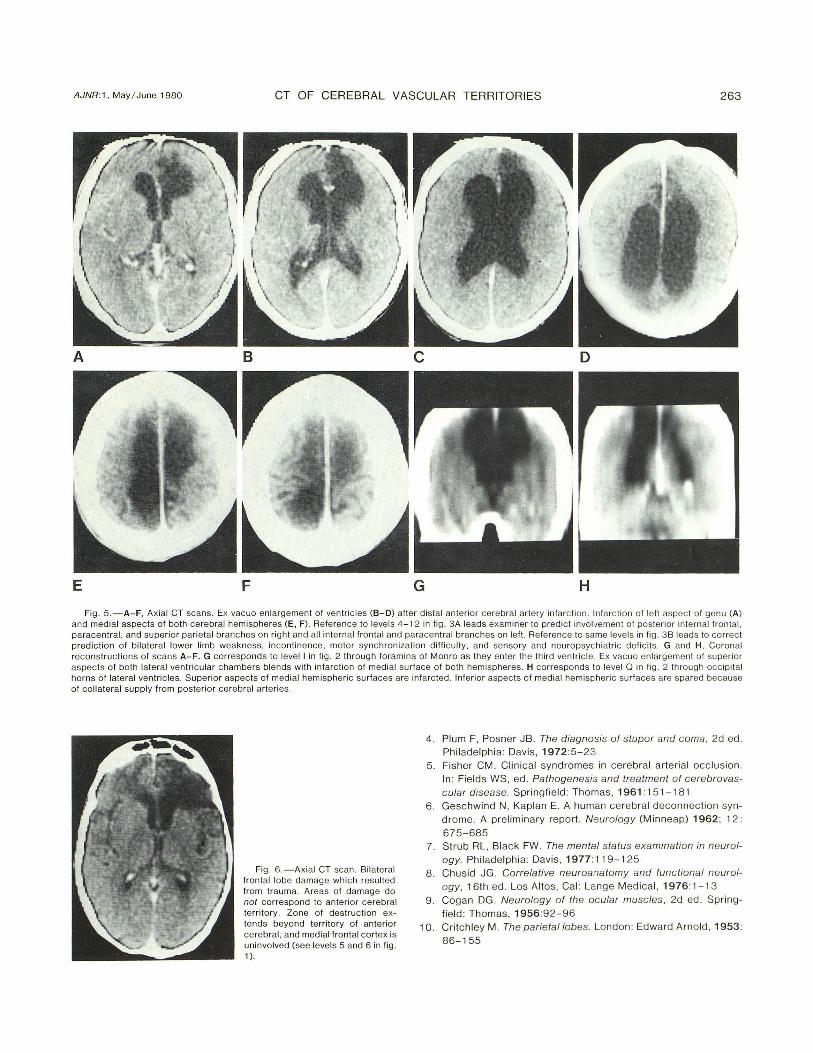
Fig . 5. -A-F, Axial CT scans. Ex vacuo en largement of ventric les (8-0) after dista l anterior cerebral artery infarction . Infarcti on of left aspec t of genu (A) and medial aspects o f both cerebral hemispheres (E, F) . Reference to levels 4 -1 2 in fig . 3A leads examiner to predict involvement of posterior internal frontal, paracentral, and superior parietal branches on ri ght and all in ternal frontal and paracentral branches on left. Reference to same levels in fig. 3B leads to correct prediction of bilateral lower limb weakness, incontinence, motor synchronization difficulty , and sensory and neuropsychiatric defi cits. G and H, Coronal reconstructions o f scans A-F. G corresponds to level I in fig . 2 through foramina of Monro as they enter the third ventricl e. Ex vacuo enlargement of superior aspects of both lateral ventricu lar chambers blends with infarction of medial surface of both hemispheres. H corresponds to level 0 in fig. 2 through occ ipital horns of lateral ventricles. Superior aspects of medial hem ispheric surfaces are infarcted. Inferior aspects of medial hemispheri c surfaces are spared because of co llateral supply from posterior ce rebra l arteries.
Fig. 6. -Axial CT scan. Bilateral frontal lobe damage wh ich resulted from trauma. Areas of damage do not correspond to anterior cerebral territory . Zone of destruction extends beyond territory of anteri or cerebral, and medial frontal cortex is uninvolved (see levels 5 and 6 in fig . 1).
4. Plum F, Posner JB. Th e diagnosis of stupor and coma, 2d ed. Philadelphia: Davis , 1972:5-23
5. Fisher CM. Clinica l syndromes in cerebral arterial occ lusion. In: Fields WS, ed. Pathogenesis and treatment of cerebrovascular disease. Springfield : Thomas , 1961 : 151 -1 8 1
6 . Gesc hwind N, Kaplan E. A human cerebral deconnect ion syndrome. A prelimin ary report. Neurology (Minneap) 1962; 12:
675-685 7. Strub RL , Blac k FW. Th e mental status examination in neurol
ogy. Ph iladelphia: Davis, 1977: 119-125 8. Chusid JG . Correlative neuroana tomy and fun c tional neurol
ogy, 16th ed. Los Altos, Cal: Lange Medical, 1976:1 -1 3 9. Cogan DG . Neurology of the ocular muscles, 2d ed . Spring
fi eld: Thomas , 1956:92-96 10. Critchley M. Th e parie tal lobes. London: Edward Arnold , 1953:
86-155