Correcting radial keratotomy- Refractive epidemic of …gulanivision.com/pdfs/GlovesOff/GV -...
5
Vol. 39, No. 16 - September 1st, 2014 Correcting radial keratotomy: Refractive epidemic of future In the lion’s cage: From cornea-based corrections to lens-based surgeries Gloves Off With Gulani by Arun C. Gulani, MD In my personal experience of having seen probably every kind of radial keratotomy (RK) presentation over the past decade of correcting such cases from all over the world, the first thing I want to address here, like we always do in these columns, is the “mindset.” First and foremost, I do not want you to feel overwhelmed by the appearance of these RK corneas. They can range from simple, well-cut, regular, 2-cut RK to 32-cut RK and even crisscross RK, irregular RK, RK with Hex-K—you name it (see Figures 1 and 2). The appearance should not deter us from applying it into our 5S system (see Table). These patients can actually see and have the potential to see better. We will divide this topic into two columns. This first part will address the entire corneal aspects of correcting RK, whereas the next column will address the lens-based surgeries from phakic to pseudophakic when they are associated with RK correction. —Arun C. Gulani, MD As mentioned in my previous columns, the 5S system (see page 3) can be used as a “coin sorter” to help collect all correctable elements of these RK corneas and unfold a plan for vision. Managing RK cases through the 5S system, when we look at these cases that are purely cornea-oriented (meaning the lens is normal, not cataractous, and not contributory to refractive error), manual, streak retinoscopy refraction is the “gold” for which I look. Refraction is what starts my thought process with every patient. It doesn’t matter if the cornea has opacity. The refraction is what we concentrate on. That’s what I want your mentality to be with these patients as well. Don’t give up! Refraction helps me provide a pathway to improve someone’s vision notwithstanding the number, pattern, or irregularity and density of cuts. Using the 5S system, cornea-based RK presentations always have Sight (meaning these patients can see). This may even be through a hard contact lens correction, but they always have sight. All they’re telling us is there is potential to see, so we should not give up. The next “S” in the 5S system that is affected is Shape. These patients are either too flat from RK and became hyperopic (they also in most cases have presbyopia due to age), and many of them have astigmatism either uncorrected or due to incorrect RK surgery.
Transcript of Correcting radial keratotomy- Refractive epidemic of …gulanivision.com/pdfs/GlovesOff/GV -...
Vol. 39, No. 16 - September 1st, 2014
Correcting radial keratotomy: Refractive epidemic of futureIn the lion’s cage: From cornea-based corrections to lens-based surgeries
Gloves Off With Gulani by Arun C. Gulani, MD
In my personal experience of having seen probably every kind of radial keratotomy (RK) presentation over the past decade of correcting such cases from all over the world, the first thing I want to address here, like we always do in these columns, is the “mindset.”
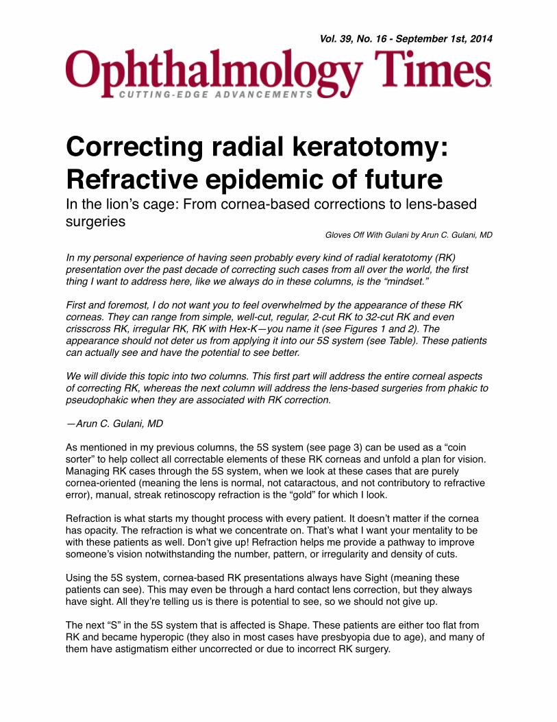
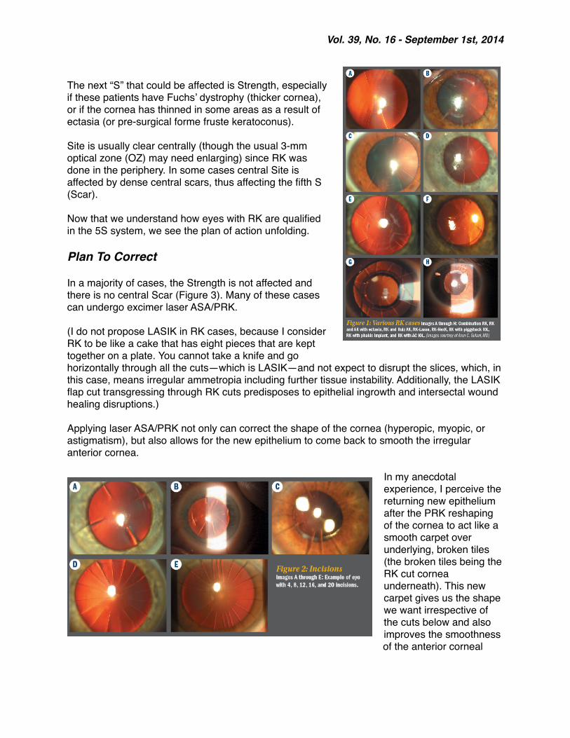
First and foremost, I do not want you to feel overwhelmed by the appearance of these RK corneas. They can range from simple, well-cut, regular, 2-cut RK to 32-cut RK and even crisscross RK, irregular RK, RK with Hex-K—you name it (see Figures 1 and 2). The appearance should not deter us from applying it into our 5S system (see Table). These patients can actually see and have the potential to see better.
We will divide this topic into two columns. This first part will address the entire corneal aspects of correcting RK, whereas the next column will address the lens-based surgeries from phakic to pseudophakic when they are associated with RK correction.
—Arun C. Gulani, MD
As mentioned in my previous columns, the 5S system (see page 3) can be used as a “coin sorter” to help collect all correctable elements of these RK corneas and unfold a plan for vision. Managing RK cases through the 5S system, when we look at these cases that are purely cornea-oriented (meaning the lens is normal, not cataractous, and not contributory to refractive error), manual, streak retinoscopy refraction is the “gold” for which I look.
Refraction is what starts my thought process with every patient. It doesn’t matter if the cornea has opacity. The refraction is what we concentrate on. That’s what I want your mentality to be with these patients as well. Don’t give up! Refraction helps me provide a pathway to improve someone’s vision notwithstanding the number, pattern, or irregularity and density of cuts.
Using the 5S system, cornea-based RK presentations always have Sight (meaning these patients can see). This may even be through a hard contact lens correction, but they always have sight. All they’re telling us is there is potential to see, so we should not give up.
The next “S” in the 5S system that is affected is Shape. These patients are either too flat from RK and became hyperopic (they also in most cases have presbyopia due to age), and many of them have astigmatism either uncorrected or due to incorrect RK surgery.
Vol. 39, No. 16 - September 1st, 2014
The next “S” that could be affected is Strength, especially if these patients have Fuchs’ dystrophy (thicker cornea), or if the cornea has thinned in some areas as a result of ectasia (or pre-surgical forme fruste keratoconus).
Site is usually clear centrally (though the usual 3-mm optical zone (OZ) may need enlarging) since RK was done in the periphery. In some cases central Site is affected by dense central scars, thus affecting the fifth S (Scar).
Now that we understand how eyes with RK are qualified in the 5S system, we see the plan of action unfolding.
Plan To Correct
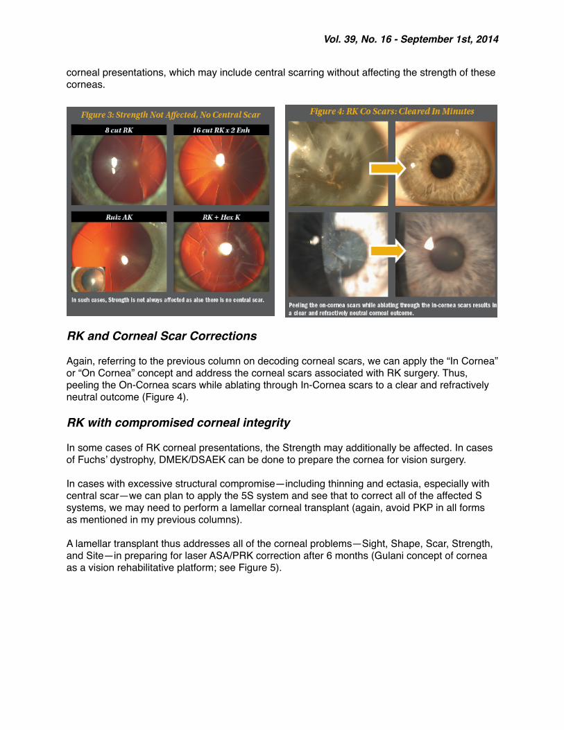
In a majority of cases, the Strength is not affected and there is no central Scar (Figure 3). Many of these cases can undergo excimer laser ASA/PRK.
(I do not propose LASIK in RK cases, because I consider RK to be like a cake that has eight pieces that are kept together on a plate. You cannot take a knife and go horizontally through all the cuts—which is LASIK—and not expect to disrupt the slices, which, in this case, means irregular ammetropia including further tissue instability. Additionally, the LASIK flap cut transgressing through RK cuts predisposes to epithelial ingrowth and intersectal wound healing disruptions.)
Applying laser ASA/PRK not only can correct the shape of the cornea (hyperopic, myopic, or astigmatism), but also allows for the new epithelium to come back to smooth the irregular anterior cornea.
In my anecdotal experience, I perceive the returning new epithelium after the PRK reshaping of the cornea to act like a smooth carpet over underlying, broken tiles (the broken tiles being the RK cut cornea underneath). This new carpet gives us the shape we want irrespective of the cuts below and also improves the smoothness of the anterior corneal
Vol. 39, No. 16 - September 1st, 2014
surface. Additionally, it decreases the refractive fluctuation by holding the underlying RK incisions much more tightly.
This concept along with the laser ASA/PRK reshaping then expands the optical zone (most RK was done with 3 mm OZ) and also translates to improve night vision and halos associated with RK.
This concept follows our Corneoplastique theme (see my previous column in Ophthalmology Times on decoding corneal scars, http://bit.ly/1lUnJKL) wherein the anterior corneal curvature overrides the posterior curvature and imperfections (including scars or RK incisions).
Using this concept, very much like corneal scars, we can disregard the RK incisions/pattern or number and concentrate on re-shaping the front cornea.
Gulani’s 5S System- Sight—Is there potential for vision? If yes, it is our duty to fight for the patient and take them
higher.- Shape—All refractive surgeries are about shape.- Scar—Is the cornea clear or does it have an opacity?- Strength—Is the cornea thicker or thinner than normal?- Site—Is the center affected or is the periphery? Don’t worry about the periphery as long as
the patient can see through the center.
Procedure
The procedure of laser ASA/PRK is standard with some RK-specific pearls:
Manual epithelium removal since manual removal allows us to feel the cornea, the RK incisions and their delicate architecture (No alcohol needed for patient or for
surgeon).Keep the spatula movements light in pressure and yet very swift in movementMove along the RK incisions (and change in directions of associated AK incisions) to avoid crossing or straddling them as you may inadvertently open an incisionKeep epithelium removal minimal and yet to allow entire ablation area including peripheral RK-knee, i.e., make it just beyond your planed laser OZ for faster healingAll other parameters of surgery are same as in any case including bandage contact lens application and most of these cases heal by day 5 postoperatively with no additional pain or discomfort.
Of course, all of these laser ASA/PRK procedures can be followed by crosslinking of the cornea to decrease any remaining fluctuation and create a permanent visual outcome.
Having addressed the majority of the presenting RK corneal or refractive errors with the above-mentioned concepts—addressing the Shape and Sight—let’s move on to the less common RK-
Vol. 39, No. 16 - September 1st, 2014
corneal presentations, which may include central scarring without affecting the strength of these corneas.
RK and Corneal Scar Corrections
Again, referring to the previous column on decoding corneal scars, we can apply the “In Cornea” or “On Cornea” concept and address the corneal scars associated with RK surgery. Thus, peeling the On-Cornea scars while ablating through In-Cornea scars to a clear and refractively neutral outcome (Figure 4).
RK with compromised corneal integrity
In some cases of RK corneal presentations, the Strength may additionally be affected. In cases of Fuchs’ dystrophy, DMEK/DSAEK can be done to prepare the cornea for vision surgery.
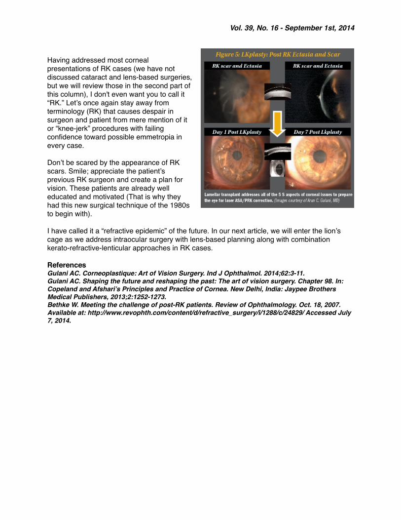
In cases with excessive structural compromise—including thinning and ectasia, especially with central scar—we can plan to apply the 5S system and see that to correct all of the affected S systems, we may need to perform a lamellar corneal transplant (again, avoid PKP in all forms as mentioned in my previous columns).
A lamellar transplant thus addresses all of the corneal problems—Sight, Shape, Scar, Strength, and Site—in preparing for laser ASA/PRK correction after 6 months (Gulani concept of cornea as a vision rehabilitative platform; see Figure 5).
Vol. 39, No. 16 - September 1st, 2014
Having addressed most corneal presentations of RK cases (we have not discussed cataract and lens-based surgeries, but we will review those in the second part of this column), I don't even want you to call it “RK.” Let’s once again stay away from terminology (RK) that causes despair in surgeon and patient from mere mention of it or “knee-jerk” procedures with failing confidence toward possible emmetropia in every case.
Don’t be scared by the appearance of RK scars. Smile; appreciate the patient’s previous RK surgeon and create a plan for vision. These patients are already well educated and motivated (That is why they had this new surgical technique of the 1980s to begin with).
I have called it a “refractive epidemic” of the future. In our next article, we will enter the lion’s cage as we address intraocular surgery with lens-based planning along with combination kerato-refractive-lenticular approaches in RK cases.
ReferencesGulani AC. Corneoplastique: Art of Vision Surgery. Ind J Ophthalmol. 2014;62:3-11.Gulani AC. Shaping the future and reshaping the past: The art of vision surgery. Chapter 98. In: Copeland and Afshari’s Principles and Practice of Cornea. New Delhi, India: Jaypee Brothers Medical Publishers, 2013;2:1252-1273.Bethke W. Meeting the challenge of post-RK patients. Review of Ophthalmology. Oct. 18, 2007. Available at: http://www.revophth.com/content/d/refractive_surgery/i/1288/c/24829/ Accessed July 7, 2014.