CorpusCallosumMicrostructuralChangesCorrelatewith...
8
Hindawi Publishing Corporation Multiple Sclerosis International Volume 2011, Article ID 304875, 7 pages doi:10.1155/2011/304875 Clinical Study Corpus Callosum Microstructural Changes Correlate with Cognitive Dysfunction in Early Stages of Relapsing-Remitting Multiple Sclerosis: Axial and Radial Diffusivities Approach Carolina de Medeiros Rimkus, 1 Thiago de Faria Junqueira, 2 Katarina Paz Lyra, 1 Marcel P. Jackowski, 3 Melissa A. R. Machado, 4 Eliane C. Miotto, 4 Dagoberto Callegaro, 2 Maria Concepci ´ on Garc´ ıa Otaduy, 1 and Claudia da Costa Leite 1 1 Department of Radiology, Laboratory of Medical Investigation (LIM-44), School of Medicine, University of S˜ ao Paulo (FM-USP), 05403-000 S˜ ao Paulo, SP, Brazil 2 Department of Neurology, Clinical Hospital, School of Medicine, University of S˜ ao Paulo (FM-USP), 05403-000 S˜ ao Paulo, SP, Brazil 3 Department of Computational Sciences, Institute of Mathematics and Statistics, University of S˜ ao Paulo (IME-USP), 05403-000 S˜ ao Paulo, SP, Brazil 4 Department of Neurology, Division of Neuropsychology, Clinical Hospital, School of Medicine University of S˜ ao Paulo (FM-USP), 05403-000 S˜ ao Paulo, SP, Brazil Correspondence should be addressed to Carolina de Medeiros Rimkus, [email protected] Received 15 November 2010; Revised 11 April 2011; Accepted 28 April 2011 Academic Editor: Angelo Ghezzi Copyright © 2011 Carolina de Medeiros Rimkus et al. This is an open access article distributed under the Creative Commons Attribution License, which permits unrestricted use, distribution, and reproduction in any medium, provided the original work is properly cited. The corpus callosum is the largest fiber bundle in the central nervous system and it takes part in several cognitive pathways. It can be affected by multiple sclerosis (MS) early in the disease. DTI is capable of infering the microstructural organization of the white matter. The vectorial analysis of the DTI offers the more specific indices of axial diffusivity (AD) and radial diffusivity (RD), which have shown to be useful to discriminate myelin damage from axon loss, respectively. This study presents DTI results (mean diffusivity (MD), fractional anisotropy (FA), RD, and AD) of 23 relapsing-remitting MS patients and its correlation with cognitive performance. There were 47.8% of cognitive impaired patients (MS CI). We found signs of demyelination, reflected by increased RD, and incipient axon loss, reflected by AD increase, which was slightly higher in the MS CI. The cognitive changes correlated with the DTI parameters, suggesting that loss of complexity in CC connections can impair neural conduction. Thus, cognitive impairment can be related to callosal disconnection, and DTI can be a promising tool to evaluate those changes. 1. Introduction Cognitive impairment is one of the major factors affect- ing the social functioning and quality of life of multiple sclerosis (MS) patients, being described in 45–65% of the clinical cases [1, 2]. The scales most commonly used for clinical classification of MS-related disability, such as the expanded disability status scale (EDSS) [3], are focused on locomotor dysfunction. Probably because of the multifocal characteristic of MS lesions, these traditional disability scales correlate poorly with cognitive disease-related changes in MS [4, 5]. In the last decade, it has been proposed to include the application of multimodality neuropsychological tests (NPT) in the assessment of clinical stage of MS, which resulted in a more comprehensive scale, the multiple sclerosis functional composite (MSFC) [6, 7]. Great efforts are being done in medical and biomedical studies aiming to design a map of cognitive functions [8–10]. However, given the complexity of cognitive pathways and cortical and subcortical connectivity, it is difficult to identify a single isolated domain for each cognitive skill. The corpus callosum (CC) is the major fiber bundle in the central nervous system (CNS) and plays a critical role in the interhemispheric communication, taking part in most of the cognitive pathways [5, 11, 12]. Its fibers connect the cortical and subcortical regions of the brain hemispheres
Transcript of CorpusCallosumMicrostructuralChangesCorrelatewith...
Hindawi Publishing CorporationMultiple Sclerosis InternationalVolume 2011, Article ID 304875, 7 pagesdoi:10.1155/2011/304875
Clinical Study
Corpus Callosum Microstructural Changes Correlate withCognitive Dysfunction in Early Stages of Relapsing-RemittingMultiple Sclerosis: Axial and Radial Diffusivities Approach
Carolina de Medeiros Rimkus,1 Thiago de Faria Junqueira,2 Katarina Paz Lyra,1
Marcel P. Jackowski,3 Melissa A. R. Machado,4 Eliane C. Miotto,4 Dagoberto Callegaro,2
Maria Concepcion Garcıa Otaduy,1 and Claudia da Costa Leite1
1 Department of Radiology, Laboratory of Medical Investigation (LIM-44), School of Medicine, University of Sao Paulo (FM-USP),05403-000 Sao Paulo, SP, Brazil
2 Department of Neurology, Clinical Hospital, School of Medicine, University of Sao Paulo (FM-USP), 05403-000 Sao Paulo, SP, Brazil3 Department of Computational Sciences, Institute of Mathematics and Statistics, University of Sao Paulo (IME-USP),05403-000 Sao Paulo, SP, Brazil
4 Department of Neurology, Division of Neuropsychology, Clinical Hospital, School of Medicine University of Sao Paulo (FM-USP),05403-000 Sao Paulo, SP, Brazil
Correspondence should be addressed to Carolina de Medeiros Rimkus, [email protected]
Received 15 November 2010; Revised 11 April 2011; Accepted 28 April 2011
Academic Editor: Angelo Ghezzi
Copyright © 2011 Carolina de Medeiros Rimkus et al. This is an open access article distributed under the Creative CommonsAttribution License, which permits unrestricted use, distribution, and reproduction in any medium, provided the original work isproperly cited.
The corpus callosum is the largest fiber bundle in the central nervous system and it takes part in several cognitive pathways. Itcan be affected by multiple sclerosis (MS) early in the disease. DTI is capable of infering the microstructural organization of thewhite matter. The vectorial analysis of the DTI offers the more specific indices of axial diffusivity (AD) and radial diffusivity (RD),which have shown to be useful to discriminate myelin damage from axon loss, respectively. This study presents DTI results (meandiffusivity (MD), fractional anisotropy (FA), RD, and AD) of 23 relapsing-remitting MS patients and its correlation with cognitiveperformance. There were 47.8% of cognitive impaired patients (MS CI). We found signs of demyelination, reflected by increasedRD, and incipient axon loss, reflected by AD increase, which was slightly higher in the MS CI. The cognitive changes correlatedwith the DTI parameters, suggesting that loss of complexity in CC connections can impair neural conduction. Thus, cognitiveimpairment can be related to callosal disconnection, and DTI can be a promising tool to evaluate those changes.
1. Introduction
Cognitive impairment is one of the major factors affect-ing the social functioning and quality of life of multiplesclerosis (MS) patients, being described in 45–65% of theclinical cases [1, 2]. The scales most commonly used forclinical classification of MS-related disability, such as theexpanded disability status scale (EDSS) [3], are focused onlocomotor dysfunction. Probably because of the multifocalcharacteristic of MS lesions, these traditional disability scalescorrelate poorly with cognitive disease-related changes in MS[4, 5]. In the last decade, it has been proposed to includethe application of multimodality neuropsychological tests
(NPT) in the assessment of clinical stage of MS, whichresulted in a more comprehensive scale, the multiple sclerosisfunctional composite (MSFC) [6, 7].
Great efforts are being done in medical and biomedicalstudies aiming to design a map of cognitive functions [8–10].However, given the complexity of cognitive pathways andcortical and subcortical connectivity, it is difficult to identifya single isolated domain for each cognitive skill.
The corpus callosum (CC) is the major fiber bundle inthe central nervous system (CNS) and plays a critical rolein the interhemispheric communication, taking part in mostof the cognitive pathways [5, 11, 12]. Its fibers connect thecortical and subcortical regions of the brain hemispheres
2 Multiple Sclerosis International
interconnecting the auditory, sensory-motor, and memoryinformation [13]. The CC is one of the sites most frequentlyaffected by demyelinating lesions in MS, and the onset ofthose lesions happens very early in the disease [14, 15].Additionally, CC atrophy is very common in MS [16, 17], andhistopathologic studies have detected signs of axon loss anddecreased fibers density in the CC of subjects with relapsing-remitting (RR) and secondary progressive (SP) MS [18].There are also evidences that regional axonal loss in the CCis related to white matter lesions projected in the cerebralhemispheres [19, 20]. For all these reasons, it is conceivablethat cognitive impairment in MS can be partially related tocommissural disconnection [21].
Conventional MR imaging is useful to demonstrate MSmacroscopic white matter (WM) lesions, but more recentresearch failed to prove direct relationship of clinical pro-gression of the disability and brain atrophy with cumulativelesion volume [22]. In consideration with those findings,other factors such as Wallerian degeneration, axon dysfunc-tion, and microscopic degeneration are gaining importancein the evaluation of the disease process, and several MRItechniques are being developed to access them [18, 23, 24].
Diffusion MRI techniques are based on the evaluationof random motion of water molecules and can provideinsights about the tissue organization. DTI has evolvedfrom those techniques, improving the evaluation of tissuemicrostructure and allowing the reconstruction of whitematter tracts [25–27]. The main scalar parameters, obtainedby DTI, fractional anisotropy (FA), and mean diffusivity(MD), are known to characterize alterations in myelin sheathand cell membrane integrity, but they are not specific fordemyelination or axon loss. The coordinates of the tensormatrix can be diagonalized to extract the three Eigenvalues,λ1, λ2, and λ3, derived from the main eigenvectors of diffu-sion elements [26]. The combination of those eigenvaluesdefines two other and more specific parameters, the axialdiffusivity (AD), parallel with the axon fibers, and the radialdiffusivity (RD), perpendicular to axon fibers. The increasesin those values are believed to represent axon loss anddemyelination, respectively [25, 28].
The purpose of this study is to determine by DTI whetherthe microstructure of the CC is already altered in MS patientswith short disease duration, and to establish if these DTIparameters are correlated with observed cognitive disability.
2. Materials and Methods
2.1. Subjects. Twenty-three clinically defined MS patients (15females) with relapsing-remitting course from an outpatientclinic were selected for the study. The mean age of the patientgroup was 31.95 ± 9.2 years (range 18–48 years), with amean disease duration of 2.4 ± 1.4 years (range 0.4–4.74years) and expanded disability status scale (EDSS) of 1.37 ±1.2 (range 0–4.5).
Out of twenty-two, twelve patients received immuno-modulatory therapy for at least six months (4 patients onglatiramer acetate, 3 on interferon beta-1a, and 5 on inter-feron beta-1b), without presenting significant side effects. All
patients were out of clinical relapse and were steroid-free forat least three months prior to MRI examination.
A control group of twelve healthy volunteers (HC) (9females) with age and educational level matched to ourpatients was also studied. The mean age of the HC was27.75 ± 5.4 years (range 20–37 years), without significantdifference with the MS patients (P = 0.12). There was nosignificant difference in the educational level between MSpatients (14.23 ± 3.3 years of school education) and healthysubjects (14.87± 3.4 years of school education) (P = 0.87).
Subjects with significant depression measured by theBeck Depression Inventory (score 14 or higher) were ex-cluded from the study.
The study protocol was approved by the local ethicalcommittee (CAPPESQ 0450/09). All subjects agreed to par-ticipate in the study by signing a written informed consent.
2.2. Magnetic Resonance and Diffusion Tensor Imaging. BrainMRI exams were performed in a 3.0 T magnetic resonancescanner (Intera Achieva, PHILIPS Healthcare, Best, TheNetherlands) with an 8-channel head coil. Sixty to sixty-seven contiguous 2 mm axial slices were acquired coveringthe whole brain, using a diffusion-weighted spin-echo (SE)single shot echo planar imaging (EPI) sequence, with a b-value of 0 and 1000 s/mm2, 32 diffusion encoding directions,field of view (FOV) = 256 × 256 mm, and a matrix size of128× 128.
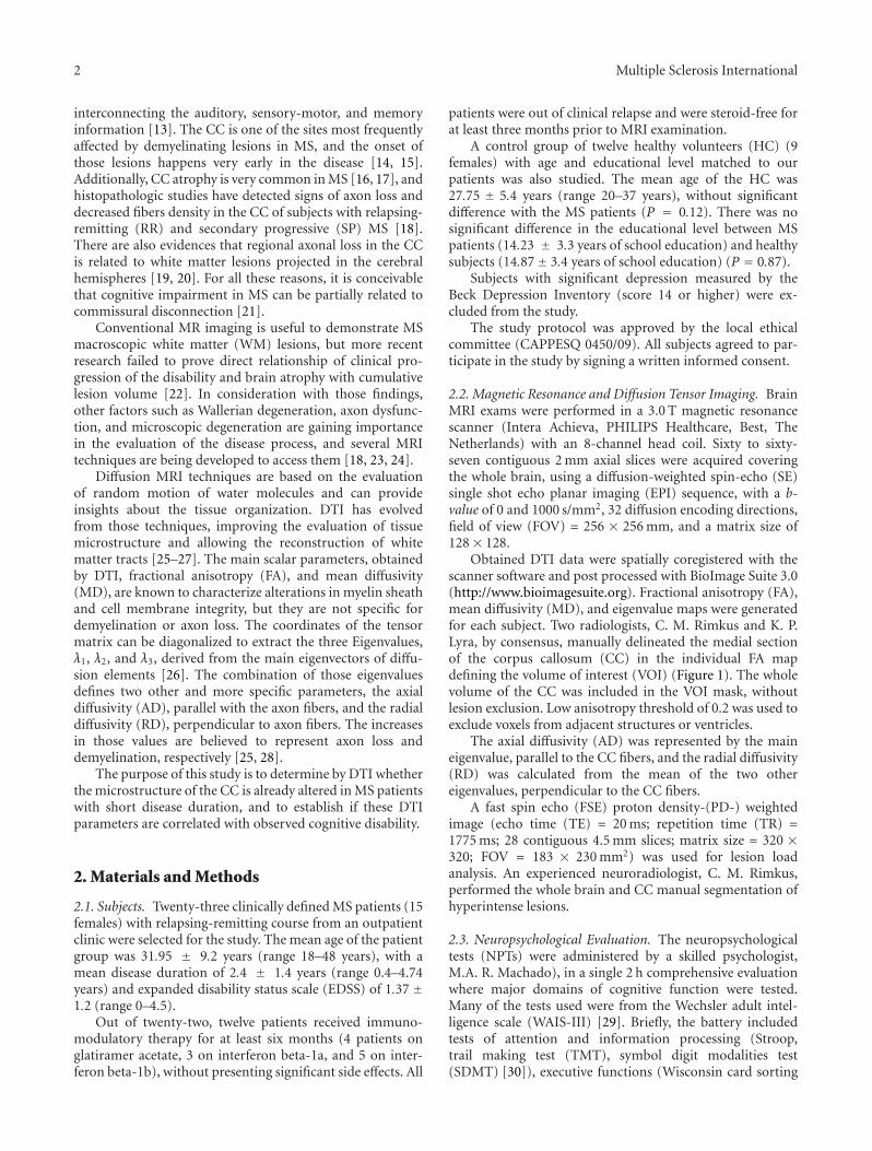
Obtained DTI data were spatially coregistered with thescanner software and post processed with BioImage Suite 3.0(http://www.bioimagesuite.org). Fractional anisotropy (FA),mean diffusivity (MD), and eigenvalue maps were generatedfor each subject. Two radiologists, C. M. Rimkus and K. P.Lyra, by consensus, manually delineated the medial sectionof the corpus callosum (CC) in the individual FA mapdefining the volume of interest (VOI) (Figure 1). The wholevolume of the CC was included in the VOI mask, withoutlesion exclusion. Low anisotropy threshold of 0.2 was used toexclude voxels from adjacent structures or ventricles.
The axial diffusivity (AD) was represented by the maineigenvalue, parallel to the CC fibers, and the radial diffusivity(RD) was calculated from the mean of the two othereigenvalues, perpendicular to the CC fibers.
A fast spin echo (FSE) proton density-(PD-) weightedimage (echo time (TE) = 20 ms; repetition time (TR) =1775 ms; 28 contiguous 4.5 mm slices; matrix size = 320 ×320; FOV = 183 × 230 mm2) was used for lesion loadanalysis. An experienced neuroradiologist, C. M. Rimkus,performed the whole brain and CC manual segmentation ofhyperintense lesions.
2.3. Neuropsychological Evaluation. The neuropsychologicaltests (NPTs) were administered by a skilled psychologist,M.A. R. Machado), in a single 2 h comprehensive evaluationwhere major domains of cognitive function were tested.Many of the tests used were from the Wechsler adult intel-ligence scale (WAIS-III) [29]. Briefly, the battery includedtests of attention and information processing (Stroop,trail making test (TMT), symbol digit modalities test(SDMT) [30]), executive functions (Wisconsin card sorting
Multiple Sclerosis International 3
Table 1: Results of DTI scalar maps (FA and MD) and vectorial diffusivity (AD and RD) in MS patients and healthy controls.
MS patients MS CP MS CI HC
MD (10−3 mm2/s) 0.8611 ± 0.05 0.8492 ± 0.04 0.8741 ± 0.05 0.791 ± 0.04
FA 0.6879 ± 0.03 0.6899 ± 0.03 0.6858 ± 0.03 0.7206 ± 0.03
AD (10−3 mm2/s) 1.6788 ± 0.06 1.6591 ± 0.06 1.7003 ± 0.05 1.6102 ± 0.08
RD (10−3 mm2/s) 0.4487 ± 0.04 0.4451 ± 0.04 0.4528 ± 0.04 0.3818 ± 0.03
Whole brain lesion load 10.16 mL ± 9.8 9.54 mL ± 6.5 10.9 mL ± 12.8 —
CC lesion load 0.65 mL ± 0.7 0.77 mL ± 0.6 0.51 mL ± 0.7 —
Table 2: Pearson’s correlation test evaluating possible correlationsbetween DTI parameters and demographic characteristics of thesample.
AgeEducational
levelDuration ofthe disease
EDSS
MD r = −0.05 r = −0.19 r = 0.37 r = 0.45∗
FA r = 0.22 r = 0.25 r = −0.02 r = −0.31
AD r = 0.05 r = −0.09 r = 0.29 r = 0.38
RD r = −0.09 r = −0.25 r = 0.26 r = 0.45∗∗
Significant correlation (P = 0.03).
test (WCST) [31], verbal fluency with phonemic associationtests (FAS), and category association [32]), memory func-tions including short-term and working memory (Digit SpanForward and Backward and Letter-Number Sequencing fromWAIS-III), long-term memory (Logical Memory, Hopkinsverbal learning tests (HVLT) [33], Rey-Osterrieth complexfigure (ROCF) delayed recall [34] and brief visual spatialmemory test (BVMT) [35]), visuospatial functions (BlockDesign Test from WAIS III and ROCF copy), and verbal skills(Boston test and WAIS-III Vocabulary [29]). The intellectualfunction was also estimated by WAIS III-revised full-scaleintelligence quotient (WAIS III-R FSIQ).
The individual score for each test was normalized basedon the mean score and standard deviation of healthy adultpopulation, obtaining the z-score [36]. The results below the5th percentile were defined as abnormal.
Patients with three or more abnormal tests were classifiedas cognitive impaired.
No subject in the HC group was considered cognitiveimpaired, mild impairment was observed in four subjects forone task and in another one for two tasks.
The Beck depression inventory was also applied toexclude subjects with significant depression levels, whichcould interfere in the NPT.
2.4. Statistical Analysis. Descriptive statistics of demographicdata such as age, sex, gender, and scholar level were analyzedto discriminate the distribution, frequency, and percentageof each of those variables in the sample and to characterizedifferences between the groups. Possible bias determinedby the sample’s demographic characteristics in the DTIparameters was tested by the Pearson’s correlation test. Thiscorrelation test was also used to explore the correlationbetween DTI parameters and NPT and between them andMS patients’ whole brain and CC lesion loads.
(a)
(b)
Figure 1: Sagital view of an individual FA map with the demarca-tion of the CC (a). In detail is the obtained CC mask (b).
The MS patients were divided in cognitive impaired(MS CI) and cognitive preserved (MS CP). Mann-Whitneynonparametric t-test was used to evaluate significant differ-ences in DTI parameters between MS patients and HC andbetween MS CI and MS CP. A P value <0.05was consideredsignificant.
The cognitive tasks were grouped by functional domain,and the relative frequency of abnormal tests in each domainin the MS patients’ group was demonstrated.
3. Results
3.1. Diffusion Tensor Imaging. In Table 1, DTI resultsobtained from the CC of MS patients, HC, MS CP, and MS CIgroups and the respective lesion loads are shown. Comparedto HC, MS patients presented in the CC significantly highermean MD (P < 0.0001) and lower mean FA (P = 0.002). TheRD (P < 0.0001) and AD (P = 0.009) were also significantlyincreased in MS patients. Both MS CP and MS CI presentedsignificant differences in all DTI parameters compared toHC. The AD was slightly higher in MS CI compared to MSCP (P = 0.048).

4 Multiple Sclerosis International
Table 3: Frequency of subjects with abnormal tests in the main cognitive domains in the MS patient subgroups.
MS patients MS CP MS CI
Short-term and working memory 8.7% 0.0% 18.2%
Long-term memory 60.9% 33.3% 90.9%
Executive functions 21.7% 8.3% 36.4%
Attention and information processing speed 60.9% 25.0% 100.0%
Verbal skills 0.0% 0.0% 0.0%
Visual spatial processing and construction 4.3% 0.0% 9.1%
Table 4: Pearson’s correlation test between the main cognitive tasks and DTI parameters (considering z scores of all MS patients).
MD FA AD RD
HVLT-Immediate recallr 0.09 0.25 0.12 −0.04
P-value 0.7 0.2 0.6 0.9
HVLT-Delayed recallr 0.33 −0.32 0.04 0.45
P-value 0.1 0.1 0.8 0.03∗
ROCF-Delayed recallr −0.10 0.15 −0.05 −0.014
P-value 0.6 0.5 0.8 0.5
Logical memory Ir −0.07 0.16 0.05 −0.21
P-value 0.7 0.6 0.8 0.3
Logical memory IIr −0.02 0.41 0.2 −0.21
P-value 0.9 0.05∗∗ 0.3 0.3
Symbol Digit (SDMT)r −0.37 0.46 −0.17 −0.37
P-value 0.07∗∗ 0.02∗ 0.4 0.07∗∗
TMTr −0.31 −0.01 −0.37 −0.18
P-value 0.1 0.9 0.08∗∗ 0.4
Stroopr −0.33 0.36 −0.12 −0.39
P-value 0.1 0.08∗∗ 0.6 0.06∗∗
FASr −0.29 0.2 −0.17 −0.24
P-value 0.2 0.3 0.4 0.3
Category associationr −0.09 −0.04 −0.24 0.03
P-value 0.7 0.9 0.3 0.9
Wisconsin r 0.03 −0.15 −0.18 0.14
P-value 0.9 0.5 0.3 0.5∗
Significant correlation between cognitive test and DTI parameter.∗∗Cognitive tests that presented a tendency of correlation with the respective DTI parameter.
All patients, except one, presented lesions in the CC(range 0 to 35 lesions, mean 10.2 ± 7.7). There was nosignificant difference between MS CP and MS CI lesion loads(Figures 2 and 3).
Pearson’s test showed no correlation of DTI results andwhole brain or CC lesion loads. There was also no correlationwith disease duration, educational, or age level. A correlationof MD and RD with EDss (P = 0.03) was found (Table 2).
3.2. Neuropsychological Characteristics. Twelve patients(52.2%) were classified as MS CP and eleven (47.8%) asMS CI. Table 3 summarizes the frequency of subjects withabnormal tests in each of the principal functional domains.In the most affected domains, the performance of thecognitive impaired patients was worse both qualitatively andquantitatively. MS CI had 1 to 3 tests below the 5th percentileof z scores in the sustained attention and informationprocessing speed (IPS) domain (mean 1.7 ± 0.8) whileMS CP had up to 1 test abnormal (mean 0.25 ± 0.45). In
long-term memory tests, MS CI had up to 5 abnormal tests(mean 2.5± 1.5) while MS CP had up to 2 (mean 0.4 ± 0.7).In executive functions, MS CI had up to 2 abnormal tests(mean 0.5 ± 0.7) and in the MS CP, only one subject had 1abnormal test (mean 0.1± 0.3).
Table 4 summarizes the correlation between the principalaffected cognitive tasks and the DTI parameters.
The intellectual performances (WAISS III-R FSIQ) of theMS patients and HC were within the normal rates of thepopulation.
There was no correlation between the NPT performancesand the lesion loads.
4. Discussion
Cognitive decline can affect MS patients very early in thedisease. In a group of MS patients with less than five yearsof disease, 47.8% were found to be cognitive impaired. Thepatients in this study presented deficits in sustained attention

Multiple Sclerosis International 5
and IPS, long-term memory, and executive functions. TheMS CI group showed also mild impairment in the short-termand working memory, visual-spatial, and construction tests.
The cognitive dysfunction was concomitant with DTIchanges in the CC. MS group of patients showed decreasedFA and increased MD compared to the matched HC, whichcan be interpreted as loss of complexity in the white mattertracts in the initial pathological process. The analysis ofthe principal eigenvalue (AD), parallel to axon fibers, andthe average of the second and third eigenvalues (RD),perpendicular to fiber tracts, showed significant increase inRD and AD in MS patients.
It is believed that the increased RD reflects the demyeli-nation degree while increased AD represents signs of axonloss [25, 28]. Although MS is classified as a myelin-relateddisease, several studies have reported loss of axons bothin MS plaques as well as in the normal-appearing whitematter (NAWM). Axon loss can be less prominent thandemyelination, but its importance is recognized in thepathogenesis of symptoms [37, 38].
The observation of slightly increased AD in the MS CIpatients reinforces the role of axon loss in the cognitivedysfunction. We neither have found significant differencesin RD between MS CP and MS CI nor the DTI parametershave correlated with whole brain or CC lesion loads. Despitethe fact that our relatively small sample can underestimateRD or lesion load differences, it is conceivable to hypoth-esize that other factors than demyelination itself or glioticlesions can lead to cellular death and axon degeneration.Traditionally, the loss of axons in MS was associated withfiber transection due to inflammation and demyelination inthe plaques, possibly conducting to Wallerian degeneration[38, 39], but more recent studies point that it might be anunderlying diffuse axonopathy that contributes to the proc-ess of neurodegeneration [18, 20, 38].
The pattern of cognitive impairment is similar to thefindings of previous studies [5, 40, 41]. Although there aresome variations in the NPT results due to differences in thetests applied, the sample sizes, and patients’ characteristics,the main abnormalities were found to be in sustained atten-tion and IPS, executive functions, and memory domains.The functions more frequently related to CC microscopiclesion are in the attention and IPS and executive domains,suggesting anterior commissural damage with compromiseof frontal and prefrontal pathways [20].
In this study, the stronger correlation was presentbetween SDMT (attention and IPS) with FA (P = 0.02) andHVLT-delayed recall with RD (P = 0.03). The sustainedattention and IPS domain presented other tests with atendency to correlate with the DTI parameters: SDMT withMD and RD, TMT with AD, and Stroop test with FA andRD. The transverse characteristic of this study only providesa static picture of a dynamic process. It is not possible topredict the previous cognitive performance and the preciseneuropsychological decline of the patients, and some of themmight have greater functional reserve, diminishing the powerof those correlations- in this sample. Additionally, slightdifferences in the AD within the MS patients were present,which suggests different degrees of axon loss. These findings
(a)
(b)
Figure 2: Female patient with high lesion load and no significantcognitive deficit. The PD axial image at the level of the body of thelateral ventricles and CC (a) shows lesions at the periventricular andsubcortical white matter. The detail of mid-sagital FLAIR (b) showsthe abundance of macroscopic lesion in the CC and callosal-septalinterface.
can reflect incipient heterogeneity of the sample, softeningthe direct correlations between DTI and NPT.
Neither the duration of the disease nor the age level of thepatients correlated with the DTI parameters. Only subjectswith short duration of the disease composed the sample; so,it was expected that this variable would not interfere in theimage parameters. Previous studies showed that the peakof highest FA and lower MD in the CC happens betweenthe ages of 21–44 years [42], and the main age-related DTIchanges are described after 7th and 8th decades of life [43].So, in our population with age range from 18 to 48 years, theDTI findings are more likely to be related with the MS diseasethan with the patients’ ages.
However, MD and RD showed correlation with EDSS,suggesting possible relationship between callosal demyeli-nation and sensory-motor dysfunction. The primary motorfunction takes part in the cortical spinal tract, but the inter-hemispheric communication of this pathway depends onmidbody CC fibers [20, 38]. Hypothetically, the callosal

6 Multiple Sclerosis International
Figure 3: Female patient with low lesion load and significantcognitive deficit. The PD axial image illustrates the presence offew lesions in the supratentorial white matter, despite the cognitivedeficit.
disconnection can partially impair this neural conductionand influence the EDSS.
5. Conclusions
Cognitive impairment can be observed early in MS diseaseprocess, but it affects the MS population in differentways. The MS patients present signs of demyelination andloss of complexity of the CC, but patients with cognitivedysfunction might present earlier and more prominent axonloss, which could be related to incipient differences in theclinical and histological profile.
The cognitive decline is not directly associated to themacroscopic lesion load. Thus, advanced neuroimage studiesare demanded to access microscopic abnormalities in theprincipal fiber tracts and functional pathways. The vectorialcharacteristic of the diffusion tensors makes it possible toexplore signs of demyelination and axon loss, making of it apromising and noninvasive tool to evaluate MS pathologicalchanges.
Acknowledgments
The authors thank FAPESP (2005/56464-9)—PROGRAMACINAPCE—for financial support. They also thank MiriamHarumi Tsunemi and Gilson Vieira for helping with thestatistical analysis of this study.
References
[1] S. M. Rao, G. J. Leo, L. Ellington, T. Nauertz, L. Bernardin,and F. Unverzagt, “Cognitive dysfunction in multiple sclerosis.II. Impact on employment and social functioning,” Neurology,vol. 41, no. 5, pp. 692–696, 1991.
[2] M. P. Amato, V. Zipoli, B. Goretti et al., “Benign multiplesclerosis: cognitive, psychological and social aspects in a
clinical cohort,” Journal of Neurology, vol. 253, no. 8, pp. 1054–1059, 2006.
[3] J. F. Kurtzke, “Rating neurologic impairment in multiple scle-rosis: an expanded disability status scale (EDSS),” Neurology,vol. 33, no. 11, pp. 1442–1452, 1983.
[4] M. P. Amato and E. Portaccio, “Clinical outcome measuresin multiple sclerosis,” Journal of the Neurological Sciences, vol.259, no. 1–2, pp. 118–122, 2007.
[5] S. Mesaros, M. A. Rocca, G. Riccitelli et al., “Corpus callosumdamage and cognitive dysfunction in benign MS,” HumanBrain Mapping, vol. 30, no. 8, pp. 2656–2666, 2009.
[6] R. Rudick, J. Antel, C. Confavreux et al., “Recommendationsfrom the national multiple sclerosis society clinical outcomesassessment task force,” Annals of Neurology, vol. 42, no. 3, pp.379–382, 1997.
[7] M. Hoogs, S. Morrow, and R. H. B. Benedict, “Utility ofroutine neuropsychological assessment for early identificationof cognitive impairment in MS,” International MS Journal, vol.17, no. 1, pp. 6–11, 2010.
[8] P. A. Arnett, S. M. Rao, L. Bernardin, J. Grafman, F. Z. Yetkin,and L. Lobeck, “Relationship between frontal lobe lesionsand Wisconsin card sorting test performance in patients withmultiple sclerosis,” Neurology, vol. 44, no. 3, pp. 420–425,1994.
[9] J. Foong, L. Rozewicz, G. Quaghebeur et al., “Executive func-tion in multiple sclerosis. The role of frontal lobe pathology,”Brain, vol. 120, no. 1, pp. 15–26, 1997.
[10] R. A. Sperling, C. R. G. Guttmann, M. J. Hohol et al., “Regionalmagnetic resonance imaging lesion burden and cognitivefunction in multiple sclerosis: a longitudinal study,” Archivesof Neurology, vol. 58, no. 1, pp. 115–121, 2001.
[11] S. D. Moffat, E. Hampson, and D. H. Lee, “Morphology of theplanum temporale and corpus callosum in left handers withevidence of left and right hemisphere speech representation,”Brain, vol. 121, no. 12, pp. 2369–2379, 1998.
[12] F. Aboitiz, A. B. Scheibel, R. S. Fisher, and E. Zaidel, “Fibercomposition of the human corpus callosum,” Brain Research,vol. 598, no. 2, pp. 143–153, 1992.
[13] M. Hines, L. Chiu, L. A. McAdams, P. M. Bentler, andJ. Lipcamon, “Cognition and the corpus callosum: verbalfluency, visuospatial ability, and language lateralization relatedto midsagittal surface areas of callosal subregions,” BehavioralNeuroscience, vol. 106, no. 1, pp. 3–14, 1992.
[14] A. D. Gean-Marton, L. G. Vezina, K. I. Marton et al.,“Abnormal corpus callosum: a sensitive and specific indicatorof multiple sclerosis,” Radiology, vol. 180, no. 1, pp. 215–221,1991.
[15] J. H. Simon, S. L. Holtas, R. B. Schiffer et al., “Corpus callosumand subcallosal-periventricular lesions in multiple sclerosis:detection with MR,” Radiology, vol. 160, no. 2, pp. 363–367,1986.
[16] F. Barkhof, M. Elton, J. Lindeboom et al., “Functionalcorrelates of callosal atrophy in relapsing-remitting multiplesclerosis patients. A preliminary MRI study,” Journal ofNeurology, vol. 245, no. 3, pp. 153–158, 1998.
[17] M. Juha, S. Leszek, F. Sten et al., “Progression of non-age-related callosal brain atrophy in multiple sclerosis: a 9-year longitudinal MRI study representing four decades ofdisease development,” Journal of Neurology, Neurosurgery andPsychiatry, vol. 78, no. 4, pp. 375–380, 2007.
[18] N. Evangelou, D. Konz, M. M. Esiri, S. Smith, J. Palace, and P.M. Matthews, “Quantitative pathological evidence for axonalloss in normal appearing white matter in multiple sclerosis,”Annals of Neurology, vol. 47, no. 3, pp. 391–395, 2000.
Multiple Sclerosis International 7
[19] M. C. de Lacoste, J. B. Kirkpatrick, and E. D. Ross, “Topogra-phy of the human corpus callosum,” Journal of Neuropathologyand Experimental Neurology, vol. 44, no. 6, pp. 578–591, 1985.
[20] N. Evangelou, D. Konz, M. M. Esiri, S. Smith, J. Palace,and P. M. Matthews, “Regional axonal loss in the corpuscallosum correlates with cerebral white matter lesion volumeand distribution in multiple sclerosis,” Brain, vol. 123, no. 9,pp. 1845–1849, 2000.
[21] J. W. Pan, L. B. Krupp, L. E. Elkins, and P. K. Coyle,“Cognitive dysfunction lateralizes with NAA in multiplesclerosis,” Applied Neuropsychology, vol. 8, no. 3, pp. 155–166,2001.
[22] M. Rovaris, G. Iannucci, M. Falautano et al., “Cognitive dys-function in patients with mildly disabling relapsing-remittingmultiple sclerosis: an exploratory study with diffusion tensorMR imaging,” Journal of the Neurological Sciences, vol. 195, no.2, pp. 103–109, 2002.
[23] D. Cox, D. Pelletier, C. Genain et al., “The unique impact ofchanges in normal appearing brain tissue on cognitive dys-function in secondary progressive multiple sclerosis patients,”Multiple Sclerosis, vol. 10, no. 6, pp. 626–629, 2004.
[24] G. J. M. Parker, K. E. Stephan, G. J. Barker et al., “Initialdemonstration of in vivo tracing of axonal projections in themacaque brain and comparison with the human brain usingdiffusion tensor imaging and fast marching tractography,”NeuroImage, vol. 15, no. 4, pp. 797–809, 2002.
[25] C. A. M. Wheeler-Kingshott and M. Cercignani, “About“axial” and “radial” diffusivities,” Magnetic Resonance inMedicine, vol. 61, no. 5, pp. 1255–1260, 2009.
[26] J. L. Y. Cheong, D. K. Thompson, H. X. Wang et al., “Abnormalwhite matter signal on MR imaging is related to abnormaltissue microstructure,” American Journal of Neuroradiology,vol. 30, no. 3, pp. 623–628, 2009.
[27] M. D. Budde, J. H. Kim, H. F. Liang, J. H. Russell, A. H. Cross,and S. K. Song, “Axonal injury detected by in vivo diffusiontensor imaging correlates with neurological disability in amouse model of multiple sclerosis,” NMR in Biomedicine, vol.21, no. 6, pp. 589–597, 2008.
[28] S. K. Song, S. W. Sun, M. J. Ramsbottom, C. Chang, J. Russell,and A. H. Cross, “Dysmyelination revealed through MRI asincreased radial (but unchanged axial) diffusion of water,”NeuroImage, vol. 17, no. 3, pp. 1429–1436, 2002.
[29] J. D. Matarazzo, Wechsler’s Measurements and Appraisal ofAdult Intelligence, Williams and Wilkins, Oxford, UK, 5thedition, 1972.
[30] A. Smith, “The symbol-digit modalities test: a neuropsy-chologic test of learning and other cerebral disorders,” inLearning Disorders, J. Helmuth, Ed., pp. 83–91, Special ChildPublications, Seattle, Wash, USA, 1968.
[31] R. H. Heaton, Wisconsin Card Sorting Test (Manual), Psycho-logical Assessment Resources, Odessa, Fla, USA, 1981.
[32] J. Raven, J. C. Raven, and J. H. Court, “Advanced progressivematrices,” in Raven Manual, Oxford Psychologists Press,Oxford, UK, 4th edition, 1998.
[33] G. Kuslansky, M. Katz, J. Verghese et al., “Detecting dementiawith the Hopkins verbal learning test and the mini-mentalstate examination,” Archives of Clinical Neuropsychology, vol.19, no. 1, pp. 89–104, 2004.
[34] P. Caffarra, G. Vezzadini, F. Dieci, F. Zonato, and A. Venneri,“Rey-Osterrieth complex figure: normative values in an Italianpopulation sample,” Neurological Sciences, vol. 22, no. 6, pp.443–447, 2002.
[35] R. H. B. Benedict, L. Groninger, D. Schretlen, M. Dobraski,and B. Shpritz, “Revision of the brief visuospatial memory
test: studies of normal performance, reliability, and, validity,”Psychological Assessment, vol. 8, no. 2, pp. 145–153, 1996.
[36] E. Strauss, E. M. S. Sherman, and O. Spreen, A Compendium ofNeuropsychological Tests: Administration, Norms and Commen-tary, Oxford University Press, New York, NY, USA, 3rd edition,2006.
[37] J. G. Greenfield and L. S. King, “Observations on thehistopathology of the cerebral lesions in disseminated sclero-sis,” Brain, vol. 59, no. 4, pp. 445–458, 1936.
[38] G. C. DeLuca, K. Williams, N. Evangelou, G. C. Ebers, and M.M. Esiri, “The contribution of demyelination to axonal loss inmultiple sclerosis,” Brain, vol. 129, no. 6, pp. 1507–1516, 2006.
[39] B. D. Trapp, J. Peterson, R. M. Ransohoff, R. Rudick, S. Mork,and L. Bo, “Axonal transection in the lesions of multiplesclerosis,” The New England Journal of Medicine, vol. 338, no.5, pp. 278–285, 1998.
[40] R. A. Dineen, J. Vilisaar, J. Hlinka et al., “Disconnection asa mechanism for cognitive dysfunction in multiple sclerosis,”Brain, vol. 132, no. 1, pp. 239–249, 2009.
[41] S. D. Roosendaal, J. J. G. Geurts, H. Vrenken et al., “RegionalDTI differences in multiple sclerosis patients,” NeuroImage,vol. 44, no. 4, pp. 1397–1403, 2009.
[42] C. Lebel, S. Caverhill-Godkewitsch, and C. Beaulieu, “Age-related regional variations of the corpus callosum identifiedby diffusion tensor tractography,” NeuroImage, vol. 52, no. 1,pp. 20–31, 2010.
[43] E. V. Sullivan, T. Rohlfing, and A. Pfefferbaum, “Longitudinalstudy of callosal microstructure in the normal adult agingbrain using quantitative DTI fiber tracking,” DevelopmentalNeuropsychology, vol. 35, no. 3, pp. 233–256, 2010.

Submit your manuscripts athttp://www.hindawi.com
Stem CellsInternational
Hindawi Publishing Corporationhttp://www.hindawi.com Volume 2014
Hindawi Publishing Corporationhttp://www.hindawi.com Volume 2014
MEDIATORSINFLAMMATION
of
Hindawi Publishing Corporationhttp://www.hindawi.com Volume 2014
Behavioural Neurology
EndocrinologyInternational Journal of
Hindawi Publishing Corporationhttp://www.hindawi.com Volume 2014
Hindawi Publishing Corporationhttp://www.hindawi.com Volume 2014
Disease Markers
Hindawi Publishing Corporationhttp://www.hindawi.com Volume 2014
BioMed Research International
OncologyJournal of
Hindawi Publishing Corporationhttp://www.hindawi.com Volume 2014
Hindawi Publishing Corporationhttp://www.hindawi.com Volume 2014
Oxidative Medicine and Cellular Longevity
Hindawi Publishing Corporationhttp://www.hindawi.com Volume 2014
PPAR Research
The Scientific World JournalHindawi Publishing Corporation http://www.hindawi.com Volume 2014
Immunology ResearchHindawi Publishing Corporationhttp://www.hindawi.com Volume 2014
Journal of
ObesityJournal of
Hindawi Publishing Corporationhttp://www.hindawi.com Volume 2014
Hindawi Publishing Corporationhttp://www.hindawi.com Volume 2014
Computational and Mathematical Methods in Medicine
OphthalmologyJournal of
Hindawi Publishing Corporationhttp://www.hindawi.com Volume 2014
Diabetes ResearchJournal of
Hindawi Publishing Corporationhttp://www.hindawi.com Volume 2014
Hindawi Publishing Corporationhttp://www.hindawi.com Volume 2014
Research and TreatmentAIDS
Hindawi Publishing Corporationhttp://www.hindawi.com Volume 2014
Gastroenterology Research and Practice
Hindawi Publishing Corporationhttp://www.hindawi.com Volume 2014
Parkinson’s Disease
Evidence-Based Complementary and Alternative Medicine
Volume 2014Hindawi Publishing Corporationhttp://www.hindawi.com