Corporate Policy & Procedures Manual ... - eLearning...
26
Peripheral IV Catheter Corporate Policy & Procedures Manual Number: VII-B-390 Date Approved December 15, 2014 Approved by: Vice President and Chief Medical Officer Vice President and Chief Operating Officer Date Effective January 9, 2015 Next Review (3 years from Effective Date) January 2018 Purpose To outline the educational requirements for clinical competency for initiation of peripheral intravenous catheters. Policy Statement An order is required for insertion of a peripheral IV catheter. The order must include the type of IV solution, the rate of infusion, the most responsible health practitioner's signature, and date. Health care professionals shall have successfully completed theory and a demonstration of competency prior to independently initiating a peripheral intravenous catheter. (See Requirements: Education / Demonstrated Skills section below.) If the health care professional has made two unsuccessful attempts at peripheral IV catheter insertion, he/she shall contact the health care professional on the unit, or within the facility, with the most advanced IV skills to evaluate the patient's venous access. Further insertion attempts should be made only if venous access is deemed adequate. If the patient has limited venous access, the most responsible health practitioner should be notified and an assessment made whether another type of vascular access device should be established; or alternative routes for medication administration need to be evaluated. (For example, dehydrated patients may require hypodermoclysis. Once hydrated, the patient may have adequate venous access.) Insertion attempts should be limited to four unless emergent situations and patient safety will be compromised. Multiple unsuccessful attempts limit future vascular access and cause unnecessary trauma to the patient Health care professionals are encouraged to report any adverse events or occurrences that result from infusion therapy treatment via the Reporting and Learning System (RLS) (resource @ http://www.compassionnet.ca/ie/Page576.aspx . Adverse events include complications noted in Section 6 in the procedure, or occurrences that cause the patient undue pain; extends the patient's hospital stay; or results in remedial treatment. Refer also to Covenant Health policy #III-45, Responding to Adverse Events, Close Calls and Hazards. Applicability This policy and procedure applies to all Covenant Health facilities, staff, members of the medical staff, students and any other persons acting on behalf of Covenant Health.
Transcript of Corporate Policy & Procedures Manual ... - eLearning...

Peripheral IV Catheter
Corporate Policy & Procedures Manual
Number: VII-B-390 Date Approved
December 15, 2014 Approved by: Vice President and Chief Medical Officer Vice President and Chief Operating Officer
Date Effective January 9, 2015
Next Review (3 years from Effective Date)
January 2018 Purpose To outline the educational requirements for clinical competency for
initiation of peripheral intravenous catheters.
Policy Statement
An order is required for insertion of a peripheral IV catheter. The order must include the type of IV solution, the rate of infusion, the most responsible health practitioner's signature, and date.
Health care professionals shall have successfully completed theory and a demonstration of competency prior to independently initiating a peripheral intravenous catheter. (See Requirements: Education / Demonstrated Skills section below.) If the health care professional has made two unsuccessful attempts at peripheral IV catheter insertion, he/she shall contact the health care professional on the unit, or within the facility, with the most advanced IV skills to evaluate the patient's venous access. Further insertion attempts should be made only if venous access is deemed adequate. If the patient has limited venous access, the most responsible health practitioner should be notified and an assessment made whether another type of vascular access device should be established; or alternative routes for medication administration need to be evaluated. (For example, dehydrated patients may require hypodermoclysis. Once hydrated, the patient may have adequate venous access.) Insertion attempts should be limited to four unless emergent situations and patient safety will be compromised. Multiple unsuccessful attempts limit future vascular access and cause unnecessary trauma to the patient Health care professionals are encouraged to report any adverse events or occurrences that result from infusion therapy treatment via the Reporting and Learning System (RLS) (resource @ http://www.compassionnet.ca/ie/Page576.aspx. Adverse events include complications noted in Section 6 in the procedure, or occurrences that cause the patient undue pain; extends the patient's hospital stay; or results in remedial treatment. Refer also to Covenant Health policy #III-45, Responding to Adverse Events, Close Calls and Hazards.
Applicability This policy and procedure applies to all Covenant Health facilities, staff, members of the medical staff, students and any other persons acting on behalf of Covenant Health.

Peripheral IV Catheter Date Effective Jan. 9, 2015
Policy No. VII-B-390
Page 2 of 26
Responsibility Venipuncture for the initiation of peripheral IV access is a skill that requires
practice and frequency to maintain competency. After initial competency is demonstrated, it is the health care professional's responsibility to maintain their knowledge and ability so they are able to safely implement the skill at all times.
Principles Venipuncture done to initiate a peripheral IV is commonly seen by patients as one of the most painful and frequently performed invasive procedure done by nurses and other staff. Multiple venipuncture attempts can heighten patient anxiety and suffering, delay vital treatment and increase costs. Failed attempts can compromise the trust and confidence the patient and family has in the nursing staff. The Reporting & Learning System (RLS) is a quick and easy way for staff to document potential and actual patient safety issues (adverse events, close calls and hazards). It is a provincial system used by both Alberta Health Services (AHS) and Covenant Health. Accountable leaders can use RLS reports to monitor peripheral IV outcomes (eg. infiltration/extravasation, infection and phlebitis rates) and to implement facility monitoring/audits as appropriate.
Requirements: Education /
Demonstrated Skills
Prior to initiation of a peripheral intravenous catheter, health care professionals shall have successfully completed a course of study consisting of theory which covers the following content:
• IV therapy and Infection Control • Selection of venipuncture site • Selection of IV device • Preparing the patient for venipuncture • Insertion of the catheter • Securing the device • Care of the IV site • Identification, prevention and management of local and systemic
complications of IV therapy • Practice on an artificial arm or simulator to become familiar with the
procedure and the equipment • Documentation
The theory portion may be completed by self study including a written exam. Once theory has been successfully completed, the individual must demonstrated competency by successfully inserting a minimum of three successful insertions prior to independently initiating peripheral IV catheters. The Clinical Nurse Educator (CNE) or designate may indicate that additional IV starts may be required to obtain competency.

Peripheral IV Catheter Date Effective Jan. 9, 2015
Policy No. VII-B-390
Page 3 of 26
If theory and practicum have been completed at an educational institution, or another hospital, a letter or certificate will be accepted as proof of completion. At least one successful insertion must be demonstrated prior to performing the skill independently. Demonstration of additional insertions may be required at the discretion of the CNE. It is the health care professional's responsibility to identify and communicate when they are no longer qualified to initiate peripheral IV catheters. If this skill is a unit expectation, notify the unit supervisor or clinical nurse educator so that further education can be provided.
Definitions Catheter flush is a technique whereby the solution is pushed through the catheter into the bloodstream (no dwell time) Catheter lock is a technique by which a solution is injected into the catheter lumen dead space until it is filled and then allowed to dwell for a period of time, until the catheter is accessed again.
Health care professional means an individual who is a member of a regulated health discipline, as defined by the Health Disciplines Act [Alberta] or the Health Professions Act [Alberta], and who practices within scope and role. Most responsible health practitioner means the health practitioner who has responsibility and accountability for the specific treatment/procedure(s) provided to a patient and who is authorized by Alberta Health Services to perform the duties required to fulfill the delivery of such a treatment/procedure(s), within the scope of his/her practice. An order means a direction given by a regulated health care professional to carry out specific activity(-ies) as part of the diagnostic and/or therapeutic care and treatment, to the benefit of a patient. An order may be written (including handwritten and or electronic), verbal, by telephone or facsimile Scrub the hub means: Each time the injection cap is entered it must be cleaned with an alcohol or chlorhexidine/alcohol wipe. Scrub the injection cap with the wipe for 15 seconds using friction and allow the solution to dry.
Related Documents
Resource documents are included with the policy @ http://www.compassionnet.ca/Page2099.aspx
References Alexander, M. (Jan/Feb 2011) Infusion nursing standards of practice. Journal of Infusion Nursing, supplement, Vol. 34, #1S, S110.
Alexander, M., Corrigan, A., Gorski, L., Hankins, J., Perucca, R. Infusion Nurses
Society: Infusion Nursing: An evidence-based Approach. 3rd Edition. 2010 Centres for Disease Control and Prevention. (2011). Guidelines for the
prevention of intravascular catheter-related infections, 2011. Self-

Peripheral IV Catheter Date Effective Jan. 9, 2015
Policy No. VII-B-390
Page 4 of 26
published.
Doellman, D. et al (2009). Infiltration and Extravasation: Update on prevention and
management. Journal of Infusion Nursing. Vol. 32, No. 4. Hadaway, L.(2007) Emergency: Infiltration and Extravasation: Preventing a
complication of IV catheterization. American Journal of Nursing. Vol. 107, No. 8.
Infectious Diseases Society of America. (April 1, 2011) Guidelines for the
Prevention of intravascular catheter-related infections. Oxford University Press. CID 2011:52 (1 May).
Infusion Nurses Society. Policies and procedures for infusion nursing. 4th edition.
2011 Infusion Nurses Society. Position Paper: Recommendations for frequency of
assessment of the short peripheral catheter site. 2012 Powell J, Gahan Tarnow K, Perucca R. (2008). The relationship between
peripheral intravenous catheter dwell time and the incidence of phlebitis. Journal of Infusion Nursing. Vol 31, No. 1 Jan/Feb.
Registered Nurses Association of Ontario. (2004). Nursing Best Practice
Guideline: Assessment and Device Selection for Vascular Access Registered Nurses Association of Ontario. (2005). Nursing Best Practice
Guideline: Care and Maintenance to reduce vascular access Complications Rickard C. McCann D, Munnings J, McGrail M. (2010) Routine resite of peripheral
intravenous devices every 3 days did not reduce complications compared with clinically indicated resite: a randomised controlled trial. BioMed Central 8:53
The Canadian Patient Safety Institute. Getting started kit; prevent central line
infections. Central line associated – blood stream infections. Safer healthcare now! Campaign. February 2009,.
The Society for Healthcare Epidemiology of America. (October 2008) Strategies
to prevent central line-associated bloodstream infections in acute care hospitals. The University of Chicago Press.
Walsh, G. (2008) Difficult Peripheral Venous Access: Recognizing and managing
the patient at risk. Journal of the Association for Vascular Access. Vol. 13, No. 4.
Webster J, Osborne S, Rickard C, Hall J. (2010) Clinically-indicated replacement
versus routine replacement of peripheral venous catheter (Review). The Cochrane Collaboration. The Cochrane Library 2010, Issue 3.
Revisions N/A

Peripheral IV Catheter Date Effective Jan. 9, 2015
Policy No. VII-B-390
Page 5 of 26
PROCEDURE
Table of Contents 1.0 GENERAL INFORMATION 2.0 PROCEDURE
2.1 General 2.2 Selection of Intravenous Site and Vein
Table 1 - Guidelines for Vein and Site Selection 2.3 Selection of IV Catheter
Table 2 - Guidelines for Selection IV Catheter Gauge 2.4 Methods of Vein Dilation
2.4.1. Tourniquet Application Method 2.4.2. Dangling the Arm Method 2.4.3. Heat Method 2.4.4. Relaxation Method
2.5 Site / Equipment Preparation 2.6 Insertion Method 2.7 Application of Transparent Dressing 2.8 Monitoring 2.9 Vessicant / Irritant Information and Management 2.10 Saline Locking 2.11 Replacing Injection Cap on Peripheral IV Catheters 2.12 Establishing an Intermittent IV Infusion
3.0 DOCUMENTATION 4.0 PATIENT TEACHING 5.0 REMOVAL OF IV CATHETER 6.0 COMPLICATIONS

Peripheral IV Catheter Date Effective Jan. 9, 2015
Policy No. VII-B-390
Page 6 of 26
1.0 GENERAL INFORMATION 1.1 An IV catheter may be inserted for the following reasons:
• To correct or maintain fluid and electrolyte balance. • To administer blood or blood components. • To correct or maintain nutritional state. • To administer continuous or intermittent medication. • To establish venous access in case of emergency. • To maintain a route for the purpose of administering general
anaesthesia or diagnostic reagents. 1.2 Consider alternate devices:
• If the patient does not have three possible sites for peripheral catheter placement.
• The infusate is greater than 600 mOsm/L or pH less than 5 or greater than 9.
• The patient will require IV therapy for more than six days. • If the patient is receiving a vesicant/irritant medication, advocate
for central venous access as soon as possible. See 'Resources' (located with the policy on compassionNET @ http://www.compassionnet.ca/ie/Page142.aspx) for prevention of extravasation. Note: Refer to parenteral monograph for vesicant /irritant information and management
• Consider using vein visualization technology (eg. near infrared technology)
1.3 Two patient identifiers must be used prior to initiation of procedure per
Covenant Policy #II-38, Identifying Patients Using Two Identifiers. 1.4 Always explain procedure and obtain verbal consent from patient. Do
not attempt insertion if patient refuses to cooperate with procedure.
1.5 Venipuncture in lower extremities should be reserved for unusual or emergent situations. Intraosseous infusion should be considered in emergency situations if unable to quickly obtain IV access. Lower extremity peripheral veins should be AVOIDED due to sluggish circulation and increased risk of frequency of complications such as pulmonary embolism and thrombophlebitis. Do not use the lower extremity in diabetic patients.

Peripheral IV Catheter Date Effective Jan. 9, 2015
Policy No. VII-B-390
Page 7 of 26
If a catheter is inserted in the lower extremity of an adult patient, it
should be changed as soon as central venous access or an appropriate site in an upper extremity can be established.
1.6 An IV catheter should not be left in if any complications are observed; i.e. tenderness, redness, swelling, leaking, pain, or phlebitis. Remove the cannula at the first sign of complications.
1.7 IV catheter should be removed as soon as it is no longer required.
1.8 It is strongly recommended that an IV inserted under emergency
situations should be resited as soon as patient is stabilized but within 24 hours of the emergency.
1.9 For administration of vesicant medications, gently aspirate blood to
confirm patency. Additional ways of determining if the peripheral catheter is within the vein and patent is to:
• Allow non-medicated solution to infuse by gravity with the clamp
open and occlude the vein. If solution continues to infuse, the catheter is no longer in the vein. Resite the IV in the other arm or above the previous site.
• Flush with a 10 mL prefilled saline syringe and assess. If pain, redness, edema, or blanching is observed the catheter is no longer in the vein. Do not flush if resistance if met. Resite the IV in the other arm or above the previous site.
2.0 PROCEDURE 2.1 General
• Perform hand hygiene with alcohol based hand rub, or if hand are visibly soiled, soap and water. Assemble equipment and bring to bedside. Ensure equipment is placed on clean area at bedside (eg. clean tray/freshly washed bedside table).
• Identify the patient using two identifiers. Ensure patient has
armband on.
• Explain procedure to patient. Ensure the patient is comfortable and is screened for privacy.
• Remove any garment that will be difficult to remove following
insertion or that impedes IV flow.

Peripheral IV Catheter Date Effective Jan. 9, 2015
Policy No. VII-B-390
Page 8 of 26
2.2 Selection of Intravenous Site and Vein
Examine both arms carefully. Care must be taken when selecting a site in order that insertion is successful and that veins are protected for future IV therapy. Use the smallest gauge cannula in the largest vein to decrease complications. See "Guidelines for Vein and Site Selection" (Table 1).
- TABLE 1 – GUIDELINES FOR VEIN AND SITE SELECTION
A. Suitable Location
· Avoid hand veins in the elderly.
· Avoid areas of flexion (wrist or antecubital fossa).
· Use hand and forearm veins (do not perform Venipuncture in lower extremities in adults due to the risk of phlebitis). Appropriate veins are metacarpal, cephalic and basilic of the forearm. Avoid upper arm veins.
Choose sites in the distal areas of the upper extremities with subsequent cannulation made proximal to previous sites.
Choose site above infiltration, phlebitis or hematomas or away from traumatized tissue.
· Avoid using an arm with diminished sensation or mobility (i.e. hemiplegia, circulation, neurological impairment, burn area, amputated limb, etc.)
· DO NOT USE affected arm of a post-mastectomy with node removal patient.
· DO NOT USE arm with arteriovenous (AV), graft, shunt, or fistula for dialysis.
· DO NOT USE the palm side of the wrist because the radial nerve is located near the vein, causing excessive pain during insertion and potentially causing nerve damage.
AVOID forearm and upper arm veins to preserve veins for potential AV fistula in patients who are stage 4-5 kidney disease.
Use the opposite extremity after infiltration or extravasation has occurred if possible

Peripheral IV Catheter Date Effective Jan. 9, 2015
Policy No. VII-B-390
Page 9 of 26
If the IV is to be inserted pre-operatively, the location is determined by the type of surgery.
Cannulation should be made proximal to a previously cannulated site. B. Condition of the Vein
· Avoid previously used veins.
· Select a vein that is large enough to allow adequate blood flow around the catheter.
· Choose soft and “bouncy” veins.
· Avoid hard, cordlike or discoloured veins (sclerosed), bruised areas and tender/painful veins.
· Avoid vein bifurcation.
· Avoid valves (detected by hard lump or knot-like feeling).
· Avoid antecubital veins for routine IV therapy.
· Do not cannulate veins of lower extremities in adults because of the increased risk of phlebitis.
C. Purpose of the Infusion
· Therapies with an osmolality greater than 600 mOsm/L, vesicant therapy, Parenteral Nutrition, and infusates with a pH less than 5 or greater than 9 are not appropriate for peripheral-short catheters. Consider CVC if these therapies are required.
Small veins may be used for short term IV fluids.
· Choose a large vein if a Pressure Infuser is used.
· A large vein and a small catheter (22 gauge) is required for hypertonic solutions (i.e. peripheral parenteral nutrition), viscous solutions (i.e. packed cells), and irritating medications (i.e. antineoplastic drugs, and antibiotics).
D. Duration of IV Therapy
· If prolonged therapy, Consider a CVC if therapy is longer than 6 days.
Always Use distal veins in the arm first and save the AC site for urgent situations or for CT.
Peripheral IV Catheter Date Effective Jan. 9, 2015
Policy No. VII-B-390
Page 10 of 26
E. Patient Considerations
Perform a Venous assessment to determine if multiple potential IV sites are available or if patient venous depleted
· Patient cooperation/comfort.
· Patient age - avoid use of metacarpal veins in elderly patients.
Patient condition, diagnosis, vein integrity, size and location and infusion history (eg. steroids, chemotherapy, chronic infusions)
Consider topical anaesthetic for patients who are needle phobic. An order is required.
Patient preference, if possible. F. Preoperative Considerations
• IVs for surgical patients should be 20 gauge or larger. If veins will not accommodate this gauge, contact anaesthesia.
• Initiation attempts are restricted to two on a single patient. Attempts are restricted to one for each patient if only one arm can be used because of surgical site or mastectomy with nodes.
• IV tubing should be securely seated in the IV catheter before tightening the leur connection to prevent leakage.
• The IV must be secured with a manufactured securement device. Only sterile equipment may be used under the dressing (eg. steristrips, sterile tape).
• Use a macro-bore extension set for high flow infusions.
2.3 Selection of IV Catheter:
· Choose the smallest gauge and shortest length that will
accommodate the prescribed therapy, and insert into a large vein. Using the smallest/ shortest catheter reduces trauma to the vein, promotes proper hemodilution of the infusate and allows blood flow around the cannula.
· When determining the size of catheter needed, consider: The
size and location of the vein; the condition and age of the patient;

Peripheral IV Catheter Date Effective Jan. 9, 2015
Policy No. VII-B-390
Page 11 of 26
the purpose of the infusion. See "Guidelines for Selecting IV Catheter Gauge" - Table 2.
- TABLE 2 -
GUIDELINES FOR SELECTION IV CATHETER GAUGE 14 - 18 g - life threatening emergencies, trauma, surgery, rapid infusions of
blood components*, rapid infusion of large volumes 20 g - surgery or viscous infusates (eg. contrast) 22 g - TPN, intermittent medication administration, blood administration or
general infusions, children and elderly, infusions of vesicants/ irritants if CVC not in place
24 g - fragile veins for intermittent or general infusions, children or elderly * Blood components may be infused through a 24g catheter if necessary, but
rate will be slower (the unit may be divided by the Blood Bank at your request). • Gauge of IV catheter must be smaller than lumen of the vein entered to
ensure adequate hemodilution and blood flow around the catheter. • Always use a short catheter.
2.4 Methods of Vein Dilation Assess each patient to determine which method should be used.
Further vein dilation may not be required for patients who are muscular or severely hypertensive.
2.4.1 Tourniquet Application Method
• Avoid using a tourniquet on patients that are severely
hypertensive or have fragile or sclerosed veins (eg. elderly) as tourniquet may cause the wall of vein to rupture.
• Apply enough pressure to impede the venous flow while
arterial flow is maintained.
• Protect the fragile skin of patients that bruise easily by using the gown sleeve or a pad under the tourniquet

Peripheral IV Catheter Date Effective Jan. 9, 2015
Policy No. VII-B-390
Page 12 of 26
• Initially apply tourniquet above antecubital fossa to allow
visualization and assessment of entire limb.
• Apply tourniquet 10 - 15 cm (4-6") above the intended puncture site.
• A new tourniquet MUST be used for each patient.
Tourniquets are single use items, discard after use.
• Ensure tourniquet is snug but not painful or too tight. A radial pulse should ALWAYS be palpable. Allow veins time to fill.
• NEVER leave tourniquet in place for longer than 2-3 minutes.
During site preparation, remove tourniquet to minimize discomfort for patient and reapply before actual Venipuncture.
• If pressure exerted by the tourniquet does not fill the veins
sufficiently, the patient may open and close the fist. The action of the muscles will force the blood into the veins, causing them to distend considerably more.
• For fragile veins (such as elderly patients and patients
receiving anticoagulant therapy), tourniquet may be applied loosely - only if necessary.
2.4.2 Dangling the Arm Method
• Lower the extremity below the heart level to increase the
blood supply to the veins and have patient open and close the fist.
2.4.3 Heat Method
• When veins are difficult to visualize the application of heat
allows vasodilation and vein relaxation.
• Wrap a warm, dry towel around entire extremity and apply an outer covering, secure covering with tape. (Ensure tourniquet has been removed.)
• Remove after 5-10 minutes, reapply tourniquet and reassess
the veins.
• This method may be very useful to patients who are cold or anxious.

Peripheral IV Catheter Date Effective Jan. 9, 2015
Policy No. VII-B-390
Page 13 of 26
2.4.4 Relaxation Method
• Deep breathing, distraction and visualization may help relax the frightened or anxious patient. Anxiety may cause vasoconstriction.
• Administering a warm liquid or warm blanket (dry heat) can
also promote vasodilation.
2.5 Site / Equipment Preparation
2.5.1 Prepare equipment. Note: Gloves must be worn during the insertion of Peripheral IV catheters.
2.5.2 If the patient's skin is not clean prior to insertion, wash it with
soap and water until skin is no longer visibly soiled. Allow to dry thoroughly.
2.5.3 Clip excessive hair at the site with surgical clippers or scissors.
To avoid cross-contamination, DO NOT SHAVE as this may cause micro abrasions and predisposes the patient to infection.
NOTE: To avoid cross-contamination, clipper heads are single patient use.
2.5.4 Perform hand hygiene with alcohol based hand rub, or if hand
are visibly soiled, soap and water and put on gloves. Use sterile gloves if you must palpate the site after cleansing. If there is a potential for blood splash, wear goggles and protective gown. Standard precautions and aseptic no-touch technique is required during insertion.
2.5.5 Cleanse site with chlorhexidine/alcohol swabs using friction in
back and forth motion from intended venipuncture site outward for at least 30 seconds in a 5 - 7 cm (2-4 inches) square. Allow at least 30 seconds for the chlorhexidine to air dry. If patient is allergic to chlorhexidine gluconate povidone iodine (must dry 2 minutes) or 70% Isopropyl alcohol [alcohol swabs] may be substituted to clean the skin.
2.5.6 Do not contaminate site by touching once site has been
prepared. 2.5.7 If you must palpate the vein at the insertion site after the
skin has been prepped, you must wear sterile gloves
Peripheral IV Catheter Date Effective Jan. 9, 2015
Policy No. VII-B-390
Page 14 of 26
2.6 IV Insertion Method
2.6.1 Remove needle cover from IV catheter and inspect carefully.
Discard if defective or product integrity compromised
2.6.2 Anchor the vein by placing your non-dominant thumb below the site of insertion to stretch the skin taut against the direction of insertion. Maintain good traction on skin. Skin stabilization is an important element of successful venipuncture to prevent veins from rolling.
2.6.3 "GO LOW". Hold flashback chamber of IV catheter with the
bevel up and enter the skin directly on top of the vein at a 10 to 30 degree angle in the direction of venous flow. A vein located superficially requires a smaller cannula angle. A vein located deeper in subcutaneous tissue requires a greater angle. "GO SLOW". A less aggressive approach to the vein prevents posterior wall penetration. NOTE: Catheter is very sharp.
2.6.4 Alternate Method: Indirect method of vein entry can be
alternately used when veins are small and rolling or skin is tough or fragile. The IV catheter enters at a 10º - 30º angle piercing the skin beside the vein. The catheter is inserted laterally into the vein once blood is visualized in flashback chamber.
2.6.5 Upon blood flashback visualization stop, then drop the angle of
the IV catheter to almost a horizontal position to the skin and advance the IV catheter 3 mm (1/8") to ensure catheter tip as well as stylet tip is in the vein. Do not push against resistance
2.6.6 Use "hooding" technique as follows to minimize the risk of
posterior wall penetration and advance catheter into vein.
Pull stylet out 6 mm (¼") from catheter hub then advance entire "unit" into the vein by grasping catheter hub and advancing into the vein.
NURSING ALERT:· DO NOT reinsert stylet. · A sterile catheter is used for each attempt.
2.6.7 Hold IV catheter steady and release tourniquet. 2.6.8 If unsuccessful in two attempts - STOP. Refer to policy.

Peripheral IV Catheter Date Effective Jan. 9, 2015
Policy No. VII-B-390
Page 15 of 26
2.6.9 Place sterile 5 x 5 cm gauze under the hub of the IV
catheter. Activate needle safety device before removing stylet.
See 'Resources' (located with the policy on compassionNET @
http://www.compassionnet.ca/ie/Page142.aspx) for posters for directions for enabling safety devices for ProtectIV® Safety I.V. Catheter, Accuvance® Safety IV Catheter, and BD Insyte™ Autoguard™ Shielded IV Catheters, BD Nexiva™.
2.6.10 Remove the stylet while applying firm digital pressure on the vein above the catheter at the end of the cannula with one finger. Dispose of stylet in sharps container.
• If blood leakage, spill or splash is observed, clean the
area so no blood remains in the environment to reduce the risk of mycocutaneous blood exposure to staff and visitors.
2.6.11 Attach a primed IV extension set with injection cap. In some
outpatient clinic situations (eg. Endoscopy) when the IV will be indwelling for a few hours, the IV administration set may be directly connected to the catheter hub. Remove digital pressure. Remove gauze and dispose.
NURSING TIPS: · You MUST apply a securement dressing to IV insertion
site prior to connecting IV administration set. · To make a luer lock connection, first turn the tubing
anti-clockwise until it enters a groove, and then tighten clockwise.
· To avoid contamination do not touch end of hub or
insertion site.
2.6.12 Open clamp on administration set slowly or flush and lock IV catheter. Observe the site for infiltration or leaking.
2.6.13 Regulate the infusion rate. You may tape administration set tubing to arm to secure the tubing prior to dressing application to minimize the risk of accidental dislodgement of the IV catheter.
2.6.14 All peripheral IV catheters must be secured with a
manufactured securement device. Attach securement

Peripheral IV Catheter Date Effective Jan. 9, 2015
Policy No. VII-B-390
Page 16 of 26
device. If using a securement device that does not include a dressing protect the IV catheter with 6 x 7 cm IV transparent dressing. See 'Resources' (located with the policy on compassionNET @ http://www.compassionnet.ca/ie/Page142.aspx) for directions for use of Tegaderm™ IV Advanced Securement dressing or StatLock Premium securement device.
2.6.15 Only sterile equipment may be used under the dressing.
2.7 Application of Securement Dressing
2.7.1 Ensure site is clean and dry.
2.7.2 Apply the dressing to top edge of the hub. Refer to resources “3M™ “Tegaderm™” I.V. Advanced Securement Dressings, Application and Removal Guide available on-line @ http://multimedia.3m.com/mws/mediawebserver?mwsId=66666UgxGCuNyXTtOxT2oXTtEVtQEcuZgVs6EVs6E666666--&fn=70-2010-8383-2.pdf
2.7.3 Do not stretch the dressing during application. Relax the film
around the hub so the dressing edges can be pinched together under the catheter to assure a good seal.
2.7.4 Smooth down the entire dressing from the centre out to the edges,
using firm pressure to enhance adhesion.
NURSING TIP: No Sting® Barrier swab may be used on the skin to form a protective barrier (for fragile skin) and to increase dressing adhesion and comfort. To apply No Sting® Barrier, simply swab skin in a “painting” motion to form a single layer of barrier. Avoid 1 cm surrounding the insertion site. Allow to dry. Apply transparent dressing.
2.7.5 Anchor IV administration set with tape. Secure IV tubing with one tape below insertion site and one tape above insertion site. Do not place tape over the transparent dressing.
2.7.6 You MUST Replace dressing that are loose, damp, or soiled.

Peripheral IV Catheter Date Effective Jan. 9, 2015
Policy No. VII-B-390
Page 17 of 26
2.8 Monitoring 2.8.1 Adjust flow rate as ordered using regulating flow clamp or
programming infusion pump.
· To calculate drop rate per minute use formula:
gtts/min. = mL/hr x gtts/ml min/hr (60) · To calculate hourly infusion rate use formula:
mL/h = total volume to be administered total number of hours.
· To Calculate flow rates by dose: dose on hand
dose desired x dilution = infusion rate/hr
2.8.2 Monitoring of IV site and infusion for local and systemic complications:
2.8.3 Educate patient to report pain, swelling, or leakage of solution
immediately if patient is able.
2.8.4 Assessment to include observation and palpation:
• The site should be assessed for redness, tenderness, swelling, drainage and the presence of paresthesias, numbness or tingling at each assessment. Remove the catheter if these are observed.
• In the presence of fever, consider the possibility of an IV related blood stream infection, even if redness or swelling is not observed.
2.9 Vesicant / Irritant Information and Management 2.9.1 Refer to parenteral monograph for vesicant /irritant information
and management
2.9.2 When administering antineoplastic irritant/vesicant medications by peripheral IV, refer to Cytotoxic drug manual for monitoring requirements.
2.9.3 When administering irritant/vesicant non-antineoplastic
medications:

Peripheral IV Catheter Date Effective Jan. 9, 2015
Policy No. VII-B-390
Page 18 of 26
• Ensure IV is patent and not interstitial as described in "Resource" section (located with the policy on compassionNET @ http://www.compassionnet.ca/ie/Page142.aspx)
• If you cannot confirm that IV is in the vein, the IV must be removed.
• Consider restarting peripheral IVs older than 24 hours • Monitor every 30 -60 minutes during the infusion
2.9.4 Monitor extravasation closely. If extravasation occurs, there is no
way of knowing which case will result in significant tissue damage. Monitor the site of extravasation and seek advice early from a physician, plastic surgeon or NP. Call the most responsible health practitioner if the patient experiences severe pain after extravasation, even in the absence of other signs or symptoms of tissue damage.
2.9.5 Administration of non-irritant/non-vesicant medications/solutions
• Every 4 hours • If confused or critically ill monitor every 1-2 hours. • If cognitive or sensory deficit monitor every hour • Outpatients monitor prior to infusion, and every 4 hours while
in clinic • Locked peripheral IV catheters daily.
2.10 Saline Locking
2.10.1 To maintain patency by flushing/locking with prefilled sodium chloride 0.9% (saline) syringe.
2.10.2 Indications: To maintain a peripheral IV for:
• Intermittent therapy; eg. IV antibiotics, blood, home IV
therapy, outpatient IV therapy. • Emergency access; eg. Cardiac patients. • Patients going on pass from hospital. • Increasing patient mobility and comfort; eg. crutch
walking. • Patient on restricted fluid intake.
2.10.3 A physician’s order is required to IV lock a peripheral IV
catheter. A typical order may read “Saline Lock IV”.
2.10.4 If an order for “heparin lock” is written, nursing staff is authorized to automatically substitute saline to maintain patency of intermittent injection cap. If physician does not

Peripheral IV Catheter Date Effective Jan. 9, 2015
Policy No. VII-B-390
Page 19 of 26
wish NS to be substituted, she/he will indicate “DO NOT SUBSTITUTE” after original heparin lock order.
2.10.5 Clamp IV administration set to stop infusion. Remove
administration set from injection cap and add a sterile tip cover to tubing connection. See picture below. If tubing tip is not covered by a sterile end, tubing must be discarded
2.10.6 Scrub the hub. Attach syringe with normal saline to the injection cap and lock the catheter with minimum of 3 mL of preservative free normal saline every 24 hours or after medication injection or infusion. Observe for signs of infiltration while flushing. Remove catheter if infiltration is observed. If resistance is noted DO NOT continue with flushing and remove the IV catheter. If IV access still required, restart IV.
2.10.7 Use the positive pulsating pressure technique for flushing and
locking. This technique helps prevent reflux of blood into the cap and therefore decreases the incidence of clot formation within the catheter.
2.10.8 For neutral pressure injection caps the clamping sequence is
not important. The syringe may be removed and the extension clamp applied, or the clamp may be applied prior to syringe removal.
2.10.9 If blood is visible within the injection cap, it must be replaced.
2.10.10 If using a 24G IV catheter it may be necessary to maintain a
continuous infusion to prevent occlusion.
2.11 Replacing injection cap on peripheral IV catheter
2.11.1 Replace injection caps at regularly scheduled intervals as outline in P/P #IV-55, Maintenance of I.V./Hypodermoclysis

Peripheral IV Catheter Date Effective Jan. 9, 2015
Policy No. VII-B-390
Page 20 of 26
Equipment and if soiled, blood is visible within the cap, or if damaged.
2.11.2 Perform hand hygiene. Wear protective gloves. 2.11.3 Cleanse the connection between the catheter hub/extension hub
and the injection cap with chlorhexidine/alcohol or alcohol 70% wipe for 15 seconds. Allow to dry. Using no touch technique, remove the old injection cap and replace with a new sterile injection cap.
2.11.4 Follow the manufacturer’s recommendations for pre-filling the
injection cap prior to being attached to a peripheral IV catheter. NOTE - extension sets must be pre-filled.
2.11.5 IV lock using positive pulsating pressure technique.
2.12 Establishing an Intermittent IV Infusion 2.12.1 Scrub the hub. Gently flush intermittent injection cap with a
minimum of 3 cc of normal saline using a prefilled NS syringe to ensure patency.
NURSING ALERT: • If resistance is met when flushing is attempted, discontinue
IV. This flushing technique is not to be used to "unplug" an already clotted IV catheter doing this may dislodge a clot.
• If patient complains of discomfort at the site, remove IV.
2.12.2 Scrub the hub and attach administration set to injection cap.
NURSING ALERT: • Blood transfusions may be administered through an
injection cap. Ensure injection cap does not contain trapped blood prior to saline locking. If necessary, replace injection cap.

Peripheral IV Catheter Date Effective Jan. 9, 2015
Policy No. VII-B-390
Page 21 of 26
2.12.3 Administer IV solution or IV medication. Ensure the entire
medication dose is delivered to the patient.
• Medications given by secondary set: infusing 15 – 20 mL of primary solution to clear the tubing of medication
• IV push medications: Flush using a 10 mL prefilled saline syringe to ensure the extension tubing is cleared of medication.
2.12.4 IV lock using positive pulsating pressure technique. NURSING ALERT: • For intermittent infusion - label IV pole and IV solution
indicating patient’s name and bed number. Label IV tubing with date and time.
• Change IV administration set per Corporate Policy
#VII-B-330, Maintenance of I.V./Hypodermoclysis Equipment .
3.0 DOCUMENTATION
3.1 Patient Care Record
• Record when initiating, discontinuing or attaching a unit of fluid • catheter insertion date, gauge, location on body • unsuccessful attempts • date of dressing change • Record injection cap changes, administration sets changes Record any local or systemic complications and care given in patient care
record. Complete an RLS report. For complications of IV therapy such as phlebitis, infiltration, thrombosis, infection.
3.2 Medication Record
Record all medications added to primary or auxiliary unit.
3.3 Intake and Output Record
Record amounts of all IV fluids infused in each 8 or 12 hour period on
appropriate form used in your area. Record each special product separately (i.e. IV Fluid, TPN solutions, Mini-bag, Blood, etc.)
Inpatients on continuous IV therapy require intake and output q shift.
Peripheral IV Catheter Date Effective Jan. 9, 2015
Policy No. VII-B-390
Page 22 of 26
4.0 PATIENT TEACHING
4.1 Explain procedures to patient.
4.2 Advise patient to avoid bumping or lying on IV site.
4.3 Advise patient to keep area dry and to report any discomfort at site. 4.4 Advise patient to ensure IV extension is taped securely and not to manipulate
dressing or catheter.
4.5 Demonstrate precautions for ambulating with an IV if patient is ambulatory.
4.6 Instruct patient to inform nurse if infusion stops or infusion pump alarms. Instruct patient not to try adjusting pump on own.
4.7 Instruct patient how to bathe with IV site.
5.0 REMOVAL OF IV CATHETER 5.1 Perform hand hygiene with soap and water or alcohol based hand rub. 5.2 Assess insertion site for evidence of local complications. 5.3 apply protective gloves. 5.4 Clamp tubing, remove tape and securement dressing/device. (TIP: To
remove transparent dressing, grasp one edge of dressing and slowly peel dressing in direction of hair growth while securing IV catheter. Use alcohol swab or double-sided tape to lift edge of dressing [Figure 7] or use the tape as a tab to help you slowly peel back the dressing [Figure 8].)
5.5 Remove catheter using a slow steady movement and keeping the hub
parallel to the skin. 5.6 With extremity elevated, gently apply pressure with dry sterile gauze to
insertion site until bleeding stops. 5.7 Assess IV catheter’s integrity and length. Dispose of IV catheter into sharps
container.
NURSING ALERT: If catheter is not removed intact, notify physician/NP immediately.

6.0 COMPLICATIONS
Complication Signs and Symptoms Cause Prevention Action Phlebitis – Mechanical and/or post infusion Thrombosis / thrombophlebitis. The formation of a blood clot within a blood vessel caused by insertion of the catheter. Platelets adhere to the vessel wall and a thrombus is formed.
See attachment - BD Phlebitis poster for pictures of Phlebitis scale Phlebitis Scale 0 = No clinical symptoms 1= Erythema at access site with or without pain 2 = Pain at access site with erythema and/or edema 3= Pain at access site with erythema, streak formation, and/or palpable venous cord 4= Pain at access site with erythema, streak formation, palpable venous core > 1 inch in length and/or purulent drainage. Swelling of the extremity, tenderness and redness.
Rapid catheter advancement
-Catheter advancement without anchoring the skin and vein by holding traction on the skin
Insertion of catheter too large for lumen of vein
Insertion of catheter close to area of joint flexion without adequate support from arm board
location of catheter tip that causes impingement of tip on vein wall
Inadequate catheter stabilization, allowing for motion of catheter
Infusion of hypertonic or hypotonic solutions/ infusion of solution with pH less than 5 or greater than 9 or if infusate is greater than 600 mOsm/L. Rapid infusion of quantities too large for vessel lumen to accommodate
-Insert catheter slowly -Anchor skin and vein by holding traction on skin Insert small catheter into large vein avoid areas of flexion or use an arm board Pick area where vein is straight/avoid valves Use manufactured stabilization device manufactured specifically for peripheral IV sites. Consider PICC/CVC as alternate device Use large vein or consider alternate device such as CVC.
Remove catheter. Clean skin with normal saline then obtain culture swab if drainage from site. Cover with sterile gauze. Apply warm compresses. Do not massage site. If streak formation or palpable cord, measure and document including RLS Physician or NP may order insertion site to be treated with oral diclofenac or NSAID or /topical diclofenac. If IV still required, place catheter in the other arm if possible, or in a separate vein that does not form a tributary of the traumatized vessel. Use a new infusion set and new solution container. Access patient for systemic infection or pulmonary embolism. If infection suspected, culture the catheter tip. Notify physician or NP
Ecchymoses (Infiltration of blood into the tissue )
Bruising around insertion site
Unskilled inserter Patients receiving
-Have highly skilled professional attempt insertion
-If bleeding noted during Venipuncture, remove the catheter immediately and
Peripheral IV Catheter Date Effective Jan. 9, 2015
Policy No. VII-B-390
Page 24 of 26
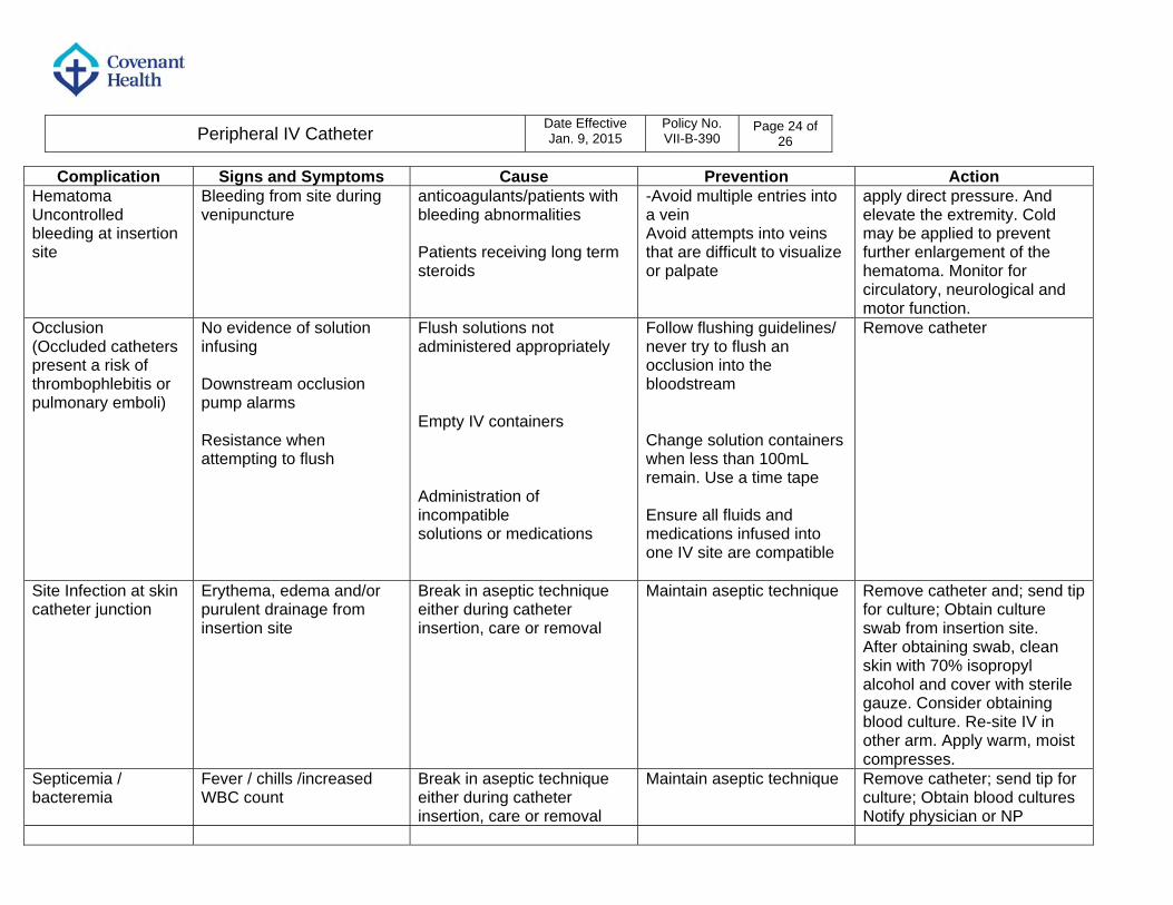
Complication Signs and Symptoms Cause Prevention Action
Hematoma Uncontrolled bleeding at insertion site
Bleeding from site during venipuncture
anticoagulants/patients with bleeding abnormalities Patients receiving long term steroids
-Avoid multiple entries into a vein Avoid attempts into veins that are difficult to visualize or palpate
apply direct pressure. And elevate the extremity. Cold may be applied to prevent further enlargement of the hematoma. Monitor for circulatory, neurological and motor function.
Occlusion (Occluded catheters present a risk of thrombophlebitis or pulmonary emboli)
No evidence of solution infusing Downstream occlusion pump alarms Resistance when attempting to flush
Flush solutions not administered appropriately Empty IV containers Administration of incompatible solutions or medications
Follow flushing guidelines/ never try to flush an occlusion into the bloodstream Change solution containers when less than 100mL remain. Use a time tape Ensure all fluids and medications infused into one IV site are compatible
Remove catheter
Site Infection at skin catheter junction
Erythema, edema and/or purulent drainage from insertion site
Break in aseptic technique either during catheter insertion, care or removal
Maintain aseptic technique Remove catheter and; send tip for culture; Obtain culture swab from insertion site. After obtaining swab, clean skin with 70% isopropyl alcohol and cover with sterile gauze. Consider obtaining blood culture. Re-site IV in other arm. Apply warm, moist compresses.
Septicemia / bacteremia
Fever / chills /increased WBC count
Break in aseptic technique either during catheter insertion, care or removal
Maintain aseptic technique Remove catheter; send tip for culture; Obtain blood cultures Notify physician or NP

Peripheral IV Catheter Date Effective Jan. 9, 2015
Policy No. VII-B-390
Page 25 of 26
Complication Signs and Symptoms Cause Prevention Action
Infiltration (leakage of fluid or non-vesicant medication into surrounding tissue) Note: Infiltration may cause significant morbidity, including skin necrosis, finger stiffness, and nerve irritation, and neuropathy and compartment syndrome.
See attachment – BD DecisIV™ Educational Brochure – Complications of Peripheral IV Access for pictures of Infiltration Scale Infiltration Scale 0 = No symptoms 1 = Skin blanched; edema <1 inch in any direction; cool to touch; with or without pain 2 = Skin blanched; edema 1-6 inches in any direction; with or without pain 3 = Skin blanched, translucent; Gross edema > 6 inches in any direction; cool to touch; mild-moderate pain; possible numbness 4 = skin blanched, translucent; skin tight, leaking; skin discoloured, bruised, swollen; gross edema > 6 inches in any direction; Dipping pitting tissue edema; circulatory impairment Moderate to severe pain; infiltration of any amount of
Catheter dislodgement caused by joint movement when catheter placed in area of flexion Previous IV sites distal to the current site Inflammation resulting from irritating solutions
-Avoid areas of flexion or use arm board to protect site Note: arm boards should be well padded and applied so that they will not cause constriction or pressure areas. -place smallest gauge and shortest length catheter to accommodate infusion -Avoid subsequent sites proximal to previous sites Infusion of hypertonic or hypotonic solutions/infusion of solution with pH less than 5 or greater than 9 or if infusate is greater than 600 mOsm/L. Stabilize the catheter to minimize in/out movement
Remove catheter immediately when signs and symptoms occur. To determine if the catheter is in the vein, apply pressure to the vein 2 inches above the insertion site, if the catheter is in the vein, the infusion will stop or slow. If the infusion continues despite the venous obstruction and infiltration has occurred. Monitor site closely for evidence of infiltration (Watch for blanching, stretched skin, firm tissues and coolness. Compare one arm to the other, watch for dependant edema)

Peripheral IV Catheter Date Effective Jan. 9, 2015
Policy No. VII-B-390
Page 26 of 26
Complication Signs and Symptoms Cause Prevention Action
blood product, irritant or vesicant.
Extravasations Is the inadvertent administration of a vesicant solution or medication into the surrounding tissue. See list of irritant/vesicant medications. – attached A vesicant is a solution or medication that can cause blistering sloughing of tissues and tissue necrosis when extravasation occurs. Irritant is a medication that may cause itching, phlebitis, or reaction along the vessel or at the injection site.
-Catheter dislodgement caused by joint movement when catheter placed in area of flexion -previous IV sites distal to the current site Previous IV sites distal to the current site Consider using a central venous catheter for infusion of vesicants
Complete assessment of the patient, the IV site, the involved extremity, and the infusion system at regular intervals during the infusion of vesicant medications The nurse must know if the patient has a history of multiple venipunctures, where they were located and how long ago the sites were used. Vesicants may seep into the tissue at previous vein entry sites Secure the catheter properly to prevent an in and out motion, which can enlarge the vein entry site and cause the vesicant to seep into the interstitial tissues, resulting in an extravasations. Avoid digits, hands, wrists and areas of flexion because of the close network of tendons and nerves that would be destroyed if extravasations occur.
Stop infusion. Call physician/NP. Consider consulting plastic surgery Extravasation/Infiltration and Care of the Patient for immediate nursing care.