Coronary Functional Abnormalities in
12
This article appeared in a journal published by Elsevier. The attached copy is furnished to the author for internal non-commercial research and education use, including for instruction at the author's institution and sharing with colleagues. Other uses, including reproduction and distribution, or selling or licensing copies, or posting to personal, institutional or third party websites are prohibited. In most cases authors are permitted to post their version of the article (e.g. in Word or Tex form) to their personal website or institutional repository. Authors requiring further information regarding Elsevier's archiving and manuscript policies are encouraged to visit: http://www.elsevier.com/authorsrights
Transcript of Coronary Functional Abnormalities in

This article appeared in a journal published by Elsevier. The attachedcopy is furnished to the author for internal non-commercial research
and education use, including for instruction at the author'sinstitution and sharing with colleagues.
Other uses, including reproduction and distribution, or selling orlicensing copies, or posting to personal, institutional or third party
websites are prohibited.
In most cases authors are permitted to post their version of thearticle (e.g. in Word or Tex form) to their personal website orinstitutional repository. Authors requiring further information
regarding Elsevier's archiving and manuscript policies areencouraged to visit:
http://www.elsevier.com/authorsrights
Listen to this manuscript’s
audio summary by
Editor-in-Chief
Dr. Valentin Fuster on
JACC.org.
J O U R N A L O F T H E A M E R I C A N C O L L E G E O F C A R D I O L O G Y V O L . 7 4 , N O . 1 9 , 2 0 1 9
ª 2 0 1 9 B Y T H E A M E R I C A N C O L L E G E O F C A R D I O L O G Y F O U N D A T I O N
P U B L I S H E D B Y E L S E V I E R
Author's Personal Copy
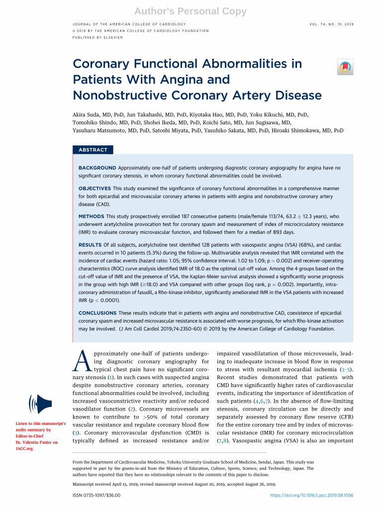
Coronary Functional Abnormalities inPatients With Angina andNonobstructive Coronary Artery Disease
Akira Suda, MD, PHD, Jun Takahashi, MD, PHD, Kiyotaka Hao, MD, PHD, Yoku Kikuchi, MD, PHD,Tomohiko Shindo, MD, PHD, Shohei Ikeda, MD, PHD, Koichi Sato, MD, Jun Sugisawa, MD,Yasuharu Matsumoto, MD, PHD, Satoshi Miyata, PHD, Yasuhiko Sakata, MD, PHD, Hiroaki Shimokawa, MD, PHDABSTRACT
ISS
Fro
su
au
Ma
BACKGROUND Approximately one-half of patients undergoing diagnostic coronary angiography for angina have no
significant coronary stenosis, in whom coronary functional abnormalities could be involved.
OBJECTIVES This study examined the significance of coronary functional abnormalities in a comprehensive manner
for both epicardial and microvascular coronary arteries in patients with angina and nonobstructive coronary artery
disease (CAD).
METHODS This study prospectively enrolled 187 consecutive patients (male/female 113/74, 63.2 � 12.3 years), who
underwent acetylcholine provocation test for coronary spasm and measurement of index of microcirculatory resistance
(IMR) to evaluate coronary microvascular function, and followed them for a median of 893 days.
RESULTS Of all subjects, acetylcholine test identified 128 patients with vasospastic angina (VSA) (68%), and cardiac
events occurred in 10 patients (5.3%) during the follow-up. Multivariable analysis revealed that IMR correlated with the
incidence of cardiac events (hazard ratio: 1.05; 95% confidence interval: 1.02 to 1.09; p ¼ 0.002) and receiver-operating
characteristics (ROC) curve analysis identified IMR of 18.0 as the optimal cut-off value. Among the 4 groups based on the
cut-off value of IMR and the presence of VSA, the Kaplan-Meier survival analysis showed a significantly worse prognosis
in the group with high IMR ($18.0) and VSA compared with other groups (log rank, p ¼ 0.002). Importantly, intra-
coronary administration of fasudil, a Rho-kinase inhibitor, significantly ameliorated IMR in the VSA patients with increased
IMR (p < 0.0001).
CONCLUSIONS These results indicate that in patients with angina and nonobstructive CAD, coexistence of epicardial
coronary spasm and increased microvascular resistance is associated with worse prognosis, for which Rho-kinase activation
may be involved. (J Am Coll Cardiol 2019;74:2350–60) © 2019 by the American College of Cardiology Foundation.
A pproximately one-half of patients undergo-ing diagnostic coronary angiography fortypical chest pain have no significant coro-
nary stenosis (1). In such cases with suspected anginadespite nonobstructive coronary arteries, coronaryfunctional abnormalities could be involved, includingincreased vasoconstrictive reactivity and/or reducedvasodilator function (2). Coronary microvessels areknown to contribute to >50% of total coronaryvascular resistance and regulate coronary blood flow(3). Coronary microvascular dysfunction (CMD) istypically defined as increased resistance and/or
N 0735-1097/$36.00
m the Department of Cardiovascular Medicine, Tohoku University Gradua
pported in part by the grants-in-aid from the Ministry of Education, Cu
thors have reported that they have no relationships relevant to the conte
nuscript received April 15, 2019; revised manuscript received August 20,
impaired vasodilatation of those microvessels, lead-ing to inadequate increase in blood flow in responseto stress with resultant myocardial ischemia (3–5).Recent studies demonstrated that patients withCMD have significantly higher rates of cardiovascularevents, indicating the importance of identification ofsuch patients (4,6,7). In the absence of flow-limitingstenosis, coronary circulation can be directly andseparately assessed by coronary flow reserve (CFR)for the entire coronary tree and by index of microvas-cular resistance (IMR) for coronary microcirculation(7,8). Vasospastic angina (VSA) is also an important
https://doi.org/10.1016/j.jacc.2019.08.1056
te School of Medicine, Sendai, Japan. This study was
lture, Sports, Science, and Technology, Japan. The
nts of this paper to disclose.
2019, accepted August 26, 2019.
AB BR E V I A T I O N S
AND ACRONYM S
ACh = acetylcholine
CAD = coronary artery disease
J A C C V O L . 7 4 , N O . 1 9 , 2 0 1 9 Suda et al.N O V E M B E R 1 2 , 2 0 1 9 : 2 3 5 0 – 6 0 Coronary Functional Abnormalities in Nonobstructive CAD Patients
2351
Author's Personal Copy
functional cardiac disorder caused by epicardial coro-nary spasm, which is caused by enhanced coronaryvasoconstricting responses (2). We have previouslydemonstrated that Rho-kinase plays a central role inthe pathogenesis of coronary spasm (2,9,10).
SEE PAGE 2361CFR = coronary flow reserve
CMD = coronary microvascular
dysfunction
IMR = index of microcirculatory
resistance
MVS = microvascular spasm
VSA = vasospastic angina
Although the importance of each component ofcoronary functional abnormalities (VSA and CMD) hasrecently emerged, comprehensive evaluation of theabnormalities in the same population remains to beexamined. In the present study, we thus aimed toevaluate the effect of epicardial coronary arteryspasm and/or abnormal microvascular resistance onlong-term prognosis and to determine if the Rho-kinase pathway is implicated in the pathogenesis ofthe functional coronary abnormalities.
METHODS
The present study was conducted following theethical principles in the Declaration of Helsinki, andthe protocol was approved by the Ethics Committeesof Tohoku University (No.2016-1-643). All patientsprovided written informed consent before studyentry.
STUDY POPULATION. The inclusion criteria of thepresent study included angina-like chest pain, non-obstructive coronary arteries, and successful perfor-mance of both coronary artery functional testing(e.g., measurement of IMR) and coronary vaso-reactivity testing (e.g., provocative testing for coro-nary spasm) to identify the origin of their chest pain(Figure 1). From November 2014 to July 2017, a total of699 patients underwent elective diagnostic coronaryangiography for evaluation of chest pain and/orelectrocardiographic abnormalities at our TohokuUniversity Hospital. Of those, 302 had no significantcoronary stenosis (luminal narrowing<70% and/orfractional flow reserve [FFR] >0.8) of the major cor-onary arteries on control angiography. Then, 243 pa-tients underwent acetylcholine (ACh) provocationtest to assess coronary vasoconstricting responsesand were also evaluated for their coronary micro-vascular vasodilatory function. We excluded patientswith proven cardiomyopathy, significant valvulardiseases (e.g., aortic stenosis), previous coronarystent implantation, relative contraindication forprovocation test (e.g., bronchial asthma), renal fail-ure, poor general condition, and unsuccessful pro-cedures during physiological measurement and/orACh provocation test. Finally, 187 consecutive pa-tients who fulfilled the inclusion criteria wereincluded in the present study (Figure 1).
ACH PROVOCATION TEST. The ACh provo-cation test was performed as previouslydescribed (9,10). Based on guidelines fromthe Japanese Circulation Society (11), thepositive provocation test for epicardial coro-nary spasm was defined as the developmentof >90% stenosis accompanied by chest painand ischemic electrocardiographic changes.In the present study, we defined microvas-cular spasm (MVS) based on the diagnosticcriteria proposed by the COVADIS (CoronaryVasomotor Disorders International Study)
group (5).CORONARY PHYSIOLOGICALMEASUREMENTS. After AChprovocation testing, we administered ISDN intra-coronarily to achieve dilatation of epicardial coronaryarteries. We then performed coronary physiologicalmeasurements for FFR, CFR, and IMR in the leftanterior descending coronary artery (LAD) duringhyperemia induced by intravenous infusion ofadenosine, as previously described (7,8). Further-more, to evaluate the involvement of Rho-kinase,after the first measurement of CFR and IMR, weadministered intracoronary fasudil (30 mg), a selec-tive Rho-kinase inhibitor (2,12), and performed thesecond measurement of IMR. We calculated %changes in IMR before and after intracoronaryfasudil as follows: (fasudil IMR � hyperemic IMR)/hyperemic IMR.
CLINICAL OUTCOME AND PATIENT FOLLOW-UP. Wedefined major adverse cardiac events (MACE) as thecomposite of cardiac death, nonfatal myocardialinfarction, and hospitalization due to unstableangina. We only counted the number of patients withthe first occurrence of an event in the MACE duringthe follow-up period. Long-term follow-up was per-formed by using a questionnaire that was sent topatients and primary physicians, in addition to theinformation available on the medical records or tele-phone surveys. The median duration of follow-upwas 893 days (interquartile range [IQR]: 637to 1,136 days).
STATISTICAL ANALYSIS. Continuous variables arepresented as mean � SD or median (interquartilerange), and categorical variables as number (%).Group comparisons for continuous variables wereperformed by the Kruskal-Wallis test for multiplegroups, and the Mann-Whitney U-test for 2 groups.The chi-square test was used for comparisons amongcategorical variables. Survival rate from cardiacevents was analyzed by the Kaplan-Meier method,and comparison between groups was performed bylog-rank tests. Given the sample size of the original

FIGURE 1 Study Flow Chart
Diagnostic cardiac catheterization forevaluation of chest pain and/or
ECG abnormalities(n = 699, November 2014-July 2017)
Acute coronary syndrome (n = 155)Significant stenosis (n = 242)
Cardiomyopathy / Valvular disease (n = 14)Previous stent implantation (n = 7)Renal failure / poor general condition (n = 59)Incomplete procedure (n = 35)
Non-VSA, (–) Epicardialspasm (n = 59)
VSA, (+) Epicardialspasm (n = 128)
Patients suspected stable angina withoutsignificant coronary stenosis (n = 302)
Patients who underwent both of AChprovocation and IMR measurement (n = 187)
IMR <18.0
Group 1 (G1)IMR <18.0 without VSA
(n = 45)
Group 2 (G2)IMR ≥18.0 without VSA
(n = 14)
Group 3 (G3)IMR <18.0 with VSA
(n = 67)
Group 4 (G4)IMR ≥18.0 with VSA
(n = 61)
IMR ≥18.0 IMR <18.0 IMR ≥18.0
ACh ¼ acetylcholine; ECG ¼ electrocardiogram; IMR ¼ index of microcirculatory resistance; VSA ¼ vasospastic angina.
Suda et al. J A C C V O L . 7 4 , N O . 1 9 , 2 0 1 9
Coronary Functional Abnormalities in Nonobstructive CAD Patients N O V E M B E R 1 2 , 2 0 1 9 : 2 3 5 0 – 6 0
2352
Author's Personal Copy
data and the significance level ¼ 0.05, the empiricalpower of the log-rank test was calculated by themethod proposed by Freedman (13). Cox proportionalhazards regression was used to calculate hazard ratios(HRs) and 95% CIs to compare between group differ-ences. The proportional hazards assumption of Coxregression was confirmed by the Schoenfeld residualstest implemented by the cox.zph command in R (14).Logistic regression was used to compute odds ratios(ORs) and 95% CI for the occurrence of the events.The prognostic significance of each variable wastested by univariable and multivariable logisticregression analyses. To select an optimal subset of
covariates for the multivariable logistic model, thebackward elimination procedure of variable selectionwith Bayesian information criterion was utilized. Theoccurrence of cardiovascular events was predicted bylogistic regression models with the presence ofVSA and/or high IMR relative to the reference model,and their performances were compared. The refer-ence model was composed of age, current smoking,and dyslipidemia. Smoking and dyslipidemia wereselected by stepwise variable selection from thelogistic model using all candidate covariates. Age wasadded to the reference because it was the indepen-dent prognostic factor of MACE in the multivariable

TABLE 1 Baseline Clinical Characteristics of Patients
Overall(n ¼ 187)
Non-VSA(n ¼ 59)
VSA(n ¼ 128) p Value
Age, yrs 63.2 � 12.3 61.9 � 14.7 63.8 � 11.0 0.35
Male 113 (60) 38 (64) 75 (59) 0.45
Hypertension 100 (53) 33 (56) 67 (52) 0.64
Dyslipidemia 66 (35) 22 (37) 44 (34) 0.70
Diabetes mellitus 52 (28) 15 (25) 37 (29) 0.62
Current smoking 52 (28) 17 (29) 35 (27) 0.84
Family history of CAD 31 (17) 13 (22) 18 (14) 0.18
Previous MI 10 (5) 2 (3) 8 (6) 0.40
Rest angina 104 (56) 31 (54) 83 (65) 0.17
Effort angina 42 (22) 14 (24) 28 (22) 0.78
Rest and effort angina 11 (6) 2 (3) 9 (7) 0.30
eGFR, ml/min/1.73 m2 73.1 � 21.3 74.6 � 27.0 72.4 � 18.2 0.51
hs-CRP, mg/ml 0.05 (0.02–0.12) 0.07 (0.02–0.32) 0.05 (0.02–0.10) 0.17
hs-TropT, ng/ml 0.007(0.004–0.011)
0.007(0.004–0.013)
0.007(0.005–0.011)
0.56
BNP, pg/ml 22.7 (9.4–46.3) 28.4 (13.8–50.3) 20.7 (8.7–42.4) 0.15
LVEF, % 65.9 � 10.5 65.1 � 10.7 66.3 � 10.4 0.50
E/eʹ 10.3 � 4.3 10.6 � 4.3 10.2 � 4.3 0.53
MVS 22 (12) 22 (37) 0 (0) <0.0001
Physiological parameters
FFR 0.9 (0.87–0.93) 0.90 (0.86–0.94) 0.90 (0.87–0.93) 0.85
CFR 2.54 (1.81–3.43) 2.66 (1.85–3.64) 2.51 (1.72–3.35) 0.34
IMR 16.2 (11.8–24.2) 14.7 (10.7–17.8) 17.5 (12.0–25.3) 0.02
Baseline Tmn, s 0.71 (0.45–1.01) 0.63 (0.40–0.96) 0.73 (0.46–1.02) 0.21
Hyperemic Tmn, s 0.26 (0.19–0.38) 0.24 (0.18–0.33) 0.27 (0.20–0.41) 0.03
Values are mean � SD, n (%), or median (interquartile range).
BNP ¼ B-type natriuretic peptide; CAD ¼ coronary artery disease; CFR ¼ coronary flow reserve; E/eʹ ¼ earlydiastolic mitral flow velocity/tissue Doppler imaging velocity; eGFR ¼ estimate glomerular filtration rate;FFR ¼ fractional flow reserve; hs-CRP ¼ high sensitivity C-reactive protein; hs-TropT ¼ high sensitivity troponin T;IMR ¼ index of microcirculatory resistance; LVEF ¼ left ventricular ejection fraction; MI ¼ myocardial infarction;MVS ¼ microvascular spasm; Tmn ¼ mean transit time.
J A C C V O L . 7 4 , N O . 1 9 , 2 0 1 9 Suda et al.N O V E M B E R 1 2 , 2 0 1 9 : 2 3 5 0 – 6 0 Coronary Functional Abnormalities in Nonobstructive CAD Patients
2353
Author's Personal Copy
Cox regression. C-statistics, which equal the areaunder the ROC curve, was used to summarize theperformance of the predicted probability of the out-comes for discrimination. The improvement of fit ofthe models relative to the reference model was eval-uated by likelihood ratio tests for the analysis ofdeviance table. To evaluate the improvement of thematured models relative to the reference model, in-tegrated discrimination improvement (IDI), andcontinuous net reclassification improvement (NRI)with the total observations, the observations with theevents and those without the event were utilized. Tocalculate NRI, censored participants were handled asno events (15). A p value <0.05 was considered to bestatistically significant. Further information is avail-able in the Online Methods.
RESULTS
CLINICAL PATIENT CHARACTERISTICS. The flowchart of the present study is shown in Figure 1. Wefinally analyzed 187 consecutive patients (113 men, 74women; age 63.2 � 12.3 years) with angina-like chestpain and nonobstructive CAD in whom we were ableto complete both ACh provocation test for coronaryspasm and physiological measurements of coronarymicrovascular functions with IMR and CFR. Clinicalcharacteristics of the patients are summarized inTable 1. All patients had a stable condition, with morethan one-half (56%) having symptoms at rest. Amongthe 187 patients, 128 (68.4%) were diagnosed ashaving VSA, and 22 patients (12.0%) had MVS andwere categorized into the non-VSA group (OnlineTable 1). The median IMR value was significantlyhigher in the VSA than non-VSA group, whereas CFRvalues were comparable between the 2 groups(Figure 2). The distributions of patients according toIMR and CFR by each type of coexisting coronaryreactivity abnormality are shown in Online Figure 1.Importantly, we found a highly negative correlationbetween IMR and CFR values in VSA patients, but notin non-VSA patients (Figure 3).
CLINICAL OUTCOMES AND PROGNOSTIC PREDICTORS.
During the median follow-up period of 893 days(IQR: 637 to 1,136 days), there were 10 MACE in overallcohorts, including cardiovascular death (n ¼ 1) andhospitalization for unstable angina (n ¼ 9). Multi-variable Cox proportional hazard analysis showedthat high IMR significantly correlated with MACE inpatients with chest pain and nonobstructive CAD(Table 2). Based on ROC curve analysis, the optimalIMR cutoff value for developing MACE was 18.0, andthe area under the ROC curve was 0.76 (OnlineFigure 2). With this value, the sensitivity and
specificity for predicting MACE were 90.0% and63.4%, respectively, while negative predictive valuewas 99.1%. In multivariable logistic regression modelfor the presence of high IMR ($18.0), proven VSA wasthe strongest correlated predictor (Table 3). Impor-tantly, there were substantial overlaps of coronaryfunctional abnormality in various combinationsamong VSA, low CFR (CFR <2.0), and high IMR(IMR $18.0) (Figure 4).
PERFORMANCE METRICS IN RISK PREDICTION
MODELS. Furthermore, as shown in Figure 1, wedivided the patients into the following 4 groupsaccording to the cut-off value of IMR (>18.0) and thepresence or absence of VSA: G1, IMR <18.0 withoutVSA (n ¼ 45); G2, IMR $18.0 without VSA (n ¼ 14); G3,IMR <18.0 with VSA (n ¼ 67); and G4, IMR $18.0 withVSA (n ¼ 61). Patient characteristics of the 4 groupsare shown in Online Table 2. Although there was nodifference in demographic profiles except maleprevalence among the 4 groups, CFR was significantlylower in G4 (VSA and high IMR). The Kaplan-Meiersurvival analysis showed that the patients of G4 had

FIGURE 2 Comparison of Coronary Physiological Parameters Between Non-VSA and VSA Groups
B
Non-VSA0
2
4
6
8
10
VSA
CFR
P = 0.34
A
Non-VSA0
20
40
60
80
VSA
IMR
P = 0.02
IMR values at hyperemic state were significantly higher in the VSA group than in the non-VSA group (A), whereas CFR values at hyperemic
state were comparable between the 2 groups (B). Results are expressed as box-and-whisker plots; the central box covers the interquartile
range, with the median indicated by the line within the box. The whiskers extend to the most extreme values within 1.5 interquartile ranges.
More extreme values are plotted individually. CFR ¼ coronary flow reserve; other abbreviations as in Figure 1.
Suda et al. J A C C V O L . 7 4 , N O . 1 9 , 2 0 1 9
Coronary Functional Abnormalities in Nonobstructive CAD Patients N O V E M B E R 1 2 , 2 0 1 9 : 2 3 5 0 – 6 0
2354
Author's Personal Copy
a significantly worse outcome compared with allother remaining patients (log-rank, p ¼ 0.002)(Figure 5). Empirical power of the log-rank test for theoccurrence of MACE in comparison with G1 and G4was 0.87. Further logistic regression analyses on
FIGURE 3 Correlation Between CFR and IMR Values in the Non-VSA
00
2
4
6
R = 0.02P = 0.89
8
A
20IMR
CFR
40 60 80
Non-VSA (n = 59)
(A) There was no significant correlation between CFR and IMR values in
between the 2 values in the VSA group. Abbreviations as in Figures 1 an
performance metrics in risk prediction models wereevaluated (Table 4). C-statistics for the prediction ofthe occurrence of MACE significantly increased (0.75[95% confidence interval (CI): 0.6 to 0.89] to 0.90[95% CI: 0.83 to 0.97]; p ¼ 0.045), when IMR and the
and VSA Groups
00
2
4
6
R = –0.30P < 0.0001
8
B
20IMR
CFR
40 60 80
VSA (n = 128)
the non-VSA group. (B) There was a significant negative correlation
d 2.
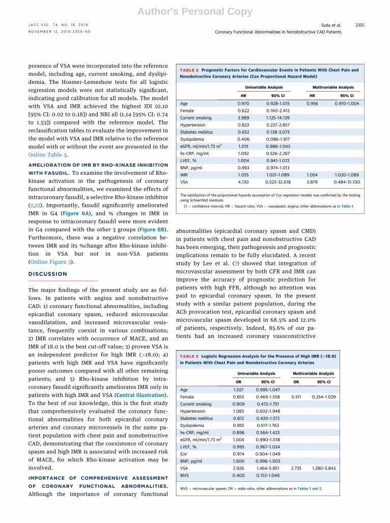
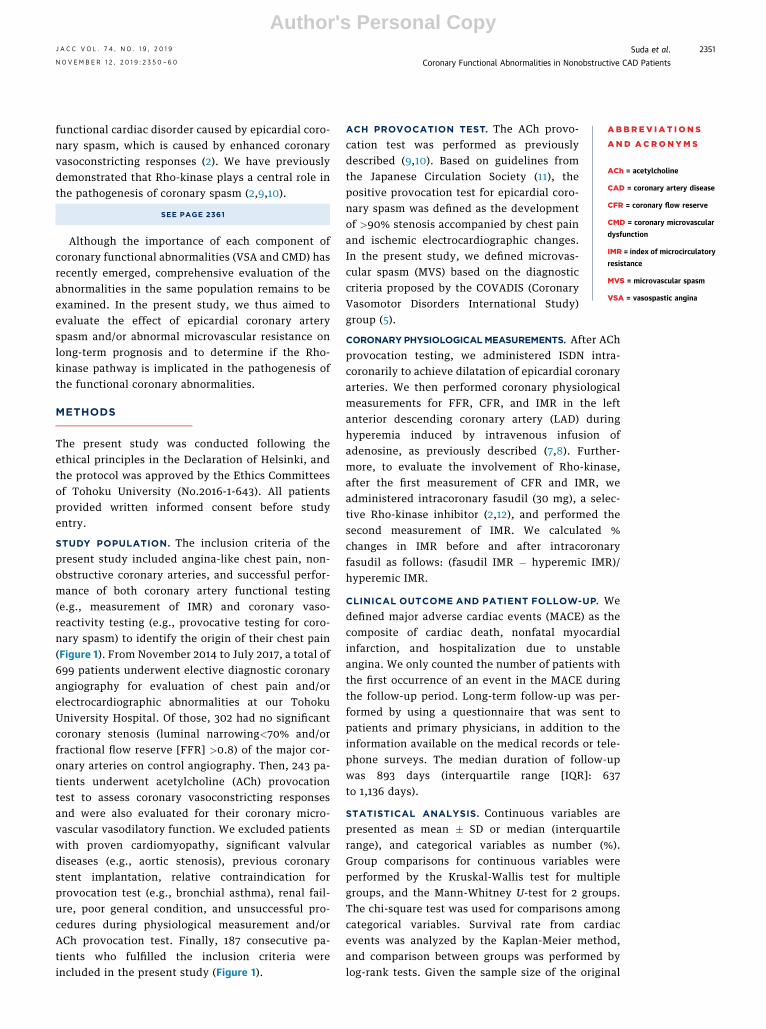
TABLE 2 Prognostic Factors for Cardiovascular Events in Patients With Chest Pain and
Nonobstructive Coronary Arteries (Cox Proportional Hazard Model)
Univariable Analysis Multivariable Analysis
HR 95% CI HR 95% CI
Age 0.970 0.928–1.015 0.956 0.910–1.004
Female 0.622 0.160–2.412
Current smoking 3.989 1.125–14.139
Hypertension 0.823 0.237–2.857
Diabetes mellitus 0.652 0.138–3.073
Dyslipidemia 0.406 0.086–1.917
eGFR, ml/min/1.73 m2 1.015 0.986–1.043
hs-CRP, mg/ml 1.092 0.526–2.267
LVEF, % 1.004 0.941–1.072
BNP, pg/ml 0.993 0.974–1.013
IMR 1.055 1.021–1.089 1.054 1.020–1.089
VSA 4.130 0.523–32.618 3.879 0.484–31.100
The satisfaction of the proportional hazards assumption of Cox regression models was confirmed by the testingusing Schoenfeld residuals.
CI ¼ confidence interval; HR ¼ hazard ratio; VSA ¼ vasospastic angina; other abbreviations as in Table 1.
TABLE 3 Logistic Regression Analysis for the Presence of High IMR ($18.0)
in Patients With Chest Pain and Nonobstructive Coronary Arteries
Univariable Analysis Multivariable Analysis
OR 95% CI OR 95% CI
Age 1.021 0.995–1.047
Female 0.855 0.469–1.558 0.511 0.254–1.029
Current smoking 0.909 0.472–1.751
Hypertension 1.083 0.602–1.948
Diabetes mellitus 0.812 0.420–1.572
Dyslipidemia 0.955 0.517–1.763
hs-CRP, mg/ml 0.896 0.564–1.423
eGFR, ml/min/1.73 m2 1.004 0.990–1.018
LVEF, % 0.995 0.967–1.024
E/eʹ 0.974 0.904–1.049
BNP, pg/ml 1.000 0.996–1.003
VSA 2.926 1.464–5.851 2.735 1.280–5.843
MVS 0.400 0.153–1.049
MVS ¼ microvascular spasm; OR ¼ odds ratio; other abbreviations as in Tables 1 and 2.
J A C C V O L . 7 4 , N O . 1 9 , 2 0 1 9 Suda et al.N O V E M B E R 1 2 , 2 0 1 9 : 2 3 5 0 – 6 0 Coronary Functional Abnormalities in Nonobstructive CAD Patients
2355
Author's Personal Copy
presence of VSA were incorporated into the referencemodel, including age, current smoking, and dyslipi-demia. The Hosmer-Lemeshow tests for all logisticregression models were not statistically significant,indicating good calibration for all models. The modelwith VSA and IMR achieved the highest IDI (0.10[95% CI: 0.02 to 0.18]) and NRI all (1.14 [95% CI: 0.74to 1.53]) compared with the reference model. Thereclassification tables to evaluate the improvement inthe model with VSA and IMR relative to the referencemodel with or without the event are presented in theOnline Table 3.
AMELIORATION OF IMR BY RHO-KINASE INHIBITION
WITH FASUDIL. To examine the involvement of Rho-kinase activation in the pathogenesis of coronaryfunctional abnormalities, we examined the effects ofintracoronary fasudil, a selective Rho-kinase inhibitor(2,12). Importantly, fasudil significantly amelioratedIMR in G4 (Figure 6A), and % changes in IMR inresponse to intracoronary fasudil were more evidentin G4 compared with the other 3 groups (Figure 6B).Furthermore, there was a negative correlation be-tween IMR and its %change after Rho-kinase inhibi-tion in VSA but not in non-VSA patients(Online Figure 3).
DISCUSSION
The major findings of the present study are as fol-lows. In patients with angina and nonobstructiveCAD: 1) coronary functional abnormalities, includingepicardial coronary spasm, reduced microvascularvasodilatation, and increased microvascular resis-tance, frequently coexist in various combinations;2) IMR correlates with occurrence of MACE, and anIMR of 18.0 is the best cut-off value; 3) proven VSA isan independent predictor for high IMR (>18.0); 4)patients with high IMR and VSA have significantlypoorer outcomes compared with all other remainingpatients; and 5) Rho-kinase inhibition by intra-coronary fasudil significantly ameliorates IMR only inpatients with high IMR and VSA (Central Illustration).To the best of our knowledge, this is the first studythat comprehensively evaluated the coronary func-tional abnormalities for both epicardial coronaryarteries and coronary microvessels in the same pa-tient population with chest pain and nonobstructiveCAD, demonstrating that the coexistence of coronaryspasm and high IMR is associated with increased riskof MACE, for which Rho-kinase activation may beinvolved.
IMPORTANCE OF COMPREHENSIVE ASSESSMENT
OF CORONARY FUNCTIONAL ABNORMALITIES.
Although the importance of coronary functional
abnormalities (epicardial coronary spasm and CMD)in patients with chest pain and nonobstructive CADhas been emerging, their pathogenesis and prognosticimplications remain to be fully elucidated. A recentstudy by Lee et al. (7) showed that integration ofmicrovascular assessment by both CFR and IMR canimprove the accuracy of prognostic prediction forpatients with high FFR, although no attention waspaid to epicardial coronary spasm. In the presentstudy with a similar patient population, during theACh provocation test, epicardial coronary spasm andmicrovascular spasm developed in 68.5% and 12.0%of patients, respectively. Indeed, 85.6% of our pa-tients had an increased coronary vasoconstrictive
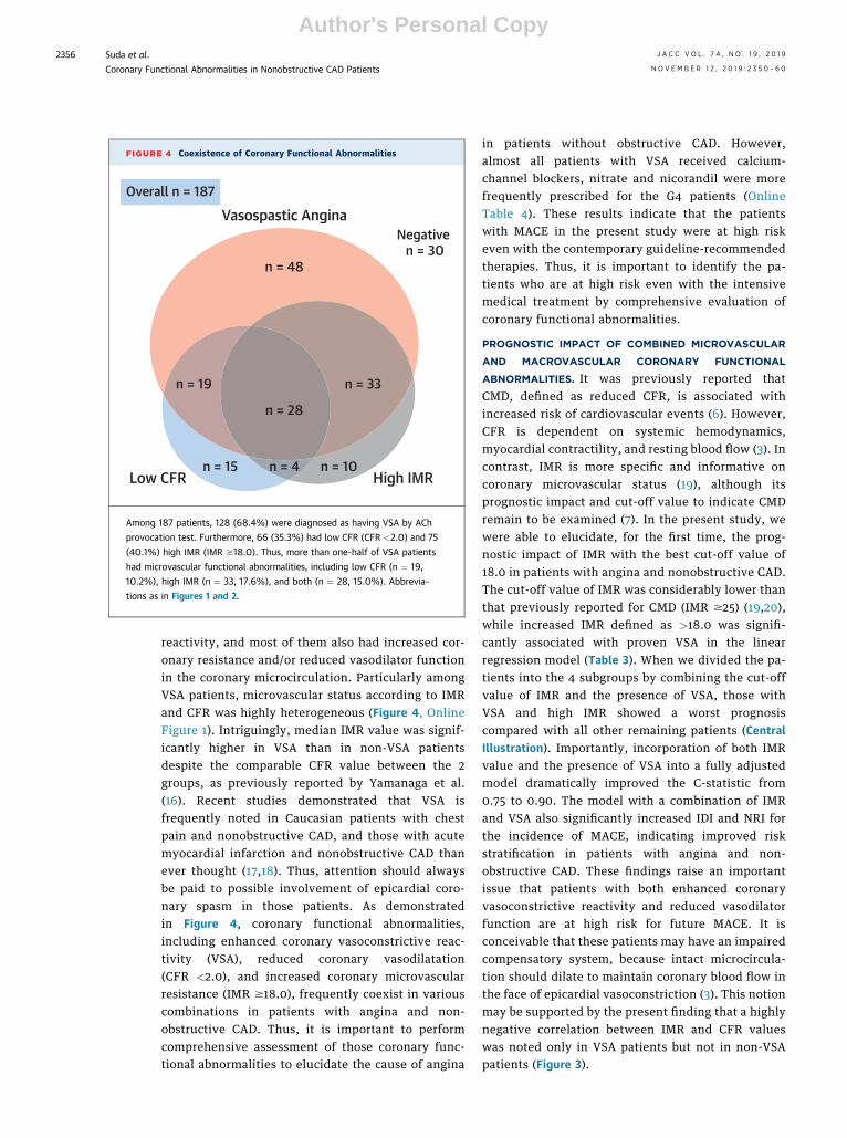
FIGURE 4 Coexistence of Coronary Functional Abnormalities
n = 19
n = 15
n = 28
n = 4 n = 10High IMRLow CFR
n = 33
n = 48
Vasospastic AnginaNegative
n = 30
Overall n = 187
Among 187 patients, 128 (68.4%) were diagnosed as having VSA by ACh
provocation test. Furthermore, 66 (35.3%) had low CFR (CFR <2.0) and 75
(40.1%) high IMR (IMR $18.0). Thus, more than one-half of VSA patients
had microvascular functional abnormalities, including low CFR (n ¼ 19,
10.2%), high IMR (n ¼ 33, 17.6%), and both (n ¼ 28, 15.0%). Abbrevia-
tions as in Figures 1 and 2.
Suda et al. J A C C V O L . 7 4 , N O . 1 9 , 2 0 1 9
Coronary Functional Abnormalities in Nonobstructive CAD Patients N O V E M B E R 1 2 , 2 0 1 9 : 2 3 5 0 – 6 0
2356
Author's Personal Copy
reactivity, and most of them also had increased cor-onary resistance and/or reduced vasodilator functionin the coronary microcirculation. Particularly amongVSA patients, microvascular status according to IMRand CFR was highly heterogeneous (Figure 4, OnlineFigure 1). Intriguingly, median IMR value was signif-icantly higher in VSA than in non-VSA patientsdespite the comparable CFR value between the 2groups, as previously reported by Yamanaga et al.(16). Recent studies demonstrated that VSA isfrequently noted in Caucasian patients with chestpain and nonobstructive CAD, and those with acutemyocardial infarction and nonobstructive CAD thanever thought (17,18). Thus, attention should alwaysbe paid to possible involvement of epicardial coro-nary spasm in those patients. As demonstratedin Figure 4, coronary functional abnormalities,including enhanced coronary vasoconstrictive reac-tivity (VSA), reduced coronary vasodilatation(CFR <2.0), and increased coronary microvascularresistance (IMR $18.0), frequently coexist in variouscombinations in patients with angina and non-obstructive CAD. Thus, it is important to performcomprehensive assessment of those coronary func-tional abnormalities to elucidate the cause of angina
in patients without obstructive CAD. However,almost all patients with VSA received calcium-channel blockers, nitrate and nicorandil were morefrequently prescribed for the G4 patients (OnlineTable 4). These results indicate that the patientswith MACE in the present study were at high riskeven with the contemporary guideline-recommendedtherapies. Thus, it is important to identify the pa-tients who are at high risk even with the intensivemedical treatment by comprehensive evaluation ofcoronary functional abnormalities.
PROGNOSTIC IMPACT OF COMBINED MICROVASCULAR
AND MACROVASCULAR CORONARY FUNCTIONAL
ABNORMALITIES. It was previously reported thatCMD, defined as reduced CFR, is associated withincreased risk of cardiovascular events (6). However,CFR is dependent on systemic hemodynamics,myocardial contractility, and resting blood flow (3). Incontrast, IMR is more specific and informative oncoronary microvascular status (19), although itsprognostic impact and cut-off value to indicate CMDremain to be examined (7). In the present study, wewere able to elucidate, for the first time, the prog-nostic impact of IMR with the best cut-off value of18.0 in patients with angina and nonobstructive CAD.The cut-off value of IMR was considerably lower thanthat previously reported for CMD (IMR $25) (19,20),while increased IMR defined as >18.0 was signifi-cantly associated with proven VSA in the linearregression model (Table 3). When we divided the pa-tients into the 4 subgroups by combining the cut-offvalue of IMR and the presence of VSA, those withVSA and high IMR showed a worst prognosiscompared with all other remaining patients (CentralIllustration). Importantly, incorporation of both IMRvalue and the presence of VSA into a fully adjustedmodel dramatically improved the C-statistic from0.75 to 0.90. The model with a combination of IMRand VSA also significantly increased IDI and NRI forthe incidence of MACE, indicating improved riskstratification in patients with angina and non-obstructive CAD. These findings raise an importantissue that patients with both enhanced coronaryvasoconstrictive reactivity and reduced vasodilatorfunction are at high risk for future MACE. It isconceivable that these patients may have an impairedcompensatory system, because intact microcircula-tion should dilate to maintain coronary blood flow inthe face of epicardial vasoconstriction (3). This notionmay be supported by the present finding that a highlynegative correlation between IMR and CFR valueswas noted only in VSA patients but not in non-VSApatients (Figure 3).

TABLE 4 Performance of the Logistic Regression Models for Cardiovascular Events
ReferenceModel
ModelWith VSA
ModelWith IMR
Model WithVSA and IMR
Discrimination
C-statistics 0.75(0.60–0.89)
0.82(0.71–0.92)
0.90(0.85–0.96)
0.90(0.83–0.97)
p value Reference 0.025 0.018 0.045
Calibration
Hosmer-Lemeshow p value 0.788 0.698 0.910 0.975
AIC 78.409 77.235 67.768 68.510
Likelihood ratio p value Reference 0.075 0.075 0.075
Reclassification
IDI Reference 0.01(�0.03–0.05)
0.08(0.01–0.14)
0.10(0.02–0.18)
NRI (all) Reference 0.45(0.05–0.85)
1.07(0.67–1.47)
1.14(0.74–1.53)
NRI with event Reference 0.80(0.43–1.17)
0.80(0.43–1.17)
0.80(0.43–1.17)
NRI with no event Reference �0.35(�0.49 to �0.21)
0.27(0.13–0.41)
0.34(0.20–0.48)
Reference model includes age, current smoking, and dyslipidemia, which were selected from age, female sex,current smoking, hypertension, diabetes mellitus, dyslipidemia, eGFR, hs-CRP, LVEF, and BNP. Model with VSA:Reference model þ VSA. Model with IMR: Reference model þ IMR. All p values versus the reference model.
AIC ¼ Akaike information criterion; IDI ¼ integrated discrimination improvement; NRI ¼ net reclassificationimprovement; other abbreviations as in Tables 1 and 2.
FIGURE 5 Kaplan-Meier Curves for MACE by Patient Group According to IMR and VSA
Log rank test for overall comparisonP = 0.002
00
20
40
60
200Follow-Up (Days)
Cum
ulat
ive
Inci
denc
e of
MAC
E (%
)
400 600 800 1,000 1,200 1,400
45 43 42 36 25 19 8 113 13 13 12 12 10 3 167 67 67 54 36 29 10 160 59 58 50 40 28 14 2
No. at RiskNegative
VSA AloneIMR ≥18.0 Alone
VSA with IMR ≥18.0
Hazard ratio (95% CI) P value
NANA
NANA
0.036.23 (1.21–118.48)
Group 2Group 3Group 4
Group 1 Reference -
Group 1, IMR <18.0 without VSA (n ¼ 45); Group 2, IMR $18.0 without VSA (n ¼ 14); Group 3, IMR <18.0 with VSA (n ¼ 67); and Group 4,
IMR$18.0 with VSA (n ¼ 61). The long-term prognosis was significantly worse in Group 4 compared with the other 3 groups. CI ¼ confidence
interval; MACE ¼ major adverse cardiovascular events; other abbreviations as in Figure 1.
J A C C V O L . 7 4 , N O . 1 9 , 2 0 1 9 Suda et al.N O V E M B E R 1 2 , 2 0 1 9 : 2 3 5 0 – 6 0 Coronary Functional Abnormalities in Nonobstructive CAD Patients
2357
Author's Personal Copy
BENEFICIAL EFFECTS OF FASUDIL ON CORONARY
FUNCTIONAL ABNORMALITIES. In the presentstudy, a close correlation was noted between coro-nary spasm and impaired coronary vasodilator re-sponses in patients with chest pain andnonobstructive CAD, suggesting the presence of acommon underlying mechanism for the abnormal-ities. Importantly, we were able to demonstrate that aselective Rho-kinase inhibitor, fasudil, significantlyameliorated impaired microvascular resistance inpatients with increased IMR ($18.0) and proven VSAcompared with other 3 groups. We have previouslydemonstrated that Rho-kinase activation plays acentral role not only for epicardial coronary spasm(9,10), but also for coronary microvascular spasm (12).Furthermore, the present study demonstrates forthe first time that Rho-kinase activation is simulta-neously involved in epicardial coronary spasmand increased coronary microvascular resistance.Furthermore, a negative correlation was notedbetween IMR and its %change after Rho-kinase inhi-bition only in VSA patients, indicating that increasedIMR is associated with Rho-kinase activation in the
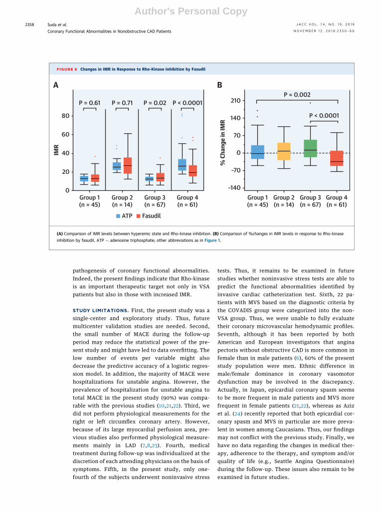
FIGURE 6 Changes in IMR in Response to Rho-Kinase Inhibition by Fasudil
Group 1(n = 45)
0
ATP Fasudil
20
40
–140Group 1(n = 45)
Group 2(n = 14)
Group 3(n = 67)
Group 4(n = 61)
–70
0
70
140
210P = 0.002
P < 0.0001
% C
hang
e in
IMR
60
IMR
80
P = 0.61 P = 0.71 P = 0.02 P < 0.0001
A B
Group 2(n = 14)
Group 3(n = 67)
Group 4(n = 61)
(A) Comparison of IMR levels between hyperemic state and Rho-kinase inhibition. (B) Comparison of %changes in IMR levels in response to Rho-kinase
inhibition by fasudil. ATP ¼ adenosine triphosphate; other abbreviations as in Figure 1.
Suda et al. J A C C V O L . 7 4 , N O . 1 9 , 2 0 1 9
Coronary Functional Abnormalities in Nonobstructive CAD Patients N O V E M B E R 1 2 , 2 0 1 9 : 2 3 5 0 – 6 0
2358
Author's Personal Copy
pathogenesis of coronary functional abnormalities.Indeed, the present findings indicate that Rho-kinaseis an important therapeutic target not only in VSApatients but also in those with increased IMR.
STUDY LIMITATIONS. First, the present study was asingle-center and exploratory study. Thus, futuremulticenter validation studies are needed. Second,the small number of MACE during the follow-upperiod may reduce the statistical power of the pre-sent study and might have led to data overfitting. Thelow number of events per variable might alsodecrease the predictive accuracy of a logistic regres-sion model. In addition, the majority of MACE werehospitalizations for unstable angina. However, theprevalence of hospitalization for unstable angina tototal MACE in the present study (90%) was compa-rable with the previous studies (10,21,22). Third, wedid not perform physiological measurements for theright or left circumflex coronary artery. However,because of its large myocardial perfusion area, pre-vious studies also performed physiological measure-ments mainly in LAD (7,8,23). Fourth, medicaltreatment during follow-up was individualized at thediscretion of each attending physicians on the basis ofsymptoms. Fifth, in the present study, only one-fourth of the subjects underwent noninvasive stress
tests. Thus, it remains to be examined in futurestudies whether noninvasive stress tests are able topredict the functional abnormalities identified byinvasive cardiac catheterization test. Sixth, 22 pa-tients with MVS based on the diagnostic criteria bythe COVADIS group were categorized into the non-VSA group. Thus, we were unable to fully evaluatetheir coronary microvascular hemodynamic profiles.Seventh, although it has been reported by bothAmerican and European investigators that anginapectoris without obstructive CAD is more common infemale than in male patients (6), 60% of the presentstudy population were men. Ethnic difference inmale/female dominance in coronary vasomotordysfunction may be involved in the discrepancy.Actually, in Japan, epicardial coronary spasm seemsto be more frequent in male patients and MVS morefrequent in female patients (21,22), whereas as Azizet al. (24) recently reported that both epicardial cor-onary spasm and MVS in particular are more preva-lent in women among Caucasians. Thus, our findingsmay not conflict with the previous study. Finally, wehave no data regarding the changes in medical ther-apy, adherence to the therapy, and symptom and/orquality of life (e.g., Seattle Angina Questionnaire)during the follow-up. These issues also remain to beexamined in future studies.
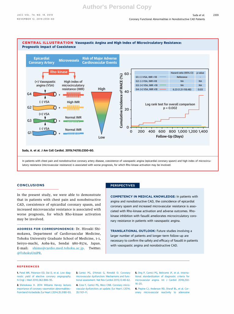
CENTRAL ILLUSTRATION Vasospastic Angina and High Index of Microcirculatory Resistance:Prognostic Impact of Coexistence
0Low
High
EpicardialCoronary Artery
Rho-kinase
(+) Vasospasticangina (VSA)
+
+
+
+
High index ofmicrocirculatoryresistance (IMR)
High IMR
Normal IMR
Normal IMR
(–) VSA
(–) VSA
G4
G2
G3
G1
Microvessels Risk of Major AdverseCardiovascular Events
0
20
40
G1: (–) VSA, IMR <18
Hazard ratio (95% CI)
Reference
p value
–NA NANA NA
6.23 (1.21-118.48) 0.03
G2: (–) VSA, IMR ≥18
G4: (+) VSA, IMR ≥18G3: (+) VSA, IMR <18
60
Cum
ulat
ive
Inci
denc
e of
MAC
E (%
)
200 400 600 800Follow-Up (Days)
1,000 1,200 1,400
(+) VSA
Log rank test for overall comparisonp = 0.002
Suda, A. et al. J Am Coll Cardiol. 2019;74(19):2350–60.
In patients with chest pain and nonobstructive coronary artery disease, coexistence of vasospastic angina (epicardial coronary spasm) and high index of microcircu-
latory resistance (microvascular resistance) is associated with worse prognosis, for which Rho-kinase activation may be involved.
PERSPECTIVES
COMPETENCY IN MEDICAL KNOWLEDGE: In patients with
angina and nonobstructive CAD, the coincidence of epicardial
coronary spasm and increased microvascular resistance is asso-
ciated with Rho-kinase activation and adverse outcomes. Rho-
kinase inhibition with fasudil ameliorates microcirculatory coro-
nary resistance in patients with vasospastic angina.
TRANSLATIONAL OUTLOOK: Future studies involving a
larger number of patients and longer-term follow-up are
necessary to confirm the safety and efficacy of fasudil in patients
with vasospastic angina and nonobstructive CAD.
J A C C V O L . 7 4 , N O . 1 9 , 2 0 1 9 Suda et al.N O V E M B E R 1 2 , 2 0 1 9 : 2 3 5 0 – 6 0 Coronary Functional Abnormalities in Nonobstructive CAD Patients
2359
Author's Personal Copy
CONCLUSIONS
In the present study, we were able to demonstratethat in patients with chest pain and nonobstructiveCAD, coexistence of epicardial coronary spasm, andincreased microvascular resistance is associated withworse prognosis, for which Rho-kinase activationmay be involved.
ADDRESS FOR CORRESPONDENCE: Dr. Hiroaki Shi-mokawa, Department of Cardiovascular Medicine,Tohoku University Graduate School of Medicine, 1-1,Seiryo-machi, Aoba-ku, Sendai 980-8574, Japan.E-mail: [email protected]. Twitter:@TohokuUniPR.
RE F E RENCE S
1. Patel MR, Peterson ED, Dai D, et al. Low diag-nostic yield of elective coronary angiography.N Engl J Med 2010;362:886–95.
2. Shimokawa H. 2014 Williams Harvey lecture:importance of coronary vasomotion abnormalities—frombenchtobedside.EurHeart J2014;35:3180–93.
3. Camici PG, D’Amati G, Rimoldi O. Coronarymicrovascular dysfunction: Mechanisms and func-tional assessment. Nat Rev Cardiol 2015;12:48–62.
4. Crea F, Camici PG, Merz CNB. Coronary micro-vascular dysfunction: an update. Eur Heart J 2014;35:1101–11.
5. Ong P, Camici PG, Beltrame JF, et al. Interna-tional standardization of diagnostic criteria formicrovascular angina. Int J Cardiol 2018;250:16–20.
6. Pepine CJ, Anderson RD, Sharaf BL, et al. Cor-onary microvascular reactivity to adenosine

Suda et al. J A C C V O L . 7 4 , N O . 1 9 , 2 0 1 9
Coronary Functional Abnormalities in Nonobstructive CAD Patients N O V E M B E R 1 2 , 2 0 1 9 : 2 3 5 0 – 6 0
2360
Author's Personal Copy
predicts adverse outcome in women evaluated forsuspected ischemia. Results from the NationalHeart, Lung and Blood Institute WISE (Women’sIschemia Syndrome Evaluation) Study. J Am CollCardiol 2010;55:2825–32.
7. Lee JM, Jung JH, Hwang D, et al. Coronary flowreserve and microcirculatory resistance in patientswith intermediate coronary stenosis. J Am CollCardiol 2016;67:1158–69.
8. Kobayashi Y, Fearon WF, Honda Y, et al. Effectof sex differences on invasive measures of coro-nary microvascular dysfunction in patients withangina in the absence of obstructive coronary ar-tery disease. J Am Coll Cardiol Intv 2015;8:1433–41.
9. Kikuchi Y, Yasuda S, Aizawa K, et al.Enhanced Rho-kinase activity in circulatingneutrophils of patients with vasospastic angina:a possible biomarker for diagnosis and diseaseactivity assessment. J Am Coll Cardiol 2011;58:1231–7.
10. Nihei T, Takahashi J, Hao K, et al. Prognosticimpacts of Rho-kinase activity in circulating leu-cocytes in patients with vasospastic angina. EurHeart J 2018;39:952–9.
11. JCS Joint Working Group. Guidelines for diag-nosis and treatment of patients with vasospasticangina (coronary spastic angina) (JCS 2008). Circ J2010;74:1745–62.
12. Mohri M, Shimokawa H, Hirakawa Y, et al. Rho-kinase inhibition with intracoronary fasudilprevents myocardial ischemia in patients withcoronary microvascular spasm. J Am Coll Cardiol2003;41:15–9.
13. Freedman LS. Tables of the number of patientsrequired in clinical trials using the log-rank test.Stat Med 1982;1:121–9.
14. Grambsch PM, Therneau TM. Proportionalhazards tests and diagnostics based on weightedresiduals. Biometrika 1994;81:515–26.
15. Leening MJ, Vedder MM, Witteman JC, et al.Net reclassification improvement: computation,interpretation, and controversies: a literature re-view and clinician’s guide. Ann Intern Med 2014;160:122–31.
16. Yamanaga K, Tsujita K, Komura N, et al. Single-wire pressure and flow velocity measurement forquantifying microvascular dysfunction in patientswith coronary vasospastic angina. Am J Physiol2015;308:H478–84.
17. Ong P, Athanasiadis A, Borgulya G, et al. Highprevalence of a pathological response to acetyl-choline testing in patients with stable anginapectoris and unobstructed coronary arteries: TheACOVA study (abnormal coronary vasomotion inpatients with stable angina and unobstructedcoronary arteries. J Am Coll Cardiol 2012;59:655–62.
18. Montone RA, Niccoli G, Fracassi F, et al. Pa-tients with acute myocardial infarction andnon-obstructive coronary arteries: Safety andprognostic relevance of invasive coronary pro-vocative tests. Eur Heart J 2018;39:91–8.
19. Ford TJ, Corcoran D, Berry C. Stable coronarysyndromes: pathophysiology, diagnostic advancesand therapeutic need. Heart 2018;104:284–92.
20. Kaski JC, Crea F, Gersh BJ, et al. Reappraisalof ischemic heart disease: fundamental role of
coronary microvascular dysfunction in the patho-genesis of angina pectoris. Circulation 2018;138:1463–80.
21. Takagi Y, Yasuda S, Tsunoda R, et al. Clinicalcharacteristics and long-term prognosis of vaso-spastic angina patients who survived out-of-hospital cardiac arrest: multicentre registry studyof the Japanese Coronary Spasm Association. CircArrhythmia Electrophysiol 2011;4:295–302.
22. Sato K, Kaikita K, Nakayama N, et al.Coronary vasomotor response to intracoronaryacetylcholine injection, clinical features, andlong-term prognosis in 873 consecutive pa-tients with coronary spasm: analysis of asingle-center study over 20 years. J Am HeartAssoc 2013;2:1–12.
23. Yang HM, Khush K, Luikart H, et al. Invasiveassessment of coronary physiology predicts latemortality after heart transplantation. Circulation2016;133:1945–50.
24. Aziz A, Hansen HS, Sechtem U, et al.Sex-related differences in vasomotor function inpatients with angina and unobstructed coronaryarteries. J Am Coll Cardiol 2017;70:2349–58.
KEY WORDS coronary spasm, IMR,microvascular dysfunction, nonobstructivecoronary artery disease, Rho-kinase
APPENDIX For an expanded Methods sectionas well as supplemental tables and figures,please see the online version of this paper.