Corona Virus - COVID 19: Situation in Numbers in the ECO ...

Omkar. World Journal of Pharmaceutical Research
www.wjpr.net │ Vol 10, Issue 6, 2021. │ ISO 9001:2015 Certified Journal │
850
CORONA VIRUS: A BRIEF REVIEW ON COVID-19
Atre Omkar*
Pravara Rural College of Pharmacy, Pravaranagar, Dist. Ahmednagar (Maharashtra) 423107
India.
ABSTRACT
A novel coronavirus, named Severe Acute Respiratory Syndrome
Coronavirus 2 (SARS-CoV-2) or (2019-nCoV) with unknown origin
spread in Guangdong market province of China. The pandemic was
initially caused in local market of Wuhan in dec-19 and thus it was
named as coronavirus 19. this infection of covid-19 was soon reached
all over the world in no time. Corona virus causes respiratory infection
including pneumonia, cold, sneezing and coughing while in animal it
causes diarrhea and upper respiratory diseases. Corona virus enters in
human cell through membrane ACE-2 exopeptidase receptor. The
presence of COVID-19 was manifested by several symptoms, ranging
from asymptomatic/mild symptoms to severe illness and death.
However, due to the worldwide spread of the virus, COVID-19 has become a serious concern
in the medical community and was declared as novel covid19 by WHO in2019. Currently
some antiviral medications are given as a supportive treatment for covid 19 which gave
appreciable results in clinical trials along with some emergency approval of the vaccines for
treatment. The number of covid cases now has raised to 15,20,00,000 (march-april). Thus, the
main purpose of this review is to provide an overview of covid-19 along with diseases current
treatment and future prospects of vaccine development and government`s strategy for
management to covid-19.
KEYWORDS: transmembrane protease, covid-19, angiotensin converting enzyme, bat borne
virus.
History and origin of disease
In December 2019, an outbreak of pneumonia with unknown origin began in China’s wuhan
Province, raised global health concerns due to the ease of transmission. Initially it was
*Corresponding Author
Atre Omkar
Pravara Rural College of
Pharmacy, Pravaranagar,
Dist. Ahmednagar
(Maharashtra) 423107 India.
Article Received on
16 April 2021,
Revised on 06 May 2021,
Accepted on 27 May 2021
DOI: 10.20959/wjpr20216-20650
World Journal of Pharmaceutical Research SJIF Impact Factor 8.084
Volume 10, Issue 6, 850-890. Review Article ISSN 2277– 7105

Omkar. World Journal of Pharmaceutical Research
www.wjpr.net │ Vol 10, Issue 6, 2021. │ ISO 9001:2015 Certified Journal │
851
stated that the virus arised from the local market of wuhan through some workers around
there. All of these workers were said to cause infection in a rapid sense due to the high
crowding market. A phylogenetic network analysis of 160 early coronavirus genomes
sampled from December 2019 to February 2020 showed that the virus type most closely
related to the bat coronavirus was most abundant in Guangdong, China, and designated type
"A". The predominant type among samples from Wuhan, "B", is more distantly related to the
bat coronavirus than the ancestral type "A". Further the strain that was isolated from the
infected person was similar to with that of the virus W1V16 that was obtained from the cave
of wuhan province of china and thus bats were said to be primary cause of infection of SARS
covid-19. further researchers stated that the virus isolated from pangolin species was only
72% similar to Covid 19 genome sequencing and thus may be said as intermediate of
injection of covid 19. Research into the natural reservoir of the virus that caused the 2002–
2004 SARS outbreak has resulted in the discovery of many SARS-like bat coronaviruses,
most originating in the Rhinolophus genus of horseshoe bats. Phylogenetic analysis
indicates that samples taken from Rhinolophus sinicus show a resemblance of 80% to
SARS-CoV-2. Phylogenetic analysis also indicates that a virus from Rhinolophus affinis,
collected in Yunnan province and designated RaTG13, has a 96% resemblance to SARS-CoV-
2. The RaTG13 virus sequence is the closest known sequence to SARS- CoV-2. All available
evidence suggests that SARS-CoV-2 has a natural animal origin and is not genetically
engineered. Nevertheless, early in the pandemic, conspiracy theories spread on social media
claiming that the virus was bio-engineered by China at the Wuhan Institute of Virology.
While some, including former CDC director Robert R. Redfield, have claimed that the virus
may have been studied by and escaped from the Institute, virologists who have studied
coronaviruses consider the possibility very remote, and the March 2021 WHO report on the
joint WHO-China study stated that such an explanation is "extremely unlikely". To quickly
diagnose and control the highly infectious disease, suspected people were isolated and
diagnostic/ therapeutic procedures were developed via patients’ epidemiological and clinical
data. After numerous studies, a novel severe acute respiratory syndrome coronavirus 2
(SARS-CoV-2) was identified as the cause of the disease, and the disease was dubbed
―coronavirus-19″ (COVID 19) by Chinese Scientists. The presence of COVID-19 is
manifested by several symptoms, ranging from asymptomatic/mild symptoms to severe
illness and death. Common symptoms include cough, fever, and shortness of breath. Other
reported symptoms are weakness, malaise, respiratory distress, muscle pain, sore throat, loss of
taste and/or smell. On 11 February 2020, the International Committee on Taxonomy of

Omkar. World Journal of Pharmaceutical Research
www.wjpr.net │ Vol 10, Issue 6, 2021. │ ISO 9001:2015 Certified Journal │
852
Viruses adopted the official name "severe acute respiratory syndrome coronavirus 2" (SARS-
CoV-2). To avoid confusion with the disease SARS, the WHO sometimes refers to SARS-
CoV-2 as "the COVID-19 virus" in public health communication.
Microbiology of covid virus
SARS-CoV-2 has four structural proteins, known as the S (spike), E (envelope), M
(membrane), and N (nucleocapsid) proteins; the N protein holds the RNA genome, and the S,
E, and M proteins together create the viral envelope. The spike protein, is the protein
responsible for allowing the virus to attach to and fuse with the membrane of a host cell
specifically, its S1 subunit catalyzes attachment, the S2 subunit fusion. Protein modeling
experiments on the spike protein of the virus soon suggested that SARS-CoV-2 has sufficient
affinity to the receptor angiotensin converting enzyme 2 (ANG II) on human cells to use them
as a mechanism of cell entry. By 22 January 2020, a group in China working with the full virus
genome and a group in the United States using reverse genetics methods independently and
experimentally demonstrated that ANG II could act as the receptor for SARS-CoV-2. Studies
have shown that SARS-CoV-2 has a higher affinity to human ANG II than the original SARS
virus. SARS-CoV-2 may also use basigin to assist in cell entry. Initial spike protein priming
by transmembrane protease, serine 2 (TMPRSS2) is essential for entry of SARS-CoV-2. The
host protein neuropilin 1 (NRP1) may aid the virus in host cell entry using ANG II. After a
SARS-CoV-2 virion attaches to a target cell, the cell's protease TMPRSS2 cuts open the spike
protein of the virus, exposing a fusion peptide in the S2 subunit, and the host receptor ANG II.
After fusion, an endosome forms around the virion, separating it from the rest of the host cell.
The virion escapes when the pH of the endosome drops or when cathepsin, a host cysteine
protease, cleaves it. The virion then releases RNA into the cell and forces the cell to produce
and disseminate copies of the virus, which infect more cells.
Fig 1: Microbiology of covid virus with spike proteins.

Omkar. World Journal of Pharmaceutical Research
www.wjpr.net │ Vol 10, Issue 6, 2021. │ ISO 9001:2015 Certified Journal │
853
Pathogenesis of the 2019-nCoV
The lungs are the primary site of 2019-nCoV infection. Ang II is found in many types of cells
and tissues, including the lungs, blood vessels, heart, liver, kidneys, and gastrointestinal tract.
It is also present in the epithelium lining the lung, the nose, and mouth. The chest CT of the
infected patients usually shows bilateral ground-glass opacity lesions in the posterior and
peripheral lungs that are reported as the characteristic of 2019-nCoV pneumonia. The human
angiotensin-converting enzyme 2 (Ang II), known as the major receptor for the viral S protein
provides the entry point for 2019-nCoV to capture and infect a wide range of human cells.
DC-SIGN (CD209), CD147, and L-SIGN (CD209L) are also other entry receptors for 2019-
nCoV. Thus, drugs that interfere with the interactions of the spike protein/ Ang II, CD147,
DCSIGN or L-SIGN or with their gene expression may inhibit viral invasion. Occupying the
ANG II receptor by SARS-CoV-2 prevents it from performing its normal function, and
breaking the Ang I and Ang II peptides. Naturally, there is a high concentration of ACE in
the lung tissue. Thus, in ANG II deficiency, ACE will be more active due to more available
Ang I which is changed into Ang II. Increased local Ang II levels damage blood vessel
linings and cause inflammation and tissue injury. For this reason, it is claimed that the renin-
angiotensin- system has a serious role in COVID-19 pathogenesis. So, it can be claimed that
the main destructive factor in the patients with severe COVID-19 is abnormal and high
activity of local Ang II. Drugs that inhibit ACE or ACE inhibitors (ACEI) such as ramipril,
lisinopril, and enalapril may prevent the injuries caused by Ang II via inhibiting its
production without blocking the actions of ANG II. In addition to ANG II, there are other
enzymes capable to hydrolyse Ang-I or Ang (1–9) to Ang (1–7) such as Neprilysin,
Prolylcarboxypeptidase, and Prolylendopeptidase. It appears that if the activity of these
enzymes is up-regulated in the lungs of people with COVID-19, the effects of reduced ANG
II may be compensated. Among the mentioned enzymes, higher expression levels of
Neprilysin have been detected in lung tissue, especially in the membrane of pulmonary
epithelial cells. In addition to the negative effect on Ang II production, it cleaves and
inactivates some other vasoactive peptides such as substance P, and endothelin. It degrades
and inactivates bradykinin. Bradykinin is identified as a potent vasodilator and lowers blood
pressure, but causes contraction in the non-vascular smooth muscle of the bronchi and
intestines and may play a role in the pain mechanism. So, Neprilysin can be considered as a
potential target to control the severity of COVID-19 disease. Both Prolylcarboxypeptidase
and Prolylendopeptidase are lysosomal and cytosolic peptidase, respectively, that have been
mainly expressed in white blood cells. They have also been detected in lung, liver, and

Omkar. World Journal of Pharmaceutical Research
www.wjpr.net │ Vol 10, Issue 6, 2021. │ ISO 9001:2015 Certified Journal │
854
kidney tissues. In addition to their role in the destruction or maturation of a variety of
peptides, both enzymes may be considered as protective agents against AngII induced
injuries due to the conversion of AngII to Ang (1–7). Prolylcarboxypeptidase also named
angiotensinase C, activates bradykinin, and hydrolyses plasma prekallikrein to active
kallikrein. However, some studies have reported an inflammatory role for
Prolylcarboxypeptidase in the lungs and other tissues.
Fig 2: The phylogenetic illustration of the receptor-binding domain (RBD) in various
betacoronaviruses a. The structure of RBD in SARS-CoV b. 2019-nCoV c. MERS-
CoV d.SARS COVID-19.
Based on the cells that are likely infected, COVID-19 can be divided into three phases that
correspond to different clinical stages of the disease.
Stage 1: Asymptomatic state (initial 1–2 days of infection
The inhaled virus SARS-CoV-2 likely binds to epithelial cells in the nasal cavity and starts
replicating. ANG II is the main receptor for both SARS-CoV2 and SARS-CoV. In vitro data
with SARS-CoV indicate that the ciliated cells are primary cells infected in the conducting
airways. However, this concept might need some revision, since single-cell RNA indicates
low level of ANG II expression in conducting airway cells and no obvious cell type
preference. There is local propagation of the virus but a limited innate immune response. At
this stage the virus can be detected by nasal swabs. Although the viral burden may be low,
these individuals are infectious. The RT-PCR value for the viral RNA might be useful to
predict the viral load and the subsequent infectivity and clinical course. Perhaps super
spreaders could be detected by these studies. For the RT-PCR cycle number to be useful, the
sample collection procedure would have to be standardised. Nasal swabs might be more
sensitive than throat swabs.

Omkar. World Journal of Pharmaceutical Research
www.wjpr.net │ Vol 10, Issue 6, 2021. │ ISO 9001:2015 Certified Journal │
855
Stage 2: Upper airway and conducting airway response (next few days)
The virus propagates and migrates down the respiratory tract along the conducting airways,
and a more robust innate immune response is triggered. Nasal swabs or sputum should yield
the virus (SARS-CoV-2) as well as early markers of the innate immune response. At this
time, the disease COVID-19 is clinically manifest. The level of CXCL10 (or some other
innate response cytokine) may be predictive of the subsequent clinical course. Viral infected
epithelial cells are a major source of beta and lambda interferons. CXCL10 is an interferon
responsive gene that has an excellent signal to noise ratio in the alveolar type II cell response to
both SARS-CoV and influenza. CXCL10 has also been reported to be useful as disease marker
in SARS. Determining the host innate immune response might improve predictions on the
subsequent course of the disease and need for more aggressive monitoring. For about 80% of
the infected patients, the disease will be mild and mostly restricted to the upper and
conducting airways. These individuals may be monitored at home with conservative
symptomatic therapy.
Stage 3: Hypoxia, ground glass infiltrates, and progression to ARDS
Unfortunately, about 20% of the infected patients will progress to stage 3 disease and will
develop pulmonary infiltrates and some of these will develop very severe disease. Initial
estimates of the fatality rate are around 2%, but this varies markedly with age. The fatality and
morbidity rates may be revised once the prevalence of mild and asymptomatic cases is better
defined. The virus now reaches the gas exchange units of the lung and infects alveolar type II
cells. Both SARS-CoV and influenza preferentially infect type II cells compared to type I
cells. The infected alveolar units tend to be peripheral and subpleural. SARS-CoV propagates
within type II cells, large number of viral particles are released, and the cells undergo
apoptosis and die. The end result is likely a self- replicating pulmonary toxin as the released
viral particles infect type II cells in adjacent units. I suspect areas of the lung will likely lose
most of their type II cells, and secondary pathway for epithelial regeneration will be
triggered. Normally, type II cells are the precursor cells for type I cells. This postulated
sequence of events has been shown in the murine model of influenza pneumonia. The
pathological result of SARS and COVID-19 is diffuse alveolar damage with fibrin rich hyaline
membranes and a few multinucleated giant cells. The aberrant wound healing may lead to
more severe scarring and fibrosis than other forms of ARDS. Recovery will require a
vigorous innate and acquired immune response and epithelial regeneration. From my
perspective, similar to influenza, administrating epithelial growth factors such as KGF might

Omkar. World Journal of Pharmaceutical Research
www.wjpr.net │ Vol 10, Issue 6, 2021. │ ISO 9001:2015 Certified Journal │
856
be detrimental and might increase the viral load by producing more ANG II expressing cells.
Elderly individuals are particularly at risk because of their diminished immune response and
reduced ability to repair the damaged epithelium. The elderly also has reduced mucociliary
clearance, and this may allow the virus to spread to the gas exchange units of the lung more
readily.
Fig 3: Human alveolar type II cells infected with SARS-CoV. Human type II cells were
isolated, cultured in vitro, and then infected with SARS-CoV. Viral particles are seen in
double membrane vesicles in the type II cells (a) and along the apical microvilli (b).
Reproduced with permission from the American Thoracic Society.
There are significant knowledge gaps in the pathogenesis of COVID-19 that will be filled in
over the next few months. We do not know if there are alternate receptors for viral entry.
CD209L is an alternative receptor for SARS-CoV. We await detailed studies on infection and
the innate immune response of differentiated primary human lung cells. The apical cilia on
airway cells and microvilli on type II cells may be important for facilitating viral entry. In
conclusion, COVID-19 confined to the conducting airways should be mild and treated
symptomatically at home. However, COVID- 19 that has progressed to the gas exchange
units of the lung must be monitored carefully and supported to the best of our ability, as we
await the development and testing of specific antiviral drugs.
Transmission (Causative agents) of the 2019-nCoV
The first known infections from SARS-CoV-2 were discovered in Wuhan, China. A
phylogenetic network analysis of 160 early coronavirus genomes sampled from December
2019 to February 2020 showed that the virus type most closely related to the bat
coronavirus was most abundant in Guangdong, China, and designated type "A". The
predominant type among samples from Wuhan, "B", is more distantly related to the bat

Omkar. World Journal of Pharmaceutical Research
www.wjpr.net │ Vol 10, Issue 6, 2021. │ ISO 9001:2015 Certified Journal │
857
coronavirus than the ancestral type "A". Research into the natural reservoir of the virus that
caused the 2002–2004 SARS outbreak has resulted in the discovery of many SARS-like bat
coronaviruses, most originating in the Rhinolophus genus of horseshoe bats. Phylogenetic
analysis indicates that samples taken from Rhinolophus sinicus show a resemblance of
80% to SARS-CoV-2. The original source of viral transmission to humans remains unclear, as
does whether the virus became pathogenic before or after the spillover event. Because many of
the early infectives were workers at the Huanan Seafood Market, it has been suggested that
the virus might have originated from the market. However, other research indicates that
visitors may have introduced the virus to the market, which then facilitated rapid expansion
of the infections.
Fig 4: Samples taken from Rhinolophus sinicus, a species of horseshoe bats, show an
80% resemblance to SARS-CoV-2.
Phylogenetic analysis also indicates that a virus from Rhinolophus affinis, collected in
Yunnan province and designated RaTG13, has a 96% resemblance to SARS-CoV-2. The
RaTG13 virus sequence is the closest known sequence to SARS-CoV-2. Bats are
considered the most likely natural reservoir of SARS-CoV-2, but differences between the bat
coronavirus and SARS-CoV-2 suggest that humans were infected via an intermediate host.
Evidence against this hypothesis includes the fact that pangolin virus samples are too distant
to SARS-CoV-2: isolates obtained from pangolins seized in Guangdong were only 92%
identical in sequence to the SARS-CoV-2 genome. In addition, despite similarities in a few
critical amino acids, pangolin virus samples exhibit poor binding to the human ANG II
receptor. The routes of human-to-human transmission of 2019-nCoV among individuals
include direct inhalation of contaminated droplets released into the environment by sneezing
or coughing, and contact transmission via oral, nasal, and eye mucous. Although a 6-ft
distance is emphasized to protect against the spread of the disease, it is not enough. Microbes

Omkar. World Journal of Pharmaceutical Research
www.wjpr.net │ Vol 10, Issue 6, 2021. │ ISO 9001:2015 Certified Journal │
858
in droplets < 5 μm in diameter can stay in the air for a long time and can be transmitted to
others over distances of more than 1 m. There is no evidence that 2019-nCoV is spread
through water in pools, rivers, lakes. To date, no reports of positive crowns have been
received from water play places; however, it cannot be said that it is completely 100% safe.
Intestinal infection and the presence of 2019-nCoV in feces have been reported, but there is
not enough evidence for fecal-oral transmission of 2019-nCoV. Song et al. examined the
presence of 2019-nCoV in testicular biopsy and semen of COVID-19 patients and did not
find positive RT- PCR. They stated that 2019-nCov does not infect the testes and the virus
may not be sexually transmitted by infected men. Some studies have shown the presence of
asymptomatic viral carriers with normal laboratory and chest CT findings.
Comparison of coronavirus with other 5 class of beta viruses and mode of action in
causing viral infection
David Tyrrell who was a british physician insolated and told about coronavirus. Accordingly,
there were 6 viruses who belonged to the class of beta coronaviruses which were as
following.
Human coronavirus 229E (HCoV-229E).
Human coronavirus OC43 (HCoV-OC43).
Human coronavirus HKU1 (HCoV-HKU1).
Middle East respiratory syndrome-related coronavirus (MERS-CoV).
Severe acute respiratory syndrome coronavirus (SARS-CoV-1).
Severe acute respiratory syndrome coronavirus 2 (SARS-CoV-2).
Human coronavirus 229E (HCoV-229E).
Identified in late 2004 in a small child of age 7 years who had bronchiolitis in netherlands. this
virus was originated from palm civets and bats with same binding to the ACE-2 as like covid-
19. Entry mechanism was same as like the COVID-19 strain along with additional
endocytosis and plasma membrane cell fusion. Treatment for the HCoV-NL63 virus was
dependent on the severity of associated symptomology. Most mild to moderate infections had
gone away on their own. Symptoms was relieved by taking a pain reliever or fever
medication, taking a hot shower, or using a humidifier. Antiviral treatment was necessary for
infected patients that end up in the intensive care unit (ICU)due to acute respiratory infection.
Intravenous immunoglobulin was FDA approved HCoV-NL63 inhibitor that is also used to
treat primary immune deficiency, RSV, and Kawasaki disease.

Omkar. World Journal of Pharmaceutical Research
www.wjpr.net │ Vol 10, Issue 6, 2021. │ ISO 9001:2015 Certified Journal │
859
Human coronavirus OC43 (HCoV-OC43).
The morphology was same as COVID-19 virus with positive sense single strained, enveloped
with binding to N-Acetyl neuraminic acid receptor. in 1889-1890 the Russian flu caused
same pandemic as like covid 19 with the killing about 1 million population worldwide. the
causative agent for this was not known. Some stated that it was Influenza virus of H2N3 or
H3N3 strain. Further in Nov-2020 Danish researcher stated that symptoms was similar to that
of COVID-19. In 2002-2004 SARS outbreak which was stated to have same common
ancestor as like the Russian flu in late 19s. orifin of these strains was said to be most likely
from rodents and then to cattle and then towards the host i.e. humans.
Human coronavirus HKU1 (HCoV-HKU1).
Discovered in Jan 2004 from one of man from hongkong the structure is same with
enveloped and ssRNA but the binding is towards 9-O Acetylnuerominic acid receptor. It also
has an additional hemagglutinin esterase binding ability which make it different from other
class of viruses. These viruses were seen in a man who returned from Shenzhen china. These
viruses were not isolated but was genetically sequenced from the virus with similar structure
with mouse hepatitis virus. It was said to arise from rodents.
Middle East respiratory syndrome-related coronavirus (MERS-CoV).
It is single strained positive spiked ssRNA with binding ability to DPP4 receptor. First case was
identified on April 2002 in Jeddah, Saudi Arabia. Evolution was said to be from the bats of
species Tylonycteris bat coronavirus and Pipisterellus bat covid HKU5 and HKU4. First
source of covid in human of these type was due to the initial contact from quatar and
dromedary camels from south Africa where it as source of infection in human beings before bat
coronavirus. The person who got infected died in Nov 2013. later on, June 2014 around 689
cases were found with 283 death number.
Severe acute respiratory syndrome coronavirus (SARS-CoV-1).
Said to have originated from wuhan market as majority of initial cases was found over there.
the virus was closely resembeled the structure as like SARS covid 19 where it infected only
epithelial cells of lungs who binds to ACE-2 who infects human bats and plam civets. The
host viral genome was isolated by university of hong kong. the two species of rhinophus were
closely related to covid 19 strain.
1. Rhinolophus Sinicus- about 80% resembalace.

Omkar. World Journal of Pharmaceutical Research
www.wjpr.net │ Vol 10, Issue 6, 2021. │ ISO 9001:2015 Certified Journal │
860
2. Rhinolophus affinis -96% similar to SARS covid 2.
Until now RaTG13 virus sequence is the closest resembalace of covid 19 strain.
Total variants for covid up to date
Notable variants
In early November 2020, Cluster 5, also referred to as FVI-spike by the Danish State Serum
Institute (SSI), was discovered in Northern Jutland, Denmark, and is believed to have been
spread from minks to humans via mink farms. On 4 November 2020, it was announced that
the mink population in Denmark would be culled to prevent the possible spread of this
mutation and reduce the risk of new mutations happening. A lockdown and travel restrictions
were introduced in seven municipalities of Northern Jutland to prevent the mutation from
spreading, which could compromise national or international responses to the COVID-19
pandemic. By 5 November 2020, some 214 mink-related human cases had been detected.
The World Health Organization (WHO) has stated that cluster 5 has a "moderately decreased
sensitivity to neutralizing antibodies". SSI warned that the mutation could reduce the effect of
COVID-19 vaccines under development, although it was unlikely to render them useless.
Following the lockdown and mass-testing, SSI announced on 19 November 2020 that cluster
5 in all probability had become extinct. As of 1 February 2021, authors to a peer-reviewed
paper, all of whom were from the SSI, assessed that cluster 5 was not in circulation in the
human population.
1. Lineage B.1.1.7 / Variant of Concern 20DEC-01
First detected in October 2020 during the COVID-19 pandemic in the United Kingdom from
a sample taken the previous month in Kent, Lineage B.1.1.7, was previously known as the
first Variant Under Investigation in December 2020 and later notated as VOC-202012/01. It
is also known as lineage B.1.1.7 or 20I/501Y.V1 (formerly 20B/501Y.V1). Since then, its
prevalence odds have doubled every 6.5 days, the presumed generational interval. It is
correlated with a significant increase in the rate of COVID-19 infection in United Kingdom,
associated partly with the N501Y mutation. There is some evidence that this variant has 40%–
80% increased transmissibility (with most estimates lying around the middle to higher end of
this range), and early analyses suggest an increase in lethality. More recent work has found
no evidence of increased virulence.

Omkar. World Journal of Pharmaceutical Research
www.wjpr.net │ Vol 10, Issue 6, 2021. │ ISO 9001:2015 Certified Journal │
861
2. Variant of Concern 21FEB-02
Variant of Concern 21FEB-02 (previously written as VOC-202102/02), described by Public
Health England (PHE) as "B.1.1.7 with E484K"is of the same lineage in the Pango
nomenclature system, but has an additional E484K mutation. As of 17 March 2021, there are
39 confirmed cases of VOC-21FEB-02 in the UK. On 4th
march scientists reported B.1.1.7
with E484K mutations in the state of Oregon. In 13 test samples analysed, one had this
combination, which appeared to have arisen spontaneously and locally, rather than being
imported.
3. Lineage B.1.1.207
First sequenced in August 2020 in Nigeria,[80] the implications for transmission and
virulence are unclear but it has been listed as an emerging variant by the US Centers for
Disease Control. Sequenced by the African Centre of Excellence for Genomics of Infectious
Diseases in Nigeria, this variant has a P681H mutation, shared in common with UK's Lineage
B.1.1.7. It shares no other mutations with Lineage B.1.1.7 and as of late December 2020 this
variant accounts for around 1% of viral genomes sequenced in Nigeria, though this may rise.
By March 2021, Lineage B.1.1.207 had been detected in Peru, Germany, Singapore, Hong
Kong, Vietnam, Costa Rica, South Korea, Canada, Australia, Japan, France, Italy, Ecuador,
Mexico, UK and the USA.
4. Lineage B.1.1.317
While B.1.1.317 is not considered a variant of concern, Queensland Health forced 2 people
undertaking hotel quarantine in Brisbane, Australia to undergo an additional 5 days
quarantine on top of the mandatory 14 days after it was confirmed they were infected with this
variant.
5. Lineage B.1.1.318
Lineage B.1.1.318 was designated by PHE as a VUI (VUI-21FEB-04, previously VUI-
202102/04) on 24 February 2021. 16 cases of it have been detected in the UK.
6. Lineage B.1.351
On 18 December 2020, the 501.V2 variant, also known as 501.V2, 20H/501Y.V2 (formerly
20C/501Y.V2), VOC-20DEC-02 (formerly VOC-202012/02), or lineage B.1.351, was first
detected in South Africa and reported by the country's health department. Researchers and
officials reported that the prevalence of the variant was higher among young people with no

Omkar. World Journal of Pharmaceutical Research
www.wjpr.net │ Vol 10, Issue 6, 2021. │ ISO 9001:2015 Certified Journal │
862
underlying health conditions, and by comparison with other variants it is more frequently
resulting in serious illness in those cases. The South African health department also indicated
that the variant may be driving the second wave of the COVID-19 epidemic in the country due
to the variant spreading at a more rapid pace than other earlier variants of the virus. Scientists
noted that the variant contains several mutations that allow it to attach more easily to human
cells because of the following three mutations in the receptor-binding domain (RBD) in the
spike glycoprotein of the virus: N501Y, K417N, and E484K. The N501Y mutation has also
been detected in the United Kingdom.
CDC has listed B.1.429 and the related B.1.427 as "variants of concern," and cites a preprint
for saying that they exhibit a ~20% increase in viral transmissibility, have a "Significant
impact on neutralization by some, but not all," therapeutics that have been given Emergency
Use Authorization (EUA) by FDA for treatment or prevention of COVID-19, and moderately
reduce neutralization by plasma collected by people who have previously infected by the
virus or who have received a vaccine against the virus. B.1.429 was first observed in July
2020 by researchers at the Cedars-Sinai Medical Center, California, in one of 1,230 virus
samples collected in Los Angeles County since the start of the COVID-19 epidemic. It was
not detected again until September when it reappeared among samples in California, but
numbers remained very low until November. In November 2020, the CAL. Europe, Asia and
Australia.
7. Lineage B.1.525
B.1.525, also called VUI-21FEB-03 (previously VUI-202102/03) by Public Health England
(PHE) and formerly known as UK1188, does not carry the same N501Y mutation found in
B.1.1.7, 501.V2 and P.1, but carries the same E484K-mutation as found in the P.1, P.2, and
501.V2 variants, and also carries the same H69/V70 deletion (a deletion of the amino acids
histidine and valine in positions 69 and 70) as found in B.1.1.7, N439K variant (B.1.141 and
B.1.258) and Y453F variant (Cluster 5). B.1.525 differs from all other variants by having
both the E484K-mutation and a new F888L mutation (a substitution of phenylalanine (F)
with leucine (L) in the S2 domain of the spike protein). As of March 5, it had been detected in
UK, Denmark, Finland, Norway, Netherlands, Belgium, France, Spain, Nigeria, Ghana, Jorda
n, Japan, Singapore, Australia, Canada, Germany, Italy, Slovenia, Austria, Malaysia,
Switzerland , the Republic of Ireland and the US. It has also been reported in Mayotte, the
overseas department/region of France. The first cases were detected in December 2020 in the

Omkar. World Journal of Pharmaceutical Research
www.wjpr.net │ Vol 10, Issue 6, 2021. │ ISO 9001:2015 Certified Journal │
863
UK and Nigeria, and as of 15 February, it had occurred in the highest frequency among
samples in the latter country. As of 24 February, 56 cases were found in the UK. Denmark,
which sequence all their COVID-19 cases, found 113 cases of this variant from January 14 to
February 21, of which seven were directly related to foreign travels to Nigeria. UK experts
are studying it to understand how much of a risk it could be. It is currently regarded as a
"variant under investigation", but pending further study, it may become a "variant of
concern". Prof Ravi Gupta, from the University of Cambridge spoke to the BBC and said
B.1.525 appeared to have "significant mutations" already seen in some of the other newer
variants, which is partly reassuring as their likely effect is to some extent more predictable.
8. Lineage B.1.526
In November 2020, a mutant variant was discovered in New York City, which was named
B.1.526. As of April 11, 2021, the variant has been detected in at least 48 U.S. states and 18
countries.
9. Lineage B.1.617
In October 2020, a new variant was discovered in India, which was named B.1.617. There
were very few detections until January 2021 and by April it had spread to at least 20 countries
in all continents except Antarctica and South America. Among some 15 defining mutations, it
has spike mutations D111D (synonymous), G142D, P681R, E484Q and L452R, the latter two
of which may cause it to easily avoid antibodies. In an update on 15 April 2021, PHE
designated B.1.617 as a 'Variant under investigation', VUI-21APR-01. On 29 April 2021,
PHE added two further variants, VUI-21APR-02 and VUI-21APR-03, effectively B.1.617.2
and B.1.617.3.
10. Lineage B.1.618
In October 2020, this variant was first isolated. It has a mutation called E484K which is the
same mutation which South African variant has. It is growing significantly in recent months in
West Bengal. As of 23 April 2021, the CoV-Lineages database showed 135 sequences
detected in India, with single-figure numbers in each of eight other countries worldwide.
11. Lineage P.3
On 18 February 2021, the Department of Health of the Philippines confirmed the detection of
two mutations of COVID-19 in Central Visayas after samples from patients were sent to
undergo genome sequencing. The mutations were later named as E484K and N501Y, which

Omkar. World Journal of Pharmaceutical Research
www.wjpr.net │ Vol 10, Issue 6, 2021. │ ISO 9001:2015 Certified Journal │
864
were detected in 37 out of 50 samples, with both mutations co-occurrent in 29 out of these.
There were no official names for the variants and the full sequence was yet to be identified.
On 13 March, the Department of Health confirmed the mutations constitutes a variant which
was designated as lineage P.3. On the same day, it also confirmed the first Lineage P.1
COVID-19 case in the country. Although the P.1 and P.3 variants stem from the same lineage
B.1.1.28, the department said that P.3 variant's impact on vaccine efficacy and
transmissibility is yet to be ascertained. The Philippines had 98 cases of P.3 variant on 13
March. On 12 March it was announced that P.3 had also been detected in Japan. On 17 March,
the United Kingdom confirmed its first two cases, where PHE termed it VUI-21MAR-02. On
30 April 2021, Malaysia detected 8 cases of P.3 variant in Sarawak.
Notable missense mutations 1. D614G
D614G is a missense mutation that affects the spike protein of SARS-CoV-2. The frequency
of this mutation in the viral population has increased during the pandemic. G (glycine) has
replaced D (aspartic acid) at position 614 in many countries, especially in Europe though
more slowly in China and the rest of East Asia, supporting the hypothesis that G increases the
transmission rate, which is consistent with higher viral titers and infectivity in vitro.
Researchers with the PANGOLIN tool nicknamed this mutation "Doug". In July 2020, it was
reported that the more infectious D614G SARS-CoV-2 variant had become the dominant form
in the pandemic. PHE confirmed that the D614G mutation had a "moderate effect on
transmissibility" and was being tracked internationally. The global prevalence of D614G
correlates with the prevalence of loss of smell (anosmia) as a symptom of COVID-19,
possibly mediated by higher binding of the RBD to the ANG II receptor or higher protein
stability and hence higher infectivity of the olfactory epithelium. Variants containing the
D614G mutation are found in the G clade by GISAID[4] and the B.1 clade by the
PANGOLIN tool.
2. E484K
The name of the mutation, E484K, refers to an exchange whereby the glutamic acid (E) is
replaced by lysine (K) at position 484. is nicknamed "Eeek".E484K has been reported to be an
escape mutation (i.e., a mutation that improves a virus's ability to evade the host's immune
system) from at least one form of monoclonal antibody against SARS-CoV-2, indicating there
may be a "possible change in antigenicity". The P.1. lineage described in Japan and Manaus,
the P.2 lineage (also known as B.1.1.28.2 lineage, Brazil) and 501.V2 (South Africa) exhibit
Omkar. World Journal of Pharmaceutical Research
www.wjpr.net │ Vol 10, Issue 6, 2021. │ ISO 9001:2015 Certified Journal │
865
this mutation. A limited number of B.1.1.7 genomes with E484K mutation have also been
detected. Monoclonal and serum-derived antibodies are reported to be from 10 to 60 times
less effective in neutralizing virus bearing the E484K mutation. On 2 February 2021, medical
scientists in the United Kingdom reported the detection of E484K in 11 samples (out of
214,000 samples), a mutation that may compromise current vaccine effectiveness.
3. N501Y
N501Y denotes a change from asparagine (N) to tyrosine (Y) in amino-acid position 501.
N501Y has been nicknamed "Nelly". This change is believed by PHE to increase binding
affinity because of its position inside the spike glycoprotein's receptor-binding domain, which
binds ANG II in human cells; data also support the hypothesis of increased binding affinity
from this change. Molecular interaction modeling and the free energy of binding calculations
has demonstrated that the mutation N501Y has the highest binding affinity in variants of
concern RBD to hANG II. Variants with N501Y include P.1 (Brazil/Japan), Variant of
Concern 20DEC-01 (UK), 501.V2 (South Africa), and COH.20G/501Y (Columbus, Ohio).
This last became the dominant form of the virus in Columbus in late December 2020 and
January and appears to have evolved independently of other variants.
4. S477G/N
A highly flexible region in the receptor binding domain (RBD) of SARS-CoV-2, starting
from residue 475 and continuing up to residue 485, was identified using bioinformatics and
statistical methods in several studies. The University of Graz and the Biotech Company
Innophore have shown in a recent publication that structurally, the position S477 shows the
highest flexibility among them.
At the same time, S477 is hitherto the most frequently exchanged amino acid residue in the
RBDs of SARS-CoV-2 mutants. By using molecular dynamics simulations of RBD during the
binding process to hANG II, it has been shown that both S477G and S477N strengthen the
binding of the SARS-COV-2 spike with the hANG II receptor. The vaccine developer
BioNTech referenced this amino acid exchange as relevant regarding future vaccine design in
a preprint published in February 2021.
5. P681H
In January 2021, scientists reported in a preprint that the mutation 'P681H', a characteristic
feature of the significant novel SARS-CoV-2 variants detected in the U.K. (B.1.1.7) and

Omkar. World Journal of Pharmaceutical Research
www.wjpr.net │ Vol 10, Issue 6, 2021. │ ISO 9001:2015 Certified Journal │
866
Nigeria (B.1.1.207), is showing a significant exponential increase in worldwide frequency,
similar to the now globally prevalent 'D614G'.
6. E484Q
The name of the mutation, E484Q, refers to an exchange whereby the glutamic acid (E) is
replaced by glutamine (Q) at position 484. India is seeing a significant surge of COVID-19
starting 2021 caused by a "double mutant". This "double mutant" strain has been named
B.1.617. E484Q is a key mutation in this strain that enhances ANG II receptor binding ability
and reduces existing antibodies from attaching to this mutated, hence differently folded, spike
protein.
7. L452R
The name of the mutation, L452R, refers to an exchange whereby the leucine (L) is
replaced by arginine (R) at position 452. There has been a significant surge of COVID-19
starting 2021 all across India caused by a "double mutant". This "double mutant" strain has
been named B.1.617. L452R is a relevant mutation in this strain that enhances ANG II
receptor binding ability and reduces existing antibodies from attaching to this mutated, hence
differently folded, spike protein. L452R, some studies show, could even make the coronavirus
resistant to T cells, that are class of cells necessary to target and destroy virus-infected cells.
They are different from antibodies that are useful in blocking coronavirus particles and
preventing it from proliferating.
8. P681R
The name of the mutation, P681R, refers to an exchange whereby the proline (P) is
replaced by arginine (R) at position 681. Indian SARS-CoV-2 Genomics Consortium
(INSACOG) found that other than the two mutations E484Q and L452R, there is also a third
significant mutation, P681R in B.1.617. All three concerning mutations are on the spike
protein, the operative part of the coronavirus that binds to receptor cells of the body.
9. N440K
This mutation is said to increase infectivity of virus by 10 to 1000 times than current
circulating strains. It is involved in current rapid surge of Covid cases in India. India has
largest proportion of N440K mutated variants followed by the US and Germany.

Omkar. World Journal of Pharmaceutical Research
www.wjpr.net │ Vol 10, Issue 6, 2021. │ ISO 9001:2015 Certified Journal │
867
Mode of Spreading
Peoples can get the infection through close contact with a person who has symptoms from the
virus includes cough and sneezing. Generally, corona virus was spread via airborne zoonotic
droplets. Virus was replicated in ciliated epithelium that caused cellular damage and infection
at infection site. According to a study published in 2019. Angiotensin converting enzyme 2
(ACE.2), a membrane exopeptidase in the receptor used by corona virus in entry to human
cell. Respiratory infections can be transmitted through droplets of different sizes: when the
droplet particles are >5-10 μm in diameter they are referred to as respiratory droplets, and
when then are <5μm in diameter, they are referred to as droplet nuclei.1 According to current
evidence, COVID-19 virus is primarily transmitted between people through respiratory
droplets and contact routes. In an analysis of 75,465 COVID-19 cases in China, airborne
transmission was not reported. Droplet transmission occurs when a person is in in close contact
(within 1 m) with someone who has respiratory symptoms (e.g., coughing or sneezing) and is
therefore at risk of having his/her mucosae (mouth and nose) or conjunctiva (eyes) exposed to
potentially infective respiratory droplets. Transmission may also occur through fomites in the
immediate environment around the infected person.8 Therefore, transmission of the COVID-
19 virus can occur by direct contact with infected people and indirect contact with surfaces in
the immediate environment or with objects used on the infected person (e.g., stethoscope or
thermometer). Airborne transmission is different from droplet transmission as it refers to the
presence of microbes within droplet nuclei, which are generally considered to be particles
<5μm in diameter, can remain in the air for long periods of time and be transmitted to others
over distances greater than 1 m. In the context of COVID-19, airborne transmission may be
possible in specific circumstances and settings in which procedures or support treatments that
generate aerosols are performed; i.e., endotracheal intubation, bronchoscopy, open suctioning,
administration of nebulized treatment, manual ventilation before intubation, turning the patient
to the prone position, disconnecting the patient from the ventilator, non-invasive positive-
pressure ventilation, tracheostomy, and cardiopulmonary resuscitation. There is some
evidence that COVID-19 infection may lead to intestinal infection and be present in faeces.
However, to date only one study has cultured the COVID-19 virus from a single stool
specimen. There have been no reports of faecal−oral transmission of the COVID-19 virus to
date.

Omkar. World Journal of Pharmaceutical Research
www.wjpr.net │ Vol 10, Issue 6, 2021. │ ISO 9001:2015 Certified Journal │
868
Fig 5: Mode of spreading of COVID-19.
Effect of COVID-19 on Other Organs
COVID-19. According to several studies, 2019-nCoV infection, similar to some viral
infections, may be accompanied by cardiac injury. A study of 400 patients hospitalized in
Wuhan, China, found that about one-fifth of patients with COVID-19 developed heart
disease, which increased the mortality rate in patients. Severe and sudden inflammation of the
heart muscle causes arrhythmias and impairs the heart’s ability to efficiently pump blood.
Therefore, patients with a history of cardiovascular disease and with high blood pressure are
at higher risk of death than normal individuals. Oxygen deficiency due to trauma in the lungs
damages the lining of the heart and blood vessels. Besides, fatty plaques in the arteries of the
heart of people with or without symptoms of cardiovascular disease may become unstable due
to fever and inflammation, leading to vascular obstruction and cardiovascular problems. Other
possible disorders seen in hospitalized patients with COVID-19 are abnormal blood
clotting and venous thromboembolism, which necessitate the administration of
anticoagulants or thromboprophylaxis for these patients. The secretion of various types of
inflammatory cytokines in these conditions can exacerbate these complications. Thus,
cytokine inhibitors may be effective in reducing the severity of the disease. Some studies
have reported that COVID-19 may damage CNS. Some observed symptoms include losing the

Omkar. World Journal of Pharmaceutical Research
www.wjpr.net │ Vol 10, Issue 6, 2021. │ ISO 9001:2015 Certified Journal │
869
senses of smell, taste or vision, and decreasing alertness. Also, seizures, stroke, and acute
necrotizing haemorrhagic encephalopathy have been reported in patients with severe
COVID-19 infection.
Fig 6: Effect of COVID-19 on Other Organs.
Fig 7: New symptoms of COVID-19.
Therapeutic results have shown that neurological symptoms gradually decrease in patients
receiving viral encephalitis treatment. About half of patients with COVID-19 show evidence
of protein or blood in the urine, which indicates early renal damage. It has been reported that
15 to 30% of hospitalized patients with COVID-19 in China and New York need to receive
renal treatments or dialysis. However, the direct attack of the virus on the kidneys is still
being debated. The presence of the virus in the fecal samples of some patients with COVID-
19 indicates that the virus can reach the human gastrointestinal tract. About half of all patients

Omkar. World Journal of Pharmaceutical Research
www.wjpr.net │ Vol 10, Issue 6, 2021. │ ISO 9001:2015 Certified Journal │
870
suffer from vomiting, diarrhoea, and other gastrointestinal disorders. Acute viral hepatitis has
also been found in some of these patients. After developing symptoms of fever and cough,
physicians connect gastrointestinal disorders with COVID-19.
Treatment of the COVID-19
Outpatient Management of Acute COVID-19
Outpatient management of acute COVID-19 should include providing supportive care, taking
steps to reduce the risk of SARS-CoV-2 transmission (including isolating the patient), and
advising patients on when to contact a health care provider and seek an in-person evaluation.
Specific Therapy for Outpatients.
1. With Mild to Moderate COVID-19
The COVID-19 Treatment studies recommends using one of the following combination anti-
SARSCoV-2 monoclonal antibodies to treat outpatients with mild to moderate COVID-19
who are at high risk of clinical progression, as defined by the Emergency Use Authorization
criteria.
Bamlanivimab 700 mg plus etesevimab 1,400 mg.
Casirivimab 1,200 mg plus.
Imdevimab 1,200 mg.
The studies recommend against the use of chloroquine or hydroxychloroquine with or
without azithromycin. The use of dexamethasone or other systemic glucocorticoids in
outpatients in the absence of another indication. There is currently a lack of safety and
efficacy data on the use of these agents in outpatients with COVID-19, and systemic
glucocorticoids may cause harm in these patients. the use of antibacterial therapy (e.g.,
azithromycin, doxycycline) in the absence of another indication.
Outpatient Management of Patients With COVID-19 in an Ambulatory Care Setting:
Approximately 80% of patients with COVID-19 have mild illness that does not warrant
medical intervention or hospitalization. Most patients with mild COVID-19 (defined as the
absence of viral pneumonia and hypoxemia) can be managed in an ambulatory care setting or
at home.
Patients with moderate COVID-19 (those with viral pneumonia but without hypoxemia) and
severe COVID-19 (those with dyspnea, hypoxemia, or lung infiltrates >50%) need in-person

Omkar. World Journal of Pharmaceutical Research
www.wjpr.net │ Vol 10, Issue 6, 2021. │ ISO 9001:2015 Certified Journal │
871
evaluation and close monitoring, as pulmonary disease can progress rapidly and require
hospitalization. Older patients and those with chronic medical conditions have a higher risk
for hospitalization and death; however, SARS-CoV-2 infection may cause severe disease and
death in patients of any age, even in the absence of any risk factors. The decision to monitor a
patient in the outpatient setting should be made on a case-by-case basis.
2. Therapeutic Management
Anti-SARS-CoV-2 Monoclonal Antibodies The studies recommends using one of the
following combination anti-SARS-CoV-2 monoclonal antibodies to treat outpatients with
mild to moderate COVID-19 who are at high risk of clinical progression as defined by
the Emergency Use Authorization (EUA) criteria.
• Bamlanivimab 700 mg + etesevimab 1,400 mg; or.
• Casirivimab 1,200 mg + imdevimab 1,200 mg.
Treatment should be started as soon as possible after the patient receives a positive result on a
SARS-CoV-2 antigen test or a nucleic acid amplification test and within 10 days of symptom
onset.. Two combination anti-SARS-CoV-2 monoclonal antibody products.
Bamlanivimab plus Etesevimab.
Casirivimab plus Imdevimab.
have received EUAs from the Food and Drug Administration (FDA) for the treatment of mild
to moderate COVID-19 in outpatients who are at high risk of clinical progression. In
laboratory studies, some SARS-CoV-2 variants of concern or interest that harbor certain
mutations have markedly reduced susceptibility to bamlanivimab and may have lower
sensitivity to etesevimab and casirivimab.
1. Remdesivir
Remdesivir is currently the only drug approved by the FDA for the treatment of COVID-19.
It is recommended for use in hospitalized patients who require supplemental oxygen. In some
cases, a hospital bed may not be available for patients who require supplemental oxygen; for
these patients, remdesivir should only be administered in health care settings that can provide
a similar level of care to an inpatient hospital.
2. Dexamethasone
The Studies recommends against the use of dexamethasone or other systemic glucocorticoids

Omkar. World Journal of Pharmaceutical Research
www.wjpr.net │ Vol 10, Issue 6, 2021. │ ISO 9001:2015 Certified Journal │
872
to treat outpatients with mild to moderate COVID-19 (AIII). There is currently a lack of
safety and efficacy data on the use of these agents in outpatients with COVID-19, and
systemic glucocorticoids may cause harm in these patients. In hospitalized patients with
COVID-19, dexamethasone was shown to reduce mortality in patients who required
supplemental oxygen.
There was no observed benefit of dexamethasone in hospitalized patients who did not receive
oxygen support.26 Outpatients with mild to moderate COVID-19 were not included in this
trial; thus, the safety and efficacy of corticosteroids in this population have not been
established. The Studies recommends against the use of corticosteroids in this population as
there are no clinical trial data to support their use. Moreover, the use of corticosteroids can
lead to adverse effects, such as hyperglycemia, neuropsychiatric symptoms, and secondary
infections, all of which may be difficult to detect and monitor in an outpatient setting. In
some cases, a hospital bed may not be available for patients who require supplemental
oxygen; for these patients, clinicians can consider administering dexamethasone only if the
patient is placed in a health care setting that can provide a similar level of care to an inpatient
hospital.
3. Antithrombotic Therapy
Anticoagulants and antiplatelet therapy should not be initiated in the outpatient setting for the
prevention of venous thromboembolism (VTE) or arterial thrombosis unless the patient has
other indications for the therapy or is participating in a clinical trial (AIII). For more
information, see Antithrombotic Therapy in Patients With COVID-19. Patients should be
encouraged to ambulate, and activity should be increased according to the patient’s tolerance.
4. Antibacterial Therapy
The use of antibacterial therapy (e.g., azithromycin, doxycycline) for outpatient treatment of
covid-19 in the absence of another indication.
Managing pregnant outpatients with COVID-19
Is similar to managing nonpregnant patients. Clinicians should offer supportive care, take
steps to reduce the risk of SARS-CoV-2 transmission, and provide guidance for when to seek
an in-person evaluation. In pregnant patients, SpO2 should be maintained at 95% or above at
sea level; therefore, the threshold for monitoring pregnant patients in an inpatient setting may
be lower than in nonpregnant patients. In general, there are no changes to fetal monitoring

Omkar. World Journal of Pharmaceutical Research
www.wjpr.net │ Vol 10, Issue 6, 2021. │ ISO 9001:2015 Certified Journal │
873
recommendations in the outpatient setting, and fetal management should be similar to that
provided to other pregnant patients with medical illness.31 However, these monitoring
strategies can be discussed on a case- by-case basis with an obstetrician.
Considerations in Children
Children and adolescents with acute COVID-19 are less likely than adults to require medical
intervention or hospitalization, and most can be managed in an ambulatory care setting or at
home. In general, the need for ED evaluation or hospitalization should be based on the
patient’s vital signs, physical exam findings (e.g., dyspnea), and risk factors for progression
to severe illness. Certain groups, including young infants, children with risk factors, or those
with presentations that overlap with multisystem inflammatory syndrome in children (MIS-C),
may require hospitalization for more intensive monitoring. However, this should be
determined on a case-by- case basis. Most children with mild or moderate COVID-19, even
those with risk factors, will not progress to more severe illness and will recover without
specific therapy. There are insufficient pediatric data to recommend either for or against the
use of anti-SARS-CoV-2 monoclonal antibody products in children with COVID-19 who are
not hospitalized but who have risk factors for severe disease. Based on adult studies.
bamlanivimab plus etesevimab.
casirivimab plus imdevimab.
May be considered on a case-by-case basis for non-hospitalized children who meet the EUA
criteria, especially those who meet more than one criterion or are aged ≥16 years. The Studies
recommends consulting a pediatric infectious disease specialist in such cases. In general,
pediatric patients should not continue receiving remdesivir, dexamethasone, or other
COVID19-directed therapies following discharge from an ED or an inpatient setting.
5. Oxygenation and Ventilation
For adults with COVID-19 and acute hypoxemic respiratory failure despite conventional
oxygen therapy, the Studies recommends high-flow nasal cannula (HFNC) oxygen over
noninvasive positive pressure ventilation (NIPPV) (BIIa). In the absence of an indication for
endotracheal intubation, the Studies recommends a closely monitored trial of NIPPV for
adults with COVID- 19 and acute hypoxemic respiratory failure and for whom HFNC is not
available (BIIa). For patients with persistent hypoxemia despite increasing supplemental
oxygen requirements in whom endotracheal intubation is not otherwise indicated, the Studies

Omkar. World Journal of Pharmaceutical Research
www.wjpr.net │ Vol 10, Issue 6, 2021. │ ISO 9001:2015 Certified Journal │
874
recommends considering a trial of awake prone positioning to improve oxygenation (CIIa).
The Studies recommends against using awake prone positioning as a rescue therapy for
refractory hypoxemia to avoid intubation in patients who otherwise meet the indications for
intubation and mechanical ventilation (AIII). If intubation becomes necessary, the procedure
should be performed by an experienced practitioner in a controlled setting due to the enhanced
risk of severe acute respiratory syndrome coronavirus 2 exposure to health care practitioners
during intubation (AIII).
6. Acute Kidney Injury and Renal Replacement Therapy
For critically ill patients with COVID-19 who have acute kidney injury and who develop
indications for renal replacement therapy, the Studies recommends continuous renal
replacement therapy (CRRT), if available (BIII). If CRRT is not available or not possible due
to limited resources, the Studies recommends prolonged intermittent renal replacement
therapy rather than intermittent hemodialysis.
List of Antiviral Drugs That Are Approved or Under Evaluation for the Treatment
of COVID-19
Remdesivir is the only Food and Drug Administration-approved drug for the treatment of
COVID-19. the COVID-19 Treatment Guidelines provides recommendations for using
antiviral drugs to treat COVID-19 based on the available data. As in the management of any
disease, treatment decisions ultimately reside with the patient and their health care provider.
1. Remdesivir.
2. Chloroquine or Hydroxychloroquine With or Without Azithromycin.
3. Lopinavir/Ritonavir and Other HIV Protease Inhibitors.
4. Ivermectin.
5. Fluvoxamine.
6. Interferons (Alfa, Beta).
7. Interleukin-1 Inhibitors.
8. Interleukin-6 Inhibitors.
9. Kinase Inhibitors: Baricitinib and Other Janus Kinase Inhibitors, and Bruton’s Tyrosine
Kinase Inhibitors.
10. Supplements.
11. Vitamin C • There are insufficient data for the COVID-19 Treatment studies to
recommend either for or against the use of vitamin C for the treatment of COVID-19.

Omkar. World Journal of Pharmaceutical Research
www.wjpr.net │ Vol 10, Issue 6, 2021. │ ISO 9001:2015 Certified Journal │
875
12. Vitamin D • There are insufficient data for the experts to recommend either for or against
the use of vitamin D for the treatment of COVID-19.
13. Zinc • There are insufficient data for the Studies to recommend either for or against the
use of zinc for the treatment of COVID-19. • The Studies recommends against using zinc
supplementation above the recommended dietary allowance for the prevention of
COVID-19, except in a clinical trial.
AYUSH treatment for covid-19
Ayurveda, Siddha, Unani, and Homeopathy (referred to as AYUSH) are Indian medicinal
systems that use natural drugs of plant, animal, and mineral origin for treatment. At present
pandemic, AYUSH has recommended that the Homeopathy and Ayurveda as immune-
boosters have sufficient potential to prevent and treat COVID-19. Considering the success of
AYUSH systems in managing several epidemics and restoring health, AYUSH system
recommends some herbs including Chyavanprash, Herbal tea, and Turmeric milk as immune
boosters and has suggested some herbal formulations for treating COVID-19. In Iran, an
herbal formula called Imam Kazem has been reported by Islamic medicine, which is said to be
effective in treating colds and influenza. This herbal formula was used for COVID-19 patients
and they claimed that it could prevent the disease from getting worse and reduce the
symptoms of the disease. The composition of this herbal formula in summer is Terminalia
chebula, Foeniculum vulgare, and red sugar, and in winter it consists of Terminalia chebula,
Pistacia lentiscus and red sugar (Red sugar means sugarcane sugar that has not been processed
industrially). Despite the positive reports of this herbal formula in the treatment of lung
diseases in our country, no experimental or clinical reports were found to refer. Rosewater as
other herbal products is flavored water made by steeping rose petals in water. In addition to
oral consumption, it has antimicrobial properties and can be sprayed on surfaces and space as
a disinfectant solution. Other examples of herbal medicine used in COVID-19 treatment
include: Ginseng (Panax ginseng) regulates the activity of immune cells including T cells,
and B cells, macrophages, dendritic cells, natural killer cells - Ginger (Zingiber officinale)
has anti- apoptotic, anti-inflammatory, anti-tumor activities, anti-tumorigenic, anti-
hyperglycaemic, antioxidant, and analgesic properties, garlic (Allium sativum) product is a
strong immune stimulator, Echinacea extract (Echinacea purpurea (L.) Moench) with
antimicrobial and antioxidant activities is used to improve the immune system and to treat
pulmonary symptoms caused by bacterial infections. Despite such treatments, there are few
reports of improved patients with negative COVID-19 test, that after a while, their COVID-19

Omkar. World Journal of Pharmaceutical Research
www.wjpr.net │ Vol 10, Issue 6, 2021. │ ISO 9001:2015 Certified Journal │
876
test has been positive again. The hospital reports have confirmed, it is possible that improved
patients with negative COVID-19 tests become positive again for 2019-nCoV RNA, although
a small portion of discharged COVID- 19 has shown recurrent recurrences. There are reports
of improved people that showed second recurrences with positive PCR tests after discharge
from the hospital or during quarantine, and were hospitalized again.
Worldwide covid-19 candidate vaccines
In order to respond quickly and effectively to the COVID-19 pandemic, a broad range of
candidate COVID-19 vaccines are being investigated globally using various technologies and
platforms. These include viral-vectored, protein subunit, nucleic acid (DNA, RNA), live
attenuated and inactivated vaccines. Some of these candidates have entered clinical trials.
1. COVAXIN
COVAXIN, India's indigenous COVID-19 vaccine Bharat Biotech is developed in
collaboration with the Indian Council of Medical Research (ICMR) - National Institute of
Virology (NIV).
This indigenous, inactivated vaccine is developed and manufactured in Bharat Biotech's BSL-
3 (Bio-Safety Level 3) high containment facility.
Fig 8: Covaxin Vaccine.
The vaccine received approval from Drug Controller General of India (DCGI) for Phase I &
II Human Clinical Trials and an Adaptive, Seamless Phase I, Followed by Phase II
Randomized, Double blind, Multicentre Study to Evaluate the Safety, Reactogenicity,
Tolerability and Immunogenicity of the Whole-Virion Inactivated SARS-CoV-2 Vaccine
(BBV152).

Omkar. World Journal of Pharmaceutical Research
www.wjpr.net │ Vol 10, Issue 6, 2021. │ ISO 9001:2015 Certified Journal │
877
2. Covishield
The Serum Institute of India (SII) and Indian Council of Medical Research are jointly
conducting a Phase II/III, Observer-Blind, Randomized, Controlled Study to Determine the
Safety and Immunogenicity of Covishield (COVID-19 Vaccine).
Fig 9: covishield vaccine.
3. ZyCoV-D
Zydus Cadila, focused on discovering and developing NCEs, Novel Biologicals, Biosimilars
and Vaccines, announced that its plasmid DNA vaccine to prevent COVID-19, ZyCoV-D.
Safety in Phase I clinical trial of ZyCoV-D in healthy subjects established as endorsed by the
independent Data Safety Monitoring Board (DSMB). Zydus commenced Phase II trial.
Fig 10: ZyCov-D vaccine.
4. Sputinik
Dr Reddys Laboratories Limited and Sputnik LLC are jointly conducting Multi-centre, phase

Omkar. World Journal of Pharmaceutical Research
www.wjpr.net │ Vol 10, Issue 6, 2021. │ ISO 9001:2015 Certified Journal │
878
II/III adaptive clinical trial to assess safety and immunogenicity of Gam-COVID-Vac
combined vector vaccine.
Fig 11: Sputinik vaccine.
5. BBV154 - Intranasal vaccine
Bharat Biotech is conducting Multicenter Study to Evaluate the Reactogenicity, Safety, and
Immunogenicity of an Intranasal Adenoviral vector COVID-19 vaccine (BBV154) in Healthy
Volunteers. BBV154 is an intranasal vaccine stimulates a broad immune response –
neutralizing IgG, mucosal IgA, and T cell responses. Immune responses at the site of
infection (in the nasal mucosa) – essential for blocking both infection and transmission of
COVID-19.
6. COVOVAX
Indian Council of Medical Research and Serum Institute of India jointly performing a phase
2/3, observer-blind, randomized, controlled study to determine the safety and
immunogenicity of COVOVAX [SARS-CoV-2 recombinant spike protein nanoparticle
vaccine (SARS-CoV-2 rS) with Matrix-M1™ adjuvant] in Indian adults.
Fig 12: covovax.

Omkar. World Journal of Pharmaceutical Research
www.wjpr.net │ Vol 10, Issue 6, 2021. │ ISO 9001:2015 Certified Journal │
879
7. mRNA based vaccine (HGCO19)
Randomized, Phase I/II, Placebo-controlled, Dose-Ranging, study to evaluate the Safety,
Tolerability and Immunogenicity of the candidate HGCO19 (COVID-19 vaccine) in healthy
adult subjects. The trial is being conducted by Gennova Biopharmaceuticals Limited.
8. Pfizer-BioNTech
Based on evidence from clinical trials, the Pfizer-BioNTech vaccine was 95% effective at
preventing laboratory-confirmed COVID-19 illness in people without evidence of previous
infection. CDC will continue to provide updates as we learn more about how well the Pfizer-
BioNTech vaccine works in real-world conditions.
Fig 13: Pfizer.
9. Moderna
Based on evidence from clinical trials, the Moderna vaccine was 94.1% effective at
preventing laboratory-confirmed COVID-19 illness in people who received two doses who
had no evidence of being previously infected. The vaccine appeared to have high
effectiveness in clinical trials (efficacy) among people of diverse age, sex, race, and ethnicity
categories and among persons with underlying medical conditions. Although few people in
the clinical trials were admitted to the hospital, this happened less often in the people who got
the Moderna vaccine compared to people who got the saline placebo. CDC will continue to
provide updates as we learn more about how well the Moderna vaccine works in real-world
conditions.

Omkar. World Journal of Pharmaceutical Research
www.wjpr.net │ Vol 10, Issue 6, 2021. │ ISO 9001:2015 Certified Journal │
880
Fig 14: Moderna.
10. Johnson & Johnson’s janssen
The J&J/Janssen vaccine was 66.3% effective in clinical trials (efficacy) at preventing
laboratory-confirmed COVID-19 illness in people who had no evidence of prior infection 2
weeks after receiving the vaccine. People had the most protection 2 weeks after getting
vaccinated. The vaccine had high efficacy at preventing hospitalization and death in people
who did get sick. No one who got COVID-19 at least 4 weeks after receiving the J&J/Janssen
vaccine had to be hospitalized. Early evidence suggests that the J&J/Janssen vaccine might
provide protection against asymptomatic infection, which is when a person is infected by the
virus that causes COVID-19 but does not get sick. CDC will continue to provide updates as
we learn more about how well the J&J/Janssen vaccine works in real-world conditions.
Fig 15: Johnson & Johnson’s janssen.
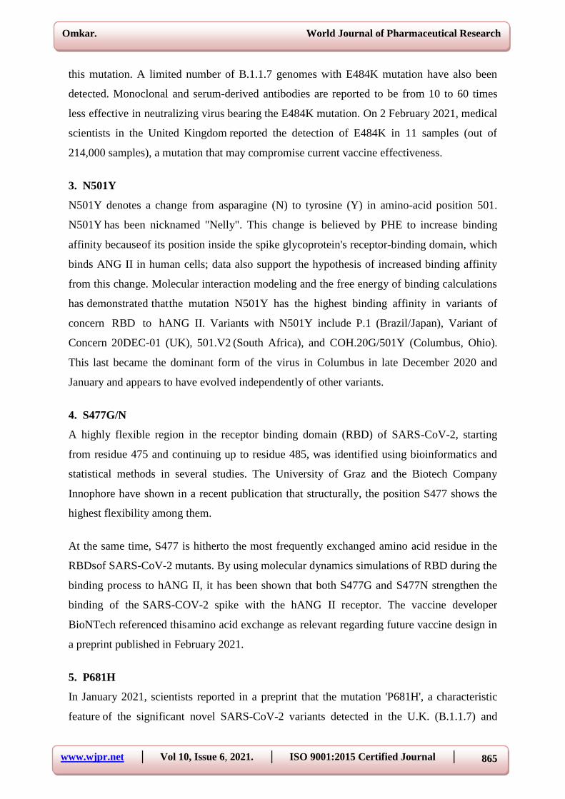
11. mRNA COVID-19 Vaccines
mRNA vaccines are a new type of vaccine to protect against infectious diseases. To trigger an
immune response, many vaccines put a weakened or inactivated germ into our bodies. Not
mRNA vaccines. Instead, they teach our cells how to make a protein—or even just a piece of
a protein—that triggers an immune response inside our bodies. That immune response, which
produces antibodies, is what protects us from getting infected if the real virus enters our
Omkar. World Journal of Pharmaceutical Research
www.wjpr.net │ Vol 10, Issue 6, 2021. │ ISO 9001:2015 Certified Journal │
881
bodies. Researchers have been studying and working with mRNA vaccines for decades.
Interest has grown in these vaccines because they can be developed in a laboratory using
readily available materials. This means the process can be standardized and scaled up,
making vaccine development faster than traditional methods of making vaccines. mRNA
vaccines have been studied before for flu, Zika, rabies, and cytomegalovirus (CMV). As soon
as the necessary information about the virus that causes COVID-19 was available, scientists
began designing the mRNA instructions for cells to build the unique spike protein into an
mRNA vaccine. Future mRNA vaccine technology may allow for one vaccine to provide
protection for multiple diseases, thus decreasing the number of shots needed for protection
against common vaccine- preventable diseases. Beyond vaccines, cancer research has used
mRNA to trigger the immune system to target specific cancer cells.
12. Viral vector vaccines
Scientists began creating viral vectors in the 1970s. Besides being used in vaccines, viral
vectors have also been studied for gene therapy, to treat cancer, and for molecular biology
research. For decades, hundreds of scientific studies of viral vector vaccines have been done
and published around the world. Some vaccines recently used for Ebola outbreaks have used
viral vector technology, and a number of studies have focused on viral vector vaccines against
other infectious diseases such as Zika, flu, and HIV.
Fig 8: The phylogenetic illustration of the receptor-binding domain (RBD) in various
betacoronaviruses a. The structure of RBD in SARS-CoV b, 2019-nCoV c, and MERS-
CoVd.

Omkar. World Journal of Pharmaceutical Research
www.wjpr.net │ Vol 10, Issue 6, 2021. │ ISO 9001:2015 Certified Journal │
882
CONCLUSION
There are hundreds of coronaviruses, most of which circulate in animals. Only seven of these
viruses infect humans and four of them cause symptoms of the common cold. But, three
times in the last 20 years, a coronavirus has jumped from animals to humans to cause severe
disease. SARS, a beta coronavirus emerged in 2002 and was controlled mainly by aggressive
public health measures. There have been no new cases since 2004. MERS emerged in 2012,
still exists in camels, and can infect people who have close contact with them. COVID-19, a
new and sometimes deadly respiratory illness that is believed to have originated in a live
animal market in China, has spread rapidly throughout that country and the world. The new
coronavirus was first detected in Wuhan, China in December 2019. Tens of thousands of
people were infected in China, with the virus spreading easily from person-to-person in many
parts of that country. The novel coronavirus infections were at first associated with travel
from Wuhan, but the virus has now established itself in 177 countries and territories around the
world in a rapidly expanding pandemic. Health officials in the United States and around the
world are working to contain the spread of the virus through public health measures such as
social distancing, contact tracing, testing, quarantines and travel restrictions. Scientists are
working to find medications to treat the disease and to develop a vaccine. The World Health
Organization declared the novel coronavirus outbreak ―a public health emergency of
international concern‖ on January 30. On March 11, 2020 after sustained spread of the disease
outside of China, the World Health Organization declared the COVID-19 epidemic a
pandemic. Public health measures like ones implemented in China and now around the world,
will hopefully blunt the spread of the virus while treatments and a vaccine are developed to
stop it.
REFERENCES
1. Sutton D, Fuchs K, D'Alton M, Goffman D (April 2020). "Universal Screening for
SARS-CoV-2 in Women Admitted for Delivery". The New England Journal of Medicine,
382(22): 2163–2164. doi:10. 1056/NEJMc2009316.PMC 7175422. PMID 32283004.
2. "Dutch study suggests 3% of population may have coronavirus antibodies". Reuters. 16
April 2020. Retrieved 20 April 2020.
3. "Interactive Serology Dashboard for Commercial Laboratory Surveys". cdc. gov. 21 July
2020. Retrieved 24 July 2020.
4. Vogel G (21 April 2020). "Antibody surveys suggesting vast undercount of coronavirus
infections may be unreliable". Science. doi:10.1126/science.abc3831.S2CID218794298.

Omkar. World Journal of Pharmaceutical Research
www.wjpr.net │ Vol 10, Issue 6, 2021. │ ISO 9001:2015 Certified Journal │
883
5. "China: age distribution of novel coronavirus patients 2020". Statista. Retrieved 11 April
2020.
6. Scott D (23 March 2020). "The Covid-19 risks for different age groups, explained". Vox.
Retrieved 12 April 2020.
7. Bi, Qifang; Wu, Yongsheng; Mei, Shujiang; Ye, Chenfei; Zou, Xuan; Zhang, Zhen; Liu,
Xiaojian; Wei, Lan; Truelove, Shaun A; Zhang, Tong; Gao, Wei; Cheng, Cong; Tang,
Xiujuan; Wu, Xiaoliang; Wu, Yu; Sun, Binbin; Huang, Suli; Sun, Yu; Zhang, Juncen;
Ma, Ting; Lessler, Justin; Feng, Tiejian (August 2020). "Epidemiology and transmission
of COVID-19 in 391 cases and 1286 of their close contacts in Shenzhen, China: a
retrospective cohort study". The Lancet Infectious Diseases, 20(8): 911–919. doi:10.
1016/S1473-3099(20)302875. PMC 7185944. PMID 32353347.
8. "Statement on the meeting of the International Health Regulations (2005) Emergency
Committee regarding the outbreak of novel coronavirus 2019 (n-CoV) on 23 January
2020". World Health Organization (WHO). Retrieved 9 April 2020.
9. Sanche S, Lin YT, Xu C, Romero-Severson E, Hengartner N, Ke R (April 2020). "High
Contagiousness and Rapid Spread of Severe Acute Respiratory Syndrome Coronavirus
2". Emerging Infectious Diseases, 26(7): 1470–1477. doi:10.3201/eid2607.200282.PMC
73 23562. PMID 32255761. S2CID 215410037.
10. Roberts L (8 May 2020). "The importance of the coronavirus R rate in other countries
across the globe". The Daily Telegraph. Retrieved 14 May 2020. Jump up to: a b c
"European Centre for Disease Prevention and Control". Retrieved 31 December 2020.
11. "Italy's coronavirus deaths could be underestimated in data: Official". Reuters. 31 March
2020.
12. "Coronavirus: Is Covid-19 really the cause of all the fatalities in Italy?". Stuff. Retrieved
16 April 2020.
13. Wu J, McCann A, Katz J, Peltier E. "28, 000 Missing Deaths: Tracking the True Toll of
the Coronavirus Crisis". The New York Times. ISSN 0362-4331. Retrieved 22 April
2020.
14. "Tracking covid-19 excess deaths across countries". The Economist. ISSN 0013- 0613.
Retrieved 22 April 2020.
15. "What 'Excess Deaths' Do and Don't Tell Us About COVID-19". Reason. 29 April 2020.
Retrieved 4 May 2020.
16. Meyerowitz-Katz G, Merone L (December 2020). "A systematic review and meta-
analysis of published research data on COVID-19 infection fatality rates". International

Omkar. World Journal of Pharmaceutical Research
www.wjpr.net │ Vol 10, Issue 6, 2021. │ ISO 9001:2015 Certified Journal │
884
Journal of Infectious Diseases, 101: 138–148. doi:10. 1016/j. ijid. 2020. 09. 1464.
PMC7524446. PMID 33007452.
17. Yang W, Kandula S, Huynh M, Greene S, Van Wye G, Li W, Chan H, McGibbon E,
Yeung A, Olson D, Fine A, Shaman J (19 October 2020). "Estimating the infection-
fatality risk of SARS-CoV-2 in New York City during the spring 2020 pandemic wave: a
model-based analysis". Lancet Infectious Diseases, 21(2): "Tracking covid-19 excess
203–212. doi:10. 1016/S1473-3099(20)30769-6. PMC 7572090. PMID 33091374.
18. Pastor-Barriuso R, Pérez-Gómez B, Hernán M, Pérez-Olmeda M, Yotti R, Oteo-Iglesias
J, Sanmartín J, León-Gómez I, Fernández- García A, Fernández-Navarro P, Cruz I,
Martín M, Delgado-Sanz C, Fernández N, León J, Muñoz-Montalvo J, Blanco F, Larrauri
A, Pollán M (27 November 2020).
19. "Infection fatality risk for SARS- CoV-2 in community dwelling population of Spain:
nationwide seroepidemiological study". British Medical Journal. 371: m4509. doi:10.
1136/bmj. m4509. PMC 7690 290. PMID 33246972. Fox, Justin (24 April 2020).
20. "The Coronavirus Isn't Just the Flu". Bloomberg. Verity, Robert; Okell, Lucy C;
Dorigatti, Ilaria; Winskill, Peter; Whittaker, Charles; Imai, Natsuko; Cuomo-Dannenburg,
Gina; Thompson, Hayley; Walker, Patrick G T; Fu, Han; Dighe, Amy; Griffin, Jamie T;
Baguelin, Marc; Bhatia, Sangeeta; Boonyasiri, Adhiratha; Cori, Anne; Cucunubá, Zulma;
FitzJohn, Rich; Gaythorpe, Katy; Green, Will; Hamlet, Arran; Hinsley, Wes; Laydon,
Daniel; Nedjati-Gilani, Gemma; Riley, Steven; van Elsland, Sabine; Volz, Erik; Wang,
Haowei; Wang, Yuanrong; Xi, Xiaoyue; Donnelly, Christl A; Ghani, Azra C; Ferguson,
Neil M (June 2020).
21. "Estimates of the severity of coronavirus disease 2019: a model-based analysis". The
Lancet Infectious Diseases, 20(6): 669–677. doi:10.1016/S1473-3099(20)30243-7.PMC
7158570.PMID 32240634.
22. "COVID-19 Pandemic Planning Scenarios" (PDF). CDC. 10 July 2020. Retrieved 29
September 2020. Meyerowitz-Katz, Gideon; Merone, Lea (7 July 2020).
23. "A systematic review and meta- analysis of published research data on COVID-19
infection-fatality rates". MedRxiv, 101: 138–148. doi:10.1101/2020.05.03.20089854.
PMC 7524446. PMID 33007452.
24. Lisboa Bastos, Mayara; Tavaziva, Gamuchirai; Abidi, Syed Kunal; Campbell, Jonathon
R; Haraoui, Louis-Patrick; Johnston, James C; Lan, Zhiyi; Law, Stephanie; MacLean,
Emily; Trajman, Anete; Menzies, Dick; Benedetti, Andrea; Ahmad Khan, Faiz (1 July
2020). "Diagnostic accuracy of serological tests for covid-19: systematic review and

Omkar. World Journal of Pharmaceutical Research
www.wjpr.net │ Vol 10, Issue 6, 2021. │ ISO 9001:2015 Certified Journal │
885
meta-analysis". BMJ. 370: m2516. doi:10.1136/bmj.m2516.PMC7327913. PMID
32611558.
25. Spencer, Elizabeth; Henighan, Carl (1 September 2020). "Overview of BMJ: Diagnostic
accuracy of serological tests for covid-19: systematic review and meta- analysis". CEBM.
Retrieved 24 September 2020. Spencer, Elizabeth; Jefferson, Tom; Brassey, Jon;
Heneghan, Carl (11 September 2020). "When is Covid, Covid?". CEBM. Retrieved 19
September 2020. 2020).
26. "Prevalence and Characteristics of Taste Disorders in Cases of COVID-19: A Meta-
analysis of 29, 349 Patients". Otolaryngology–Head and Neck Surgery: 1–10. doi:10.
1177/0194599820981018. PMID33320033.
27. Islam MA (April 2021). "Prevalence and characteristics of fever in adult and paediatric
patients with coronavirus disease 2019 (COVID-: A systematic review and meta- analysis
of 17515 patients". PLOS ONE. 16 (4): e0249788. doi:10. 1371/journal. pone. 024978 8.
PMC 8023501. PMID 33822812.
28. "Clinical characteristics of COVID- 19". European Centre for Disease Prevention and
Control. Retrieved 29 December 2020. Niazkar HR, Zibaee B, Nasimi A, Bahri N (July
2020). "The neurological manifestations of COVID-19: a review article". Neurological
Sciences, 41(7): 1667–1671. doi:10. 1007/s10072-020-04486- 3. PMC 7262683. PMID
32483687.
29. "Interim Clinical Guidance for Management of Patients with Confirmed Coronavirus
Disease (COVID-19)". U. S. Centers for Disease Control and Prevention (CDC). 6 April
2020. Archivedfrom the original on 2 March 2020. Retrieved 19 April 2020. Oran DP,
Topol EJ (January 2021).
30. "The Proportion of SARS-CoV-2 Infections That Are Asymptomatic: A Systematic
Review". Annals of Internal Medicine. doi:10. 7326/M20- 6976. PMC 7839426. PMID
33481642.
31. "Transmission of COVID-19". European Centre for Disease Prevention and Control.
Retrieved 6 December 2020. Wiersinga WJ, Rhodes A, Cheng AC, Peacock SJ, Prescott
HC (August 2020). "Pathophysiology, Transmission, Diagnosis, and Treatment of
Coronavirus Disease 2019 (COVID-19): A Review". JAMA, 324(8): 782–793.
doi:10.1001/jama.2020.12839.PMID32648899. S2CID220465311.CDC(11 February
2020).
32. "COVID-19 and Your Health". Centers for Disease Control and Prevention. Retrieved 23
January 2021.

Omkar. World Journal of Pharmaceutical Research
www.wjpr.net │ Vol 10, Issue 6, 2021. │ ISO 9001:2015 Certified Journal │
886
33. "Transmission of COVID- 19". www. ecdc. europa. eu. 7 September 2020. Retrieved 14
October 2020. Fox D (January 2020). "What you need to know about the novel
coronavirus". Nature. doi:10.1038/d41586-020-00209-y.PMID33483684.
34. Zhu N, Zhang D, Wang W, Li X, Yang B, Song J, et al. (February 2020). "A Novel
Coronavirus from Patients with Pneumonia in China, 2019". The New England Journal of
Medicine, 382(8): 727–733. doi:10.1056/NEJMoa2001017. PMC 70 92803. PMID
31978945.
35. Gorbalenya AE, Baker SC, Baric RS, de Groot RJ, Drosten C, Gulyaeva AA, et al.
(March 2020). "The species Severe acute respiratory syndrome-related coronavirus:
classifying 2019-nCoV and naming it SARS- CoV-2". Nature Microbiology, 5(4): 536–
544. doi:10.1038/s41564-020-0695- z. PMC 7095448. PMID 32123347.
36. Li C, Zhao C, Bao J, Tang B, Wang Y, Gu B (November 2020). "Laboratory diagnosis of
coronavirus disease-2019 (COVID- 19)". Clinica Chimica Acta; International Journal of
Clinical Chemistry. 510: 35 46. doi:10.1016/j.cca.2020.06.045.PMC7329657.PMID
32621814.
37. "2019 Novel Coronavirus (2019-nCoV) Situation Summary". U. S. Centers for Disease
Control and Prevention (CDC). 30 January 2020. Archived from the original on 26
January 2020. Retrieved 30 January 2020.
38. "Coronavirus disease (COVID-19) technical guidance: Laboratory testing for 2019-nCoV
in humans". World Health Organization(WHO). Archived from the original on 15 March
2020. Retrieved 14 March 2020. Bullard J, Dust K, Funk D, Strong JE, Alexander D,
Garnett L, et al. (December 2020).
39. "Predicting Infectious Severe Acute Respiratory Syndrome Coronavirus 2 From Qin,
Amy (7 March 2020). "China May Be Beating the Coronavirus, at a Painful Cost". The
New York Times. McCurry J, Ratcliffe R, Davidson H (11 March 2020).
40. "Mass testing, alerts and big fines: the strategies used in Asia to slow coronavirus". The
Guardian. Diagnostic Samples". Clinical Infectious Diseases, 71(10): 2663–2666. doi:10.
1093/cid/ciaa638.PMC 73141 98.PMID 32442256.
41. Palmieri L, Andrianou X, Barbariol P, Bella A, Bellino S, Benelli E, et al. (22 July 2020).
Characteristics of SARS-CoV-2 patients dying in Italy Report based on available data on
July 22nd, 2020 (PDF)(Report). Istituto Superiore di Sanità. Retrieved 4 October 2020.
42. "Fever Screening | IntelliSEC | Durban, Johannesburg, Cape Town". IntelliSEC. Baird RP
(11 March 2020). "What It Means to Contain and Mitigate the Coronavirus". The New
Yorker.

Omkar. World Journal of Pharmaceutical Research
www.wjpr.net │ Vol 10, Issue 6, 2021. │ ISO 9001:2015 Certified Journal │
887
43. "Impact of non-pharmaceutical interventions (NPIs) to reduce COVID19 mortality and
healthcare demand" (PDF). Imperial College COVID-19 Response Team. 16 March
2020.
44. "Expert interview: What is contact tracing?". Blog: Public Health Matters. Public Health
England, Government of the United Kingdom. Retrieved 28 February 2020.
45. ingram D, Ward J (7 April 2020). "Behind the global efforts to make a privacy-first
coronavirus tracking app". NBC News. Retrieved 10 April 2020.
46. "Apple and Google are launching a joint COVID-19 tracing tool for iOS and Android".
TechCrunch. Retrieved 10 April 2020.
47. Privacy-Preserving Contact Tracing". Apple. 10 April 2020. "Apple and Google partner
on COVID-19 contact tracing technology". 10 April 2020. Retrieved 10 April 2020.
48. "Symptoms of Coronavirus". U. S. Centers for Disease Control and Prevention (CDC). 22
February 2021. Archived from the original on 4 March 2021. Retrieved 4 March 2021.
49. Grant MC, Geoghegan L, Arbyn M, Mohammed Z, McGuinness L, Clarke EL, Wade RG
(23 June 2020). "The prevalence of symptoms in 24, 410 adults infected by the novel
coronavirus (SARS-CoV-2; COVID- 19): A systematic review and meta-analysis of 148
studies from 9 countries". PLOS ONE, 15(6): e0234765. Bibcode: 2020PLoSO.
1534765G. doi:10. 1371/journal. pone. 0234765. PMC 73 10678. PMID 32574165.
S2CID 220046286.
50. Islam MA (November 2020). "Prevalence of Headache in Patients With Coronavirus
Disease 2019 (COVID-19): A Systematic Review and Meta-Analysis of 14, 275
Patients". Frontiers in Neurology, 11: 562634. doi:10.3389/fneur.2020.562634. P MC
7728918. PMID 33329305.
51. Saniasiaya J, Islam MA (April 2021). "Prevalence of Olfactory Dysfunction in
Coronavirus Disease 2019 (COVID-19): A Meta-analysis of 27, 492 Patients". The
Laryngoscope, 131(4): 865–878. doi:10.1002/lary.29286.PMC7753439.PMID33219539.
52. Chan JF-W, Yuan S, Kok K-H, To KK-W, Chu H, Yang J, et al. A familial cluster of
pneumonia associated with the 2019 novel coronavirus indicating person-to-person
transmission: a study of a family cluster. The Lancet. 24 January 2020. 18.
53. European Centre for Disease Prevention and Control (ECDC). Case definition and
European surveillance for human infection with novel coronavirus (2019-nCoV) [14 feb
2020]. Available from: https: //www. ecdc. europa. eu/en/case- definition-and-european-
surveillance- human-infection-novelcoronavirus-2019- ncov 19.
54. European Centre for Disease Prevention and Control (ECDC). Health emergency

Omkar. World Journal of Pharmaceutical Research
www.wjpr.net │ Vol 10, Issue 6, 2021. │ ISO 9001:2015 Certified Journal │
888
preparedness for imported cases of high consequence infectious diseases 2019 [14 feb
2020]. Available from: https: //www. ecdc. europa. eu/en/publications- data/ health-
emergency-preparedness- imported-cases-highconsequence-infectious- diseases.
55. Zhao Z, Zhang F, Xu M, Huang K, Zhong W, Cai W, et al. Description and clinical
treatment of an early outbreak of severe acute respiratory syndrome (SARS) in
Guangzhou, PR China. J Med Microbiol, 2003; 52: 715– 20.
56. Chan JF, Chan KH, Kao RY, To KK, Zheng BJ, Li CP, et al. Broadspectrum antivirals
for the emerging Middle East respiratory syndrome coronavirus. J Inf Secur, 2013; 67:
606–16.
57. Al-Tawfiq JA, Momattin H, Dib J, Memish ZA. Ribavirin and interferon therapy in
patients infected with the Middle East respiratory syndrome coronavirus: an observational
study. Int J Infect Dis, 2014; 20: 42–6.
58. Al-Qahtani AA, Lyroni K, Aznaourova M, Tseliou M, Al-Anazi MR, Al-Ahdal MN, et
al. Middle East respiratory syndrome corona virus spike glycoprotein suppresses
macrophage responses via DPP4-mediated induction of IRAK-M and PPARγ.
Oncotarget, 2017; 8(6): 9053–66. doi:10. 18632/oncotarget. 14754.
59. Bin Saeed AA, Abedi GR, Alzahrani AG, Salameh I, Abdirizak F, Alhakeem R, et al.
Surveillance and testing for Middle East respiratory syndrome Coronavirus, Saudi Arabia,
April 2015–February 2016. Emerg Infect Dis, 2017; 23(4): 682–5. doi:10. 3201/eid2304.
161793.
60. "Interim Guidelines for Collecting, Handling, and Testing Clinical Specimens from
Persons for Coronavirus Disease 2019 (COVID-19)". U. S. Centers for Disease Control
and Prevention (CDC). 11 February 2020. Archived from the original on 4 March 2020.
Retrieved 26 March 2020.
61. "Real-Time RT-PCR Studies for Detection 2019-nCoV". U. S. Centers for Disease
Control and Prevention (CDC). 29 January 2020. Archived from the original on 30
January 2020. Retrieved 1 February 2020.
62. "Laboratory testing for 2019 novel coronavirus (2019-nCoV) in suspected human cases".
World Health Organization (WHO). Archived from the original on 17 March 2020.
Retrieved 13 March 2020.
63. "Viral Load Exposure Factors". ReallyCorrect.com.
64. Ding S, Liang TJ. Is SARS-CoV-2 Also an Enteric Pathogen with Potential Fecal-Oral
Transmission: A COVID-19 Virological and Clinical Review. Gastroenterology, 2020:
S0016-5085(20)30571-0.
Omkar. World Journal of Pharmaceutical Research
www.wjpr.net │ Vol 10, Issue 6, 2021. │ ISO 9001:2015 Certified Journal │
889
65. Fung TS, Liu DX. Human coronavirus: host- pathogen interaction. Ann Rev Microbiol.
2019; 73: 529–57.
66. Wu R, Wang L, Kuo H-CD, Shannar A, Peter R, Chou PJ, et al. An update on current
therapeutic drugs treating COVID-19. Curr Pharmacol Rep, 2020; 1: 1- 15
67. Sawalha AH, Zhao M, Coit P, Lu Q. Epigenetic dysregulation of ANG II and interferon-
regulated genes might suggest increased COVID-19 susceptibility and severity in lupus
patients. Clin Immun, 2020; 108410.
68. Gattinoni L, Coppola S, Cressoni M, Busana M, Rossi S, Chiumello D. Covid19 does not
lead to a ―typical‖ acute respiratory distress syndrome. Am J Resp Crit Care Med, 2020;
201(10): 1299–300.
69. Perales C, Domingo E. Antiviral strategies based on lethal mutagenesis and error
threshold. Quasispecies, 2015; 323–39.
70. Pruijssers AJ, Denison MR. Nucleoside analogues for the treatment of coronavirus
infections. Curr Opinion Virol, 2019; 35: 57– 62.
71. Lu H. Drug treatment options for the 2019- new coronavirus (2019-nCoV). Biosci
Trends, 2020; 14(1): 69–71.
72. He X, Lau EH, Wu P, Deng X, Wang J, Hao X, et al. Temporal dynamics in viral
shedding and transmissibility of COVID-19. Nat Med, 2020; 26(5): 672–5.
73. Vincent MJ, Bergeron E, Benjannet S, Erickson BR, Rollin PE, Ksiazek TG, et al.
Chloroquine is a potent inhibitor of SARS coronavirus infection and spread. Virol J.
2005; 2: 69.
74. Singh AK, Singh A, Shaikh A, Singh R, Misra A. Chloroquine and hydroxychloroquine
in the treatment of COVID-19 with or without diabetes: A systematic search and a
narrative review with a special reference to India and other developing countries. Diab
Metab Syndr, 2020; 14(3): 241–6.
75. Prajapati S, Kumar N. SARS-CoV-2 pandemic: an opportunity for Indian traditional
medicines (AYUSH). Int J Complement Alt Med, 2020; 13(3): 103–5.
76. Khanal P, Duyu T, Dey YN, Patil B, Pasha I, Wanjari M. Network pharmacology of
AYUSH recommended immune-boosting medicinal plants against COVID-19. 2020.
77. Boskabady MH, Shafei MN, Saberi Z, Amini S. Pharmacological effects of rosa
damascena. Iran J Basic Med Sci, 2011; 14(4): 295–307.
78. Kang S, Min H. Ginseng, the 'Immunity Boost': The Effects of Panax ginseng on Immune
System. J Ginseng Res, 2012; 36(4): 354–68.
79. Shahrajabian MH, Sun W, Cheng Q. The power of natural Chinese medicine, ginger and

Omkar. World Journal of Pharmaceutical Research
www.wjpr.net │ Vol 10, Issue 6, 2021. │ ISO 9001:2015 Certified Journal │
890
ginseng root in an organic life. Middle- East J Sci Res, 2019; 27(1): 64–71.
80. Daliri EBM, Kim SH, Park BJ, Kim HS, Kim JM, Kim HS, et al. Effects of different
processing methods on the antioxidant and immune stimulating abilities of garlic. Food
Sci Nutr, 2019; 7(4): 1222–9.
81. Sharifi-Rad M, Mnayer D, Morais-Braga MFB, Carneiro JNP, Bezerra CF, Coutinho
HDM, et al. Echinacea plants as antioxidant and antibacterial agents: From traditional
medicine to biotechnological applications. Phytother Res, 2018; 32(9): 1653–63.
82. Chen D, Xu W, Lei Z, Huang Z, Liu J, Gao Z, Peng L. Recurrence of positive SARS-
CoV-2 RNA in COVID-19: A case report. Int J Infect Dis, 2020; 93: 297–9.
83. Hoang VT, Dao TL, Gautret P. Recurrence of positive SARS-CoV-2 in patients
recovered from COVID-19. J Med Virol. 2020. https: //doi. org/10. 1002/jmv. 26056.
84. Hoang VT, Dao TL, Gautret P. Recurrence of positive SARS-CoV-2 in patients
recovered from COVID-19. J Med Virol. 2020. Publisher’s Note Springer Nature remains
neutral with regard to jurisdictional. Skalny AV, Rink L, Ajsuvakova OP, Aschner M,
Gritsenko VA, Alekseenko SI, et al. Zinc and respiratory tract infections: Perspectives for
COVID-19. Int J Mol Med, 2020; 46(1): 17– 26.
85. Seftel J. Profile: Irene Pepperberg & Alex. Boston: WGBH; 2011. [cited March 11,
2013].


![Research Article Abundant Transmission of Corona Virus Nsp2 … · 2021. 2. 24. · Corona virus [6-8]. COVID-19 is related to SARS and MERS Corona viruses [9]. The mutation rate](https://static.fdocuments.in/doc/165x107/611cbe5e2580095b1343aa8e/research-article-abundant-transmission-of-corona-virus-nsp2-2021-2-24-corona.jpg)