Corbet Et Al-2009-Australian Dental Journal
17
Australian Dental Journal 2009; 54:(1 Suppl): S27–S43 doi: 10.1111/j.1834-7819.2009.01141.x Radiographs in periodontal disease diagnosis and management EF Corbet,* DKL Ho,* SML Lai* *Periodontology, Faculty of Dentistry, The University of Hong Kong. ABSTRACT Radiographs are an integral component of a periodontal assessment for those with clinical evidence of periodontal destruction. A close consideration of the current approach to periodontal diagnosis compatible with the current classification of periodontal diseases reveals that radiographs only inform with respect to diagnosis for a small proportion of conditions. The area in periodontal assessment in which radiographs play a pivotal role is in treatment planning. A variety of radiographic exposure types assist in the development of periodontal treatment plans. This ‘‘therapeutic yield’’ can be achieved by panoramic oral radiographs supplemented by selective intra-oral views. Digital panoramic oral radiographs viewed on screen appear to offer advantages over printouts or films. Newer imaging approaches, such as cone-beam computed (digital volume) tomography, may come to show some usefulness but experience has shown that digital subtraction radiography will probably remain a research tool without much clinical application. Keywords: Radiographs, periodontitis, diagnosis, treatment, imaging. Abbreviations and acronyms: CADIA = computer-assisted densitometric image analysis; CEJ = cemento-enamel junction; CT = computed tomography; DSR = digital subtraction radiography; DVT = digital volume tomography; GTR = guided tissue regeneration. INTRODUCTION It is generally widely accepted that radiographs supplement clinical examination in establishing the diagnosis and guiding the treatment plan for a patient affected by those periodontal diseases which have contributed to destruction of the periodontal attach- ment. A range of findings of relevance to clinically evident periodontal conditions can become apparent on radiographs. Radiographs can provide key infor- mation of relevance to periodontal decision making which is not capable of being captured by clinical examination, such as length of root(s) with remaining bony support. Published reviews on radiographs and imaging in periodontology this decade – 2000 onwards There have been at least four previous reviews on radiographs and imaging in periodontology since the turn of this century. Each of these reviews has had a different emphasis and each will be introduced briefly. Each review rewards careful study and each has taken a different approach to this topic. Tugnait and colleagues 1 in 2000 reviewed the usefulness of radiographs in diagnosis and management of periodontal diseases. Their review aimed to cover periodontally significant diagnostic information obtain- able from conventional radiography and to consider how, with respect to periodontal therapy, radiographs may influence patient management. The studies reviewed were selected on the basis of offering infor- mation on the role of radiographs in the diagnosis of periodontal diseases and in guiding management of periodontal diseases at various stages of treatment. Furthermore, evidence for the value added by the viewing of radiographs was critically reviewed. That review concluded that various features of periodontal diagnostic interest are apparent on radiographs, that the visualization of these may be dependent on the radiographic view chosen, that a relationship exists between clinical attachment and radiographic bone height, and that radiographs can be used in all stages of periodontal care, although some decisions may be made following clinical assessment only. That thought pro- voking review, however, noted that any evidence of the benefit gained from radiographs taken for periodontal patients was, up to the year 2000, sparse. Further, the ª 2009 Australian Dental Association S27
-
Upload
raja-purba -
Category
Documents
-
view
13 -
download
0
description
Jurnal
Transcript of Corbet Et Al-2009-Australian Dental Journal

Australian Dental Journal 2009; 54:(1 Suppl): S27–S43
doi: 10.1111/j.1834-7819.2009.01141.x
Radiographs in periodontal disease diagnosis andmanagement
EF Corbet,* DKL Ho,* SML Lai*
*Periodontology, Faculty of Dentistry, The University of Hong Kong.
ABSTRACT
Radiographs are an integral component of a periodontal assessment for those with clinical evidence of periodontaldestruction. A close consideration of the current approach to periodontal diagnosis compatible with the currentclassification of periodontal diseases reveals that radiographs only inform with respect to diagnosis for a small proportion ofconditions. The area in periodontal assessment in which radiographs play a pivotal role is in treatment planning. A varietyof radiographic exposure types assist in the development of periodontal treatment plans. This ‘‘therapeutic yield’’ can beachieved by panoramic oral radiographs supplemented by selective intra-oral views. Digital panoramic oral radiographsviewed on screen appear to offer advantages over printouts or films. Newer imaging approaches, such as cone-beamcomputed (digital volume) tomography, may come to show some usefulness but experience has shown that digitalsubtraction radiography will probably remain a research tool without much clinical application.
Keywords: Radiographs, periodontitis, diagnosis, treatment, imaging.
Abbreviations and acronyms: CADIA = computer-assisted densitometric image analysis; CEJ = cemento-enamel junction; CT = computedtomography; DSR = digital subtraction radiography; DVT = digital volume tomography; GTR = guided tissue regeneration.
INTRODUCTION
It is generally widely accepted that radiographssupplement clinical examination in establishing thediagnosis and guiding the treatment plan for a patientaffected by those periodontal diseases which havecontributed to destruction of the periodontal attach-ment. A range of findings of relevance to clinicallyevident periodontal conditions can become apparenton radiographs. Radiographs can provide key infor-mation of relevance to periodontal decision makingwhich is not capable of being captured by clinicalexamination, such as length of root(s) with remainingbony support.
Published reviews on radiographs and imaging inperiodontology this decade – 2000 onwards
There have been at least four previous reviews onradiographs and imaging in periodontology since theturn of this century. Each of these reviews has had adifferent emphasis and each will be introduced briefly.Each review rewards careful study and each has taken adifferent approach to this topic.
Tugnait and colleagues1 in 2000 reviewed theusefulness of radiographs in diagnosis and managementof periodontal diseases. Their review aimed to coverperiodontally significant diagnostic information obtain-able from conventional radiography and to considerhow, with respect to periodontal therapy, radiographsmay influence patient management. The studiesreviewed were selected on the basis of offering infor-mation on the role of radiographs in the diagnosis ofperiodontal diseases and in guiding management ofperiodontal diseases at various stages of treatment.Furthermore, evidence for the value added by theviewing of radiographs was critically reviewed. Thatreview concluded that various features of periodontaldiagnostic interest are apparent on radiographs, thatthe visualization of these may be dependent on theradiographic view chosen, that a relationship existsbetween clinical attachment and radiographic boneheight, and that radiographs can be used in all stages ofperiodontal care, although some decisions may be madefollowing clinical assessment only. That thought pro-voking review, however, noted that any evidence of thebenefit gained from radiographs taken for periodontalpatients was, up to the year 2000, sparse. Further, the
ª 2009 Australian Dental Association S27

literature reviewed poorly addressed the extent towhich radiographs influenced treatment decisions andtreatment outcomes. The authors concluded that clini-cians should critically appraise the traditional role ofradiographs in the diagnosis and management ofperiodontal disease to ensure that all radiographs doindeed provide clearly defined benefits to patients.
Hausmann2 in 2000 reviewed radiographs anddigital imaging in periodontal practice. Hausmann’sreview first considered the terminologies ‘‘accuracy’’and ‘‘reproducibility’’ in imaging, and covered how toproduce standardized X-radiographs and how to man-age serial X-radiographs once these have been digitized.Then he considered what alveolar bone height indicatedno bone loss, taken as 1.9 mm from the cemento-enamel junction (CEJ) in molar sites on bitewingradiographs3 and what cut-off can be used to indicatea change in alveolar bone height, taken as 0.71 mm forroutine paralleling periapical radiographs.4 He notedthe correlation between the radiographic bone heightand clinical attachment level and then dealt withmethods of digital image subtraction and consideredwhat investigations such approaches may allow for. Heconcluded his review optimistically by forecasting thatlinear radiographic measurements of digitized andcomputer managed images, rather than just visualinspection of radiographs, will in the not-too-distantfuture, measured from the year 2000, be commonplacein the management of patients with periodontal dis-eases. He noted that subtraction radiography (beingable to tell differences in structures recorded betweenone standardized digital or digitized radiograph andanother) could be of great use to the practisingperiodontist.
Mol5 in 2004 extensively reviewed imaging methodsin periodontology covering why and when to use thefollowing imaging: intra-oral and extra-oral radiogra-phy, digital radiography, digital subtraction radiogra-phy, computed tomography (CT) and ‘‘new frontier’’imaging including cone-beam CT. In considering‘‘where do we go from here?’’, Mol notes that thedigital era is in its infancy but that current non-digitalapproaches to handling radiographic images can beimproved upon, nonetheless concluding that there islittle doubt that periodontists of the future will be usingmore advanced imaging modalities.
Tugnait and Carmichael6 in 2005 reviewed the use ofradiographs in the diagnosis of periodontal disease.That review, written basically for general practitioners,had as a focus the selection of radiographs followingclinical examination and taken only on the basis ofclinical findings, noting that each exposure should bejustified.
Bragger,7 also in 2005, reviewed radiographicparameters, their biological significance and clinicaluse. His review considered conventional versus digital
imaging methods, the radiographic parameters obtain-able in daily practice – linear measurements fromlandmarks to alveolar bone crest and tooth and rootlengths, angular defects, defect angles, furcation radio-lucencies – noting the influence of methodologicalerrors. Bragger considered the perception of biologicalprocesses which can be derived from radiographicimages and dealt in some detail with the clinical use ofradiographs, reviewing the role that radiographs havein establishing a periodontal diagnosis, creating atreatment plan, estimating disease risk, and document-ing tissue stability, breakdown or remodelling. Henoted that image processing, such as digital subtraction,is a pure research tool, a different conclusion to that ofHausmann.2
Thus, there is a series of recent reviews to whichreaders of the Australian Dental Journal can refer inbuilding up a picture of the utilities of radiographs (andnewer imaging methods) in the diagnosis of periodontaldiseases and to some extent in the treatment of thesediseases. However, some questions remain, questionsraised directly in these recent reviews or issues notthemselves directly considered heretofore.
This review raises for consideration issues to do withperiodontal diagnosis, questioning the exact role ofradiographic imaging, covers the usefulness of pano-ramic radiography in periodontal assessment and intreatment planning decision making, considers thepracticalities of digital imaging in periodontology,shares experiences with digital subtraction radiographyand considers possible utilities of cone-beam computed(digital volume) tomography in periodontology.
Radiographs in periodontal disease diagnosis
In this issue, Highfield8 has dealt with the currentsituation regarding diagnosis based on the outcome ofan International Workshop for Classification of Peri-odontal Diseases.9 The word ‘‘diagnosis’’ is derivedthrough Latin from the two Greek words, romanized as‘‘dia’’ meaning ‘‘to split’’ or ‘‘apart’’, and ‘‘gnosis’’meaning ‘‘to learn’’. Thus, diagnosis really impliesbeing able to separate one (or more) conditions fromanother (or others). In medicine this telling apart ofdifferent departures from normal, or health, in a personconstitutes diagnosis. In Periodontology 2000, Armit-age10 proposed that a ‘‘periodontal diagnosis’’ is a‘‘label’’ which clinicians place on a person’s periodontalcondition or disease. This label given to a person’speriodontal condition (if departing from what isconsidered normal form for a given racial group) ordisease should conform, or be convertible, to currentclassification of periodontal diseases (and conditions).Highfield8 has provided a more convenient and simpli-fied summary of the current classification. The use ofradiographs in arriving at the ‘‘label’’ to attach to a
S28 ª 2009 Australian Dental Association
EF Corbet et al.

person’s periodontal condition or disease is consideredaccording to the structure given by Highfield.8
I. Gingival diseases11
Both A. Plaque induced, and B. Non-plaque induced,gingival diseases can be diagnosed on the basis ofclinical findings, and the results of further investiga-tions, without the need for radiographs. Also, there isrecognition in the 1999 classification that plaqueinduced gingivitis may occur on a reduced periodon-tium which is not undergoing progressive destruction.11
It is possible, if clinical records (chartings and studycasts) are comprehensive and accurate, for stability of areduced periodontium to be assessed without the needfor radiographs, although radiographs should of coursereveal further alveolar bone loss, but such radiogra-phic evidence would in normal clinical circumstancesonly be a confirmatory finding of new or recurrentperiodontitis.
II. Chronic periodontitis12
Both A. Localized and B. Generalized chronic peri-odontitis are characterized by pocket formation and ⁄ orgingival recession, both clinically detectable withoutradiographs. Chronic periodontitis can be divided intolocalized if less than 30 per cent of available sitesdisplay clinical attachment loss, and generalized if morethan 30 per cent of sites display clinical attachmentloss. This differentiation is made on the basis of clinicalfindings and so radiographs are not required, althoughradiographs may be used but may mislead. Chronicperiodontitis can be further characterized by variousdegrees of severity on the basis of measures of clinicalattachment loss. Therefore, for the assessment of theseverity of chronic periodontitis, radiographs are notrequired although radiographs may be used, but arenot essential. The manner in which radiographs maymislead in the assessment of extent is that the chronicperiodontitis may have been treated and while theradiographs may show (and here because conventionalradiographs do not allow for an interpretation ofbuccal and lingual sites and so only interproximal sitescan be assessed and calculated) the extent of thenumber of interproximal sites, or teeth, with bone losshowever it is the presence or absence of the clinicalsigns apparent only on clinical examination whichindicate extent of current chronic periodontitis. Simi-larly in the determination of the severity of chronicperiodontitis from the estimation of alveolar boneheights shown on conventional radiographs, thechronic periodontitis might have been previously suc-cessfully treated and so the assessment of severity is anassessment of the severity of the previous chronicperiodontitis and not of the current status.
The diagnosis of chronic periodontitis is made on thebasis of periodontal pockets and ⁄ or recession. In someclinical situations restorations may impede the accessi-bility of the periodontal probe into a pocket and ⁄ ormay obscure the CEJ and so compromise the clinicalassessment of the presence and severity of chronicperiodontitis. In such a situation radiographic evidenceof alveolar bone loss may be helpful. Similarly,subgingival calculus or root surface topographies ormalformations may impede the passage of the peri-odontal probe. In these situations radiographic evi-dence of alveolar bone loss may be helpful as itmay direct the attention of the examining clinician toprobe carefully sites or teeth with evident radiographicbone loss.
III. Aggressive periodontitis13
Both A. Localized and B. Generalized aggressiveperiodontitis share the common features of chronicperiodontitis, pockets and ⁄ or recession. However, thereis or has been rapid attachment loss and bonedestruction and, where possibly noted, a familialaggregation can be elicited, and apart from theperiodontitis the patients are otherwise clinicallyhealthy. It was suggested13 that the diagnosis may bebased on clinical, radiographic and historical data,although it can be questioned whether radiographs arerequired for the diagnosis. The issue is that thediagnosis of aggressive periodontitis can be made withrecourse to laboratory testing.13 The differentiationbetween A. Localized: first molar ⁄ incisor presentationwith interproximal clinical attachment loss on at leasttwo permanent teeth, one of which is a molar andinvolving no more than two teeth other than firstmolars and incisors; and B. Generalized: interproximalclinical attachment loss affecting at least three perma-nent teeth other than first molars and incisors, needs tobe made clinically and radiographs are not required.The diagnosis ought not be made on the basis ofradiographs alone, and given that the key feature isclinical attachment loss, radiographic examination isnot required for the diagnosis of aggressive perio-dontitis, although the localized form may present witha very characteristic ‘‘mirror image’’ pattern of bonedestruction.
IV. Periodontitis as a manifestation of systemic disease
Highfield8 notes that this classification, periodontitis asa manifestation of systemic disease, proposes only thosediseases in which the periodontitis is a manifestation ofthe disease process and does not include disease statesor medications which modify existing periodontitis.Periodontitis as a manifestation of a disease process canbe diagnosed and characterized on the basis of the
ª 2009 Australian Dental Association S29
Radiographs for diagnosis and management

findings from a clinical examination without the needfor radiographs.
V. Necrotizing periodontal diseases14
Necrotizing periodontal diseases are divided into A.Necrotizing ulcerative gingivitis, an infection charac-terized by gingival necrosis presenting as ‘‘punched-out’’ papillae, with gingival bleeding and pain. Acharacteristic foetid breath and pseudomembranescovering the ulcerations, which themselves bleed read-ily on being disturbed, may be noted; and B. Necrotiz-ing ulcerative periodontitis in which there is not onlynecrosis of gingival tissues but also necrosis of peri-odontal ligament, and alveolar bone. Both of thesediagnoses are established on the basis of the symptomsand the clinical signs, and radiographs are not required.
VI. Abscesses of periodontium15
Abscesses affecting periodontal tissues were dividedinto: A. Gingival abscess, a localized prevalent infectionthat involves the marginal gingiva or interdentalpapilla; B. Periodontal abscess, a localized purulentinfection within the tissues adjacent to a periodontalpocket; and C. Pericoronal abscess, a localized purulentinfection within the tissue surrounding the crown of apartially erupted tooth. A periodontal abscess may leadto the destruction of periodontal ligament and alveolarbone whereas a gingival abscess and a pericoronalabscess probably will not give rise to radiographicallydetectable bone loss. Conventional radiographs may,therefore, allow for the differentiation between agingival abscess and a periodontal abscess but clinicalfindings alone should be sufficient to allow for thisdifferentiation. Highfield8 notes that periodontalabscesses may result from root fractures or cementaltears. Such misfortunes may not be apparent radio-graphically, but if detected on radiographic examina-tion than the radiograph(s) can be said to have been anaid in determining the underlying cause for thediagnosed condition, but any radiograph(s) did notserve, as is often implied, as an ‘‘aid to diagnosis’’,because the diagnosis of a periodontal abscess wasmade on the basis of the clinical findings.
VII. Periodontitis associated with endodontic lesions16
Simply reducing all categories of combined periodontal-endodontic lesions, without any need to determinewhich component preceded or were the cause or theresult of the other, is a pragmatic approach which hasbeen adopted for the diagnosis of lesions in which thereis any coalescence of endodontic and periodontalpathologies. A primary periodontal lesion which mim-ics an endodontic lesion is still just solely a periodontal
lesion and so is not a ‘‘perio-endo’’ lesion. Similarly, anendodontic lesion with a sinus draining through theperiodontal ligament, which after successful endodontictherapy completely resolves without the need for anyperiodontal therapeutic intervention, is only an end-odontic lesion and is not a ‘‘perio-endo’’ lesion. Hence,all that needs to be diagnosed is a coalescence ofperiodontal and endodontic pathologies and this istermed a combined periodontal-endodontic lesion. Forthis diagnosis the standard conventional intra-oralradiographic exposures for diagnosing periapical peri-odontitis, usually periapical radiographs, are required,and if periodontal pathology is evident clinically a long-cone paralleling technique17 is preferred for the takingof the periapical radiographs. The use of gutta-perchacones inserted into any sinus opening to trace the originof a draining lesion is a very useful approach at the timeof exposing a paralleling periapical radiograph in thediagnosis of combined lesions.
VIII.Developmentaloracquireddeformitiesandconditions
As Highfield8 notes, this category of the currentclassifications seems to have been added for complete-ness. Another interpretation is that these seem to beincluded in the classification because clinical periodon-tology devotes some time and energy to correcting, orat least managing these, and if these were not includedin the classification then the justification for theexpenditure of clinical effort and the use of clinicaltime could be questioned. It is only for tooth-relatedconditions18 that there may be a periodontal diagnosticimperative for radiographic examination.
Whether radiographs are required for the establish-ment of a diagnosis compatible with the current classi-fication system (I to VIII) is summarized in Table 1.
Clinical assessment of the need for radiographicexamination in periodontal patients
The Australian Radiation Protection and NuclearSafety Agency’s Code of Practice for Radiation Protec-tion in Dentistry19 and its Safety Guide for RadiationProtection in Dentistry20 both make it abundantly clearthat there is a responsibility for clinical assessment forthe need for dental radiography to be performed, unlessan emergency situation dictates otherwise, and for thisto precede the radiographic exposure. Radiographymust not be a substitute for clinical investigation,and routine use of X-radiographs as a component ofperiodic examinations or at any given frequency cannotbe condoned. This code of practice and safety guidepresumably offer the most definitive available advice todentists in Australia on radiography in dentistry and theadvice should be heeded, specifically all the advice givenon minimizing exposure to ionizing radiation.
S30 ª 2009 Australian Dental Association
EF Corbet et al.
Many of the clinically important features of peri-odontal diseases are not evident on radiographs(Table 2), but nonetheless radiographic investigationis only ever warranted after careful clinical examina-tion and recording. In the event of clinical signs ofperiodontitis, probing pocket depths and ⁄ or recession,being encountered in a clinical examination, radio-graphic examination yields some information on:evident bone levels; evident patterns of bone loss, evenor angular; tooth-root lengths, morphologies andtopographies; and importantly length of tooth-rootradiographically surrounded by alveolar bone. Clinicalattachment loss (probing pocket depth plus recession,or probing depth from the detected CEJ to the pocketdepth when there is no recession) is the diagnosticyardstick for periodontitis and also the calculating toolfor determining clinical severity of periodontitis, butremaining tooth-root support is the major complemen-tary estimation provided only by radiographic imaging,which while not diagnostic, is of pivotal concern intreatment planning decisions, in prognosis estimation,and, in fact, in contributing hugely to the eventualoutcome, the retention of periodontitis affected teeth inacceptable function for life.
Some findings which relate to a predisposition toperiodontal disease are only evident radiographically(Table 3).
Diagnostic yield of radiographs in clinicalperiodontology
Surprisingly, there is really only one published studywhich has investigated what was called ‘‘diagnosticyield’’21 in periodontology. This study involved clinical
examination of 300 subjects, 55 of whom had previousradiographs, and a first treatment plan was developed.Then a full-mouth radiographic survey (parallelingperiapical and bitewing radiographs) was performedand a second treatment plan was developed. The firstand second treatment plans were compared, and‘‘diagnostic yield’’ was the term given to the differencebetween the first and second treatment plans. Fromwhat was proposed by Armitage,10 a periodontaldiagnosis is the label put on a person’s periodontaldisease or condition. This means that unless ‘‘hopelesstooth ⁄ teeth’’ is a label, which is not put on aperiodontal condition in any event, then this study didnot investigate ‘‘diagnostic yield’’ but rather investi-gated the impact of radiographs on treatment planningin clinical periodontology, which could perhaps bebetter termed a ‘‘therapeutic yield’’. The major differ-ences in treatment plans between the first and secondplans were to do with teeth to be extracted, and thenrestorative, endodontic and prosthodontic treatmentdecisions. It appears from this study that radiography inclinical periodontology informs treatment planningmore than it does diagnosis and, apart from decisionsto do with extractions, the therapeutic yield lies in theother aspects of treatment needs: endodontic, restor-ative and prosthodontic. Hence, in compliance withAustralian guidelines19,20 radiographic examination inclinical periodontology is only justified if changes intreatment plans from those treatment plans developedon the basis of clinical examination supplemented byany already available radiographs are anticipated.
Often radiographs are prescribed to confirm alreadyestablished treatment decisions, and while sometimesthis may be justified to form a basis for informedconsent for instance, confirmatory radiographs are notusually to be condoned. If the clinical findings, for
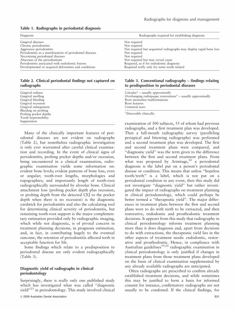
Table 1. Radiographs in periodontal diagnosis
Diagnosis Radiographs required for establishing diagnosis
Gingival diseases Not requiredChronic periodontitis Not requiredAggressive periodontitis Not required but sequential radiographs may display rapid bone lossPeriodontitis as a manifestation of periodontal diseases Not requiredNecrotizing periodontal diseases Not requiredAbscesses of the periodontium Not required but may reveal causePeriodontitis associated with endodontic lesions Required, as if for endodontic diagnosisDevelopmental or acquired deformities and conditions Required really only for some tooth related
Table 2. Clinical periodontal findings not captured onradiographs
Gingival rednessGingival swellingGingival bleedingGingival recessionGingival enlargementBleeding on probingProbing pocket depthsTooth hypermobilitySuppuration
Table 3. Conventional radiography – findings relatingto predisposition to periodontal diseases
Calculus* – usually approximallyOverhanging radiopaque restorations* – usually approximallyRoot anomalies ⁄ malformationsRoot featuresCemental tears
*Detectable clinically.
ª 2009 Australian Dental Association S31
Radiographs for diagnosis and management

example, indicate that a tooth is hopeless due toextreme and symptomatic hypermobility accompaniedby advanced loss of clinical attachment, a radiographexposed purely to document this assessment of thetooth being hopeless, based as it is on the clinicalfindings, is not justified. In reality, exposure of thepatient to additional radiation would only be war-ranted if the tooth could be salvaged following thestudy of a radiograph. The clinical findings, if accu-rately recorded in a patient’s records are sufficientdocumentation of the findings, and a ‘‘for the recordonly’’ radiograph is not advised. Further, in prescribingdental radiographs in Australia all the advice given19,20
should be followed. It has been proposed elsewhere thatfailure to minimize the x-ray dose exposure throughthe use of E-speed films and rectangular collimatingdevices may constitute a medico-legal issue.22 Indeed,the positioning device shown in Fig 1b is for acylindrical cone and not for a rectangular collimatingdevice. When manufacturers of positioning devices havebeen specifically questioned on the lack of rectangularpositioning devices, the reply has been that rectangularcollimating devices are not popular, and thus therewould not be a demand among those who purchasex-ray equipment. Certainly, in Sweden23 and Englandand Wales,24 published reports have suggested the
manufacturers’ assessments to be correct, documentingas they do that concern for minimizing radiationexposure is not widespread. However, the situation inAustralia with respect to general and specialist dentists’adherence to x-ray dose reduction practices is notknown at present. This would make a worthwhile study.
Radiographic features impacting upon treatmentdecisions
There are many features to do with bone and teethevident on conventional radiographs, in addition tothose relating to caries, endodontic, restorative andprosthodontic conditions, which can impact on peri-odontal treatment planning. These are listed in Table 4.One ‘‘paper case’’ based study with and withoutradiographs in periodontal diagnosis and treatmentplanning25 showed that the availability of radiographs,as in the earlier ‘‘diagnostic yield’’ study,21 resulted inmore extractions being planned. These two studiesstrongly support the contention that radiographsinform periodontal treatment planning by revealingnot what has been lost but what is remaining. What isremaining can be viewed from two aspects: (1) whatclinical challenges remain, in terms, for instance, ofdebriding sufficiently well the root surfaces of incom-pletely separated (fused) roots of molar teeth or teethwith radiographically evident root grooves ⁄ flutings onroots; and (2) how much length of root appears toremain embedded in alveolar bone, which estimation isallowed by radiographic examination and which is notdiscernable from the clinical examination.
Conventional radiographic views to assist periodontaltreatment planning
Conventional bitewing radiographs
Horizontal bitewing radiographs, while useful forapproximal caries detection, are not so useful ininforming periodontal treatment and treatment plan-ning if bone loss is in any way advanced. Verticalbitewing radiographs, whereby the film is placed withits long axis at 90º to the placement for horizontal
(a)
(b)
Fig 1. (a) Vertical (left) and horizontal (right) bitewing tab positions.(b) Vertical bitewing film holder.
Table 4. Conventional radiography – findings ofperiodontal interest impacting upon treatmentdecisions
Bone levelsBone loss – even or angular patternsIntra(infra) – bony defectsRoot morphologies ⁄ topographiesFurcation radiolucenciesEndodontic lesionsEndodontic mishapsDevelopmental anomaliesRoot length and shape(s) remaining in bone
S32 ª 2009 Australian Dental Association
EF Corbet et al.

bitewing radiography, can be very helpful if clinicallythere is nothing suggestive of any previous endodontictherapy or current periapical periodontitis alone or incombination with periodontal destruction. The verticalbitewing radiograph can be facilitated by placing the‘‘tab’’ on which the patient bites at 90º to its positionfor horizontal bitewings (Fig 1a), or by using a verticalbitewing holder (Fig 1b) through which, if there is acone positioning ring, a reasonable degree of reproduc-ibility can be achieved for subsequent sequentialradiographs. There is one Swiss product called theHawe Paro-Bite Centring Device which can be insertedinto a round positioning device which assists inthe positioning of rectangular cones for vertical andhorizontal bitewings (Fig 2). The assistance provided bysuch a positioning aid is advised to reduce the need forrepeat radiographs and hence the need for unnecessaryx-ray exposure.
For intact arches, probably two vertical bitewingradiographs per posterior sextant are required. Figure 3shows one vertical bitewing with bone levels anddefects apparent revealing root morphologies of rootsin need of debridement. Not all root apices are evidentand hence some periapical bone is not visible.
Conventional periapical radiographs
Periapical radiographs when exposed for periodontalpurposes should use long-cone paralleling projections,preferably with rectangular collimators. Full-mouthsurveys of paralleling periapical radiographs have beenconsidered to be a ‘‘gold standard’’ for periodontaldiagnosis and treatment planning. For some this viewstill persists. For instance, the European Federation ofPeriodontology still calls for this full-mouth series ofperiapical radiographs in case presentations by candi-dates at the conclusion of higher education and trainingin periodontology. However, there is no basis forconsidering a full-mouth series of paralleling periapical
radiographs to influence periodontal treatment deci-sions any more than, say, panoramic radiographs.The pictorial heading banner of the website* of theAustralian and New Zealand Academy of Periodontol-ogy shows a gloved hand holding a panoramic radio-graph. If a panoramic radiograph is available, havingbeen exposed for whatever purpose, that radiographmay alone be sufficient,26 or a panoramic radiographmay be supplemented by selected intra-oral radiographswhich numbered less than four per patient to reach the‘‘gold standard’’ in one study.27 It has been shown thatif seven periapical radiographs supplement a panoramicoral radiograph then the effective radiation doseexceeds that of a full-mouth series of periapicals,28
but if the number is less than four, then there is areduction in radiation exposure and yet the ‘‘goldstandard’’ in terms of information can be achieved.
Conventional panoramic oral radiographs
Modern panoramic oral radiography achieves decentimages suitable, with perhaps only modest intra-oralsupplementation, for periodontal treatment planningpurposes. The differences in any ‘‘yield’’, even with anolder generation of panoramic radiograph machines
Fig 2. A Kwikbite ⁄Parobite positioning device for a rectangularcollimating device for both vertical and horizontal bitewing
radiograph exposures, inserted into a round cone positioning ring.
Fig 3. A vertical bitewing, with bone levels and intrabony defectsapparent.
*URL: ‘http://anzap.org.au’. Accessed 20 March 2009.
ª 2009 Australian Dental Association S33
Radiographs for diagnosis and management

and technology, in comparison with periapicals,29 andbitewings,30 and bitewings and periapicals,31 andperiapicals and clinical probing32 were small, and fornewer panoramic radiograph technologies difference inany ‘‘yield’’ are apparently even smaller.33 Panoramicradiography in a group of periodontal maintenancepatients, that is patients previously affected by peri-odontal disease which had been treated and who wereundergoing supportive periodontal care, showed ‘‘greatagreement’’ with long cone intra-oral radiographs.34
Two of the recent reviews1,6 have dealt with the issue ofparalleling periapical series versus panoramic oralradiographs. The features of interest for periodontalassessment noted on periapical radiographs are alsocapable of being noted on panoramic radiographs(Table 5). For many practitioners the radiographicfeatures of interest on a panoramic, supplementedwhere necessary by a small number of intra-oral views,is sufficient for the management of periodontal diseases.Tugnait and Camichael6 note how there has been apragmatic shift by many towards panoramic radio-graphs in the investigation of patients with periodontaldiseases, in view of time efficiency, greater patienttolerance, and often a lower radiation exposure. Costsavings are also more and more an issue, and while themachinery for panoramic oral radiography is notcheap, it is nowadays relatively cheaper than formerly,and the time taken for producing a full-mouth periapi-cal series is a costly investment if there is little by way ofyield.
The American Academy of Periodontology (AAP) inits 2000 Parameter on Comprehensive PeriodontalExamination35 holds that ‘‘radiographs that are cur-rent, based on the diagnostic needs of the patient,
should be utilized for proper evaluation and interpre-tation of the status of the periodontium … Radiographsof diagnostic quality are necessary for these purposes’’.It further states ‘‘Radiographic abnormalities should benoted’’.35 Panoramic radiographs fulfil these conditions(Table 5) and allow for the identification of radio-graphic abnormalities. The point made by the AAP thatthere should be some record made of what was detectedon the radiographs is advice given in many jurisdic-tions. While available, the radiograph(s) reveal all thatcan be discerned, but if the radiograph(s) is(are) notavailable for whatever reason, then having somewritten record of the findings should obviate the needfor any additional repeat radiograph to compensate forthe temporary unavailability of the radiograph(s). TheAAP in its 2001 Position Paper on ‘‘Guidelines forperiodontal therapy’’36 holds that ‘‘interpretation of asatisfactory number of updated, diagnostic qualityperiapical and bitewing radiographs or other diagnosticimaging needed for implant therapy’’ is required. TheAAP contends that intra-oral radiographs, such asperiapical films and vertical or horizontal bitewings,provide a considerable amount of information aboutthe periodontium that cannot be obtained by any othernon-invasive means.37 Panoramic radiographs certainlydo provide a considerable amount of information(Table 5) and they also inform on treatment planning.The situation of the AAP becomes clear on viewing theAAP website* (‘‘Search Our Site’’: ‘‘Panoramic’’ in thegateway to ‘‘Members Only’’ AAP Insurance PolicyStatement – Radiographs in Periodontics), where it isstated: ‘‘The American Academy of Periodontologybelieves that panoramic radiographs have limited valuein the diagnosis of periodontal disease …’’ This,however, is only a ‘‘belief’’. A casual view of the worldreveals that not all share the same beliefs. The authorsof this review do not share the same belief as the AAP.The expense, time and physical inconvenience in havingall periodontitis patients subjected to a full-mouthseries of periapical and bitewing radiographs on thebasis of a belief can be questioned, as it is in this review.The American Board of Orthodontics is more reason-able in its advice, only requiring six intra-oral radio-graphs to supplement a panoramic view for adults forcomparison of pre-treatment and post-treatment crestalbone levels and root status.38 One earlier study showshow dearly belief systems with respect to full-mouthperiapical surveys can be held.39 In this Americanstudy, the proportion of patients who had the resultsfrom a screening clinical examination and a panoramicradiograph but who were still judged by independentexaminers to be in need of full-mouth periapical serieswas the same as for those patients who only had the
Table 5 (after Tugnait et al.1). Detectable features ofinterest on radiographs
Periapical Panoramic
Bone levels Yes YesBone loss Yes Yes
– even Yes Yes– angular Yes Yes
Furcation involvement Yes YesCalculus Yes YesRadio-opaque restorative margins Yes Yes
– deficiency Yes Yes– overhang Yes Yes
Root morphologies Yes YesRoot length embedded in alveolar bone Yes YesWidened periodontal ligament space Yes YesApproximal root caries Yes YesRoot canal fillings Yes YesPeriapical periodontitis, cysts, granulomas Yes YesImpacted third molars Yes YesRetained roots Yes YesFractured roots Yes YesCemental tears Often SometimesCysts ⁄ tumours Sometimes Yes *URL: ‘http://www.perio.org/index_pro.html.’ Accessed 20 March
2009.
S34 ª 2009 Australian Dental Association
EF Corbet et al.

clinical screening results. In that study, patients wereslated by the examiners for the full-mouth series ofperiapical radiographs on the basis of the case type intowhich they fell. Recommendations for the full-mouthperiapical radiograph series was made on the basis ofcase type, and information available from the pano-ramic was not used because in the examiners’ opinionsthat particular case type demanded the full-mouthperiapical series, because it seems that was what theyhad been taught. When dental teaching hospitals in theUnited Kingdom and Ireland were surveyed,40 the mostcommonly taken views to assess periodontal statuswere panoramic radiographs with selected periapicalradiographs. Hopefully, graduates from these dentalschools will follow their teaching, while constantlyevaluating the state of knowledge and experience in thisfield and being prepared to change practice as newevidence emerges.
Further, panoramic radiographs have been shown toreveal in a majority (63 per cent) of periodontalpatients some form of dental abnormality unrelated toperiodontal disease.41 General radiologists in Austral-asia have had recent advice on the interpretation ofdental panoramic radiographs42 and should thus beavailable for consultation, as would be other dentalspecialists in Australia, if abnormalities detected wereto be out of the ordinary.
Panoramic radiographs may not reveal alveolar bonydefects as accurately as periapical radiographs.43,44
However, that is not the issue. The issue must bewhether there is any additional therapeutic yield fromany greater accuracy in representation of alveolar bonedestruction revealed on periapical radiographs.
A small study was conducted in Hong Kong in whichpart-time clinical dental teachers were asked to developperiodontal treatment plans on the basis of, in the firstinstance, a complete periodontal charting, study castsand a panoramic radiograph. The 35 patient recordschosen were of adult patients with, what would now bediagnosed as, chronic periodontitis and who had atleast six teeth per quadrant. After a one-year wash-out
period, the same clinical records camouflaged, andstudy casts were given along with, on this secondoccasion, not the panoramic radiograph but a full-mouth series of paralleling periapical radiographswhich at the time of the charting had been prescribedby a dental surgeon in the clinic in compliance with hisprevious teaching. The individual treatment plansderived on the first and second occasion were almostidentical. Between examiners there was variation intreatment plans regarding periodontal surgery, as hasbeen reported from elsewhere,45 and extractions.However, each individual part-time clinical dentalteacher developed almost identical treatment plansfrom clinical findings and panoramic radiographs (ofan earlier generation) as they did from the clinicalfindings and the full-mouth paralleling periapicalradiograph series. Hence, there was no perceptible‘‘therapeutic yield’’ from the additional full-mouthperiapical radiograph series, such as that shown inFig 4.
The full-mouth series of periapical radiographsshown in Fig 4 is mounted on a clear, not a traditionallyblack, background. The Australian Safety Guide forRadiation Protection in Dentistry20 suggests mountingradiographs on a ‘‘mask’’ which eliminates stray lightaround the radiograph, and provision for magnificationis also suggested as being advisable. The periapicalradiographs in Fig 4 are mounted on a clear back-ground because each radiograph should be viewedagainst a light-box using a viewing box with in-builtmagnifying lens (Fig 5). Such viewing boxes areavailable and are highly recommended not only forconventional periapical and bitewing radiographs, butalso for the study of conventional panoramic oralradiographs in assessing crestal bone loss and alveolarbony defects.
While panoramic radiography may be less accurate inthe representation of bony defects than intra-oralradiography,43,44 this has little therapeutic effect inpractice. For instance, many therapeutic decisionsto do with the management of bony defects are not
Fig 4. A full-mouth periapical radiograph series.
ª 2009 Australian Dental Association S35
Radiographs for diagnosis and management

determined by the radiographic appearance, but ratherby the intra-operative appearance of the tooth-rootsand the bony defects. Guided tissue regeneration(GTR), it has been concluded in a systematic review,46
achieves 1.22 mm more gain in clinical attachmentlevel at pocket sites than open flap (access flap)debridement. Narrow and deep infrabony defects havebeen shown to respond radiographically and, to someextent at least, clinically more favourably to GTR thanwide and shallow defects, and depth was more indic-ative of favourable response than the angle of thedefect.47 This finding was confirmed in a follow-upstudy.48 GTR requires a surgical approach to thedefects. The surgical approach allows for direct intra-operative assessment of defect depths and angles. Noreliance should be made on the radiographic assessmentof the bony defect. The most that the radiographicassessment of defect depth and width might indicatewould be a preparedness to consider GTR as atherapeutic alternative. The findings with respect tothe defect on flap reflection – whether the defect iscontained or circumferential, indeed whether the toothis treatable and retainable or not – determine the
applicability of the GTR approach. This is decidedintra-operatively and not on the basis of the radio-graphic assessment alone, if indeed at all.
In a systematic review,49 Emdogain� (an enamelmatrix derivative) was found to have improved probingattachment levels by 1.2 mm and probing pocket depthreduction by 0.8 mm compared to open (access) flapdebridement, although these results have to be inter-preted with caution. The effectiveness of Emdogain� isalso dependent to an extent on defect depth and defectmorphology. Emdogain� has been shown to be verysuccessful, over a nine-year period, in deep defects50
and in angular defects.51,52 The depth of defect and itssuitability for Emdogain� regeneration are all madeintra-operatively. Questions of interest to the operator –such as ‘‘is the defect a deep defect?’’, ‘‘is it containedor circumferential?’’, ‘‘are the root surfaces amenableto debridement?’’ – can all be answered on the basis ofthe intra-operative direct assessment. The pre-surgicalradiographic assessment again may only indicate thatEmdogain� might be considered in the surgical treat-ment of that defect. Also, for Emdogain� regenerativetherapy, as for GTR, often in the clinical situation onsurgical reflection of flaps, defects reveal themselvesto be topographically well suited to regenerativeapproaches, when the pre-surgical radiographic assess-ment had not suggested such. In these clinical circum-stances, the radiographic assessment has not guided theeventual treatment approach adopted.
If an adjunctive regenerative approach had proved towork with non-surgical periodontal therapy for specificinfrabony defect depths and configurations, but not forothers, then pre-treatment radiographic accuracy inrepresenting defects would be at a premium. Sadly,however, Emdogain� has been shown to offer noadvantage when applied as an adjunct to non-surgicalperiodontal therapy.53–55 Hence for most therapeuticdecisions, and thus offering satisfactory ‘‘therapeuticyield’’, panoramic oral radiography is, notwithstandingits less accurate depiction of radiographically evidentalveolar bone defects, of great therapeutic use.
Digital panoramic radiography versus conventionalfilm panoramic radiography
There has, up to the present, been very little directcomparison between digital panoramic radiographs andconventional film panoramic radiographs in periodon-tal assessment. One study compared the efficacy of theOrthophos DS Digital panoramic system with conven-tional film obtained from the Orthophos Plus (bothSirona, Bensheim, Germany) and found that theconventional film outperformed the digital panoramicin the detection of periodontal findings.56 However,based on a couple of years practical experience ofusing digital panoramic oral radiographs, the authors’
Fig 5. A radiograph viewing box to provide magnification andto block out ambient light in use.
S36 ª 2009 Australian Dental Association
EF Corbet et al.

experience is that there is an advantage in periodontalassessment. The digital panoramic system with whichthe authors have experience uses a Kodak 8000CDigital Panoramic Machine (from Kodak-Trophy,Croissy-Beaubourg, France) using Kodak Dental Soft-ware, to feed into a patient database held on TrophyDICOM software which is transferred into the hospi-tal’s patient management system. An example of adigital panoramic is given in Fig 6a and a ‘‘zoomed-in’’portion of that image is shown in Fig 6b. The ability tomanipulate the panoramic images, such as zooming inand changing contrast, has proved a very useful featurein assessment of bony defects and root morphologies,and open (access) flap debridement surgeries haveconfirmed an impression of a greater ability in antici-pating the shape, depth and extent of bony defectsfollowing study of digital panoramic radiographscompared to viewing conventional panoramic oralradiographs. The ability to zoom in and magnifydefects and to adjust dynamically the contrast andbrightness allows for vastly improved ‘‘visualization’’of bone levels and intra-bony defects. To date one studyhas shown that periapical periodontitis was morescoreable on screen from digital panoramic radiographsthan on printed digital panoramic radiographs, sup-porting the advantages gained from manipulating thedigital panoramic oral images in assessing bony lesionsrather than interpreting only one static panoramic oralimage.57
Digital intra-oral radiography
The authors’ experience with digital intra-oral radio-graph for periodontal assessment is somewhat lessencouraging but nonetheless generally positive. Thereare two approaches to digital imaging, direct digitalimaging and indirect digital imaging. ‘‘Direct’’ isachieved by using a solid state sensor to detect x-rays.For intra-oral use these sensors are bulky and inflexible,and require a cable connection. These sensors mustconvert x-ray detection to electronic signals forsubsequent electronic processing to produce usableimages. These direct digital images can be of highquality. Readers are referred to earlier reviews2,5 for thefurther information on direct digital imaging in peri-odontal assessment. Since these reviews2,5 have beenpublished, there has been not much change to theconclusion that alveolar bone measurements are repro-ducible, using both direct digital and conventionalradiographs, and that direct digital radiographs do notenhance examiner agreement over conventional radio-graphs.58 Of course digital images, derived from directdigital imaging, or indeed also indirectly derived, can bestudied using image analyser tools. A study of one suchtool concluded that a dental image analyser tool canreliably replace conventional measuring on intra-oralfilm radiographs for measuring bone in periodontitispatients.59 Such image analyser tools may well becomemore of mainstream devices for quantifying bone loss,and also post-periodontal therapy bone gain or bonelevel stability.
Indirect digital imaging involves a latent image beingacquired using a photostimulable phosphor plate, andthen after the latent image is captured on this plate theimage is scanned by laser to produce a usable image.The scanning process either erases the latent image onthe plate so that the plate can be reused straight awaywithout there being double imaging, or else the imageon the plate is only degraded by laser scanning andmust be erased by exposure to light. There are variousadvantages of indirect digital imaging over direct digitalimaging. The plates can be the same sizes as intra-oralconventional x-ray films and so all conventional intra-oral clinical approaches can be used. Also there isno need for a cable, patient tolerance is greater, andthe expense is less. However, there are disadvantages.Depending on the approach, the image quality may notbe as good as with the direct imaging. Direct imagingproduces immediate images which is not necessarily sowith indirect digital imaging. Further, the flexibility ofthe plate means that image distortion due to platebending can occur, as with film bending in conventionalradiography. If the plates are scratched, effectively theyare ruined, and so plates must be treated with great careat all times. However, many other dental schools usingthe same hospital patient management system as used in
(a)
(b)
Fig 6. (a) Digital panoramic. (b) Zoomed-in portion of lowerleft posterior sextant.
ª 2009 Australian Dental Association S37
Radiographs for diagnosis and management

Hong Kong, into which indirect digital images are fed,report very high quality images with great utilities.Some studies on marginal alveolar bone levels doconfirm that indirect digital images had favourablemeasurement accuracy compared with film radio-graphs,60 while colourizing digital images using col-our-coding algorithms did not produce greater accuracyin this respect.61 However, indirect digital bitewingshave been shown to be no better than film bitewings inthe assessment of alveolar bone loss.62 If decent imagesare produced from indirect digital imaging, there isno evidence to suggest that indirect digital intra-oralradiographs are inferior to conventional intra-oralradiographs in periodontal assessment and treatmentplanning.
There is another approach to the management ofradiographs, sometime erroneously also referred to asindirect digital imaging, and that is the digitization ofexposed and processed conventional x-radiograph filmsusing a flatbed scanner with a transparency adaptor.This is the approach used to date in the digitalsubtraction radiography (DSR) approach, consideredbelow. The viewing possibilities, e.g., projection ofsuch images, may improve interpretation,63 and suchsubsequent management of conventional intra-oralradiographs may facilitate the interpretation of peri-odontal bone defects,64 and of course image analysistools can be used on these digitized radiographs.
Digital subtraction radiography
Digital subtraction radiography (DSR) in periodontol-ogy basically allows the detection of small changes inalveolar bone, which might otherwise go undetected.For DSR to permit this to be realized, serial radiographsneed to be taken with the best possible reproducibleprojection geometry and using standardized imageprocessing. To optimize the projection geometry cus-tom (patient-by-patient, area-by-area) bite blocks must
be made and attached to the film holders and the filmholder must be reproducibly aligned to the x-ray beamcollimating device (Fig 7). Once standardized serialperiapical radiographs have been produced, theseconventional radiographs must then be digitized usingflatbed scanning devices. Obviously, this step can beomitted if direct or indirect digital images have beencollected. Then there are various methods to deter-mine the changes (+ indicating more bone ⁄ more bonedensity, – indicating less bone ⁄ less bone density)between two radiographic images. Reference alumin-ium wedges can be captured in the images which moreeasily allow for determination of density correction.65
Computer-assisted densitometric image analysis(CADIA)66 of the digitized standardized intra-oralradiographs is the method with which the authors haveexperience.
A DSR system was developed at The University ofHong Kong which has been calibrated and validated.67
This DSR system has been used in a published study ona low-power laser system in periodontal therapy.68 Ithas also been used in clinical studies on comparingperiodontal surgical therapy with repeated non-surgical
Fig 7. A custom positioned periapical film holder with bite blockattached to a collimating device for digital subtraction radiography.
Fig 8. Cone-beam Computed Tomography Machine (i-CAT,Imaging Sciences International, Hatfield, USA).
S38 ª 2009 Australian Dental Association
EF Corbet et al.

therapy, and on non-surgical therapy on post-meno-pausal females, comparing those taking with those nottaking hormone replacement therapy. Thus, quite someexperience with this approach has been gleaned. Theconclusion would be in line with that of Bragger7 thatDSR remains primarily a research tool for clinical trials.Mol5 discusses how it is often proposed that the timeand effort involved in producing subtraction images ofhigh quality to detect small changes is prohibitive inclinical practice. There are more issues to be consid-ered. The storage of multiple custom bite blocks andholders is a very practical one. The time taken in thealignment of images prior to the CADIA is another.Slight variations in choice of the regions of interest arecapable of producing contrary results. Also, if DSRdetects a loss of bone density or volume, but clinicallyat that site there are no signs of gingival inflammation,no bleeding on probing and no probing pocket depth,then the reaction can be ‘‘so what?’’, because there isnothing in the routine clinical periodontal armamen-tarium which can be applied at such a site. Mol5 opinesthat at one level or another a price has to be paid forincreased diagnostic utility. But when the DSR ‘‘diag-nosis’’ does not suggest any clinically useful interven-tion, then not only the ‘‘price’’ but the ‘‘utility’’ can alsobe questioned. A further issue is what sites to select.Studies can focus on specific sites, such as furcations69
and can show favourable outcomes to therapy in termsof bone behaviour. A recent study of scaling and rootplaning had 13 subjects but each subject had only threesites included in the study.70 It can be questioned whatis special or representative about the sites chosen. TheDSR did reveal favourable outcomes to scaling androoting planing, but studies on scaling and root planinghave shown favourable effects on bone detectableby conventional radiography. DSR was used in thepublished study on low-power laser as an adjunct inperiodontal therapy.68 It showed a biological andmeasurable early effect on bone, which could not bedetected by clinical means, but again the questionsuggested is ‘‘so what?’’ as no clinically detectablebenefit could be observed.
A final issue in DSR is that periodontally involvedteeth are often mobile and can be displaced by the biteblock and ⁄ or the process of registering the bite prior totherapy, but such teeth can firm up, and for driftedteeth they may reposition themselves, in response totherapy. Then the custom bite block does not fit post-therapy and the serial images cannot be aligned and thealtered tooth positions in the serial images cannot becorrected for. The American Academy of Periodon-tology Position Paper on Diagnosis of PeriodontalDiseases37 very optimistically holds that future devel-opment of subtraction radiography techniques promises
Fig 9. i-CAT Vision software interface, consisting of pan-map (upper right), horizontal section (upper left), vertical sections (lower right)and reconstructed 3-dimensional model (lower left).
ª 2009 Australian Dental Association S39
Radiographs for diagnosis and management

to have a profound impact on the diagnosis ofperiodontal diseases. The authors of this review couldnot in any way agree.
Cone-beam computed tomography
Computed tomography (CT) has been used in somestudies in relation to periodontal defects.71,72 However,conventional CT does not offer any favourable cost-benefit, dose exposure or therapeutic yield advantage inperiodontal practice and is unlikely to find a routineplace.
Cone-beam geometry allows for reduced dose ofradiation. This, in combination with ‘‘fast’’ receptorsand the reduced cost of manufacturing the machines,has allowed for the introduction of cone-beam CT intodental ⁄ oral imaging. One difference between cone-beam CT and conventional CT is that the cone-beamproduces increased scatter on images making cone-beam CT unsuitable for soft tissue, a major benefit ofconventional CT, which in fact is an advantage indental ⁄ oral radiography wherein only radio-opaquestructures are generally studied. Machines specificallyfor dental ⁄ oral use have been brought to the marketand are hugely impacting the field of dental ⁄ oralimaging. Figure 8 shows one such machine, whichwhile less obtrusive than a medical CT machine, stillrequires space to accommodate it. However, it seemsunlikely that these machines will soon become routinein general dental practices. Because the cone-beamgeometry allows for a large volume of tissues to bescanned with a single sweep resulting in a digital image,cone-beam CT is also known in some quarters as digitalvolume tomography (DVT).
Cone-beam CT for assessment of periodontal defectshas been applied in in vitro studies.73–77 These have allsuggested that there ought to be an application for cone-beam CT in vivo in the imaging of periodontal defects.There has, so far, been only one clinical report, of 12patients, which suggests that cone-beam CT may providedetailed information about furcation involvements inpatients with chronic periodontitis78 and so may influ-ence treatment planning decisions. Obviously moreresearch is required. A preliminary study is underwayin Hong Kong on the utility of cone-beam CT inperiodontal assessment and in informing treatmentplanning decisions in periodontitis patients. It takeslonger for general dentists, periodontology trainees and
Fig 11. Vertical views showing lingual furcation involvement at lower left first molar (36) and an extensive defect at palatal aspect of the upper leftfirst molar (26).
Fig 10. Horizontal views of periodontal alveolar bony defects at upperleft second premolar (25) to upper left second molar (27).
S40 ª 2009 Australian Dental Association
EF Corbet et al.
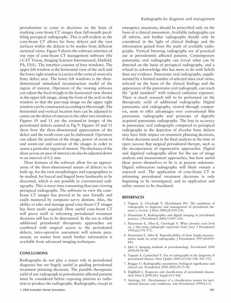
periodontists to come to decisions on the basis ofstudying cone-beam CT images than full-mouth paral-leling periapical radiographs. This is self-evident as thecone-beam CT allows for bony defects and the rootsurfaces within the defects to be studies from differentsectional views. Figure 9 shows the software interface ofone type of cone-beam CT management software (thei-CAT Vision, Imaging Sciences International, Hatfield,PA, USA). The interface consists of four windows. Theupper left window is the horizontal view of the arch andthe lower right window is a series of the vertical views of abony defect area. The lower left windows is the three-dimensional stimulated reconstruction model of theregion of interest. Operators of the viewing softwarecan adjust the focal trough in the horizontal view shownin the upper left image, along the form of the arch in thiswindow so that the pan-map image on the upper rightwindow can be constructed according to this trough. Thehorizontal and vertical location bars can be adjusted tocentre on the defect of interest in the other two windows.Figures 10 and 11 are the zoomed-in images of theperiodontal defects centred in Fig 9. Figures 10 and 11show how the three-dimensional appreciation of thedefect and the tooth-roots can be elaborated. Operatorscan adjust the position of the image, power of zoom-inand zoom-out and contrast of the images in order toassess a particular region of interest. The thickness of theslices across an area of interest can also be adjusted downto an interval of 0.2 mm.
These features of the software allow for an appreci-ation of the three-dimensional nature of defects to bebuilt up, for the root morphologies and topographies tobe studied, for buccal and lingual bony landmarks to bediscerned, which is not possible in conventional radi-ography. This is more time consuming than just viewingperiapical radiographs. The software to view the cone-beam CT images has proved to be user friendly andeasily mastered by computer savvy dentists. Also, theability to take and manage good cone-beam CT imageshas been easily acquired. How useful cone-beam CTwill prove itself in informing periodontal treatmentdecisions still has to be determined. In the era in whichadditional periodontal therapeutic approaches arecombined with surgical access to the periodontaldefects, intra-operative assessment will remain para-mount, no matter how much further information isavailable from advanced imaging techniques.
CONCLUSIONS
Radiographs do not play a major role in periodontaldiagnosis but are hugely useful in guiding periodontaltreatment planning decisions. The possible therapeuticyield of any radiograph in periodontitis affected patientsmust be considered before exposing patients to radia-tion to produce the radiographs. Radiographs, except in
emergency situations, should be prescribed only on thebasis of a clinical assessment. Available radiographs canall inform, and further radiographs should only beconsidered in the light of clinical findings and theinformation gained from the study of available radio-graphs. Vertical bitewing radiographs are of practicaluse in periodontitis affected patients. Contemporarypanoramic oral radiographs can reveal what can bedetected on the basis of periapical radiographs, and arefusal to acknowledge this has more to do with beliefsthan any evidence. Panoramic oral radiographs, supple-mented by a limited number of selected intra-oral views,selected on the basis of the clinical findings and theappearance of the panoramic oral radiograph, can reachthe ‘‘gold standard’’ with reduced radiation exposure.There is much research still to be performed on thetherapeutic yield of additional radiographs. Digitalpanoramic oral radiographs, viewed through comput-ers, seem to offer advantages over conventional filmpanoramic radiographs and printouts of digitallyacquired panoramic radiographs. The loss in accuracyin panoramic oral radiographs compared to periapicalradiographs in the depiction of alveolar bone defectsmay have little impact on treatment planning decisions,if these decisions need in the final analysis at the time ofopen (access) flap surgical periodontal therapy, such asthe incorporation of regenerative approaches. Digitaland digitized radiographs allow for the use of imageanalysis and measurement approaches, but how usefulthese prove themselves to be is at present unknown.Digital subtraction radiography will likely remain aresearch tool. The application of cone-beam CT ininforming periodontal treatment decisions is onlybeginning to be investigated, and its application andutility remain to be elucidated.
REFERENCES
1. Tugnait A, Clerehugh V, Hirschmann PN. The usefulness ofradiographs in diagnosis and management of periodontal dis-eases: a review. J Dent 2000;28:219–226.
2. Hausmann E. Radiographic and digital imaging in periodontalpractice. J Periodontol 2000;71:497–503.
3. Hausmann E, Allen K, Clerehugh V. What alveolar crest levelon a bite-wing radiograph represents bone loss? J Periodontol1991;62:570–572.
4. Hausmann E, Allen K. Reproducibility of bone height measure-ments made on serial radiographs. J Periodontol 1997;68:839–841.
5. Mol A. Imaging methods in periodontology. Periodontol 20002004;34:34–48.
6. Tugnait A, Carmichael F. Use of radiographs in the diagnosis ofperiodontal disease. Dent Update 2005;32:536–538; 541–532.
7. Bragger U. Radiographic parameters: biological significance andclinical use. Periodontol 2000 2005;39:73–90.
8. Highfield J. Diagnosis and classification of periodontal disease.Aust Dent J 2009;54(1 Suppl):S11–S26.
9. Armitage GC. Development of a classification system for peri-odontal diseases and conditions. Ann Periodontol 1999;4:1–6.
ª 2009 Australian Dental Association S41
Radiographs for diagnosis and management

10. Armitage GC. Periodontal diagnosis and classification of peri-odontal diseases. Periodontol 2000 2004;34:9–21.
11. CatonJ,GreenwellH,MahanondaR,etal.Consensusreport:dentalplaque-induced gingival diseases. Ann Periodontol 1999;4:18–19.
12. Lindhe J, Ranney R, Lamster I, et al. Consensus report: chronicperiodontitis. Ann Periodontol 1999;4:38.
13. Lang N, Bartold PM, Cullinan M, et al. Consensus report:aggressive periodontitis. Ann Periodontol 1999;4:53.
14. Lang N, Soskolne WA, Greenstein G, et al. Consensus report:necrotizing periodontal diseases. Ann Periodontol 1999;4.
15. Lang N, Soskolne WA, Greenstein G, et al. Consensus report:abscesses of the periodontium. Ann Periodontol 1999;4:83.
16. Lang NP, Soskolne WA, Greenstein G, et al. Consensus report:periodontic-endodontic lesions. Ann Periodontol 1999;4:90.
17. Updegrave WJ. The paralleling extension-cone technique inintraoral dental radiography. Oral Surg Oral Med Oral Pathol1951;4:1250–1261.
18. Ishikawa I, McGuire MK, Mealey B, et al. Consensus report:localized tooth-related factors that modify or predispose to pla-que-induced gingival diseases and periodontitis. Ann Periodontol1999;4:97.
19. Australian Radiation Protection and Nuclear Safety Agency.Code of Practice for Radiation Protection in Dentistry. Canberra:Australian Government and ARPANSA, 2005.
20. Australian Radiation Protection and Nuclear Safety Agency.Safety Guide for Radiation Protection in Dentistry. Canberra:Australian Government and ARPANSA, 2005.
21. Weems RA, Manson-Hing LR, Jamison HC, Greer DF. Diag-nostic yield and selection criteria in complete intraoral radio-graphy. J Am Dent Assoc 1985;110:333–338.
22. Zinman E. Dental and legal considerations in periodontaltherapy. Periodontol 2000 2001;25:114–130.
23. Svenson B, Soderfeldt B, Grondahl HG. Attitudes of Swedishdentists to the choice of dental X-ray film and collimator for oralradiology. Dentomaxillofac Radiol 1996;25:157–161.
24. Tugnait A, Clerehugh V, Hirschmann PN. Radiographic equip-ment and techniques used in general dental practice: a survey ofgeneral dental practitioners in England and Wales. J Dent 2003;31:197–203.
25. Tugnait A, Hirschmann PN, Clerehugh V. Validation of amodel to evaluate the role of radiographs in the diagnosis andtreatment planning of periodontal diseases. J Dent 2006;34:509–515.
26. Dundar N, Ilgenli T, Kal BI, Boyacioglu H. The frequency ofperiodontal infrabony defects on panoramic radiographs of anadult population seeking dental care. Community Dent Health2008;25:226–230.
27. Molander B, Ahlqwist M, Grondahl HG. Panoramic andrestrictive intraoral radiography in comprehensive oral radio-graphic diagnosis. Eur J Oral Sci 1995;103:191–198.
28. Jenkins WM, Brocklebank LM, Winning SM, Wylupek M,Donaldson A, Strang RM. A comparison of two radiographicassessment protocols for patients with periodontal disease.Br Dent J 2005;198:565–569; discussion 557; quiz 586.
29. Rohlin M, Kullendorff B, Ahlqwist M, Henrikson CO, HollenderL, Stenstrom B. Comparison between panoramic and periapicalradiography in the diagnosis of periapical bone lesions. Dento-maxillofac Radiol 1989;18:151–155.
30. Akesson L, Rohlin M, Hakansson J, Hakansson H, Nasstrom K.Comparison between panoramic and posterior bitewing radio-graphy in the diagnosis of periodontal bone loss. J Dent1989;17:266–271.
31. Akesson L, Rohlin M, Hakansson J. Marginal bone in peri-odontal disease: an evaluation of image quality in panoramic andintra-oral radiography. Dentomaxillofac Radiol 1989;18:105–112.
32. Akesson L, Hakansson J, Rohlin M. Comparison of panoramicand intraoral radiography and pocket probing for the measure-ment of the marginal bone level. J Clin Periodontol 1992;19:326–332.
33. Kim TS, Obst C, Zehaczek S, Geenen C. Detection of bone losswith different X-ray techniques in periodontal patients. J Peri-odontol 2008;79:1141–1149.
34. Persson RE, Tzannetou S, Feloutzis AG, Bragger U, Persson GR,Lang NP. Comparison between panoramic and intra-oral radio-graphs for the assessment of alveolar bone levels in a periodontalmaintenance population. J Clin Periodontol 2003;30:833–839.
35. American Academy of Periodontology. Parameter on compre-hensive periodontal examination. J Periodontol 2000;71:847–848.
36. Greenwell H. Committee on Research, Science and Therapy.Position paper: guidelines for periodontal therapy. J Periodontol2001;72:1624–1628.
37. Position paper: diagnosis of periodontal diseases. J Periodontol2003;74:1237–1247.
38. Grubb JE, Greco PM, English JD, et al. Radiographic and peri-odontal requirements of the American Board of Orthodontics:a modification in the case display requirements for adult andperiodontally involved adolescent and preadolescent patients. AmJ Orthod Dentofacial Orthop 2008;134:3–4.
39. Kantor ML, Slome BA. Efficacy of panoramic radiography indental diagnosis and treatment planning. J Dent Res 1989;68:810–812.
40. Tugnait A, Clerehugh DV, Hirschmann PN. Survey of radio-graphic practices for periodontal disease in UK and Irishdental teaching hospitals. Dentomaxillofac Radiol 2000;29:376–381.
41. Osborne GE, Hemmings KW. A survey of disease changesobserved on dental panoramic tomographs taken of patientsattending a periodontology clinic. Br Dent J 1992;173:166–168.
42. Boeddinghaus R, Whyte A. Dental panoramic tomography: anapproach for the general radiologist. Australas Radiol2006;50:526–533.
43. Pepelassi EA, Diamanti-Kipioti A. Selection of the most accuratemethod of conventional radiography for the assessment of perio-dontal osseous destruction. J Clin Periodontol 1997;24:557–567.
44. Pepelassi EA, Tsiklakis K, Diamanti-Kipioti A. Radiographicdetection and assessment of the periodontal endosseous defects.J Clin Periodontol 2000;27:224–230.
45. Cosyn J, De Bruyn H. Interclinician disparity in periodontaldecision making: need for consensus statements on surgicaltreatment. J Periodontal Res 2007;42:311–317.
46. Needleman IG, Worthington HV, Giedrys-Leeper E, Tucker RJ.Guided tissue regeneration for periodontal infra-bony defects.Cochrane Database Syst Rev 2006:CD001724.
47. Klein F, Kim TS, Hassfeld S, et al. Radiographic defect depth andwidth for prognosis and description of periodontal healing ofinfrabony defects. J Periodontol 2001;72:1639–1646.
48. Eickholz P, Horr T, Klein F, Hassfeld S, Kim TS. Radiographicparameters for prognosis of periodontal healing of infrabonydefects: two different definitions of defect depth. J Periodontol2004;75:399–407.
49. Esposito M, Grusovin MG, Coulthard P, Worthington HV.Enamel matrix derivative (Emdogain) for periodontal tissueregeneration in intrabony defects. Cochrane Database Syst Rev2005:CD003875.
50. Sculean A, Schwarz F, Chiantella GC, Arweiler NB, Becker J.Nine-year results following treatment of intrabony periodontaldefects with an enamel matrix derivative: report of 26 cases. Int JPeriodontics Restorative Dent 2007;27:221–229.
S42 ª 2009 Australian Dental Association
EF Corbet et al.

51. Pontoriero R, Wennstrom J, Lindhe J. The use of barrier mem-branes and enamel matrix proteins in the treatment of angularbone defects. A prospective controlled clinical study. J ClinPeriodontol 1999;26:833–840.
52. Heden G, Wennstrom J, Lindhe J. Periodontal tissue alterationsfollowing Emdogain treatment of periodontal sites with angularbone defects. A series of case reports. J Clin Periodontol1999;26:855–860.
53. Sculean A, Windisch P, Keglevich T, Gera I. Histologic evaluationof human intrabony defects following non-surgical periodontaltherapy with and without application of an enamel matrix pro-tein derivative. J Periodontol 2003;74:153–160.
54. Gutierrez MA, Mellonig JT, Cochran DL. Evaluation of enamelmatrix derivative as an adjunct to non-surgical periodontaltherapy. J Clin Periodontol 2003;30:739–745.
55. Giannopoulou C, Andersen E, Brochut P, Plagnat D, Mombelli A.Enamel matrix derivative and systemic antibiotics as adjuncts tonon-surgical periodontal treatment: biologic response. J Peri-odontol 2006;77:707–713.
56. Ramesh A, Tyndall DA, Ludlow JB. Evaluation of a new digitalpanoramic system: a comparison with film. DentomaxillofacRadiol 2001;30:98–100.
57. Ridao-Sacie C, Segura-Egea JJ, Fernandez-Palacin A, Bullon-Fernandez P, Rios-Santos JV. Radiological assessment of peri-apical status using the periapical index: comparison of periapicalradiography and digital panoramic radiography. Int Endod J2007;40:433–440.
58. Pecoraro M, Azadivatan-le N, Janal M, Khocht A. Comparison ofobserver reliability in assessing alveolar bone height on directdigital and conventional radiographs. Dentomaxillofac Radiol2005;34:279–284.
59. Teeuw WJ, Coelho L, Silva A, et al. Validation of a dental imageanalyzer tool to measure alveolar bone loss in periodontitispatients. J Periodontal Res 2009;44:94–102.
60. Li G, Engstrom PE, Nasstrom K, Lu ZY, Sanderink G, Welander U.Marginal bone levels measured in film and digital radiographscorrected for attenuation and visual response: an in vivo study.Dentomaxillofac Radiol 2007;36:7–11.
61. Li G, Engstrom PE, Welander U. Measurement accuracy ofmarginal bone level in digital radiographs with and without colorcoding. Acta Odontol Scand 2007;65:254–258.
62. Henriksson CH, Stermer EM, Aass AM, Sandvik L, Moystad A.Comparison of the reproducibility of storage phosphor and filmbitewings for assessment of alveolar bone loss. Acta OdontolScand 2008;66:380–384.
63. Lanning SK, Best AM, Temple HJ, Richards PS, Carey A,McCauley LK. Accuracy and consistency of radiographic inter-pretation among clinical instructors in conjunction with a train-ing program. J Dent Educ 2006;70:545–557.
64. Gomes-Filho IS, Sarmento VA, de Castro MS, et al. Radiographicfeatures of periodontal bone defects: evaluation of digitizedimages. Dentomaxillofac Radiol 2007;36:256–262.
65. Allen KM, Hausmann E. Analytical methodology in quantitativedigital subtraction radiography: analyses of the aluminum refer-ence wedge. J Periodontol 1996;67:1317–1321.
66. Bragger U, Pasquali L, Rylander H, Carnes D, Kornman KS.Computer-assisted densitometric image analysis in periodontal
radiography. A methodological study. J Clin Periodontol 1988;15:27–37.
67. Woo BM, Zee KY, Chan FH, Corbet EF. In vitro calibration andvalidation of a digital subtraction radiography system usingscanned images. J Clin Periodontol 2003;30:114–118.
68. Lai SM, Zee KY, Lai MK, Corbet EF. Clinical and radiographicinvestigation of the adjunctive effects of a low-power He-Ne laserin the treatment of moderate to advanced periodontal disease:a pilot study. Photomed Laser Surg 2009;27:287–293.
69. Cury PR, Araujo NS, Bowie J, Sallum EA, Jeffcoat MK. Com-parison between subtraction radiography and conventionalradiographic interpretation during long-term evaluation of peri-odontal therapy in Class II furcation defects. J Periodontol2004;75:1145–1149.
70. Hwang YJ, Fien MJ, Lee SS, et al. Effect of scaling and rootplaning on alveolar bone as measured by subtraction radiogra-phy. J Periodontol 2008;79:1663–1669.
71. Naito T, Hosokawa R, Yokota M. Three-dimensional alveolarbone morphology analysis using computed tomography. J Peri-odontol 1998;69:584–589.
72. Pistorius A, Patrosio C, Willershausen B, Mildenberger P, RippenG. Periodontal probing in comparison to diagnosis by CT-scan.Int Dent J 2001;51:339–347.
73. Mengel R, Candir M, Shiratori K, Flores-de-Jacoby L. Digitalvolume tomography in the diagnosis of periodontal defects: an invitro study on native pig and human mandibles. J Periodontol2005;76:665–673.
74. Misch KA, Yi ES, Sarment DP. Accuracy of cone beam computedtomography for periodontal defect measurements. J Periodontol2006;77:1261–1266.
75. Mol A, Balasundaram A. In vitro cone beam computed tomo-graphy imaging of periodontal bone. Dentomaxillofac Radiol2008;37:319–324.
76. Vandenberghe B, Jacobs R, Yang J. Detection of periodontal boneloss using digital intraoral and cone beam computed tomographyimages: an in vitro assessment of bony and ⁄ or infrabony defects.Dentomaxillofac Radiol 2008;37:252–260.
77. Noujeim M, Prihoda T, Langlais R, Nummikoski P. Evaluationof high-resolution cone beam computed tomography in thedetection of simulated interradicular bone lesions. Dentomax-illofac Radiol 2009;38:156–162.
78. Walter C, Kaner D, Berndt DC, Weiger R, Zitzmann NU.Three-dimensional imaging as a pre-operative tool in decisionmaking for furcation surgery. J Clin Periodontol 2009;36:250–257.
Address for correspondence:Professor Esmonde Corbet
PeriodontologyPrince Philip Dental Hospital
34 Hospital RoadHong Kong
Email: [email protected]
ª 2009 Australian Dental Association S43
Radiographs for diagnosis and management