
Copyright 2010, John Wiley & Sons, Inc. Chapter 19 The Digestive System.
59
Copyright 2010, John Wiley & Sons, Inc. Chapter 19 The Digestive System
-
date post
21-Dec-2015 -
Category
Documents
-
view
217 -
download
1
Transcript of Copyright 2010, John Wiley & Sons, Inc. Chapter 19 The Digestive System.

Copyright 2010, John Wiley & Sons, Inc.
Chapter 19
The Digestive System

Copyright 2010, John Wiley & Sons, Inc.
Functions of the Digestive System Ingestion: eating Secretion: release of water, enzymes, buffers Mixing and propulsion: movement along GI
tract Digestion: breakdown of foods
Mechanically: by movements of digestive organs Chemically: by enzymes
Absorption: moving products of digestion into the body
Defecation: dumping waste products

Copyright 2010, John Wiley & Sons, Inc.
Organs of the Digestive System Gastrointestinal (GI) tract
A tube through which foods pass and where digestion and absorption occur.
Includes: mouth, pharynx, esophagus, stomach, small intestine, large intestine
Accessory organs: Organs that help in digestion but through which
food never passes. Includes: teeth, tongue, salivary glands, liver,
gallbladder, and pancreas

Copyright 2010, John Wiley & Sons, Inc.
Organs of the Digestive System

Copyright 2010, John Wiley & Sons, Inc.
Layers of the Gastrointestinal (GI) Wall Four layers from lower esophagus to anus
1. Mucosa: epithelium in direct content with food; made of connective tissue, glands, and thin muscularis mucosae
2. Submucosa: connective tissue, blood vessels, lymphatic vessels, and enteric nervous system (ENS)

Copyright 2010, John Wiley & Sons, Inc.
Layers of the Gastrointestinal (GI) Wall3. Muscularis: inner circular layer, outer longitudinal
layer Smooth muscle in most of GI tract Except skeletal (voluntary muscle) in mouth, pharynx,
upper esophagus, and external anal sphincter
4. Serosa: visceral layer of peritoneum Also forms extensions: greater omentum and mesentery

Copyright 2010, John Wiley & Sons, Inc.
Layers of the Gastrointestinal (GI) Wall

Copyright 2010, John Wiley & Sons, Inc.
Layers of the Gastrointestinal (GI) Wall

Copyright 2010, John Wiley & Sons, Inc.
Layers of the Gastrointestinal (GI) Wall

Copyright 2010, John Wiley & Sons, Inc.
Mouth (Oral Cavity) Formed by
Cheeks and tongue Hard palate anteriorly, soft palate posteriorly
Uvula U-shaped extension of soft palate posteriorly During swallowing, uvula blocks entry of food or
drink into nasal cavity Tongue: muscular accessory organ
Maneuvers food for chewing Adjusts shape for speech and swallowing
Lingual tonsils at base of tongue

Copyright 2010, John Wiley & Sons, Inc.
Salivary Glands Exocrine glands with ducts that empty into
oral cavity Three pairs of salivary glands
Parotid Largest; inferior and anterior to ears
Submandibular In floor of mouth; medial and inferior to mandible
Sublingual Inferior to tongue and superior to submandibular
Saliva: 99.5% water, salivary amylase, mucus and other solutes Dissolves food and starts digestion of starches

Copyright 2010, John Wiley & Sons, Inc.
Salivary Glands

Copyright 2010, John Wiley & Sons, Inc.
Teeth Accessory organs in bony sockets of
mandible and maxilla Three external regions
Crown: above gums Root: part(s) embedded in socket Neck: between crown and root near gum line
Three layers of material Enamel: hardest substance in body; over crown Dentin: majority of interior of tooth Pulp cavity: nerve, blood vessel, and lymphatics

Copyright 2010, John Wiley & Sons, Inc.
Teeth

Copyright 2010, John Wiley & Sons, Inc.
Teeth Humans have two sets of teeth
The 20 deciduous teeth are replaced by the permanent teeth between ages 6 and 12 years.
The 32 permanent teeth appear between 6 years and adulthood.
Four types of teeth Incisors (8): used to cut food Cuspids (canines) (4): used to tear food Premolars (8): for crushing and grinding food Molars (12): used for crushing and grinding food
Copyright 2010, John Wiley & Sons, Inc.
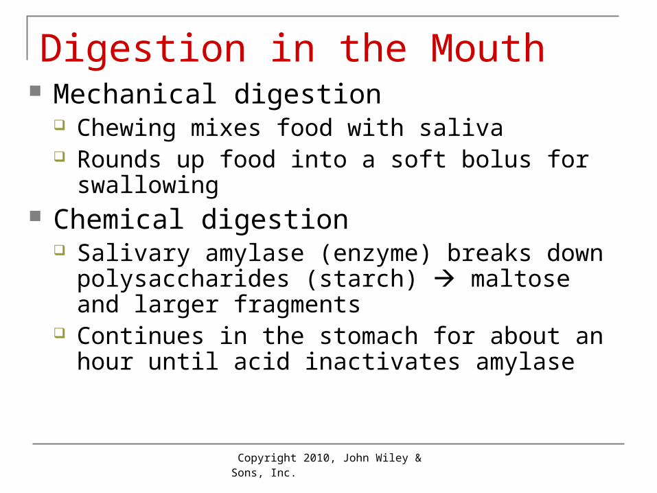
Digestion in the Mouth Mechanical digestion
Chewing mixes food with saliva Rounds up food into a soft bolus for swallowing
Chemical digestion Salivary amylase (enzyme) breaks down
polysaccharides (starch) maltose and larger fragments
Continues in the stomach for about an hour until acid inactivates amylase

Copyright 2010, John Wiley & Sons, Inc.
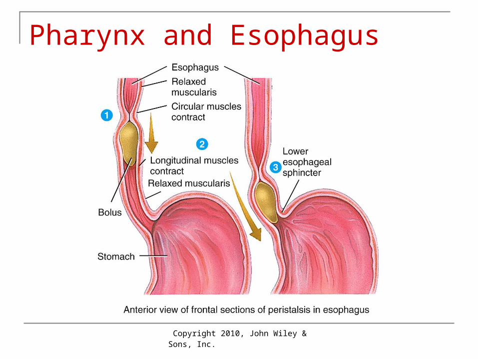
Pharynx and Esophagus Food passages from mouth stomach Swallowing: 3 stages
Voluntary stage: bolus of food oropharynx Pharyngeal stage: oropharynx esophagus
Soft palate moves up and epiglottis moves down; prevent food from entering nasopharynx and larynx
Esophageal: food stomach by peristalsis Esophageal sphincters:
Upper: controls entry esophagus Lower: controls entry stomach; GERD affects

Copyright 2010, John Wiley & Sons, Inc.
Pharynx and Esophagus
Copyright 2010, John Wiley & Sons, Inc.
Pharynx and Esophagus

Copyright 2010, John Wiley & Sons, Inc.
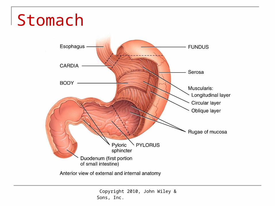
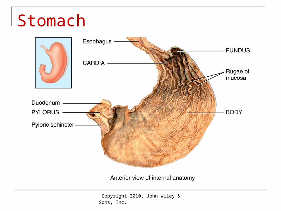
Stomach J- shaped enlargement of GI tract Mixing chamber and holding reservoir Very elastic/expandable and muscular Four regions
Cardia: surrounds upper opening Fundus: superior and to left of cardia Body: large central portion Pylorus: lower part leading to pyloric sphincter
and duodenum
Copyright 2010, John Wiley & Sons, Inc.
Stomach
Copyright 2010, John Wiley & Sons, Inc.
Stomach

Copyright 2010, John Wiley & Sons, Inc.
Stomach Wall: Four Layers1. Mucosa
Empty stomach lies in folds called rugae Epithelium: simple columnar; glands secrete
mucus Gastric glands line gastric pits
2. Secretory cells Mucous cells mucus Parietal cells HCl and intrinsic factor
These secretions collectively called gastric juice Intrinsic factor helps with vitamin B12 absorption needed
for RBC formation. If missing anemia Chief cells inactive enzyme pepsinogen G cells secrete gastrin (hormone) into blood
Copyright 2010, John Wiley & Sons, Inc.
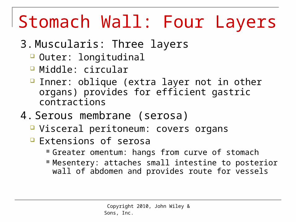
Stomach Wall: Four Layers3. Muscularis: Three layers
Outer: longitudinal Middle: circular Inner: oblique (extra layer not in other organs)
provides for efficient gastric contractions
4. Serous membrane (serosa) Visceral peritoneum: covers organs Extensions of serosa
Greater omentum: hangs from curve of stomach Mesentery: attaches small intestine to posterior wall of
abdomen and provides route for vessels

Copyright 2010, John Wiley & Sons, Inc.
Stomach Wall: Four Layers

Copyright 2010, John Wiley & Sons, Inc.
Digestion and Absorption Digestion
Mechanical digestion Stretching of stomach wall nerve impulses Secretion + mixing waves Food mixed with juice now called chyme
Chemical digestion Pepsin (pepsinogen + HCl) digests protein peptides
(small chains of amino acids) Gastric emptying through pyloric sphincter
Carbohydrates fastest, proteins next, fats last Once in duodenum feedback inhibition of stomach
Little absorption: water, ions, some drugs

Copyright 2010, John Wiley & Sons, Inc.
Pancreas Location: behind stomach
Produces pancreatic juice in acinar cells Passes into duodenum via pancreatic duct
Secretions that help digestion Sodium bicarbonate (NaHCO3): pH 7.1-8.2) Digestive enzymes: many Pancreatic lipase: fat-digesting Pancreatic amylase: starch-digesting Proteases: made in inactivated form
Activated by enterokinase from small intestine Chymotrypsinogen, trypsinogen, carboxypeptidase RNAase and DNAase

Copyright 2010, John Wiley & Sons, Inc.
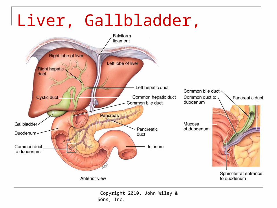
Liver and Gallbladder Weighs 1.4 kg (3 lb): 2nd largest organ in the
body; large right lobe + 3 smaller parts In right upper quadrant, below diaphragm Bile production and pathway
Hepatocytes (liver cells) make bile Bile canaliculi bile ducts hepatic duct Gallbladder (green, pear-shaped organ that stores
bile) Cystic duct common bile duct duodenum
Copyright 2010, John Wiley & Sons, Inc.
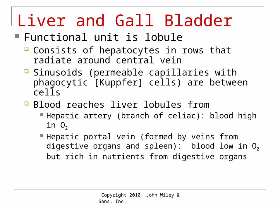
Liver and Gall Bladder Functional unit is lobule
Consists of hepatocytes in rows that radiate around central vein
Sinusoids (permeable capillaries with phagocytic [Kuppfer] cells) are between cells
Blood reaches liver lobules from Hepatic artery (branch of celiac): blood high in O2
Hepatic portal vein (formed by veins from digestive organs and spleen): blood low in O2 but rich in nutrients from digestive organs
Copyright 2010, John Wiley & Sons, Inc.
Bile Functions of bile
Emulsification: breaking apart clusters of fats so they are more digestible
Absorption of fats Formation and recycling of bile
Bilirubin from heme when RBCs broken down Bile is digested stercobilin: gives feces brown
color Bile salts reabsorbed into blood in small intestine
(ileum) portal vein liver Gallstones may form from bile
Obstruct bile ducts from gallbladder pain
Copyright 2010, John Wiley & Sons, Inc.
Liver, Gallbladder, Duodenum

Copyright 2010, John Wiley & Sons, Inc.
Liver

Copyright 2010, John Wiley & Sons, Inc.
Liver
Copyright 2010, John Wiley & Sons, Inc.
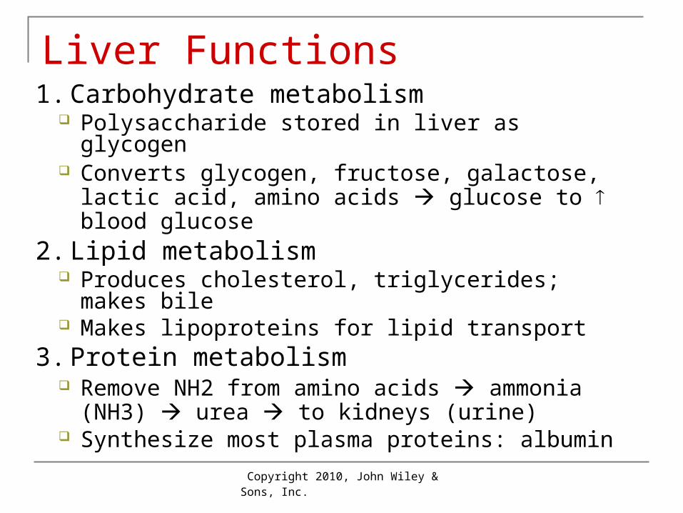
Liver Functions1. Carbohydrate metabolism
Polysaccharide stored in liver as glycogen Converts glycogen, fructose, galactose, lactic
acid, amino acids glucose to blood glucose2. Lipid metabolism
Produces cholesterol, triglycerides; makes bile Makes lipoproteins for lipid transport
3. Protein metabolism Remove NH2 from amino acids ammonia
(NH3) urea to kidneys (urine) Synthesize most plasma proteins: albumin
Copyright 2010, John Wiley & Sons, Inc.
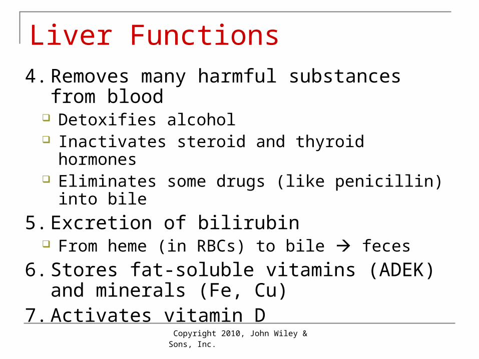
Liver Functions4. Removes many harmful substances from
blood Detoxifies alcohol Inactivates steroid and thyroid hormones Eliminates some drugs (like penicillin) into bile
5. Excretion of bilirubin From heme (in RBCs) to bile feces
6. Stores fat-soluble vitamins (ADEK) and minerals (Fe, Cu)
7. Activates vitamin D

Copyright 2010, John Wiley & Sons, Inc.
Small Intestine Length
10 feet long in living person Extends from pylorus of stomach to cecum of
large intestine Three major regions: duodenum, jejunum,
ileum Functions
Site of most of digestion Essentially all nutrient absorption occurs here
Ends in ileocecal sphincter (in RLQ)

Copyright 2010, John Wiley & Sons, Inc.
Small Intestine

Copyright 2010, John Wiley & Sons, Inc.
Small Intestine

Copyright 2010, John Wiley & Sons, Inc.
Intestinal Wall Structure Same 4 layers but with modifications Epithelium in mucosa: simple columnar
Absorptive cells with microvilli Goblet cells: secrete mucus
Intestinal glands secrete Enzymes that complete digestion Secretin, cholecystokinin (CCK), glucose-
dependent insulinotropic peptide (GIP) Lymphatic tissue within wall: defense

Copyright 2010, John Wiley & Sons, Inc.
Intestinal Wall Structure Submucosa has duodenal glands
Alkaline mucus helps neutralize stomach acid Circular folds
In mucosa and submucosa; increase surface area Villi: fingerlike projections of mucosa
Increase absorptive surface area Microvilli on absorptive cells further enhance absorption
Contain vessels that absorb nutrients: Arteriole, capillary, venule Lacteal (lymph capillary) for lipid absorption

Copyright 2010, John Wiley & Sons, Inc.
Intestinal Wall Structure

Copyright 2010, John Wiley & Sons, Inc.
Digestion in Small Intestine Mechanical digestion
Segmentation activity: for mixing Peristalsis for movement of intestinal contents
after most absorption completed: slow waves Chemical digestion: 2 L/d of secretions
Alkaline chyme due to bicarbonate From pancreas and alkaline mucus from small intestine
Enzymes produced by cells on villi Peptidases: breaks small peptides Disaccharidases: sucrase, lactase, and galactase
Copyright 2010, John Wiley & Sons, Inc.
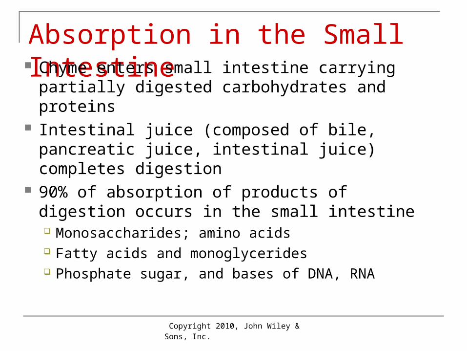
Absorption in the Small Intestine Chyme enters small intestine carrying
partially digested carbohydrates and proteins Intestinal juice (composed of bile, pancreatic
juice, intestinal juice) completes digestion 90% of absorption of products of digestion
occurs in the small intestine Monosaccharides; amino acids Fatty acids and monoglycerides Phosphate sugar, and bases of DNA, RNA
Copyright 2010, John Wiley & Sons, Inc.
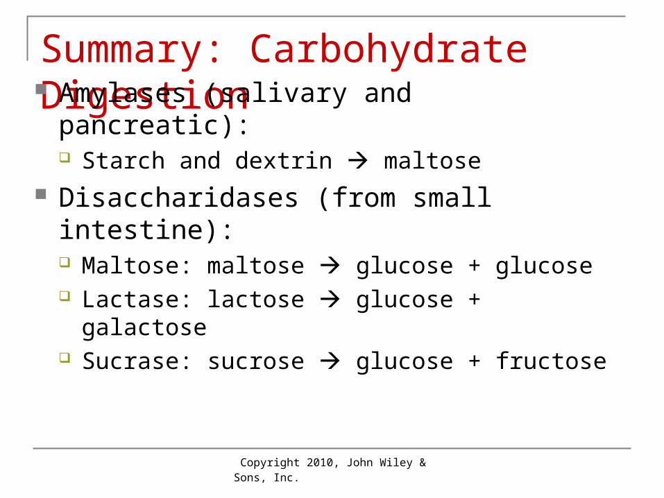
Summary: Carbohydrate Digestion Amylases (salivary and pancreatic):
Starch and dextrin maltose Disaccharidases (from small intestine):
Maltose: maltose glucose + glucose Lactase: lactose glucose + galactose Sucrase: sucrose glucose + fructose
Copyright 2010, John Wiley & Sons, Inc.
Protein and Fat Digestion Pepsin, trypsin, chymotrypsin, and
carboxypeptidase Proteins small peptides
Peptidases at surface: Peptides amino acids, dipeptides, and
tri-peptides Lipase (pancreatic)
Triglyceridesfatty acids + monoglycerides

Copyright 2010, John Wiley & Sons, Inc.
Absorption of Products of Digestion By diffusion, facilitated diffusion, osmosis and
active transport Carbohydrates monosaccharides
Via portal system (blood) to liver Proteins (jejunum + ileum) amino acids
Via portal system (blood) to liver Lipids
Short-chained fatty acids or monoglycerides or blood in villi
Larger lipids coated by proteins in chlyomicrons lacteals lymphatics (lymph) then blood

Copyright 2010, John Wiley & Sons, Inc.
Absorption of Products of Digestion Water and salt
Primarily osmotic movement that accompanies other nutrients
Vitamins Fat-soluble (A, D, E, K) absorbed with fat Water-soluble (B’s, C) with simple diffusion B12
Combines with intrinsic factor for transport through duodenum and jejunum
Finally can be absorbed by active transport in ileum

Copyright 2010, John Wiley & Sons, Inc.
Absorption of Products of Digestion

Copyright 2010, John Wiley & Sons, Inc.
Absorption of Products of Digestion
Copyright 2010, John Wiley & Sons, Inc.
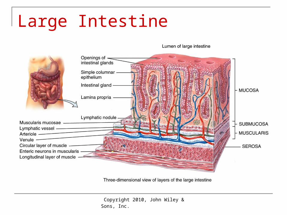
Large Intestine Structure: 4 regions
Cecum Ileocecal sphincter Appendix attached
Colon: ascending, transverse, descending and sigmoid
Rectum Anal canal with sphincters
Wall: standard 4 layers Mucosa: goblet cells secrete mucus Muscularis: incomplete longitudinal layer

Copyright 2010, John Wiley & Sons, Inc.
Large Intestine

Copyright 2010, John Wiley & Sons, Inc.
Large Intestine
Copyright 2010, John Wiley & Sons, Inc.
Large Intestine
Copyright 2010, John Wiley & Sons, Inc.
Digestion and Absorption Ileocecal sphincter limits rate of emptying of
ileum Slow peristalsis Mass peristalsis
Triggered by presence of food in stomach Wastes move from mid-colon rectum
Bacterial digestion Produce some B-vitamins + vitamin K Produce gases: flatus Colon absorbs salt + water

Copyright 2010, John Wiley & Sons, Inc.
Defecation Reflex Stretch of rectum wall neural reflex
contraction of longitudinal muscle Combined pressure + parasympathetic
activity relaxes internal anal sphincter External anal sphincter is voluntary Contraction of diaphragm and abdominal
muscles aid defecation

Copyright 2010, John Wiley & Sons, Inc.
Control: Phases of Digestion Rule: activate forward and inhibit behind Three phases: cephalic, gastric, intestinal
1. Cephalic: smell, sight, thought of food Cranial nerves VII + IX stimulate salivary glands Cranial nerve X (vagus) stimulates gastric glands
2. Gastric: stretching, pH of stomach Gastrin activates stomach and relaxes pyloric sphincter
3.Intestinal phase: intestinal hormones play key roles

Copyright 2010, John Wiley & Sons, Inc.
Control: Phases of Digestion Secretin
Released when acidic chyme enters intestine Stimulates release of pancreatic juice high in
bicarbonate to buffer acidic chyme from stomachCholecystokinin (CCK)
Released when chyme rich in amino acids and fatty acids enters intestine
Stimulates release of pancreatic juice high in digestive enzymes
Decreases gastric motility and secretion Causes gallbladder to contract and eject bile

Copyright 2010, John Wiley & Sons, Inc.
Aging Decreased GI secretion, motility, strength of
responses Loss of taste, increased risk for periodontal
disease, difficulty swallowing, hiatal hernia, gastritis, peptic ulcer disease
Increased risk for gallbladder problems, cirrhosis of liver, pancreatitis, constipation, hemorrhoids, diverticulitis
Copyright 2010, John Wiley & Sons, Inc.
End of Chapter 19
Copyright 2010 John Wiley & Sons, Inc.All rights reserved. Reproduction or translation of this work beyond that permitted in section 117 of the 1976 United States Copyright Act without express permission of the copyright owner is unlawful. Request for further information should be addressed to the Permission Department, John Wiley & Sons, Inc. The purchaser may make back-up copies for his/her own use only and not for distribution or resale. The Publishers assumes no responsibility for errors, omissions, or damages caused by the use of theses programs or from the use of the information herein.


