
Copyright © 2007 Wolters Kluwer Health | Lippincott Williams & Wilkins Neuroscience: Exploring the...
40
Copyright © 2007 Wolters Kluwer Health | Lippincott Williams & Wilkins Neuroscience: Exploring the Brain, 3e Chapter 23: Wiring the Brain
-
Upload
april-oliver -
Category
Documents
-
view
216 -
download
0
Transcript of Copyright © 2007 Wolters Kluwer Health | Lippincott Williams & Wilkins Neuroscience: Exploring the...

Copyright © 2007 Wolters Kluwer Health | Lippincott Williams & Wilkins
Neuroscience: Exploring the Brain, 3e
Chapter 23: Wiring the Brain

Copyright © 2007 Wolters Kluwer Health | Lippincott Williams & Wilkins
IntroductionIntroduction
• Operation of the brain
– Precise interconnections among 100 billion neurons
• Brain development
– Begins as a tube
– Neurogenesis, synaptogenesis, pathway formation, connections formed and modified
• Wiring in brain
– Establishing correct pathways and targets
– Fine tuning based on experience

Copyright © 2007 Wolters Kluwer Health | Lippincott Williams & Wilkins
The Genesis of NeuronsThe Genesis of Neurons• Example: Mammalian retinogeniculocortical pathway

Copyright © 2007 Wolters Kluwer Health | Lippincott Williams & Wilkins
Activity-dependent SynapticRearrangementActivity-dependent SynapticRearrangement
• Synaptic rearrangement
– Change from one pattern to another
– Consequence of neural activity/synaptic transmission before and after birth
– Critical Period

Copyright © 2007 Wolters Kluwer Health | Lippincott Williams & Wilkins
The Elimination of Cells and SynapsesThe Elimination of Cells and Synapses• Changes in Synaptic Capacity
– Synapse elimination modeled in the neuromuscular junction

Copyright © 2007 Wolters Kluwer Health | Lippincott Williams & Wilkins
The Lateral Geniculate Nucleus (LGN)
The Lateral Geniculate Nucleus (LGN)

Copyright © 2007 Wolters Kluwer Health | Lippincott Williams & Wilkins
Activity-dependent SynapticRearrangementActivity-dependent SynapticRearrangement
• Synaptic segregation
– Refinement of synaptic connections
• Segregation of Retinal Inputs to the LGN
• Retinal waves (in utero) (Carla Shatz)
• Activity of the two eyes not correlated -> segregation in LGN
• Process of synaptic stabilization
• Hebbian modifications (Donald Hebb)

Copyright © 2007 Wolters Kluwer Health | Lippincott Williams & Wilkins
Activity-dependent SynapticRearrangementActivity-dependent SynapticRearrangement
• Segregation of Retinal Inputs to the LGN (Cont’d)
– Plasticity at
‘Hebb’ synapses
– “Winner-takes-
all”

Copyright © 2007 Wolters Kluwer Health | Lippincott Williams & Wilkins
Activity-dependent SynapticRearrangementActivity-dependent SynapticRearrangement
• Segregation of LGN Inputs in the Striate Cortex
– Visual cortex has ocular dominance columns (cat, monkey) - segregated input from each eye
– Synaptic rearrangement is activity-dependent
– Plastic during critical period
– Effects of congenital cataracts (if not removed early)

Copyright © 2007 Wolters Kluwer Health | Lippincott Williams & Wilkins
Activity-dependent SynapticRearrangementActivity-dependent SynapticRearrangement
• Synaptic Convergence
– Anatomical basis of binocular vision and binocular receptive fields
– Monocular deprivation:
• Ocular dominance shift
• Plasticity of binocular connections
• Synaptic competition

Copyright © 2007 Wolters Kluwer Health | Lippincott Williams & Wilkins

Copyright © 2007 Wolters Kluwer Health | Lippincott Williams & Wilkins
Activity-dependent SynapticRearrangementActivity-dependent SynapticRearrangement
• Critical period for plasticity of binocular connections

Copyright © 2007 Wolters Kluwer Health | Lippincott Williams & Wilkins
Activity-dependent SynapticRearrangementActivity-dependent SynapticRearrangement• Effect of strabismus on cortical binocularity

Copyright © 2007 Wolters Kluwer Health | Lippincott Williams & Wilkins
Activity-dependent SynapticRearrangementActivity-dependent SynapticRearrangement
• Modulatory Influences
– Increasing age
– Before and after birth
– Enabling factors

Copyright © 2007 Wolters Kluwer Health | Lippincott Williams & Wilkins
Elementary Mechanisms of CorticalSynaptic PlasticityElementary Mechanisms of CorticalSynaptic Plasticity
• Two rules for synaptic modification
– Wire together fire together (Hebbian modifications)
– Out of sync lose their link
– Correlation: heard and validated

Copyright © 2007 Wolters Kluwer Health | Lippincott Williams & Wilkins
Elementary Mechanisms of CorticalSynaptic PlasticityElementary Mechanisms of CorticalSynaptic Plasticity• Excitatory Synaptic Transmission in the Immature Visual System
– Focus on 2 glutamate receptors (Rs):
• AMPARs: glutamate-gated ion channels
• NMDARs: Unique properties

Copyright © 2007 Wolters Kluwer Health | Lippincott Williams & Wilkins
Elementary Mechanisms of Cortical Synaptic PlasticityElementary Mechanisms of Cortical Synaptic Plasticity
• Excitatory Synaptic Transmission
– NMDA receptors have two unique properties
• Voltage-gated owing to Mg2+
• Conducts Ca2+
• Magnitude of Ca2+ flux signals level of pre- and postsynaptic activation

Copyright © 2007 Wolters Kluwer Health | Lippincott Williams & Wilkins
Elementary Mechanisms of CorticalSynaptic PlasticityElementary Mechanisms of CorticalSynaptic Plasticity
• Long-Term Synaptic Potentiation
– Monitor synaptic strength before and after episodes of strong NMDA activation
– Accounting for LTP
• AMPA insertion (“AMPAfication”)
• Splitting synapses (doubling)

Copyright © 2007 Wolters Kluwer Health | Lippincott Williams & Wilkins
Elementary Mechanisms of CorticalSynaptic PlasticityElementary Mechanisms of CorticalSynaptic Plasticity• Lasting synaptic effects of strong NMDA receptor activation

Copyright © 2007 Wolters Kluwer Health | Lippincott Williams & Wilkins
Elementary Mechanisms of CorticalSynaptic PlasticityElementary Mechanisms of CorticalSynaptic Plasticity
• Long-Term Synaptic Depression (LTD)
– Neurons fire out of sync
– Synaptic plasticity mechanism opposite of LTP
• Loss of synaptic AMPARs
• Loss of synapses? (unknown)
– Mechanism for consequences of monocular deprivation

Copyright © 2007 Wolters Kluwer Health | Lippincott Williams & Wilkins
Elementary Mechanisms of CorticalSynaptic PlasticityElementary Mechanisms of CorticalSynaptic Plasticity• Brief monocular deprivation leads to reduced visual
responsiveness
– Depends on retinal activity, NMDA activation, postsynaptic calcium

Copyright © 2007 Wolters Kluwer Health | Lippincott Williams & Wilkins
Why Critical Periods EndWhy Critical Periods End
• Why do critical periods end?
– Plasticity diminishes:
• When axon growth ceases
• When synaptic transmission matures
• When cortical activation is constrained
– Intrinsic inhibitory circuitry late to mature
– Understanding developmental regulation of plasticity may help recovery from CNS damage

Copyright © 2007 Wolters Kluwer Health | Lippincott Williams & Wilkins
Enriched environment: More complex brain structure.Enriched environment: More complex brain structure.
•Increased dendritic branching and synaptic density
•Increased transmitter levels, total protein
•Better at solving maze problems

Copyright © 2007 Wolters Kluwer Health | Lippincott Williams & Wilkins
Concluding Remarks
• Generation of brain development circuitry
– Placement of wires before birth
– Refinement of synaptic infancy
• Developmental critical periods
– Visual system and other sensory and motor systems
• Environment influences brain modification throughout life

Copyright © 2007 Wolters Kluwer Health | Lippincott Williams & Wilkins
End of Presentation

Copyright © 2007 Wolters Kluwer Health | Lippincott Williams & Wilkins
The Genesis of NeuronsThe Genesis of Neurons
• Cell Proliferation
– Neural stem cells give rise to neurons and glia

Copyright © 2007 Wolters Kluwer Health | Lippincott Williams & Wilkins
The Genesis of NeuronsThe Genesis of Neurons• Cell Proliferation (Cont’d)
– Cleavage plane during cell division determines fate of daughter cells

Copyright © 2007 Wolters Kluwer Health | Lippincott Williams & Wilkins
The Genesis of NeuronsThe Genesis of Neurons
• Cell Migration
– Pyramidal cells and astrocytes migrate vertically from ventricular zone by moving along thin radial glial fibers
– Inhibitory interneurons and oligodendroglia generate from a different site and migrate laterally

Copyright © 2007 Wolters Kluwer Health | Lippincott Williams & Wilkins
The Genesis of NeuronsThe Genesis of Neurons
• Cell Migration
– First cells to migrate take up residence in “subplate” layer which eventually disappears
– Next cells to divide migrate to the cortical plate
– The first to arrive become layer VI, followed V, IV, and so on: “inside out”

Copyright © 2007 Wolters Kluwer Health | Lippincott Williams & Wilkins
The Genesis of NeuronsThe Genesis of Neurons
• Cell Differentiation
– Cell takes the appearance and characteristics of a neuron after reaching its destination but programming occurs much earlier

Copyright © 2007 Wolters Kluwer Health | Lippincott Williams & Wilkins
The Genesis of NeuronsThe Genesis of Neurons
• Differentiation of Cortical Areas
– Adult cortical sheet is a “patchwork quilt
– Cortical “protomap” in the ventricular zone replicated by radial glial guides
– But some neurons migrate laterally
– Thalamic input contributes to cortical differentiation

Copyright © 2007 Wolters Kluwer Health | Lippincott Williams & Wilkins
The Genesis of ConnectionsThe Genesis of Connections• The three phases of pathway formation:
– (1) pathway, (2) target, (3) address

Copyright © 2007 Wolters Kluwer Health | Lippincott Williams & Wilkins
The Genesis of ConnectionsThe Genesis of Connections• The Growing Axon
– Growth cone: Growing tip of a neurite

Copyright © 2007 Wolters Kluwer Health | Lippincott Williams & Wilkins
The Genesis of ConnectionsThe Genesis of Connections
• Axon Guidance
– Challenge in wiring the brain
• Distances between connected structures
• But in early stages nervous system is a few centimeters long
– Pioneer axons stretch as nervous system expands
• Guide neighbor axons to same targets
– Pioneer neurons grow in the correct direction by “connecting the dots”

Copyright © 2007 Wolters Kluwer Health | Lippincott Williams & Wilkins
The Genesis of ConnectionsThe Genesis of Connections
• Axon Guidance
– Guidance Cues: Chemoattractant (e.g., netrin), Chemorepellent (e.g., slit)

Copyright © 2007 Wolters Kluwer Health | Lippincott Williams & Wilkins
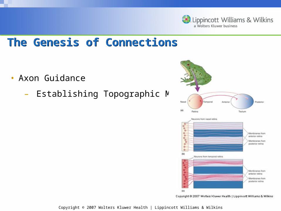
The Genesis of ConnectionsThe Genesis of Connections
• Axon Guidance
– Establishing Topographic Maps
• Choice point; Retinal axons innervate targets of LGN and superior colliculus
• Sperry (1940s): Chemoaffinity hypothesis
• CNS axons regenerate in amphibians, not in mammals
• Factors guiding retinal axons to tectum
• Ephrins/eph (repulsive signal)
Copyright © 2007 Wolters Kluwer Health | Lippincott Williams & Wilkins
The Genesis of ConnectionsThe Genesis of Connections
• Axon Guidance
– Establishing Topographic Maps

Copyright © 2007 Wolters Kluwer Health | Lippincott Williams & Wilkins
The Genesis of ConnectionsThe Genesis of Connections
• Synapse Formation
– Modeled in the neuromuscular junction

Copyright © 2007 Wolters Kluwer Health | Lippincott Williams & Wilkins
The Genesis of ConnectionsThe Genesis of Connections
• Synapse Formation
– Steps in the formation of a CNS synapse:
– Dendritic filopodium contacts axon
– Synaptic vesicles and active zone proteins recruited to presynaptic membrane
– Receptors accumulate on postsynaptic membrane

Copyright © 2007 Wolters Kluwer Health | Lippincott Williams & Wilkins
The Elimination of Cells and SynapsesThe Elimination of Cells and Synapses
• The mechanisms of pathway formation– Large-scale reduction in neurons
and synapses
• Development of brain function
– Balance between genesis & elimination of cells and synapses
• Apoptosis: Programmed Cell Death
– Importance of trophic factors, e.g., nerve growth factor