Copyright © 2013, 2010 by Saunders, an imprint of Elsevier Inc. Chapter 5 Pharmacodynamics.
Upload
pearl-blankenshipCategory
view
240download
1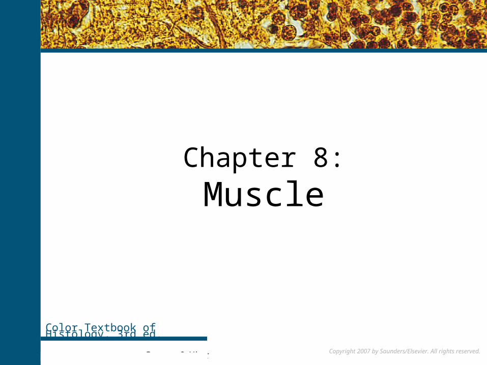
Copyright 2007 by Saunders/Elsevier. All rights reserved.
Chapter 8:
Muscle
Color Textbook of Histology, 3rd ed.
Gartner & Hiatt Copyright 2007 by Saunders/Elsevier. All rights reserved.
Copyright 2007 by Saunders/Elsevier. All rights reserved.
Muscle Tissue
Cells of muscle are elongated and are called striated or smooth muscle, depending on the respective presence or absence of a regularly repeated arrangement of myofibrillar contractile proteins, the myofilaments. Striated muscle cells display characteristic alternations of light and dark cross-bands, which are absent in smooth muscle. There are two types of striated muscle: skeletal, accounting for most of the voluntary muscle mass of the body, and involuntary cardiac muscle, limited almost exclusively to the heart.
Smooth muscle is located in the walls of blood vessels and the viscera as well as in the dermis of the skin.
Unique terms are often used to describe the components of muscle cells. Thus, muscle cell membrane is referred to as sarcolemma; the cytoplasm, as sarcoplasm; the smooth endoplasmic reticulum, as sarcoplasmic reticulum; and occasionally, the mitochondria, as sarcosomes. Because they are much longer than they are wide, muscle cells frequently are called muscle fibers; unlike collagen fibers, however, they are living entities.
For more information see Muscle in Chapter 8 of Gartner and Hiatt: Color Textbook of Histology, 3rd ed. Philadelphia, W.B. Saunders, 2007.
Figure 8–2 Three types of muscle. Top, Skeletal muscle; center, smooth muscle; bottom, cardiac muscle.
Copyright 2007 by Saunders/Elsevier. All rights reserved.
Skeletal Muscle
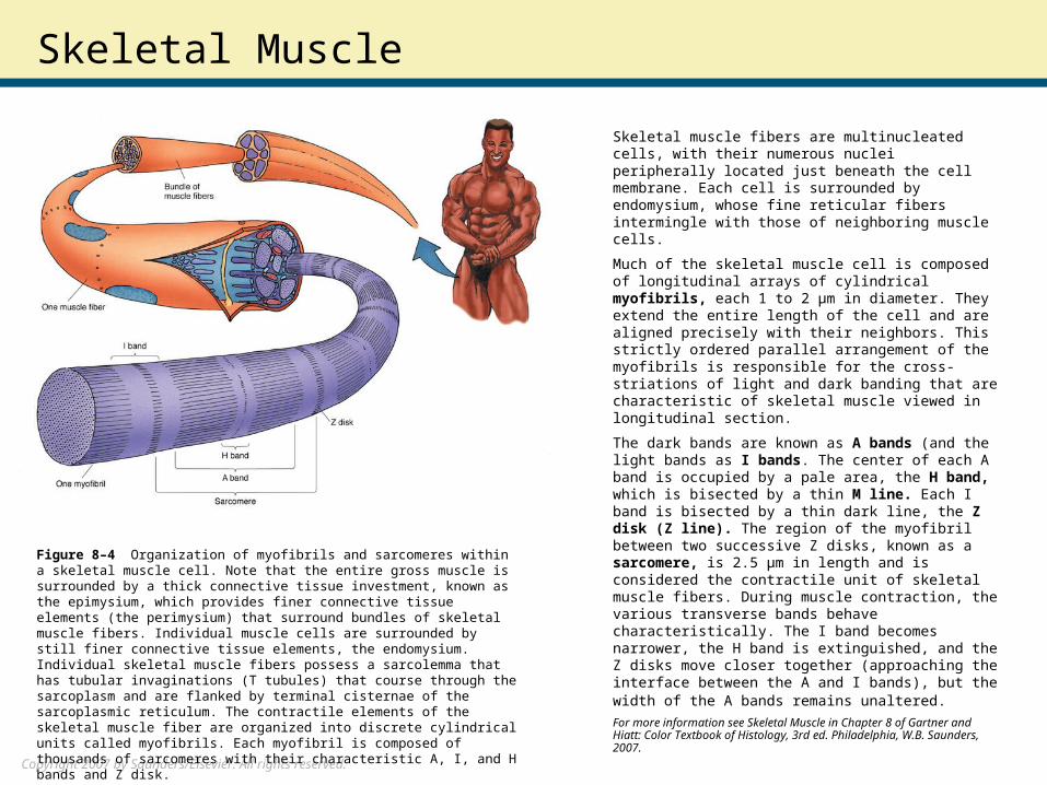
Skeletal muscle fibers are multinucleated cells, with their numerous nuclei peripherally located just beneath the cell membrane. Each cell is surrounded by endomysium, whose fine reticular fibers intermingle with those of neighboring muscle cells.
Much of the skeletal muscle cell is composed of longitudinal arrays of cylindrical myofibrils, each 1 to 2 μm in diameter. They extend the entire length of the cell and are aligned precisely with their neighbors. This strictly ordered parallel arrangement of the myofibrils is responsible for the cross-striations of light and dark banding that are characteristic of skeletal muscle viewed in longitudinal section.
The dark bands are known as A bands (and the light bands as I bands. The center of each A band is occupied by a pale area, the H band, which is bisected by a thin M line. Each I band is bisected by a thin dark line, the Z disk (Z line). The region of the myofibril between two successive Z disks, known as a sarcomere, is 2.5 μm in length and is considered the contractile unit of skeletal muscle fibers. During muscle contraction, the various transverse bands behave characteristically. The I band becomes narrower, the H band is extinguished, and the Z disks move closer together (approaching the interface between the A and I bands), but the width of the A bands remains unaltered. For more information see Skeletal Muscle in Chapter 8 of Gartner and Hiatt: Color Textbook of Histology, 3rd ed. Philadelphia, W.B. Saunders, 2007.
Figure 8–4 Organization of myofibrils and sarcomeres within a skeletal muscle cell. Note that the entire gross muscle is surrounded by a thick connective tissue investment, known as the epimysium, which provides finer connective tissue elements (the perimysium) that surround bundles of skeletal muscle fibers. Individual muscle cells are surrounded by still finer connective tissue elements, the endomysium. Individual skeletal muscle fibers possess a sarcolemma that has tubular invaginations (T tubules) that course through the sarcoplasm and are flanked by terminal cisternae of the sarcoplasmic reticulum. The contractile elements of the skeletal muscle fiber are organized into discrete cylindrical units called myofibrils. Each myofibril is composed of thousands of sarcomeres with their characteristic A, I, and H bands and Z disk.
Copyright 2007 by Saunders/Elsevier. All rights reserved.
Electron Microscopy of Skeletal Muscle
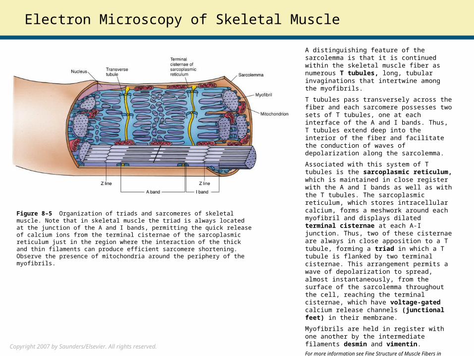
A distinguishing feature of the sarcolemma is that it is continued within the skeletal muscle fiber as numerous T tubules, long, tubular invaginations that intertwine among the myofibrils.
T tubules pass transversely across the fiber and each sarcomere possesses two sets of T tubules, one at each interface of the A and I bands. Thus, T tubules extend deep into the interior of the fiber and facilitate the conduction of waves of depolarization along the sarcolemma.
Associated with this system of T tubules is the sarcoplasmic reticulum, which is maintained in close register with the A and I bands as well as with the T tubules. The sarcoplasmic reticulum, which stores intracellular calcium, forms a meshwork around each myofibril and displays dilated terminal cisternae at each A-I junction. Thus, two of these cisternae are always in close apposition to a T tubule, forming a triad in which a T tubule is flanked by two terminal cisternae. This arrangement permits a wave of depolarization to spread, almost instantaneously, from the surface of the sarcolemma throughout the cell, reaching the terminal cisternae, which have voltage-gated calcium release channels (junctional feet) in their membrane.
Myofibrils are held in register with one another by the intermediate filaments desmin and vimentin.
For more information see Fine Structure of Muscle Fibers in Chapter 8 of Gartner and Hiatt: Color Textbook of Histology, 3rd ed. Philadelphia, W.B. Saunders, 2007.
Figure 8–5 Organization of triads and sarcomeres of skeletal muscle. Note that in skeletal muscle the triad is always located at the junction of the A and I bands, permitting the quick release of calcium ions from the terminal cisternae of the sarcoplasmic reticulum just in the region where the interaction of the thick and thin filaments can produce efficient sarcomere shortening. Observe the presence of mitochondria around the periphery of the myofibrils.

Copyright 2007 by Saunders/Elsevier. All rights reserved.
MyofilamentsElectron microscopy reveals the presence of parallel, interdigitating, rod-like thick myofilaments and thin myofilaments. The thick filaments (15 nm in diameter and 1.5 μm long) are composed of myosin, whereas the thin filaments (7 nm in diameter and 1.0 μm long) are composed primarily of actin.
Thin filaments originate at the Z disk and project toward the center of the two adjacent sarcomeres, thus pointing in opposite directions. Hence, a single sarcomere has two groups of parallel arrays of thin filaments, each attached to one Z disk, with all the filaments in each group pointing toward the middle of the sarcomere. Thick filaments also form parallel arrays, interdigitating with the thin filaments in a specific fashion.
In a relaxed skeletal muscle fiber, the thick filaments do not extend the entire length of the sarcomere and the thin filaments projecting from the two Z disks of the sarcomere do not meet in the midline. Therefore, there are regions of each sarcomere, on either side of each Z disk, where only thin filaments are present. These adjacent portions of two successive sarcomeres correspond to the I band seen by light microscopy; for instance, the region of each sarcomere that encompasses the entire length of the thick filaments is the A band, and the zone in the middle of the A band, which is devoid of thin filaments, is the H band, which is bisected by the M line.
During contraction, individual thick and thin filaments do not shorten; instead, the two Z disks are brought closer together as the thin filaments slide past the thick filaments (Huxley’s sliding filament theory). Thus, when contraction occurs, the motion of the thin filaments toward the center of the sarcomere creates a greater overlap between the two groups of filaments, effectively reducing the widths of the I and H bands without influencing the width of the A band.
In mammalian skeletal muscle, each thick filament is surrounded equidistantly by six thin filaments.
For more information see Structural organization of Myofibrils in Chapter 8 of Gartner and Hiatt: Color Textbook of Histology, 3rd ed. Philadelphia, W.B. Saunders, 2007.
Copyright 2007 by Saunders/Elsevier. All rights reserved.
Copyright 2007 by Saunders/Elsevier. All rights reserved.
Neuromuscular Junction
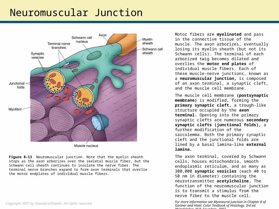
Motor fibers are myelinated and pass in the connective tissue of the muscle. The axon arborizes, eventually losing its myelin sheath (but not its Schwann cells). The terminal of each arborized twig becomes dilated and overlies the motor end plates of individual muscle fibers. Each of these muscle-nerve junctions, known as a neuromuscular junction, is composed of an axon terminal, a synaptic cleft, and the muscle cell membrane.
The muscle cell membrane (postsynaptic membrane) is modified, forming the primary synaptic cleft, a trough-like structure occupied by the axon terminal. Opening into the primary synaptic clefts are numerous secondary synaptic clefts (junctional folds), a further modification of the sarcolemma. Both the primary synaptic cleft and the junctional folds are lined by a basal lamina–like external lamina.
The axon terminal, covered by Schwann cells, houses mitochondria, smooth endoplasmic reticulum, and as many as 300,000 synaptic vesicles (each 40 to 50 nm in diameter) containing the neurotransmitter acetylcholine. The function of the neuromuscular junction is to transmit a stimulus from the nerve fiber to the muscle cell.
For more information see Myoneural Junction in Chapter 8 of Gartner and Hiatt: Color Textbook of Histology, 3rd ed. Philadelphia, W.B. Saunders, 2007.
Figure 8–13 Neuromuscular junction. Note that the myelin sheath stops as the axon arborizes over the skeletal muscle fiber, but the Schwann cell sheath continues to insulate the nerve fiber. The terminal nerve branches expand to form axon terminals that overlie the motor endplates of individual muscle fibers.

Copyright 2007 by Saunders/Elsevier. All rights reserved.
Neuromuscular Junction (cont.)
Stimulus transmission across a synaptic cleft involves the following sequence of events:
1. A stimulus depolarizes the membrane of the axon terminal, opens voltage-gated calcium channels, near dense bars.2. Calcium causes the fusion of synaptic vesicles with the presynaptic membrane (along the active sites) with subsequent release of acetylcholine synaptic cleft.3. Acetylcholine then diffuses across the synaptic cleft and binds to postsynaptic acetylcholine receptors in the muscle cell membrane. These receptors, located in the vicinity of the presynaptic active sites, are ligand-gated ion channels, which open in response to the binding of acetylcholine. The resulting ion influx leads to depolarization of the sarcolemma and generation of an action potential.4. The impulse generated spreads quickly throughout the muscle fiber via the system of T tubules, initiating muscle contraction.5. To prevent a single stimulus from eliciting multiple responses, acetylcholinesterase degrades acetylcholine into acetate and choline, thus permitting the reestablishment of the resting potential. Choline is transported back into the axon terminal by a sodium-choline symport. Within the axon terminal the acetylcholine, synthesized from activated acetate (produced in mitochondria) and the recycled choline, a reaction catalyzed by choline acetyl transferase, into newly formed synaptic vesicles.
For more information see Myoneural Junction in Chapter 8 of Gartner and Hiatt: Color Textbook of Histology, 3rd ed. Philadelphia, W.B. Saunders, 2007.
Figure 8-14 Neuromuscular junction. C, Excitatory synaptic transmission. The nerve action potential opens voltage-gated calcium channels. Entry of Ca2+ triggers fusion of a synaptic vesicle containing acetylcholine (ACh) with the plasma membrane. Acetylcholine binds and opens postsynaptic channels on the muscle cell which trigger an action potential. (From Pollard TD, Earnshaw WC: Cell Biology. Philadelphia, Elsevier/Saunders, 2004, p 156.)
Copyright 2007 by Saunders/Elsevier. All rights reserved.
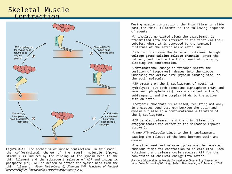
Skeletal Muscle ContractionDuring muscle contraction, the thin filaments slide past the thick filaments in the following sequence of events :
•An impulse, generated along the sarcolemma, is transmitted into the interior of the fiber via the T tubules, where it is conveyed to the terminal cisternae of the sarcoplasmic reticulum.
•Calcium ions leave the terminal cisternae through voltage-gated calcium release channels, enter the cytosol, and bind to the TnC subunit of troponin, altering its conformation.
•Conformational change in troponin shifts the position of tropomyosin deeper into the groove, unmasking the active site (myosin binding site) on the actin molecule.
•ATP present on the S1 subfragment of myosin is hydrolyzed,
but both adenosine diphosphate (ADP) and inorganic phosphate (Pi) remain attached to the S1 subfragment, and the complex
binds to the active site on actin.
•Inorganic phosphate is released, resulting not only in a greater bond strength between the actin and myosin but also in a conformational alteration of the S1 subfragment.
•ADP is also released, and the thin filament is dragged toward the center of the sarcomere (“power stroke”).
•A new ATP molecule binds to the S1 subfragment, causing the
release of the bond between actin and myosin.
•The attachment and release cycles must be repeated numerous times for contraction to be completed. Each attachment and release cycle requires ATP for the conversion of chemical energy into motion.
For more information see Muscle Contraction in Chapter 8 of Gartner and Hiatt: Color Textbook of Histology, 3rd ed. Philadelphia, W.B. Saunders, 2007.
Figure 8-10 The mechanism of muscle contraction. In this model, the conformational change of the myosin molecule (“power stroke”) is induced by the binding of the myosin head to the thin filament and the subsequent release of ADP and inorganic phosphate (Pi). ATP is needed to detach the myosin head from the thin filament. (From Meisenberg, G, Simmons WH: Principles of Medical Biochemistry, 2e. Philadelphia, Elsevier/Mosby, 2006, p 226.)
Copyright 2007 by Saunders/Elsevier. All rights reserved.
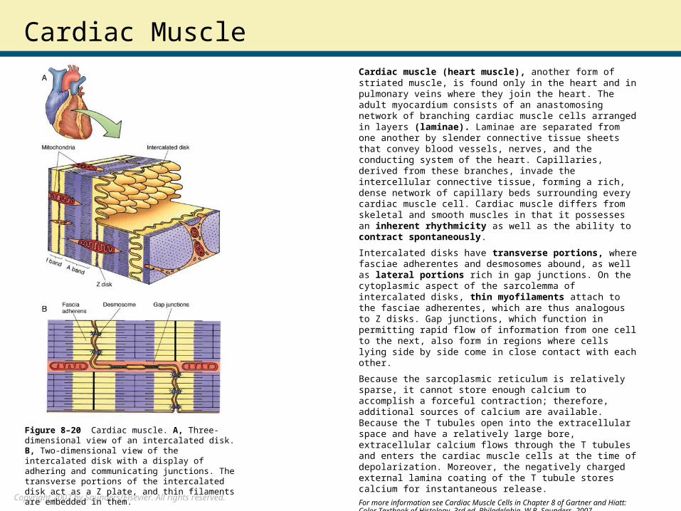
Cardiac MuscleCardiac muscle (heart muscle), another form of striated muscle, is found only in the heart and in pulmonary veins where they join the heart. The adult myocardium consists of an anastomosing network of branching cardiac muscle cells arranged in layers (laminae). Laminae are separated from one another by slender connective tissue sheets that convey blood vessels, nerves, and the conducting system of the heart. Capillaries, derived from these branches, invade the intercellular connective tissue, forming a rich, dense network of capillary beds surrounding every cardiac muscle cell. Cardiac muscle differs from skeletal and smooth muscles in that it possesses an inherent rhythmicity as well as the ability to contract spontaneously.
Intercalated disks have transverse portions, where fasciae adherentes and desmosomes abound, as well as lateral portions rich in gap junctions. On the cytoplasmic aspect of the sarcolemma of intercalated disks, thin myofilaments attach to the fasciae adherentes, which are thus analogous to Z disks. Gap junctions, which function in permitting rapid flow of information from one cell to the next, also form in regions where cells lying side by side come in close contact with each other.
Because the sarcoplasmic reticulum is relatively sparse, it cannot store enough calcium to accomplish a forceful contraction; therefore, additional sources of calcium are available. Because the T tubules open into the extracellular space and have a relatively large bore, extracellular calcium flows through the T tubules and enters the cardiac muscle cells at the time of depolarization. Moreover, the negatively charged external lamina coating of the T tubule stores calcium for instantaneous release.
For more information see Cardiac Muscle Cells in Chapter 8 of Gartner and Hiatt: Color Textbook of Histology, 3rd ed. Philadelphia, W.B. Saunders, 2007.
Figure 8–20 Cardiac muscle. A, Three-dimensional view of an intercalated disk. B, Two-dimensional view of the intercalated disk with a display of adhering and communicating junctions. The transverse portions of the intercalated disk act as a Z plate, and thin filaments are embedded in them.
Copyright 2007 by Saunders/Elsevier. All rights reserved.
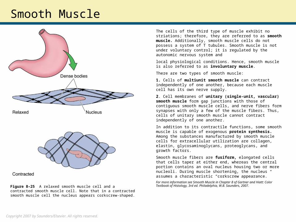
Smooth MuscleThe cells of the third type of muscle exhibit no striations; therefore, they are referred to as smooth muscle. Additionally, smooth muscle cells do not possess a system of T tubules. Smooth muscle is not under voluntary control; it is regulated by the autonomic nervous system and
local physiological conditions. Hence, smooth muscle is also referred to as involuntary muscle.
There are two types of smooth muscle:
1. Cells of multiunit smooth muscle can contract independently of one another, because each muscle cell has its own nerve supply.
2. Cell membranes of unitary (single-unit, vascular) smooth muscle form gap junctions with those of contiguous smooth muscle cells, and nerve fibers form synapses with only a few of the muscle fibers. Thus, cells of unitary smooth muscle cannot contract independently of one another.
In addition to its contractile functions, some smooth muscle is capable of exogenous protein synthesis. Among the substances manufactured by smooth muscle cells for extracellular utilization are collagen, elastin, glycosaminoglycans, proteoglycans, and growth factors.
Smooth muscle fibers are fusiform, elongated cells that cells taper at either end, whereas the central portion contains an oval nucleus housing two or more nucleoli. During muscle shortening, the nucleus assumes a characteristic “corkscrew appearance.”
For more information see Smooth Muscle in Chapter 8 of Gartner and Hiatt: Color Textbook of Histology, 3rd ed. Philadelphia, W.B. Saunders, 2007.
Figure 8–25 A relaxed smooth muscle cell and a contracted smooth muscle cell. Note that in a contracted smooth muscle cell the nucleus appears corkscrew-shaped.
Copyright 2007 by Saunders/Elsevier. All rights reserved.
Contraction of Smooth Muscle
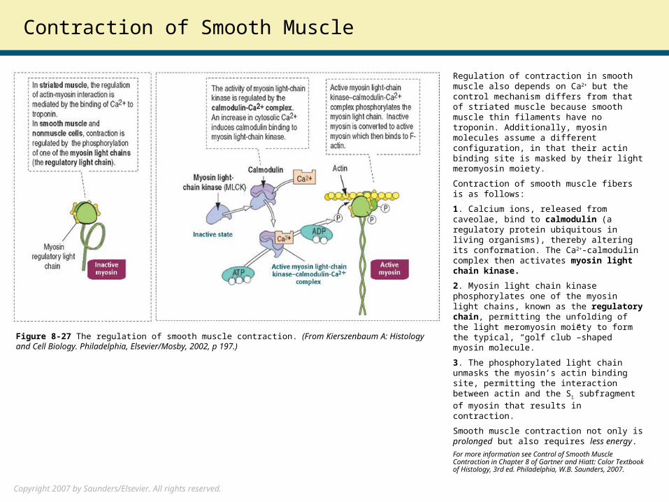
Regulation of contraction in smooth muscle also depends on Ca2+ but the control mechanism differs from that of striated muscle because smooth muscle thin filaments have no troponin. Additionally, myosin molecules assume a different configuration, in that their actin binding site is masked by their light meromyosin moiety.
Contraction of smooth muscle fibers is as follows:
1. Calcium ions, released from caveolae, bind to calmodulin (a regulatory protein ubiquitous in living organisms), thereby altering its conformation. The Ca2+-calmodulin complex then activates myosin light chain kinase.
2. Myosin light chain kinase phosphorylates one of the myosin light chains, known as the regulatory chain, permitting the unfolding of the light meromyosin moiety to form the typical, “golf club”–shaped myosin molecule.
3. The phosphorylated light chain unmasks the myosin’s actin binding site, permitting the interaction between actin and the S1
subfragment of myosin that results in contraction.
Smooth muscle contraction not only is prolonged but also requires less energy.
For more information see Control of Smooth Muscle Contraction in Chapter 8 of Gartner and Hiatt: Color Textbook of Histology, 3rd ed. Philadelphia, W.B. Saunders, 2007.
Figure 8-27 The regulation of smooth muscle contraction. (From Kierszenbaum A: Histology and Cell Biology. Philadelphia, Elsevier/Mosby, 2002, p 197.)