Cooperation of local motions in the Hsp90 …sro.sussex.ac.uk/67665/1/emss-68351.pdfCooperation of...
25
Cooperation of local motions in the Hsp90 molecular chaperone ATPase mechanism Article (Accepted Version) http://sro.sussex.ac.uk Schulze, Andrea, Beliu, Gerti, Helmerich, Dominic A, Schubert, Jonathan, Pearl, Laurence H, Prodromou, Chrisostomos and Neuweiler, Hannes (2016) Cooperation of local motions in the Hsp90 molecular chaperone ATPase mechanism. Nature Chemical Biology, 12 (8). pp. 628-635. ISSN 1552-4450 This version is available from Sussex Research Online: http://sro.sussex.ac.uk/id/eprint/67665/ This document is made available in accordance with publisher policies and may differ from the published version or from the version of record. If you wish to cite this item you are advised to consult the publisher’s version. Please see the URL above for details on accessing the published version. Copyright and reuse: Sussex Research Online is a digital repository of the research output of the University. Copyright and all moral rights to the version of the paper presented here belong to the individual author(s) and/or other copyright owners. To the extent reasonable and practicable, the material made available in SRO has been checked for eligibility before being made available. Copies of full text items generally can be reproduced, displayed or performed and given to third parties in any format or medium for personal research or study, educational, or not-for-profit purposes without prior permission or charge, provided that the authors, title and full bibliographic details are credited, a hyperlink and/or URL is given for the original metadata page and the content is not changed in any way.
Transcript of Cooperation of local motions in the Hsp90 …sro.sussex.ac.uk/67665/1/emss-68351.pdfCooperation of...

Cooperation of local motions in the Hsp90 molecular chaperone ATPase mechanism
Article (Accepted Version)
http://sro.sussex.ac.uk
Schulze, Andrea, Beliu, Gerti, Helmerich, Dominic A, Schubert, Jonathan, Pearl, Laurence H, Prodromou, Chrisostomos and Neuweiler, Hannes (2016) Cooperation of local motions in the Hsp90 molecular chaperone ATPase mechanism. Nature Chemical Biology, 12 (8). pp. 628-635. ISSN 1552-4450
This version is available from Sussex Research Online: http://sro.sussex.ac.uk/id/eprint/67665/
This document is made available in accordance with publisher policies and may differ from the published version or from the version of record. If you wish to cite this item you are advised to consult the publisher’s version. Please see the URL above for details on accessing the published version.
Copyright and reuse: Sussex Research Online is a digital repository of the research output of the University.
Copyright and all moral rights to the version of the paper presented here belong to the individual author(s) and/or other copyright owners. To the extent reasonable and practicable, the material made available in SRO has been checked for eligibility before being made available.
Copies of full text items generally can be reproduced, displayed or performed and given to third parties in any format or medium for personal research or study, educational, or not-for-profit purposes without prior permission or charge, provided that the authors, title and full bibliographic details are credited, a hyperlink and/or URL is given for the original metadata page and the content is not changed in any way.

Cooperation of local motions in the Hsp90 molecular chaperone ATPase mechanism
Andrea Schulze1, Gerti Beliu1, Dominic A. Helmerich1, Jonathan Schubert1, Laurence H. Pearl2, Chrisostomos Prodromou2, and Hannes Neuweiler1,*
1Department of Biotechnology and Biophysics, Julius-Maximilians-University Würzburg, Am Hubland, 97074 Würzburg, Germany
2Genome Damage and Stability Centre, School of Life Sciences, University of Sussex, Falmer, Brighton BN1 9RQ, United Kingdom
Abstract
The Hsp90 chaperone is a central node of protein homeostasis activating a large number of diverse
client proteins. Hsp90 functions as a molecular clamp that closes and opens in response to the
binding and hydrolysis of ATP. Crystallographic studies define distinct conformational states of
the mechanistic core implying structural changes that have not yet been observed in solution.
Here, we engineered one-nanometer fluorescence probes based on photo-induced electron transfer
into yeast Hsp90 to observe these motions. We found that the ATPase activity of the chaperone
was reflected in the kinetics of specific structural rearrangements at remote positions that acted
cooperatively. Nanosecond single-molecule fluorescence fluctuation analysis uncovered that
critical structural elements that undergo rearrangement are mobile on a sub-millisecond time scale.
We identified a two-step mechanism for lid closure over the nucleotide-binding pocket. The
activating co-chaperone Aha1 mobilizes the lid of apo Hsp90, suggesting an early role in the
catalytic cycle.
The 90-kDa heat shock protein (Hsp90) is a highly abundant and evolutionary conserved
molecular chaperone that acts at the late stages of folding where it stabilizes and activates a
plethora of structurally and functionally diverse client proteins. Many of them are essential
for signal-transduction such as steroid hormone receptors, kinases, transcription factors as
well as viral proteins1–3. Hsp90 is implicated in malignant disease where its chaperone
Users may view, print, copy, and download text and data-mine the content in such documents, for the purposes of academic research, subject always to the full Conditions of use:http://www.nature.com/authors/editorial_policies/license.html#terms*Corresponding author: [email protected]; Phone: +49-(0)931-3183872; Fax: +49-(0)931-3184509.
Author contributionsA.S. designed experiments, synthesized modified protein material, performed rapid-mixing fluorescence experiments, performed ATPase assays, analyzed data, interpreted results, and wrote the paper. G.B. synthesized modified protein material, performed PET-FCS experiments, analyzed data, and interpreted results. D.A.H. synthesized modified protein material, performed PET-FCS experiments, and analyzed data. J.S. synthesized modified protein material, performed PET-FCS experiments, and analyzed data. L.H.P. interpreted results and wrote the paper. C.P. designed experiments, interpreted results, and wrote the paper. H.N. conceptually designed the research, designed experiments, analyzed data, interpreted results, and wrote the paper.
Competing financial interests:The authors declare no competing financial interests.
Europe PMC Funders GroupAuthor ManuscriptNat Chem Biol. Author manuscript; available in PMC 2016 December 20.
Published in final edited form as:Nat Chem Biol. 2016 August ; 12(8): 628–635. doi:10.1038/nchembio.2111.
Europe PM
C Funders A
uthor Manuscripts
Europe PM
C Funders A
uthor Manuscripts

activity is responsible for the stability of several key oncoproteins and is thus a current target
of anti-cancer drug development4–6.
Hsp90 is a homo-dimer where each monomer consists of three distinct domains. The N-
terminal domain (NTD) contains the ATP-binding pocket7, which is also the binding site for
the many Hsp90 inhibitors currently in clinical development8–10. The NTD is connected to
the middle domain (MD), which is implicated in client protein binding11,12, via a long and
flexible charged linker. Constitutive dimerization is provided by the C-terminal domain
(CTD)13,14.
Hsp90 undergoes a conformational cycle coupled to a very slow inherent ATPase activity,
with time constants of the order of minutes15, which drives transient association of the two
NTDs in the dimer16. This ATPase-coupled mechanism is critical to the biological function
of Hsp90, and mutational disruption or pharmacological inhibition abolishes its molecular
chaperone activity in vivo15,17,18.
Comparison of Hsp90 crystal structures has defined the conformational differences between
the relaxed apo or ADP-bound state of the chaperone, in which the NTDs are not
constrained to interact with each other, and the closed ‘tense’ state engendered by binding of
ATP7,14,19,20. Inter-subunit dimerization of the NTDs and their close juxtaposition to the
MDs in the ATP-bound state is controlled by a set of localized conformational switches,
driven by the behavior of a critical structural element in the NTD termed the ‘lid’. The lid
closes over the nucleotide-binding pocket, trapping the bound ATP while simultaneously
exposing a hydrophobic surface that facilitates N-terminal dimerization and exchange of the
N-terminal β-strands between the interacting NTDs14,21. Full activation of the ATPase
activity requires the additional docking of the MDs onto the NTDs and remodeling of a
catalytic loop on the MD to allow a critical arginine (Arg 380 in yeast Hsp90) to project into
the top of the nucleotide pocket and interact with the γ-phosphate of ATP11,21. The
dramatic effects of mutations in the lid segment on ATPase activity16 suggests that it is
restructuring and the connected conformational changes in other parts of the protein, rather
than ATP hydrolysis itself, that limits the rate constant of the chaperone catalytic cycle20,21.
This model is supported by a number of more recent biophysical studies22–24.
Hsp90 is regulated by a number of collaborating proteins21, so-called co-chaperones,
including Hop/Sti1, Cdc37, and p23/Sba1, which all inhibit the ATPase cycle, and Aha1,
which in contrast activates Hsp9025. Structural studies of co-chaperone complexes with
Hsp9014,26,27 show that these proteins exert much of their regulatory effect by interacting
with those segments of the NTD and MD that undergo local structural changes as part of the
ATPase mechanism20–24.
To date, our understanding of the structural changes that accompany the ATPase mechanism
of Hsp90 has been mainly deduced from crystal structures and biochemical studies. Direct
observation of local conformational switching in solution has been hampered by the lack of
suitable spectroscopic probes.
Quenching of extrinsic fluorophores by the amino acid tryptophan (Trp) through photo-
induced electron transfer (PET) can measure local conformational changes at a scale of ~1
Schulze et al. Page 2
Nat Chem Biol. Author manuscript; available in PMC 2016 December 20.
Europe PM
C Funders A
uthor Manuscripts
Europe PM
C Funders A
uthor Manuscripts

nm28,29. The combination of PET with nanosecond single-molecule fluorescence
fluctuation analysis can probe rapid protein folding dynamics30–32. The 1-nm resolution of
PET complements the popular fluorescence resonance energy transfer (FRET) approach,
which is active on a 2-10 nm scale, in the exploration of protein conformation33.
Here, we engineered PET-based reporter systems into yeast Hsp90 to site-specifically probe
kinetics of local conformational changes. We found that the rate constant of ATP hydrolysis
was reflected in the rate constants of lid closure, β-strand swap, and intra-subunit association
of NTD and MD. These conformational changes, which form the catalytically active unit of
Hsp90, appeared to cooperate. We identified a two-step mechanism for lid-closure and a
previously unknown mode of action of the co-chaperone Aha1.
Results
Design of fluorescence probes for conformational changes
We developed reporter systems based on fluorescence quenching by PET to probe dynamics
of local structural changes of Hsp90. PET requires van der Waals contact between an
organic fluorophore and the indole side chain of Trp, which occurs at a distance of ≤1 nm34.
Based on crystal structures of the closed yeast Hsp90 conformation (pdb id 2CG9) and the
isolated NTD (pdb id 1AM1), which was used as a model for the open-clamp NTD
conformation, we engineered PET reporters at surface-exposed positions (Figure 1a). We
placed Trp and the extrinsic oxazine fluorophore AttoOxa11 (Oxa) such that a change of
conformation resulted in formation or disruption of the Oxa-Trp interaction, so that
fluorescence was either off or on, respectively. To this end, Trp and cysteine (Cys, C)
residues were introduced by site-directed mutagenesis. Cys provided the attachment site for
the thiol-reactive derivative of Oxa. Yeast Hsp90 has no inherent Cys that would interfere
with site-specific modification. We probed closure of the lid using two PET reporter
systems, namely S51C-A110W and A110C-S51W, and intra-subunit association of N- and
M-domains using the reporter E192C-N298W. Cross-subunit swap of the N-terminal β-
strands (β-strand swap) was monitored by placing Oxa on the N-terminus of one subunit
(A2C) and Trp in a waiting-position on the other (E162W).
Kinetics of conformational switching of Hsp90
For measurement of intra-subunit conformational changes, i.e. lid-closure and N/M-domain
association, we formed hetero-dimers consisting of one wild-type and one PET reporter-
containing subunit, thus avoiding complications in data interpretation that would arise from
two fluorescence probes located on the same Hsp90 dimer. For measurement of β-strand
swap, we formed hetero-dimers consisting of one subunit that contained fluorescently
modified A2C and one subunit that contained the E162W mutation. We triggered closure of
the molecular clamp by rapidly adding excess of the non-hydrolysable ATP-analogue
adenosine 5′-[β,γ-imido]triphosphate (AMP-PNP) to reporter-containing Hsp90 samples
and measured the time-dependence of fluorescence intensities in solution. Nucleotide binds
to both subunits of Hsp90, which is the physiologically active state35.
Schulze et al. Page 3
Nat Chem Biol. Author manuscript; available in PMC 2016 December 20.
Europe PM
C Funders A
uthor Manuscripts
Europe PM
C Funders A
uthor Manuscripts

All three reporter-systems showed a strong decrease of fluorescence intensity after binding
of AMP-PNP (Figure 1b-d). To confirm that the signal changes arose specifically from PET
quenching we conducted control measurements with samples that lacked the engineered Trp.
The controls showed no fluorescence decrease except for the lid-closure reporter S51C
where we observed an increase of fluorescence intensity upon addition of AMP-PNP (Figure
1c). Increase of fluorescence emission may be explained by a change of polarity of the
micro-environment of the environmentally sensitive label at position 51 upon structural
change. In the second reporter for lid-closure we swapped positions of Trp and Oxa: the
label was placed on the rearranging lid segment (A110C) and Trp in waiting position on the
opposite site of the nucleotide-binding pocket (S51W). Upon addition of AMP-PNP we
observed a rapid drop of fluorescence emission that was faster than the dead time of manual
mixing. This burst phase was followed by a slowly decaying signal that resulted from full
closure of the lid over the ATP-binding pocket (Figure 1c, inset). The burst phase did not
originate from PET quenching since we also observed it in the control sample without
engineered Trp, which suggests a rapid change of polarity in the microenvironment of Oxa.
Lid closure thus emerged as a two-step process in which binding of nucleotide triggered
rapid remodeling of the lid to an intermediate conformation, followed by its slow folding
over the binding pocket. Interestingly, some of the many crystal structures of Hsp90 in
complex with drugs show partially ordered lid segments, which may be reflective of this
intermediate state36. To investigate whether ADP would also induce conformational change
in apo Hsp90 we replaced AMP-PNP with ADP in rapid-mixing experiments. ADP did not
induce any detectable kinetics in all three reporter systems showing that only the
triphosphate derivative was able to trigger switches. However, the burst phase was still
present (Supplementary Results, Supplementary Figure 1). This suggested that the
nucleotide phosphate group is not required for rapid remodeling of the lid. To gain further
insights we resolved the burst phase of the lid using stopped-flow spectroscopy
(Supplementary Figure 2). We found that initial remodeling was rate-limited by diffusion-
controlled binding of nucleotide. The bi-molecular rate constant, reported by conformational
change of the lid, was 1.3±0.2 × 10⁵ M-1 s-1, a value that was in excellent agreement with
the one previously obtained from resonance energy transfer experiments using fluorescently
modified ATP37.
The slowly decaying fluorescence signals observed for all three conformational switches
required a sum of two or three single-exponential functions to describe the data accurately
(Supplementary Table 1), which raised the question as to the origin of the observed
heterogeneity in kinetics. Structural studies show that the apo-state of Hsp90 is a
heterogeneous ensemble of open-clamp conformers38,39. Different conformational ground
states each associated with a different free energy will give rise to different rate constants of
conformational change along parallel pathways to the closed clamp conformation. If the
change of brightness of the label upon transition of Hsp90 from an open-clamp to the
closed-clamp conformation is similar for the different ground states, the relative amplitudes
of kinetic phases reflect the relative populations of ground states from which the transitions
originate. We found that the mean rate constant of each motion, calculated as the inverse of
the sum of fitted time constants in a multi-exponential decay weighted by the respective
amplitudes, was similar and in good agreement with the kcat for ATP-hydrolysis (~0.2 min-1;
Schulze et al. Page 4
Nat Chem Biol. Author manuscript; available in PMC 2016 December 20.
Europe PM
C Funders A
uthor Manuscripts
Europe PM
C Funders A
uthor Manuscripts

Figure 1e). The mean rate constants of conformational switching thus accounted for the
ATPase activity of the entire ensemble of Hsp90 molecules.
Cooperation of local motions in the chaperone
To investigate cooperation of and possible allosteric communication between conformational
switches, we introduced single point mutations to alter functionality and speed of
conformational changes. We looked for allosteric effects by introducing a mutation at one
site and measuring its effect on remote sites.
Energetics of β-strand swap was modulated through stabilization of the β-strand on its own
domain using a tryptophan-zipper (TrpZip) motif. TrpZip is a designed cross-strand
interaction of two Trp side chains on neighboring β-strands, which substantially stabilizes
the β-hairpin fold40. TrpZips are widely applied in fundamental studies on mechanisms of
β-hairpin folding41. We engineered a TrpZip on the N-terminal β-strand of Hsp90 by
mutating Ala2 and Leu160 to Trp (A2W-L160W) (Figure 2a).
To modulate the process of lid-closure we used previously characterized mutations A107N
and T101I in the lid segment (Figure 1a). A107N increases ATPase activity about 5-fold
through suggested stabilization of the closed-lid conformation14,16, while T101I has the
opposite effect, substantially reducing ATPase activity16.
The crystal structure of the closed, N-terminally dimerized Hsp90 shows that Arg380 in the
catalytic loop of the M-domain interacts with the γ-phosphate of ATP bound to the NTD
and stabilizes an N/M-associated conformation14,42 (Figure 1a). Arg380 provides a
connector between the N- and the M-domain and is thought to act as an ATP sensor.
Mutation R380A causes severe decrease of ATPase activity in vitro and loss of viability in vivo11. To impair N/M-domain association, we applied mutation R380A.
We introduced each of the modifications described above in Hsp90 constructs containing
reporter systems for local motions and measured their effects on AMP-PNP binding-induced
kinetics. Fluorescence intensity time traces, data analysis, and ATPase activities are shown
in Figure 2. To measure the effect of the TrpZip, we introduced mutation E162C on one
subunit and A2W-L160W on the other, and formed hetero-dimers. For the T101I and R380A
mutations, which were expected to abolish motion, we applied reporter S51W-A110C that is
sensitive to the two-step process of lid closure, as indicated by the presence of a burst phase.
Due to the inherently low ATPase activity of Hsp90 at 25 ºC, we conducted ATPase assays at
37 ºC for all deactivating mutants thus increasing assay accuracy. Measured rate constants of
ATP hydrolysis and conformational change are provided as Supplementary Tables 1 and 2.
Figure 2e shows the relative change of mean rate constant of each motion compared with the
relative change of ATPase activity for the corresponding mutation. The TrpZip motif slowed
cross-subunit swap of β-strands by ~6-fold. This modification also altered kinetics of N/M-
domain association and lid-closure, although to a smaller extent, indicating weak coupling
of motions. Mutation A107N, by contrast, accelerated closure of the lid and N/M-domain
association by ~5-fold (Figure 2e, inset), in agreement with the predicted stabilizing effect
of this mutation14,16. The effect of A107N on β-strand swap was moderate in comparison,
in agreement with the weak coupling of β-strand swap with other motions found for the
Schulze et al. Page 5
Nat Chem Biol. Author manuscript; available in PMC 2016 December 20.
Europe PM
C Funders A
uthor Manuscripts
Europe PM
C Funders A
uthor Manuscripts

TrpZip construct. Mutations T101I and R380A abolished lid closure, β-strand swap, and
N/M-domain association altogether (Figure 2b-d). However, the lid segment still remodeled
rapidly as observed by the presence of burst phases (Figure 2c). Burst phases of small
amplitude observed for strand swap and N/M-domain association in mutants T101I and
R380A may originate from minor sub-populations of rapidly rearranging apo-Hsp90
conformers that are picked up by the environmentally sensitive label. But the amplitudes of
these signals were too small to assign a conformational change with confidence or to
investigate them further. Mutant R380A was not capable of hydrolyzing ATP, but mutant
T101I still showed some residual ATPase activity (Figure 2e). The seemingly conflicting
observation of stalled conformational change through mutation T101I but residual ATPase
activity can be explained by the fact that Hsp90 molecules that fail to form an N-terminally
dimerized state retain small but measurable activities16,43,44.
A more detailed comparison of rate constants and amplitudes of all mutants is provided as
Supplementary Figure 3. Analysis showed that the pattern of kinetics was conserved across
positions and mutations. An exception was β-strand swap that had an additional exponential
phase on a fast time scale, which vanished in the decelerating TrpZip construct but
reappeared in the accelerating mutant A107N.
Influence of Aha1 on local motions
We investigated the influence of the co-chaperone Aha1 on conformational motions in
Hsp90. Reporter-containing Hsp90 samples were pre-incubated with Aha1 and motions
were triggered by binding of AMP-PNP. Aha1 substantially accelerated conformational
changes requiring the use of stopped-flow spectroscopy to measure kinetics on fast time
scales. Fluorescence transients showed multi-exponential decays, similar to those observed
without Aha1, but on a faster time scale (Figure 3a, Supplementary Table 3). Control
measurements of constructs lacking the engineered Trp showed no decays, confirming that
the signals of reporter-containing constructs arose specifically from PET fluorescence
quenching. The mean rate constant of N/M-domain association was accelerated by ~40 fold,
in good agreement with the enhanced ATPase activity, while the mean rate constant of lid-
closure and β-strand swap was accelerated by ~20-fold (Figure 3b). Stronger acceleration of
N/M-domain association compared with other motions may be explained by pre-association
of N- and M-domains induced by Aha123,45. Equilibrium fluorescence intensities of the
N/M-domain association reporter showed a significant decrease after binding of Aha1
(Figure 3c), indicating that this preorganization of the N/M-associated state was indeed
promoted by Aha1.
Phenylalanine at position 349 (F349) forms the center of a highly conserved, surface-
exposed hydrophobic patch on the M-domain (Figure 1a). This patch is thought to be critical
for N/M-association and for correct positioning of the catalytic loop on the M-
domain11,45,46. An F349A mutation deactivates Hsp90 and causes dramatic loss of ATPase
activity, but activity can be recovered with Aha111,45. We found that Aha1 was indeed
capable of stimulating the ATPase activity of F349A to the wild-type level, in agreement
with previous observations45 (Figure 3b). Kinetics of N/M-association and β-strand swap of
this mutant were similar (Figure 3b, Supplementary Table 3). However, lid reporter S51C-
Schulze et al. Page 6
Nat Chem Biol. Author manuscript; available in PMC 2016 December 20.
Europe PM
C Funders A
uthor Manuscripts
Europe PM
C Funders A
uthor Manuscripts

A110W showed fluorescence quenching upon addition of Aha1, both for wild-type and
mutant F349A (Figure 3c), suggesting that binding of Aha1 to apo-Hsp90 influenced the lid.
As a consequence, the amplitude of AMP-PNP-triggered decay was too small to be fitted
accurately.
Early events in the N-terminal domain of Hsp90
Early events occur in the NTD where binding of ATP initiates the chaperone cycle of Hsp90.
Remodeling of the lid exposes the dimerization interface that facilitates productive cross-
subunit interaction of NTDs and swap of the terminal β-strands. To gain deeper insight into
dynamics of lid and β-strand we combined PET with nanosecond single-molecule
fluorescence fluctuation analysis, which probes sub-millisecond conformational fluctuations
in proteins (PET-FCS)32. In contrast to non-equilibrium rapid-mixing techniques, PET-FCS
measures thermally activated equilibrium motions. Fluorescence correlation spectroscopy
(FCS) analyzes fluctuations of individual molecules passing through the detection volume of
a confocal microscope setup by Brownian motion. Fluorescence time traces are recorded and
processed to calculate the second order autocorrelation function (ACF). Besides kinetics of
molecular diffusion, protein dynamics are detected from engineered PET reporters that
transform conformational fluctuations into fluorescence fluctuations30,32.
We engineered PET-FCS reporters for dynamics of the N-terminal β-strand and the lid
(Figure 4a). Reporter Q14C-A2W was designed to probe β-strand motion through
engineering Oxa and Trp on positions that result in fluorescence quenching once the strand
was released from the domain. In the folded strand, Oxa and Trp are in 3-nm distance
separation such that PET fluorescence quenching cannot occur. Once the β-strand detaches,
it forms a mobile coil that facilitates transient interaction of fluorophore and Trp. Reporter
A112C-S25W was designed to probe the lid. In A112C-S25W, Oxa and Trp are in PET
quenching-distance in the open-lid conformation (Figure 4a). Transient release of a mobile
lid from the fully open position would lead to fluorescence fluctuations detected by FCS.
We first investigated the NTD in isolation to study local dynamics without interference from
possible intra-subunit interactions in multi-domain assemblies. The isolated NTD cannot
hydrolyze ATP at any detectable rate13,16, and we therefore applied the physiological
nucleotide ATP instead of AMP-PNP. ACFs of reporter-containing NTDs recorded in
absence and presence of ATP are shown in Figures 4b and 4c. ACFs of control samples
lacking the engineered Trp showed a single decay on the ~2-ms time scale that resulted from
translational diffusion of NTDs through the detection volume. Some residual fluctuations
were observed in controls A112C and Q14C, which may arise from changes of polarity in
the micro-environment of the label associated with conformational change. Reporters Q14C-
A2W and A112C-S25W showed additional decays of substantial amplitude, which required
a bi-exponential function to describe them accurately. These decays resulted from
conformational fluctuations that were transformed into fluorescence fluctuations by PET.
The major kinetic phase was on the ~400-µs scale. A second decay was on the ~7-µs scale
but of negligible amplitude in comparison (Supplementary Table 4). To test if the ~400-µs
phase truly arose from motion of the terminal β-strand, we applied the TrpZip motif to
stabilize the β-strand on the domain (Figure 2a). Application of the TrpZip eliminated PET
Schulze et al. Page 7
Nat Chem Biol. Author manuscript; available in PMC 2016 December 20.
Europe PM
C Funders A
uthor Manuscripts
Europe PM
C Funders A
uthor Manuscripts
fluorescence fluctuations in the ACF, the decay of which was now well described by
molecular diffusion only, showing that the β-strand was immobilized (Figure 4b). Results
showed that N-terminal β-strand and lid are not rigid structures, despite their ordered
appearance in crystallographic data, but are in fact highly mobile. We estimated the
microscopic rate constants of unfolding of β-strand and lid from the observed amplitudes
and time constants assuming a two-state equilibrium between fluorescent and fluorescence-
quenched conformations (see Online Methods). The obtained rate constants were 1000±150
s-1 and 1500±70 s-1, respectively. Binding of ATP reduced the fluctuation amplitude of the
lid significantly but had marginal effects on the β-strand (Figure 4b and 4c). Binding of ATP
accelerated lid release to 2580±40 s-1, but the rate constant of β-strand release remained
within error (810±100 s-1). We asked whether binding of ATP to the isolated NTD would
induce full closure of the lid over the nucleotide-binding pocket. Fluorescence intensity time
traces of reporter A110C-S51W showed that this was apparently not the case
(Supplementary Figure 4a).
Next, we investigated the influence of the presence of the M-domain on dynamics of lid and
β-strand in the NTD. We synthesized constructs that lacked the CTD (i.e. constructs
consisting only of N- and M-domain separated by the charged linker; NM-domain), and that
were therefore monomeric. NM-domains at low nM concentrations are not capable of
hydrolyzing ATP13. ACFs of NM-domain constructs Q14C-A2W and A112C-S25W
showed additional fluorescence fluctuations over those observed in control samples that
lacked the engineered Trp, similar to the NTD, but the kinetics were more complex (Figure
4d and 4e). The main PET decay was an exponential on the 300-900 µs time scale. There
were additional fluctuations of lower amplitude on the fast ns-µs scale that most likely
resulted from transient intra-subunit interactions of N- and M-domains separated by the long
and flexible charged linker (Supplementary Table 4). We could assign with confidence the
300-900 µs kinetic phases to motions of the β-strand and the lid because they were identified
in the isolated NTD as single-exponential decays of similar time constants. Fits to the ACFs
using a model that lacked an exponential phase on the 300-900 µs time scale did not describe
the data well (Supplementary Figure 5). Such fits yielded artificially small diffusion time
constants, which arose from an erroneous description of fluorescence fluctuations from
diffusion and conformational changes on similar time scales, showing that conformational
fluctuations on the 300-900 µs scale were truly present. From the observed amplitudes and
time constants we estimated the rate constants of β-strand release and lid release to 540±130
s-1 and 1800±200 s-1, respectively (Figure 4f). Binding of ATP had no significant effect on
the β-strand but doubled the rate constant of lid release to 3600±200 s-1, similar as observed
for the NTD.
Finally, we investigated the influence of Aha1 on dynamics of β-strand and lid. Binding of
Aha1 to the isolated NM-domain26,47 increased the diffusion time constant from 2.2 ms to
2.6 ms, which was consistent with the expected increase of molecular weight. The co-
chaperone had weak effect on dynamics of the β-strand. However, it mobilized the lid
significantly (Figure 4f). We found an increase of rate constant of lid release to 5800±800 s-1
upon binding of Aha1. ATP was not able to fully close the lid over the ATP-binding pocket
in the NM-domain construct in complex with Aha1, which was evident from fluorescence
intensity time traces of reporter S51C-A110W (Supplementary Figure 4b).
Schulze et al. Page 8
Nat Chem Biol. Author manuscript; available in PMC 2016 December 20.
Europe PM
C Funders A
uthor Manuscripts
Europe PM
C Funders A
uthor Manuscripts

Discussion
A range of structural studies show that binding of ATP to Hsp90 drives a set of local
conformational switches that coordinate to the global process of domain rearrangements,
referred to as N-terminal dimerization or closure of the molecular clamp, forming the ‘tense’
catalytically active conformation of the chaperone14,19–21,45. Previous FRET
spectroscopy, which probes global domain rearrangements, shows multi-exponential kinetics
of clamp-closure triggered by nucleotide binding. Multi-exponential kinetics was interpreted
as reporting on formation of discrete intermediates along the conformational pathway23,24.
For example, a slow exponential phase was assigned to slow closure of the lid as a first
intermediate state in the catalytic cycle23. Multi-exponential kinetics in protein
conformation, however, can have various origins in general, such as heterogeneity of the
ground-state conformational ensemble from which transitions originate, population of
discrete intermediates along the pathway of conformational change, or the presence of
multiple pathways over free energy barriers of different height (Figure 5). Distinguishing
these scenarios is not straightforward. For example, identification of intermediates along a
folding pathway requires complex analysis of kinetic phases and their coupling48. Structural
studies show that the apo state of Hsp90 is a heterogeneous ensemble of conformers that
resemble beads on a string38,39. This ground-state conformational heterogeneity is thought
to be responsible for the remarkable capacity of Hsp90 to accommodate structurally diverse
clients49. Assuming ground-state conformational heterogeneity as the origin of multi-
exponential kinetics in PET experiments, we calculated the mean rate constant of each
conformational change from the sum of individual time constants weighted by the respective
amplitudes. Obtained quantities were in good agreement with detected ATPase activities. It
should be noted, however, that in vivo Hsp90 molecules progress repeatedly through the
conformational cycle, which may change the relative populations of open-clamp
conformations.
Similarity of rate constants measured from remote sites suggests that conformational
switches cooperate in formation of the catalytically active conformation. The interpretation
was supported by mutagenesis experiments that showed similar modulation of kinetics of
point mutants. Mutations that affected lid closure or docking of M- and N-domains, which
dramatically impair ATPase activity, were strongly interdependent. We found weak coupling
of β-strand swap with the other motions, suggesting an auxiliary role, possibly stabilizing
the closed conformation of the dimer under high workload.
The high, inherent mobility of N-terminal β-strand and lid, detected by PET-FCS, has
implications in the mechanisms of β-strand swap and lid closure, which are difficult to
rationalize from static structures alone. The loose and dynamic β-strand can sense the
temporarily vacant site on the neighboring subunit once the N-domains associate and then
fold onto it. Despite the weak coupling we found of strand-swap with other motions, the N-
terminus does play an important regulatory role in the chaperone. Metazoan Hsp90
homologs contain an extension of the N-terminus of 10-50 residues. These organisms have a
substantially lower ATPase activity than yeast Hsp90. Deletion of this ‘strap’ in TRAP1
leads to acceleration of ATP hydrolysis, an effect that is also shown for a Δ8-mutant of yeast
Hsp9037,50. N-terminal extensions in metazoan Hsp90s may have similar effects. The N-
Schulze et al. Page 9
Nat Chem Biol. Author manuscript; available in PMC 2016 December 20.
Europe PM
C Funders A
uthor Manuscripts
Europe PM
C Funders A
uthor Manuscripts

terminal β-strand might therefore provide a modulatory element where the energy barrier
between open and closed states can be increased or reduced through addition or removal of
stabilizing interactions on the parental subunit domain.
High mobility of the malleable lid primes the segment for rapid remodeling induced by
binding of nucleotide, which was identified as the initial step of closure over the nucleotide-
binding pocket. This initial event might release the self-association interface of the NTD
early and prepare the lid for slow closure in cooperation with other motions.
The co-chaperone Aha1 is known to stimulate ATPase activity by remodeling the catalytic
loop in the M-domain and by promoting an N-terminally closed state26,51. Aha1 consists of
two-domains: The N-terminal domain (N-Aha1), which binds to the M-domain of Hsp9026,
and the C-terminal domain (C-Aha1), which is thought to bind the nucleotide-bound form of
Hsp90 where it stabilizes the N-terminally dimerized state52. N-Aha1 in isolation can
stimulate ATPase activity to some extent while C-Aha1 cannot. Maximal stimulation of
ATPase-activity requires binding of both domains of full-length Aha121,25,26,52. The main
interaction occurs between N-Aha1 and the Hsp90 M-domain26, and is essentially
independent of nucleotide25. N-Aha1 mediates constitutive association of Aha1 with Hsp90
and anchors C-Aha1 in close proximity to the Hsp90 N-domain. Here, we observed that
Aha1 stabilized the N/M-associated state, in agreement with previous findings23,45, and
accelerated all three local motions. It has been suggested that Aha1 helps “bypassing” a
slowly formed closed-lid intermediate state of the NTD of Hsp9023. The proposal was
amended recently by a new model suggesting that Aha1 initiates a partially closed lid and
acts late on the nucleotide-bound N-terminally-dimerized conformation51. PET-FCS showed
that Aha1 mobilizes the lid early in the apo-state of Hsp90. In support of this finding, NMR
chemical shift perturbations show weak and transient interactions of C-Aha1 with the N-
domain of Hsp9052. Lid-mutation T101I is re-activated by Aha145. Lid mobilization
therefore emerges as an early mode of action of this co-chaperone.
Figure 6 integrates our findings into the conformational cycle of Hsp90. At the beginning,
Hsp90 is unconstrained and can adopt a plethora of flexible open-clamp conformations. Sub-
populations of different free energy within a heterogeneous ensemble of conformers give
rise to parallel pathways to the N-terminally dimerized, closed-clamp conformation,
encountering different activation barriers. The lid in apo Hsp90 is not a static structure but
dynamically populates an ensemble of conformers. Binding of ATP rapidly reconfigures the
lid to an intermediate state, and this process likely releases the dimerization interface. Full
closure of the lid over the nucleotide-binding pocket occurs slowly and in cooperation with
inter- and intra-subunit association of N- and M-domains. The N-terminal β-strand of apo
Hsp90 is highly mobile, which facilitates subunit swap upon association of N-domains. The
co-chaperone Aha1 releases the lid early in the catalytic cycle, adding to the established
modes of action, that is, modulation of the catalytic loop in the M-domain and stabilization
of N/M-domain interactions26,52. Swap of the terminal β-strands, closure of the lid, and
association of N- and M-domains cooperatively coordinate formation of the catalytically
active conformation that hydrolyses ATP. Opening of the molecular clamp reconstitutes
Hsp90 for the next catalytic cycle.
Schulze et al. Page 10
Nat Chem Biol. Author manuscript; available in PMC 2016 December 20.
Europe PM
C Funders A
uthor Manuscripts
Europe PM
C Funders A
uthor Manuscripts

Online Methods
Protein synthesis, mutagenesis, and fluorescence modification
Engineered constructs for bacterial expression contained the genes of yeast Aha1, yeast
Hsp82 in full-length, NTD (1-220) or NM-Domain (1-551) versions, with N-terminal His6-
tag, cloned into a pRSET A vector (Invitrogen). Single-point mutants were generated using
the QuikChange mutagenesis protocol (Stratagene). All constructs and mutants thereof were
overexpressed in E. coli C41 (DE3) cells. His6-tagged proteins were isolated from bacterial
cell lysate using Nickel-nitriloacetic acid chromatography. The eluate was loaded on an ion-
exchange POROS® HQ column (Applied Biosystems) using 20 mM Tris-HCl, pH 8.0, as
running buffer. Elution was performed applying a gradient from 0-1 M NaCl in 20 mM Tris-
HCl, pH 8.0. In case of Cys mutants, 10 mM DTT was added to the protein solution prior to
loading, and 1 mM DTT was added to running and elution buffer. Pooled fractions
containing the protein were purified to homogeneity using size exclusion chromatography
(SEC) on a Superdex 75 column (GE Healthcare), or, in case of full-length yeast Hsp90, on
a Sephacryl™ S-400 column (GE Healthcare) equilibrated with buffer A (40 mM HEPES,
pH 7.5, with the ionic strength adjusted to 200 mM using potassium chloride). In case of
Cys mutants, SEC was performed using degassed buffer A. 10 mM DTT was added to the
protein solution prior to SEC. Pooled fractions containing protein were concentrated using
10-kDa MWCO centrifugal concentrators (Vivaspin 20, Sartorius). Purity of synthesized
proteins was confirmed by SDS-PAGE.
Single-point Cys mutants were fluorescently modified using the thiol-reactive maleimide
derivative of the fluorophore AttoOxa11 (AttoTec). Labelling was carried out in buffer A
that contained a 10-fold molar excess of tris(2-carboxyethyl)phosphine (TCEP) to prevent
thiol oxidation. A 5-fold molar excess of dye and an incubating time of 2.5 hours at 25°C
was applied. Labeled protein was isolated from excess dye using Sephadex G-25 resin (GE
Healthcare) SEC.
ATPase assays
ATPase activities of Hsp90 constructs were measured using an enzyme-coupled ATPase
assay as previously described15. A regenerating pyruvate kinase/lactate dehydrogenase (PK/
LDH) linked assay, which is coupled to the oxidation of NADH to NAD+, was applied.
Activity was measured as decrease of the NADH absorbance maximum at 340 nm in direct
stoichiometry to ADP release. Assays were carried out at 25 ºC or 37 ºC in reaction buffer
containing 0.2 mM NADH, 2 mM phosphoenol pyruvate, 50 U/ml pyruvate kinase, 50 U/ml
lactate dehydrogenase, 2 mM ATP, 5 mM DTT, and 10 mM MgCl2 in buffer A. For co-
chaperone experiments 20 µM Aha1 was added. Reactions were started by addition of
Hsp90 at concentrations between 5-20 µM. The decrease in absorbance over time was
detected using a V-650 spectrophotometer (Jasco). Background ATPase activity was
recorded by inhibition of Hsp90 using geldanamycin (Cayman Chemical). The reaction
buffer was prepared using 150 µM geldanamycin and 5-20 µM Hsp90. The reaction was
started by addition of 2 mM ATP.
Schulze et al. Page 11
Nat Chem Biol. Author manuscript; available in PMC 2016 December 20.
Europe PM
C Funders A
uthor Manuscripts
Europe PM
C Funders A
uthor Manuscripts

Time-resolved fluorescence experiments
Time-dependent fluorescence intensities were measured from Hsp90 samples in a quartz
glass cuvette using a FP-6500 spectrofluorimeter (Jasco). Fluorescence was excited at 620
nm and emission intensities were recorded at a wavelength of 678 nm. Sample temperature
was adjusted to 25 ºC using a peltier thermocouple. Hsp90 samples were prepared in buffer
A containing 10 mM MgCl2 and 150 nM of AttoOxa11-labeled Hsp90 constructs. 5 µM
non-labelled wild-type or mutant Hsp90 protein was added to ensure that only one subunit in
hetero-dimeric constructs carried the fluorophore. Reactions were started by addition of 2
mM AMP-PNP or 4 mM ADP. Stock solutions of nucleotide were prepared by dissolving
dry powder (≥93% purity; Sigma) in water to a concentration of 40 mM and stored at -80 ºC.
Stopped-flow fluorescence spectroscopy
Stopped-flow experiments were carried out on a SFM-300 machine (BioLogic Instruments)
using a 639 nm diode laser as excitation source. The fluorescence signal was filtered using a
long-pass optical filter (RazorEdge® 647RU, Semrock). 200 nM Oxa-labeled Hsp90
constructs were measured in solutions containing 5 µM wild-type Hsp90 or E162W and 20
µM Aha1 in buffer A containing 10 mM MgCl2. 4 mM AMP-PNP solution was added in a
1:1 mixing ratio such that the final concentration was 2 mM AMP-PNP. For measurement of
rapid lid dynamics, samples contained 400 nM Oxa-labeled Hsp90 mutant S51W-A110C
and 5 µM wild-type Hsp90. ADP was added in varying concentrations using stopped-flow
syringes. Sample temperature was adjusted to 25 ºC using a circulating water bath.
PET-FCS experiments
PET-FCS was performed using a custom-built confocal fluorescence microscope setup
described elsewhere53. Fluorescently modified Hsp90 constructs were diluted to 1 nM
concentration in 50 mM phosphate buffer pH 7.5 containing 10 mM MgCl2 (with the
solution ionic strength adjusted to 200 mM using potassium chloride). 0.3 mg/ml Protease-
free bovine serum albumin and 0.05% Tween-20 were used as solution additives to suppress
sample/glass-surface interactions. To study the influence of effector molecules, Hsp90
constructs were incubated with 20 µM Aha1 or 2 mM ATP prior to measurement. Samples
were filtered through a 0.2 µm syringe filter, transferred onto a microscope slide, and
covered by a cover slip. A 1-nM sample yielded an average of ~20 molecules in the
detection focus of the microscope setup. Sample temperature was adjusted to 25 ºC using a
custom-built objective heater. For each sample, three individual ACFs were recorded of 10
min measurement time each.
PET-FCS data analysis
ACFs were analyzed by fitting a model for globule diffusion in two dimensions and a sum of
single-exponential relaxations:
(1)
Schulze et al. Page 12
Nat Chem Biol. Author manuscript; available in PMC 2016 December 20.
Europe PM
C Funders A
uthor Manuscripts
Europe PM
C Funders A
uthor Manuscripts
τ is the lag time, N is the average number of molecules in the detection volume, τD is the
experimental diffusion time constant, an and τn are the observed amplitude and time constant
of the nth relaxation.
Rate constants of release of the lid and the N-terminal β-strand were calculated from kinetic
quantities of the main PET decays in the ACFs (a1 and τ1; Supplementary Table 4) assuming
a two-state transition between fluorescent and non-fluorescent conformational states.
Validity of the assumption is supported by previous work on loop closure kinetics of
unstructured model peptides measured using the same technique and analysis54, and by the
fact that Oxa and Trp in aqueous solution form virtually non-fluorescent, π-π stacking
complexes34. Microscopic rate constants of transitions to fluorescent and fluorescence-
quenched states, kon and koff, respectively, were calculated from a1 and τ1:
(2)
(3)
In lid reporter A112C-S25W, the fully open-lid conformation is fluorescence-quenched
(Figure 4a) and release of the lid leads to fluorescent conformation. The rate constant of lid
release is thus kon. In β-strand reporter Q14C-A2W, the label is fluorescent as long as the β-
strand is folded on the domain (Figure 4a) and gets fluorescence-quenched once it detaches.
The rate constant of strand release is thus koff.
Supplementary Material
Refer to Web version on PubMed Central for supplementary material.
Acknowledgments
The authors thank the Deutsche Forschungsgemeinschaft (grant # NE 1201/3-1 to H.N.) and the Wellcome Trust (Senior Investigator Award 095605/Z11/Z to L.H.P). A.S. was supported by a grant of the German Excellence Initiative to the Graduate School of Life Sciences (University of Würzburg).
References
1. Taipale M, Jarosz DF, Lindquist S. HSP90 at the hub of protein homeostasis: emerging mechanistic insights. Nat Rev Mol Cell Biol. 2010; 11:515–528. [PubMed: 20531426]
2. Saibil H. Chaperone machines for protein folding, unfolding and disaggregation. Nat Rev Mol Cell Biol. 2013; 14:630–642. [PubMed: 24026055]
3. Nagy PD, Wang RY, Pogany J, Hafren A, Makinen K. Emerging picture of host chaperone and cyclophilin roles in RNA virus replication. Virology. 2011; 411:374–82. [PubMed: 21295323]
4. Whitesell L, Lindquist SL. HSP90 and the chaperoning of cancer. Nat Rev Cancer. 2005; 5:761–72. [PubMed: 16175177]
Schulze et al. Page 13
Nat Chem Biol. Author manuscript; available in PMC 2016 December 20.
Europe PM
C Funders A
uthor Manuscripts
Europe PM
C Funders A
uthor Manuscripts

5. Pearl LH, Prodromou C, Workman P. The Hsp90 molecular chaperone: an open and shut case for treatment. Biochem J. 2008; 410:439–53. [PubMed: 18290764]
6. Trepel J, Mollapour M, Giaccone G, Neckers L. Targeting the dynamic HSP90 complex in cancer. Nat Rev Cancer. 2010; 10:537–49. [PubMed: 20651736]
7. Prodromou C, et al. Identification and structural characterization of the ATP/ADP-binding site in the Hsp90 molecular chaperone. Cell. 1997; 90:65–75. [PubMed: 9230303]
8. Jhaveri K, et al. Heat shock protein 90 inhibitors in the treatment of cancer: current status and future directions. Expert Opin Investig Drugs. 2014; 23:611–28.
9. Neckers L, Workman P. Hsp90 molecular chaperone inhibitors: are we there yet? Clin Cancer Res. 2012; 18:64–76. [PubMed: 22215907]
10. Cullinan SB, Whitesell L. Heat shock protein 90: a unique chemotherapeutic target. Semin Oncol. 2006; 33:457–65. [PubMed: 16890800]
11. Meyer P, et al. Structural and functional analysis of the middle segment of hsp90: implications for ATP hydrolysis and client protein and cochaperone interactions. Mol Cell. 2003; 11:647–58. [PubMed: 12667448]
12. Vaughan CK, et al. Structure of an Hsp90-Cdc37-Cdk4 complex. Mol Cell. 2006; 23:697–707. [PubMed: 16949366]
13. Richter K, Muschler P, Hainzl O, Buchner J. Coordinated ATP Hydrolysis by the Hsp90 Dimer. J Biol Chem. 2001; 276:33689–33696. [PubMed: 11441008]
14. Ali MM, et al. Crystal structure of an Hsp90-nucleotide-p23/Sba1 closed chaperone complex. Nature. 2006; 440:1013–7. [PubMed: 16625188]
15. Panaretou B, et al. ATP binding and hydrolysis are essential to the function of the Hsp90 molecular chaperone in vivo. EMBO J. 1998; 17:4829–36. [PubMed: 9707442]
16. Prodromou C, et al. The ATPase cycle of Hsp90 drives a molecular 'clamp' via transient dimerization of the N-terminal domains. EMBO J. 2000; 19:4383–92. [PubMed: 10944121]
17. Maloney A, et al. Gene and protein expression profiling of human ovarian cancer cells treated with the heat shock protein 90 inhibitor 17-allylamino-17-demethoxygeldanamycin. Cancer Res. 2007; 67:3239–53. [PubMed: 17409432]
18. Roe SM, et al. Structural basis for inhibition of the Hsp90 molecular chaperone by the antitumor antibiotics radicicol and geldanamycin. J Med Chem. 1999; 42:260–6. [PubMed: 9925731]
19. Shiau AK, Harris SF, Southworth DR, Agard DA. Structural Analysis of E. coli hsp90 Reveals Dramatic Nucleotide-Dependent Conformational Rearrangements. Cell. 2006; 127:329–340. [PubMed: 17055434]
20. Pearl LH, Prodromou C. Structure and mechanism of the Hsp90 molecular chaperone machinery. Annu Rev Biochem. 2006; 75:271–94. [PubMed: 16756493]
21. Prodromou C. The 'active life' of Hsp90 complexes. Biochim Biophys Acta. 2012; 1823:614–23. [PubMed: 21840346]
22. Graf C, Stankiewicz M, Kramer G, Mayer MP. Spatially and kinetically resolved changes in the conformational dynamics of the Hsp90 chaperone machine. EMBO J. 2009; 28:602–13. [PubMed: 19165152]
23. Hessling M, Richter K, Buchner J. Dissection of the ATP-induced conformational cycle of the molecular chaperone Hsp90. Nat Struct Mol Biol. 2009; 16:287–293. [PubMed: 19234467]
24. Mickler M, Hessling M, Ratzke C, Buchner J, Hugel T. The large conformational changes of Hsp90 are only weakly coupled to ATP hydrolysis. Nat Struct Mol Biol. 2009; 16:281–6. [PubMed: 19234469]
25. Panaretou B, et al. Activation of the ATPase activity of hsp90 by the stress-regulated cochaperone aha1. Mol Cell. 2002; 10:1307–18. [PubMed: 12504007]
26. Meyer P, et al. Structural basis for recruitment of the ATPase activator Aha1 to the Hsp90 chaperone machinery. EMBO J. 2004; 23:511–9. [PubMed: 14739935]
27. Roe SM, et al. The Mechanism of Hsp90 regulation by the protein kinase-specific cochaperone p50(cdc37). Cell. 2004; 116:87–98. [PubMed: 14718169]
Schulze et al. Page 14
Nat Chem Biol. Author manuscript; available in PMC 2016 December 20.
Europe PM
C Funders A
uthor Manuscripts
Europe PM
C Funders A
uthor Manuscripts
28. Neuweiler H, Sauer M. Using photoinduced charge transfer reactions to study conformational dynamics of biopolymers at the single-molecule level. Curr Pharm Biotechnol. 2004; 5:285–98. [PubMed: 15180550]
29. Doose S, Neuweiler H, Sauer M. Fluorescence quenching by photoinduced electron transfer: a reporter for conformational dynamics of macromolecules. ChemPhysChem. 2009; 10:1389–98. [PubMed: 19475638]
30. Neuweiler H, Johnson CM, Fersht AR. Direct observation of ultrafast folding and denatured state dynamics in single protein molecules. Proc Natl Acad Sci U S A. 2009; 106:18569–18574. [PubMed: 19841261]
31. Neuweiler H, Banachewicz W, Fersht AR. Kinetics of chain motions within a protein-folding intermediate. Proc Natl Acad Sci U S A. 2010; 107:22106–10. [PubMed: 21135210]
32. Sauer M, Neuweiler H. PET-FCS: probing rapid structural fluctuations of proteins and nucleic acids by single-molecule fluorescence quenching. Methods Mol Biol. 2014; 1076:597–615. [PubMed: 24108646]
33. Schuler B, Hofmann H. Single-molecule spectroscopy of protein folding dynamics-expanding scope and timescales. Curr Opin Struct Biol. 2013; 23:36–47. [PubMed: 23312353]
34. Vaiana AC, et al. Fluorescence quenching of dyes by tryptophan: interactions at atomic detail from combination of experiment and computer simulation. J Am Chem Soc. 2003; 125:14564–72. [PubMed: 14624606]
35. Mishra P, Bolon DN. Designed Hsp90 heterodimers reveal an asymmetric ATPase-driven mechanism in vivo. Mol Cell. 2014; 53:344–50. [PubMed: 24462207]
36. Roughley SD, Hubbard RE. How well can fragments explore accessed chemical space? A case study from heat shock protein 90. J Med Chem. 2011; 54:3989–4005. [PubMed: 21561141]
37. Richter K, Reinstein J, Buchner J. N-terminal residues regulate the catalytic efficiency of the Hsp90 ATPase cycle. J Biol Chem. 2002; 277:44905–44910. [PubMed: 12235160]
38. Krukenberg KA, Forster F, Rice LM, Sali A, Agard DA. Multiple conformations of E. coli Hsp90 in solution: insights into the conformational dynamics of Hsp90. Structure. 2008; 16:755–65. [PubMed: 18462680]
39. Southworth DR, Agard DA. Species-dependent ensembles of conserved conformational states define the Hsp90 chaperone ATPase cycle. Mol Cell. 2008; 32:631–40. [PubMed: 19061638]
40. Cochran AG, Skelton NJ, Starovasnik MA. Tryptophan zippers: stable, monomeric beta-hairpins. Proc Natl Acad Sci U S A. 2001; 98:5578–83. [PubMed: 11331745]
41. Santiveri CM, Jimenez MA. Tryptophan residues: scarce in proteins but strong stabilizers of beta-hairpin peptides. Biopolymers. 2010; 94:779–90. [PubMed: 20564027]
42. Cunningham CN, Southworth DR, Krukenberg KA, Agard DA. The conserved arginine 380 of Hsp90 is not a catalytic residue, but stabilizes the closed conformation required for ATP hydrolysis. Protein Sci. 2012; 21:1162–1171. [PubMed: 22653663]
43. McLaughlin SH, Ventouras LA, Lobbezoo B, Jackson SE. Independent ATPase activity of Hsp90 subunits creates a flexible assembly platform. J Mol Biol. 2004; 344:813–826. [PubMed: 15533447]
44. Wegele H, Muschler P, Bunck M, Reinstein J, Buchner J. Dissection of the contribution of individual domains to the ATPase mechanism of Hsp90. J Biol Chem. 2003; 278:39303–39310. [PubMed: 12890674]
45. Siligardi G, et al. Co-chaperone regulation of conformational switching in the Hsp90 ATPase cycle. J Biol Chem. 2004; 279:51989–98. [PubMed: 15466438]
46. Prodromou C, Pearl LH. Structure and functional relationships of Hsp90. Curr Cancer Drug Targets. 2003; 3:301–23. [PubMed: 14529383]
47. Koulov AV, et al. Biological and structural basis for Aha1 regulation of Hsp90 ATPase activity in maintaining proteostasis in the human disease cystic fibrosis. Mol Biol Cell. 2010; 21:871–84. [PubMed: 20089831]
48. Gianni S, Ivarsson Y, Jemth P, Brunori M, Travaglini-Allocatelli C. Identification and characterization of protein folding intermediates. Biophys Chem. 2007; 128:105–13. [PubMed: 17498862]
Schulze et al. Page 15
Nat Chem Biol. Author manuscript; available in PMC 2016 December 20.
Europe PM
C Funders A
uthor Manuscripts
Europe PM
C Funders A
uthor Manuscripts

49. Krukenberg KA, Street TO, Lavery LA, Agard DA. Conformational dynamics of the molecular chaperone Hsp90. Q Rev Biophys. 2011; 44:229–255. [PubMed: 21414251]
50. Lavery LA, et al. Structural Asymmetry in the Closed State of Mitochondrial Hsp90 (TRAP1) Supports a Two-Step ATP Hydrolysis Mechanism. Mol Cell. 2014; 53:330–343. [PubMed: 24462206]
51. Li J, Richter K, Reinstein J, Buchner J. Integration of the accelerator Aha1 in the Hsp90 co-chaperone cycle. Nat Struct Mol Biol. 2013; 20:326–31. [PubMed: 23396352]
52. Retzlaff M, et al. Asymmetric activation of the hsp90 dimer by its cochaperone aha1. Mol Cell. 2010; 37:344–54. [PubMed: 20159554]
53. Ries J, Schwarze S, Johnson CM, Neuweiler H. Microsecond folding and domain motions of a spider silk protein structural switch. J Am Chem Soc. 2014; 136:17136–44. [PubMed: 25382060]
54. Daidone I, Neuweiler H, Doose S, Sauer M, Smith JC. Hydrogen-bond driven loop-closure kinetics in unfolded polypeptide chains. PLoS Comput Biol. 2010; 6:e1000645. [PubMed: 20098498]
Schulze et al. Page 16
Nat Chem Biol. Author manuscript; available in PMC 2016 December 20.
Europe PM
C Funders A
uthor Manuscripts
Europe PM
C Funders A
uthor Manuscripts

Figure 1. Observation of conformational motions in Hsp90 by PET fluorescence quenching.(a) PET reporter design. Left: Structural model of apo Hsp90 based on crystallographic data
of the NTD (pdb id 1AM1) and the MC-domain (pdb id 2CGE). NTD (N), charged linker
(CL), M-domain (M), and C-domain (C) are indicated. The nucleotide-binding pocket is
indicated by an orange arrow. Right: Crystal structure of full-length Hsp90 in closed-clamp
conformation with bound AMP-PNP (pdb id 2CG9). N-terminal β-strand and lid are colored
magenta and green, respectively. Engineered Oxa and Trp are shown as red spheres and blue
sticks, respectively. Amino acid side chains that were mutated to alter function are
Schulze et al. Page 17
Nat Chem Biol. Author manuscript; available in PMC 2016 December 20.
Europe PM
C Funders A
uthor Manuscripts
Europe PM
C Funders A
uthor Manuscripts

highlighted in cyan. (b) Fluorescence intensity time traces of reporter A2C-E162W for β-
strand swap (magenta) and the corresponding control A2C (gray). AMP-PNP was added at t
= 0 min. The black line is a three-exponential fit to the data. (c) Fluorescence intensity time
traces of reporter S51C-A110W for lid closure (green) fitted using a bi-exponential function,
and of variant S51W-A110C (inset, green). Controls S51C and A110C are shown in gray.
(d) Fluorescence intensity time traces of reporter E192C-N298W for N/M-association (cyan)
fitted using a bi-exponential function. Control E192C is shown in gray. (e) ATPase activity
of wild-type Hsp90 and mean rate constants of β-strand swap, lid closure, and N/M-domain
association obtained from PET fluorescence experiments. Data represent mean values ± s.d.
of three measurements.
Schulze et al. Page 18
Nat Chem Biol. Author manuscript; available in PMC 2016 December 20.
Europe PM
C Funders A
uthor Manuscripts
Europe PM
C Funders A
uthor Manuscripts
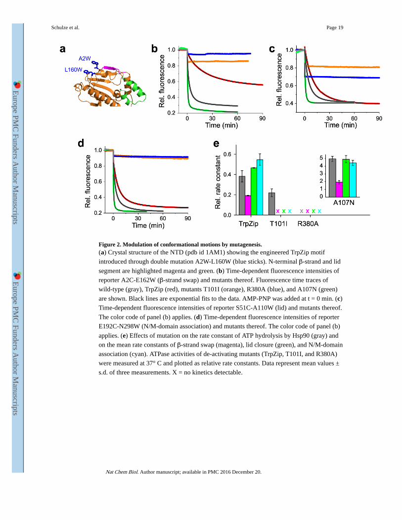
Figure 2. Modulation of conformational motions by mutagenesis.(a) Crystal structure of the NTD (pdb id 1AM1) showing the engineered TrpZip motif
introduced through double mutation A2W-L160W (blue sticks). N-terminal β-strand and lid
segment are highlighted magenta and green. (b) Time-dependent fluorescence intensities of
reporter A2C-E162W (β-strand swap) and mutants thereof. Fluorescence time traces of
wild-type (gray), TrpZip (red), mutants T101I (orange), R380A (blue), and A107N (green)
are shown. Black lines are exponential fits to the data. AMP-PNP was added at t = 0 min. (c)
Time-dependent fluorescence intensities of reporter S51C-A110W (lid) and mutants thereof.
The color code of panel (b) applies. (d) Time-dependent fluorescence intensities of reporter
E192C-N298W (N/M-domain association) and mutants thereof. The color code of panel (b)
applies. (e) Effects of mutation on the rate constant of ATP hydrolysis by Hsp90 (gray) and
on the mean rate constants of β-strand swap (magenta), lid closure (green), and N/M-domain
association (cyan). ATPase activities of de-activating mutants (TrpZip, T101I, and R380A)
were measured at 37° C and plotted as relative rate constants. Data represent mean values ±
s.d. of three measurements. X = no kinetics detectable.
Schulze et al. Page 19
Nat Chem Biol. Author manuscript; available in PMC 2016 December 20.
Europe PM
C Funders A
uthor Manuscripts
Europe PM
C Funders A
uthor Manuscripts

Figure 3. Influence of Aha1 on kinetics of local motions.(a) AMP-PNP-triggered fluorescence intensity time traces of β-strand swap (A2C+E162W,
magenta), lid closure (S51C-A110W, green), and N/M-domain association (E192C-N298W,
cyan). Samples were incubated with Aha1 before measurement and time traces were
recorded using stopped-flow spectroscopy. Data in shaded color are control samples that
lacked the engineered Trp. Fluorescence transients were fitted using a bi-exponential model
including a linear baseline drift of minor amplitude (black line). (b) Rate constants of ATP
hydrolysis (gray) by wild-type Hsp90 (wt) and mutant F349A together with the
corresponding mean rate constants of β-strand swap (magenta), lid closure (green), and
N/M-domain association (cyan) measured in absence and presence of Aha1 (X = no kinetics
detectable). Data represent mean values ± s.d. of three measurements. (c) Equilibrium
fluorescence intensities measured from reporters of N/M-domain association and of the lid
on wild-type Hsp90 and mutant F349A after incubation with Aha1.
Schulze et al. Page 20
Nat Chem Biol. Author manuscript; available in PMC 2016 December 20.
Europe PM
C Funders A
uthor Manuscripts
Europe PM
C Funders A
uthor Manuscripts

Figure 4. Equilibrium dynamics of lid and β-strand probed by PET-FCS.(a) Reporter design. N-terminal β-strand and lid of the NTD (pdb id 1AM1) are highlighted
magenta and green. Engineered pairs of Oxa (red sphere) and Trp (blue sticks) probing β-
strand (Q14C-A2W) and lid (A112C-S25W) are indicated. (b) and (c), ACFs (G(τ))
recorded from Q14C-A2W (magenta) and A112C-S25W (green), respectively, on the
isolated NTD. Data recorded after binding of ATP are shown in orange. Control samples
lacking the engineered Trp are shown in gray. Black lines are fits to the data using a model
for molecular diffusion containing two single-exponential relaxations. The ACF of the
Schulze et al. Page 21
Nat Chem Biol. Author manuscript; available in PMC 2016 December 20.
Europe PM
C Funders A
uthor Manuscripts
Europe PM
C Funders A
uthor Manuscripts
TrpZip construct (Q14C-A2W-L60W) is shown in blue in panel (b), and was described by a
molecular diffusion model without additional relaxations. All ACFs were normalized to the
average number of molecules in the detection focus for clarity. Broken lines indicate the
amplitudes of the diffusion decays. (d) and (e) ACFs of same reporters engineered on the
NM-domain. Same color code as in panels (b) and (c) applies. Black lines are fits to the data
using a model for molecular diffusion containing three single-exponential relaxations. ACFs
recorded in the presence of Aha1 are shown in cyan. (f) Rate constants of β-strand release
(magenta) and lid release (green) in NTD and NM-domain. Effects of binding of ATP and
Aha1 are shown. Data represent mean values ± s.d. of three measurements.
Schulze et al. Page 22
Nat Chem Biol. Author manuscript; available in PMC 2016 December 20.
Europe PM
C Funders A
uthor Manuscripts
Europe PM
C Funders A
uthor Manuscripts

Figure 5. Possible origins of multi-exponential kinetics in protein dynamics.Two-dimensional projection of a conformational free energy surfaces along an arbitrary
reaction coordinate. (a) Ground-state heterogeneity. Multiple open-clamp conformations
(o1-o3) of different free energy give rise to energy barriers of different height (broken
arrows) along parallel pathways to the closed-clamp conformation (c). (b) On-pathway
intermediates. Conformational change along a pathway containing a series of discrete
intermediates (i1-i3) of different free energy. (c) Pathway heterogeneity. Open-clamp
conformers of same free energy traverse to the closed-clamp conformation along different
pathways that are characterized by different energy barrier heights.
Schulze et al. Page 23
Nat Chem Biol. Author manuscript; available in PMC 2016 December 20.
Europe PM
C Funders A
uthor Manuscripts
Europe PM
C Funders A
uthor Manuscripts

Figure 6. Integration of results into the chaperone catalytic cycle.(1) At the beginning of the catalytic cycle, apo Hsp90 populates a heterogeneous ensemble
of open-clamp conformers. Lid (green) and N-terminal β-strand (magenta) are highly mobile
structural elements with sub-millisecond reconfiguration times. (2) Binding of ATP to the
NTD leads to rapid release of the lid to an intermediate conformational state. The co-
chaperone Aha1 pre-associates N- and M-domains but also remodels the lid segment for
accelerated closure. (3) Closure of the molecular clamp involves cooperative action of
conformational switches. Closure of the lid over the ATP-binding pocket, cross-subunit swap
of β-strands, and association of the N- and M-domains are slow and interdependent. Swap of
the terminal β-strands is weakly coupled with the other motions. (4) Hydrolysis of ATP
leads to a compact, ADP-bound conformation, which relaxes to an open state with
concomitant release of ADP and inorganic phosphate. Opening of the molecular clamp
reconstitutes Hsp90 for the next catalytic cycle.
Schulze et al. Page 24
Nat Chem Biol. Author manuscript; available in PMC 2016 December 20.
Europe PM
C Funders A
uthor Manuscripts
Europe PM
C Funders A
uthor Manuscripts