Conversion of Amino Acids to Specialized Products · Conversion of Amino Acids to Specialized...
32
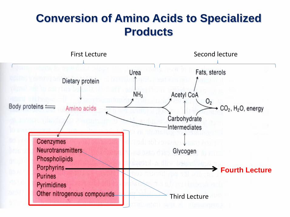
Conversion of Amino Acids to Specialized Products First Lecture Second lecture Third Lecture Fourth Lecture
Transcript of Conversion of Amino Acids to Specialized Products · Conversion of Amino Acids to Specialized...
Conversion of Amino Acids to Specialized Products
First Lecture Second lecture
Third Lecture
Fourth Lecture
Structure of porphyrins
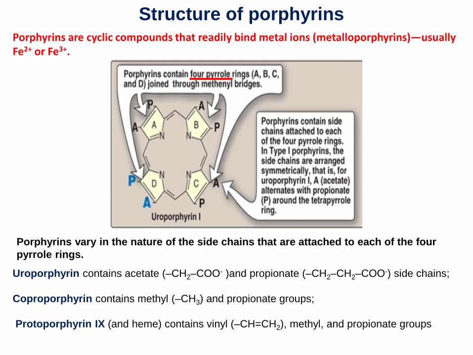
Porphyrins vary in the nature of the side chains that are attached to each of the four pyrrole rings.
Uroporphyrin contains acetate (–CH2–COO- )and propionate (–CH2–CH2–COO-) side chains;
Coproporphyrin contains methyl (–CH3) and propionate groups;
Protoporphyrin IX (and heme) contains vinyl (–CH=CH2), methyl, and propionate groups
Porphyrins are cyclic compounds that readily bind metal ions (metalloporphyrins)—usually Fe2+ or Fe3+.
Structure of Porphyrins
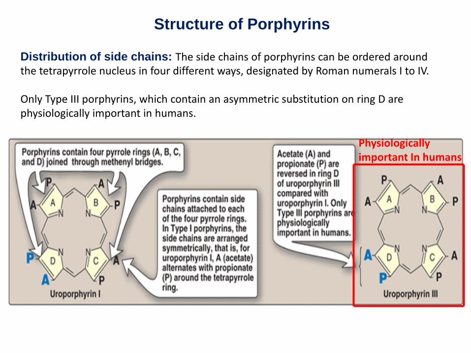
Distribution of side chains: The side chains of porphyrins can be ordered around the tetrapyrrole nucleus in four different ways, designated by Roman numerals I to IV.
Only Type III porphyrins, which contain an asymmetric substitution on ring D are physiologically important in humans.
Physiologically important In humans

Heme is a prosthetic groupfor hemoglobin, myoglobin,the cytochromes, catalase andtrptophan pyrrolase
1) Four Pyrrole rings linked together withmethenyle bridges;
2) Three types of side chains are attachedto the rings; arrangement of these side chains determines the activity;
3) Asymmetric molecule3) Porphyrins bind metal ions to form
metalloporphyrins.
Heme
propionyl
methyl vinyl

Biosynthesis of Heme
The major sites of heme biosynthesis are the liver, which synthesizes a number of heme proteins (particularly cytochrome P450), and the erythrocyte-producing cells of the bone marrow, which are active in hemoglobin synthesis.
The initial reaction and the last three steps in the formation of porphyrins occur in mitochondria, whereas the intermediate steps of the biosynthetic pathway occur in the cytosol.
[Note: Mature red blood cells lack mitochondria and are unable to synthesize heme.]
Heme synthesis occurs in all cells due to the requirement for heme as a prosthetic group on enzymes and electron transport chain.

Biosynthesis of HemeStep I: Formation of δ-aminolevulinic acid (ALA)
δ-aminolevulinicacid (ALA) synthase Pyridoxal
Phosphate
ALA synthase activity (transcription) is modulated by •Heme (inhibits ALA synthase)•Drugs (e.g. antifungals and Anticonvulsants) (enhances the ALA activity)
Cytochrome P450 monooxygenase

Biosynthesis of Heme
The condensation of two molecules of ALA to form porphobilinogen by ALA dehydratase.
Reaction is extremely sensitive to inhibition by heavy metal ions. This inhibition is, in part, responsible for the elevation in ALA and the anemia seen in lead poisoning.
δ-aminolevulinicacid (ALA) dehydratase
Step II: Formation of porphobilinogen:
Porphobilinogen (PBG) is the first pathway intermediate that includes a pyrrole ring.
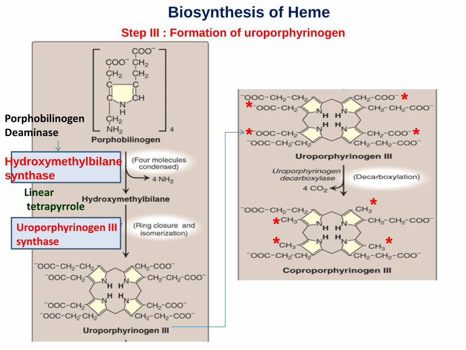
Biosynthesis of HemeStep III : Formation of uroporphyrinogen
Uroporphyrinogen III synthase
Hydroxymethylbilanesynthase
PorphobilinogenDeaminase
Lineartetrapyrrole
*
* **
*** *
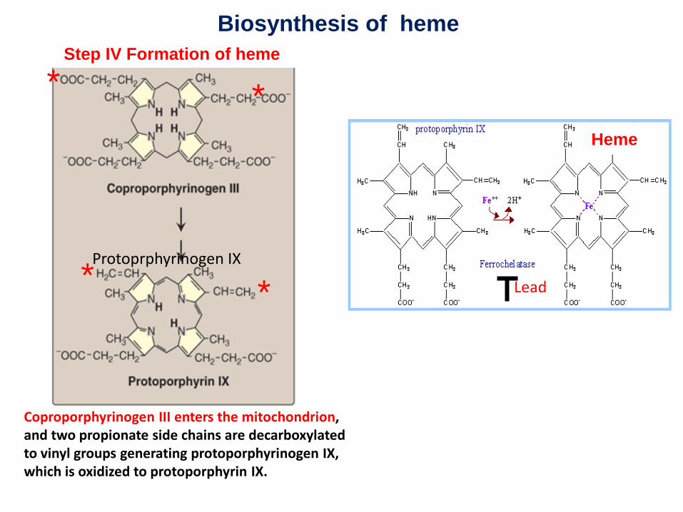
Biosynthesis of hemeStep IV Formation of heme
Protoprphyrinogen IX* *
**
Coproporphyrinogen III enters the mitochondrion, and two propionate side chains are decarboxylatedto vinyl groups generating protoporphyrinogen IX, which is oxidized to protoporphyrin IX.
Lead
Heme

Uroporphyrinogen I Coproporphyrinogen I
Overview of Heme Synthesis
Succinyl CoA + Glycine
δ-aminolevulinic acid
δ-aminolevulinic acid
Porphobilinogen Uroporphyrinogen III Coproporphyrinogen III
Coproporphyrinogen III
Protoporphyrinogen IX
Protoporphyrin IX
Heme
ALA synthase
cytoplasmmitochondrial matrix
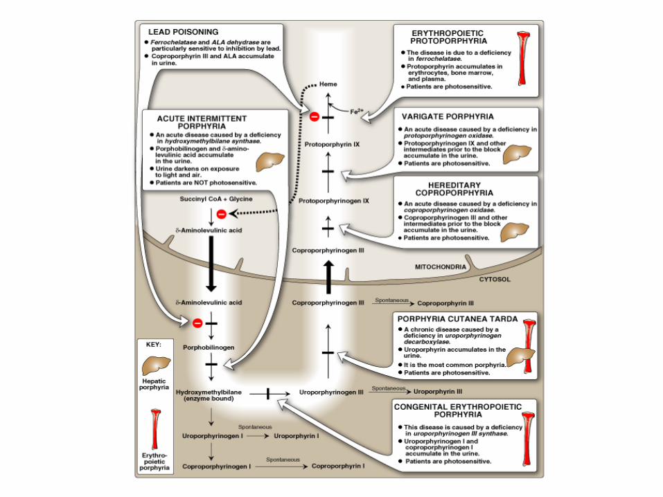
Porphyrias
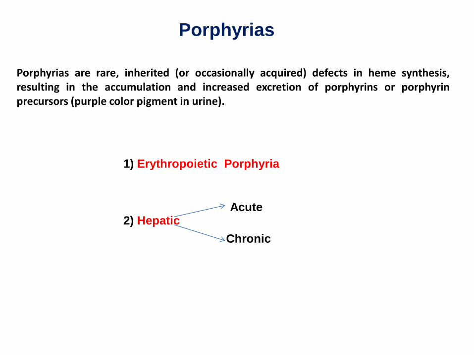
Porphyrias are rare, inherited (or occasionally acquired) defects in heme synthesis,resulting in the accumulation and increased excretion of porphyrins or porphyrinprecursors (purple color pigment in urine).
1) Erythropoietic Porphyria
2) HepaticChronic
Acute

Acute intermittent porphyria (AIP)- Uroporphyrinogen synthasedeficiency with high ALA or PBG in urine and serum.
Variegate porphyria (VP)- Protoporphyrinogen oxidase deficiency with high fecal levels of protoporphyrin and coproporphyrin.
Hereditary coproporphyria - Coproporphyrinogen oxidase deficiency with high urinary and/or fecal levels of coproporphyrins.
ALA dehydratase deficient porphyria (rare).
Erythropoietic protoporphyria (EPP)- Ferrochelatase deficiency.
Hepatic Porphyria(Acute)
Symptoms: Acute attacks of gastrointestinal, neurologic/psychiatric, and cardiovascular symptoms.
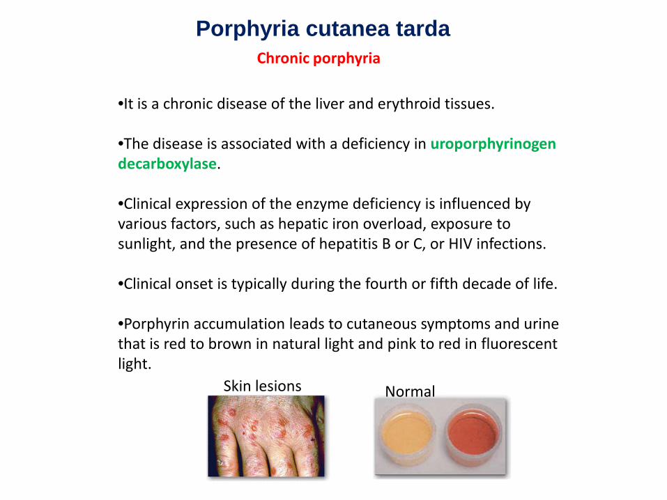
•It is a chronic disease of the liver and erythroid tissues.
•The disease is associated with a deficiency in uroporphyrinogendecarboxylase.
•Clinical expression of the enzyme deficiency is influenced by various factors, such as hepatic iron overload, exposure to sunlight, and the presence of hepatitis B or C, or HIV infections.
•Clinical onset is typically during the fourth or fifth decade of life.
•Porphyrin accumulation leads to cutaneous symptoms and urine that is red to brown in natural light and pink to red in fluorescent light.
Chronic porphyria
Porphyria cutanea tarda
NormalSkin lesions
They are characterized by skin rashes and blisters that appear in early childhood.
The diseases are complicated by cholestatic liver cirrhosis and progressive hepatic failure.
Erythropoietic porphyriasCongenital erythropoietic porphyria and erythropoietic protoporphyria)
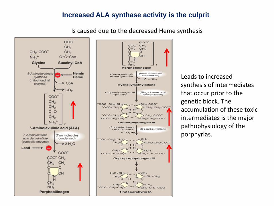
Leads to increased synthesis of intermediates that occur prior to the genetic block. The accumulation of these toxic intermediates is the major pathophysiology of the porphyrias.
Increased ALA synthase activity is the culprit
Is caused due to the decreased Heme synthesis
The severity of symptoms of the porphyrias can be diminished by intravenous injection of hemin, which decreases the synthesis of ALA synthase.
Avoidance of sunlight and ingestion of β-carotene (a free-radical scavenger) are also helpful.
Treatment
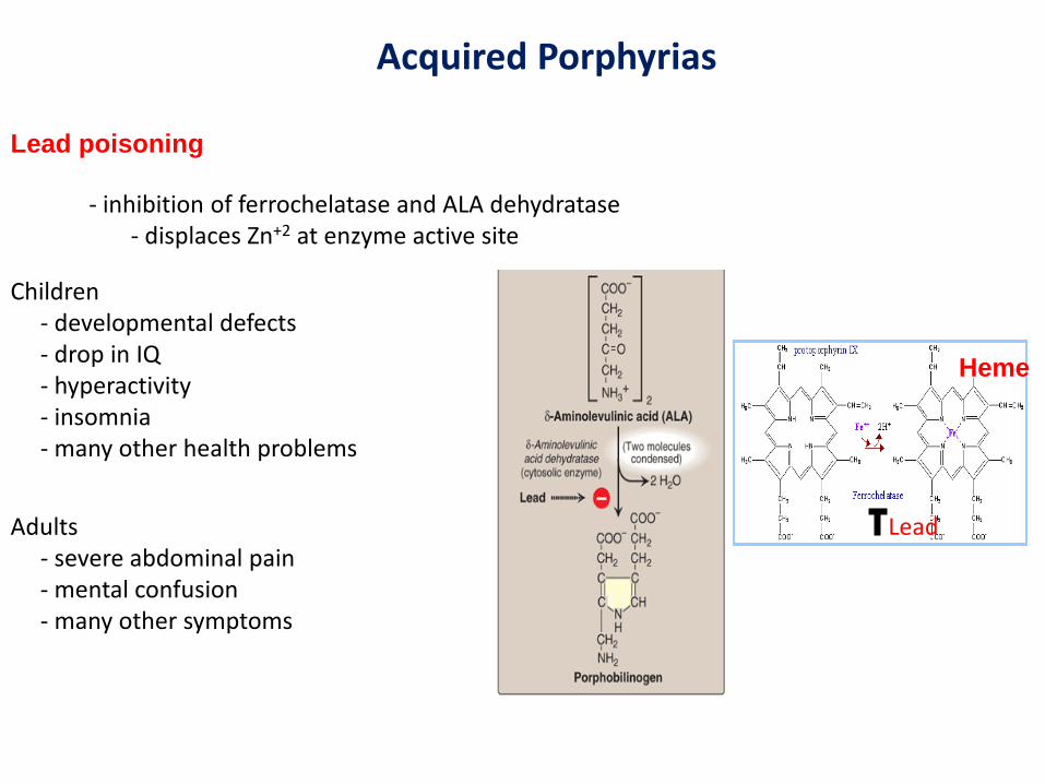
Acquired Porphyrias
Lead poisoning
- inhibition of ferrochelatase and ALA dehydratase- displaces Zn+2 at enzyme active site
Children- developmental defects- drop in IQ- hyperactivity- insomnia- many other health problems
Adults- severe abdominal pain- mental confusion- many other symptoms
Lead
Heme
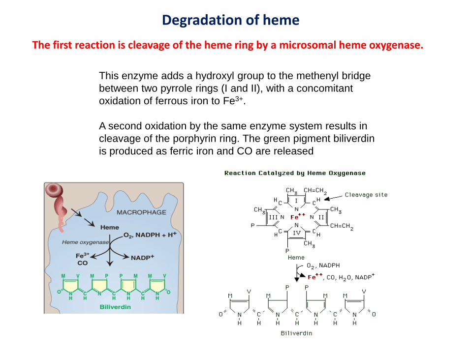
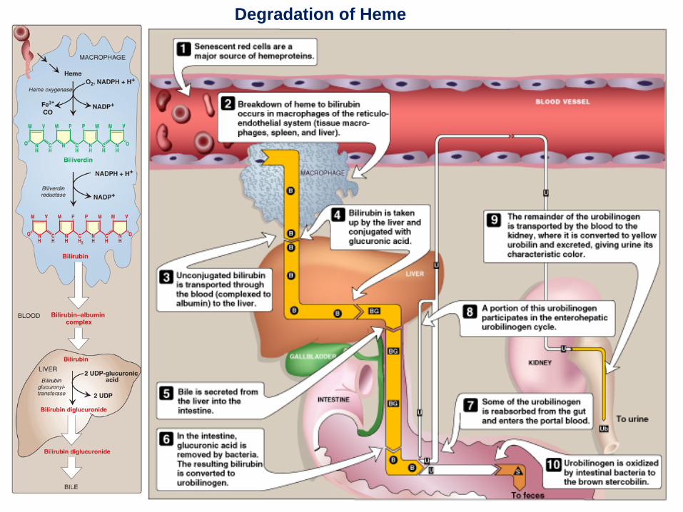
Degradation of heme
This enzyme adds a hydroxyl group to the methenyl bridge between two pyrrole rings (I and II), with a concomitant oxidation of ferrous iron to Fe3+.
A second oxidation by the same enzyme system results in cleavage of the porphyrin ring. The green pigment biliverdinis produced as ferric iron and CO are released
The first reaction is cleavage of the heme ring by a microsomal heme oxygenase.

Biliverdin is reduced, forming the red-orange bilirubin. Bilirubin and its derivatives are collectively termed bile pigments.
[Note: The changing colors of a bruise reflect the varying pattern of intermediates that occur during heme degradation.]
Degradation of HemeIn the second reaction biliverdin reductase reduces the central methenyl bridge of biliverdin, producing bilirubin.
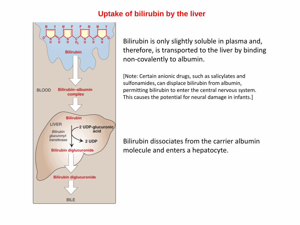
Bilirubin is only slightly soluble in plasma and, therefore, is transported to the liver by binding non-covalently to albumin.
[Note: Certain anionic drugs, such as salicylates and sulfonamides, can displace bilirubin from albumin, permitting bilirubin to enter the central nervous system. This causes the potential for neural damage in infants.]
Bilirubin dissociates from the carrier albumin molecule and enters a hepatocyte.
Uptake of bilirubin by the liver
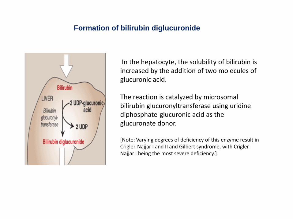
In the hepatocyte, the solubility of bilirubin is increased by the addition of two molecules of glucuronic acid.
The reaction is catalyzed by microsomalbilirubin glucuronyltransferase using uridinediphosphate-glucuronic acid as the glucuronate donor.
[Note: Varying degrees of deficiency of this enzyme result in Crigler-Najjar I and II and Gilbert syndrome, with Crigler-Najjar I being the most severe deficiency.]
Formation of bilirubin diglucuronide

Bilirubin diglucuronide (conjugated bilirubin) is actively transported into the bile.
Unconjugated bilirubin is normally not secreted.
This energy-dependent, rate-limiting step is susceptible to impairment in liver disease.
[Note: A deficiency in the protein required for transport of conjugated bilirubin out of the liver results in Dubin-Johnson syndrome.]
Secretion of bilirubin into bile

Bilirubin diglucuronide is hydrolyzed and reduced by bacteria in the gut to yield urobilinogen, a colorless compound.
Most of the urobilinogen is oxidized by intestinal bacteria to stercobilin, which gives feces the characteristic brown color.
However, some of the urobilinogen is reabsorbed from the gut and enters the portal blood. A portion of this urobilinogen participates in the enterohepatic urobilinogen cycle in which it is taken up by the liver, and then resecreted into the bile.
The remainder of the urobilinogen is transported by the blood to the kidney, where it is converted to yellow urobilin and excreted, giving urine its characteristic color.
Formation of urobilins in the intestine
Degradation of Heme

Symptoms: Yellow color of skin, nail beds, and sclerae (whites of the eyes) caused by deposition of bilirubin, secondary to increased bilirubin levels in the blood
JaundiceDefects in the degradation of Heme
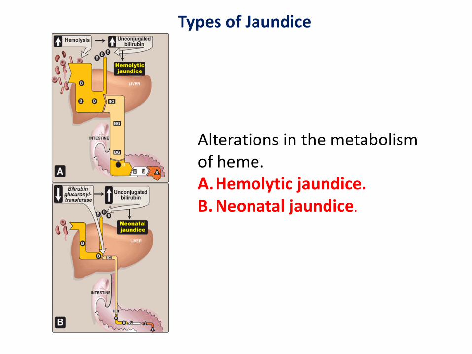
Alterations in the metabolism of heme. A.Hemolytic jaundice. B. Neonatal jaundice.
Types of Jaundice
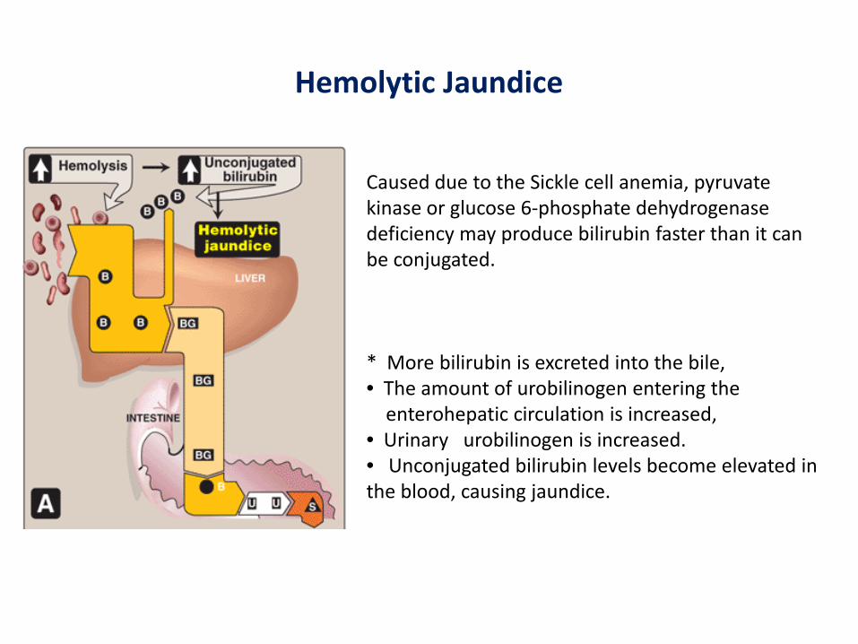
Hemolytic Jaundice
Caused due to the Sickle cell anemia, pyruvatekinase or glucose 6-phosphate dehydrogenasedeficiency may produce bilirubin faster than it can be conjugated.
* More bilirubin is excreted into the bile,• The amount of urobilinogen entering the
enterohepatic circulation is increased, • Urinary urobilinogen is increased. • Unconjugated bilirubin levels become elevated in the blood, causing jaundice.
Hepatocellular Jaundice
Damage to liver cells (for example, in patients with cirrhosis or hepatitis) can cause unconjugated bilirubin levels to increase in the blood as a result of decreased conjugation.
The bilirubin that is conjugated is not efficiently secreted into the bile, but instead diffuses (“leaks”) into the blood. Urobilinogen is increased in the urine because hepatic damage decreases the enterohepatic circulation of this compound, allowing more to enter the blood, from which it is filtered into the urine.
The urine thus becomes dark, whereas stools are a pale, clay color. Plasma levels of AST (SGOT) and ALT are elevated, and the patient experiences nausea and anorexia.
Obstructive Jaundice
In this instance, jaundice is not caused by overproduction of bilirubin or decreased conjugation, but instead results from obstruction of the bile duct. For example, the presence of a hepatic tumor or bile stones may block the bile ducts, preventing passage of bilirubin into the intestine.
Liver regurgitate conjugated bilirubin into the blood
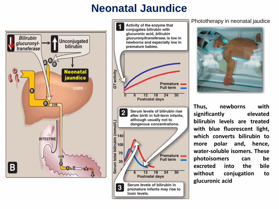
Neonatal Jaundice
Thus, newborns withsignificantly elevatedbilirubin levels are treatedwith blue fluorescent light,which converts bilirubin tomore polar and, hence,water-soluble isomers. Thesephotoisomers can beexcreted into the bilewithout conjugation toglucuronic acid
Phototherapy in neonatal jaudice
Bilirubin is most commonly determined by the van den Bergh reaction, in which diazotized sulfanilic acid reacts with bilirubin to form red azodipyrroles that are measured colorimetrically.
Determination of bilirubin concentration
Only conjugated bilirubin is water soluble and reacts directly. This is called the DIRECT bilirubin.
To measure the unconjugated bilirubin bound to albumin, alcohol is added to release it into solution, where it can now react. This is called the INDIRECT bilirubin.


![8 th lecture December 10, 2015 Specialized Connective Tissue [Bone (Osseous) Tissue]](https://static.fdocuments.in/doc/165x107/5a4d1b567f8b9ab0599a95f3/8-th-lecture-december-10-2015-specialized-connective-tissue-bone-osseous.jpg)