Conventional Endodontic Therapy of Upper Central … · Conventional Endodontic Therapy of Upper...
5
Conventional Endodontic Therapy of Upper Central Incisor Combined with Cyst Decompression: A Case Report Scott A. Martin, DDS Abstract Treatment of a maxillary central incisor with an asso- ciated cystic lesion by conventional endodontic therapy combined with decompression is reported. Although small cystic lesions frequently heal simply with en- dodontic therapy, larger lesions may need additional treatment. If surgical enucleation is elected, other teeth or structures may be damaged unnecessarily. Therefore, a case can be made for first attempting the more conservative treatment of decompression, and a work- able protocol for this is presented. In this case, 6 weeks with latex tubing in place and daily irrigation with 0.12% chlorhexidine led to complete healing with no need for further surgery or other root canal therapy on teeth initially surrounded by this lesion. At the 2-year recall, the lesion has completely resolved, and the adjacent teeth remain vital and normal. (J Endod 2007; 33:753–757) Key Words Decompression, marsupialization, radicular cyst T reatment of teeth with large cystic lesions can be problematic. For many years, it was thought that the distribution of cysts among periapical lesions was around 40% to 50% (1, 2). Subsequent studies in which complete lesions were evaluated by serial sectioning revealed that, in fact, many fewer lesions were actually cystic than previously thought (3–6). Of the 15% of lesions now considered to be truly cystic, somewhat more than half are true apical cysts (3–6). According to Nair, “...a periapical pocket cyst may heal after conventional root canal therapy whereas an apical true cyst is less likely to be resolved without surgical intervention...” (7). Treatment of suspected cysts, therefore, requires follow-up over a period of time. According to Natkin et al. (8), there is no doubt that larger lesions are more likely to be cysts and will be less likely to heal with endodontic therapy alone. Should surgical intervention become necessary, the decision point is whether to raise a flap and completely enucleate the lesion or to try “decompression” first (8 –10). Even if enucleation is still necessary later, the lesion will predictably be much smaller and present less difficulty with removal and less risk of damage to associated teeth and vital structures. As an aside, decompression with placement of tubing to maintain drainage is quite different from “marsupialization,” although the terms tend to be used interchangeably. Marsupialization is actually described as “...unroofing the outer wall of the cyst by making a surgical incision, evacuating its contents, and establishing a large permanent opening by suturing the remaining part of the cystic membrane to the mucosal surface around the periphery of the opening” (9). Decompression is favored because of lower morbidity and the fact that bony ingrowth occurs as the lesion shrinks in size, thus resulting in more normal bony contours after treatment is concluded. Case Report A healthy 13-year-old male was referred for evaluation based on the presence of a sinus tract stoma on the buccal attached gingiva over the left upper lateral incisor. The patient was asymptomatic and not aware of the stoma. A radiograph revealed part of a radiolucent lesion that appeared to be centered on tooth #10. The sinus tract was traced with gutta percha and another radiograph made so that both the full extent of the lesion could be assessed and to see where the sinus tract may have originated (Fig. 1). Sinus tract tracing may or may not reveal anything that would not be revealed with subsequent tests and examination. However, it is one more corroborating bit of evidence, is easy to perform, and carries essentially no risk. The tracing pointed to tooth #9 as the source of the lesion. Tooth #9 was indeed found to be nonvital (no response to cold or electrical stimulation), whereas all other maxillary anterior teeth were normal in all regards. In consultation with the patient’s parents, initial treatment was to be root canal therapy with an interim dressing of calcium hydroxide for a period of 4 weeks. On opening, copious drainage of very thin viscosity was noted, which persisted for some time despite repeated drying with paper points after shaping and alternately irrigating with sodium hypochlorite (NaOCl) 5%, EDTA 17%, and pure ethanol as a final rinse. Vitapex (Dia- Dent Group International, Burnaby, BC, Canada) Ca(OH) 2 paste was injected into the canal space (Fig. 2). Vitapex is Ca(OH) 2 in a silicone base with potassium iodide incorporated as an antimicrobial agent. At 1 month, the sinus tract was resolved so the Ca(OH) 2 paste was removed with a combination of irrigation as noted earlier and reinstrumentation to slightly larger apical diameter (.070 mm) and taper (12%) using stainless steel hand files and greater taper rotary files. At this visit, complete drying of the canal space was achieved, and the canal From Private Practice, Duvall, Washington; and Depart- ment of Oral Medicine, University of Washington Dental School, Duvall, Washington. Address requests for reprints to Dr. Scott A. Martin, PO Box 1180, Duvall, WA 98019. E-mail address: scott@ innovativeendo.org. 0099-2399/$0 - see front matter Copyright © 2007 by the American Association of Endodontists. doi:10.1016/j.joen.2007.01.013 Case Report/Clinical Techniques JOE — Volume 33, Number 6, June 2007 Conventional Endodontic Therapy 753
Transcript of Conventional Endodontic Therapy of Upper Central … · Conventional Endodontic Therapy of Upper...
CCS
ATccsdtoacaw0ntra3
KD
mS
Bi0
Ed
Case Report/Clinical Techniques
J
onventional Endodontic Therapy of Upper Central Incisorombined with Cyst Decompression: A Case Report
cott A. Martin, DDS
T5stthrrde
rEavdiopmbi
sprwcttpo
scwcrhDci
cd
bstractreatment of a maxillary central incisor with an asso-iated cystic lesion by conventional endodontic therapyombined with decompression is reported. Althoughmall cystic lesions frequently heal simply with en-odontic therapy, larger lesions may need additionalreatment. If surgical enucleation is elected, other teethr structures may be damaged unnecessarily. Therefore,
case can be made for first attempting the moreonservative treatment of decompression, and a work-ble protocol for this is presented. In this case, 6 weeksith latex tubing in place and daily irrigation with.12% chlorhexidine led to complete healing with noeed for further surgery or other root canal therapy oneeth initially surrounded by this lesion. At the 2-yearecall, the lesion has completely resolved, and thedjacent teeth remain vital and normal. (J Endod 2007;3:753–757)
ey Wordsecompression, marsupialization, radicular cyst
From Private Practice, Duvall, Washington; and Depart-ent of Oral Medicine, University of Washington Dental
chool, Duvall, Washington.Address requests for reprints to Dr. Scott A. Martin, PO
ox 1180, Duvall, WA 98019. E-mail address: [email protected]/$0 - see front matter
Copyright © 2007 by the American Association ofndodontists.oi:10.1016/j.joen.2007.01.013
r
OE — Volume 33, Number 6, June 2007
reatment of teeth with large cystic lesions can be problematic. For many years, it wasthought that the distribution of cysts among periapical lesions was around 40% to
0% (1, 2). Subsequent studies in which complete lesions were evaluated by serialectioning revealed that, in fact, many fewer lesions were actually cystic than previouslyhought (3– 6). Of the 15% of lesions now considered to be truly cystic, somewhat morehan half are true apical cysts (3– 6). According to Nair, “...a periapical pocket cyst mayeal after conventional root canal therapy whereas an apical true cyst is less likely to beesolved without surgical intervention...” (7). Treatment of suspected cysts, therefore,equires follow-up over a period of time. According to Natkin et al. (8), there is nooubt that larger lesions are more likely to be cysts and will be less likely to heal withndodontic therapy alone.
Should surgical intervention become necessary, the decision point is whether toaise a flap and completely enucleate the lesion or to try “decompression” first (8 –10).ven if enucleation is still necessary later, the lesion will predictably be much smallernd present less difficulty with removal and less risk of damage to associated teeth andital structures. As an aside, decompression with placement of tubing to maintainrainage is quite different from “marsupialization,” although the terms tend to be used
nterchangeably. Marsupialization is actually described as “...unroofing the outer wallf the cyst by making a surgical incision, evacuating its contents, and establishing a largeermanent opening by suturing the remaining part of the cystic membrane to theucosal surface around the periphery of the opening” (9). Decompression is favored
ecause of lower morbidity and the fact that bony ingrowth occurs as the lesion shrinksn size, thus resulting in more normal bony contours after treatment is concluded.
Case ReportA healthy 13-year-old male was referred for evaluation based on the presence of a
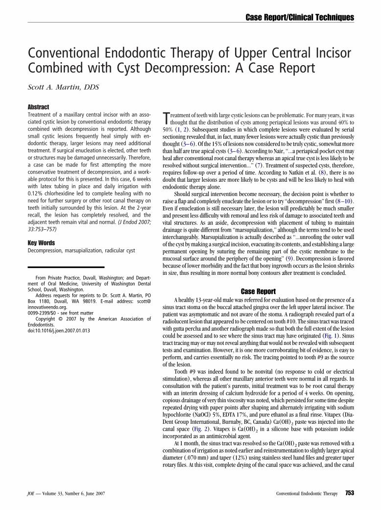
inus tract stoma on the buccal attached gingiva over the left upper lateral incisor. Theatient was asymptomatic and not aware of the stoma. A radiograph revealed part of aadiolucent lesion that appeared to be centered on tooth #10. The sinus tract was tracedith gutta percha and another radiograph made so that both the full extent of the lesionould be assessed and to see where the sinus tract may have originated (Fig. 1). Sinusract tracing may or may not reveal anything that would not be revealed with subsequentests and examination. However, it is one more corroborating bit of evidence, is easy toerform, and carries essentially no risk. The tracing pointed to tooth #9 as the sourcef the lesion.
Tooth #9 was indeed found to be nonvital (no response to cold or electricaltimulation), whereas all other maxillary anterior teeth were normal in all regards. Inonsultation with the patient’s parents, initial treatment was to be root canal therapyith an interim dressing of calcium hydroxide for a period of 4 weeks. On opening,opious drainage of very thin viscosity was noted, which persisted for some time despiteepeated drying with paper points after shaping and alternately irrigating with sodiumypochlorite (NaOCl) 5%, EDTA 17%, and pure ethanol as a final rinse. Vitapex (Dia-ent Group International, Burnaby, BC, Canada) Ca(OH)2 paste was injected into theanal space (Fig. 2). Vitapex is Ca(OH)2 in a silicone base with potassium iodidencorporated as an antimicrobial agent.
At 1 month, the sinus tract was resolved so the Ca(OH)2 paste was removed with aombination of irrigation as noted earlier and reinstrumentation to slightly larger apicaliameter (.070 mm) and taper (12%) using stainless steel hand files and greater taper
otary files. At this visit, complete drying of the canal space was achieved, and the canalConventional Endodontic Therapy 753
wP
tuwtpili
cUbcasaiwrwcb0ata
wsbp
icwa7
sttctdatb(n
k(ct
F
Case Report/Clinical Techniques
7
as obturated with warm vertical compaction of gutta-percha and AHlus sealer (Dentsply Maillefer, Ballaigues, Switzerland) (Fig. 3).
At the 3-month recall, the patient was still asymptomatic; however,he sinus tract stoma was present again. At this time, discussion wasndertaken with several endodontic specialists, and decompressionas considered as a possible treatment alternative. In consultation with
he patient and his parents, it was believed that decompression for aeriod of 6 weeks offered a reasonable chance of success and that evenf complete healing did not ensue, enucleation of the residual (smaller)esion would be less likely to cause damage to adjacent structures,ncluding teeth #10 and 11 and the floor of the nose.
After infiltration anesthesia, an approximately 1.5-cm vertical in-ision was made in between the root eminences of teeth #9 and 10.pon entry into the cyst cavity, there was slight drainage of pus followedy copious drainage of the typical straw-colored fluid associated withystic lesions. Lavage with sterile saline was accomplished, and then anpproximately 2-cm length of #10Fr radiopaque latex tubing was in-erted to the depth of the cyst cavity. One 4-0-gut suture was placedbove and below the drain. Another suture was placed through the draintself as well as through mucosa to stabilize it during initial healing. At 2eeks, healing was complete, and the drain could be removed and
einserted with minimal discomfort to patient (Figs. 4, 5). The patientas instructed to irrigate through the lumen of the drain daily withhlorhexidine 0.12%, consistent with a protocol previously describedy Brondum and Jensen (11). Their recommendation is once daily with.12% chlorhexidine gluconate delivered through “...a normal syringend a thin blunt hypodermic needle.” Irrigation is accomplishedhrough the lumen of the drain. Internal diameter of size #10Fr drain is
igure 1. Sinus tract tracing.
bout 1.5 mm so #16- or 18-gauge blunt Luer lock dispensing tips work F
54 Martin
ell with small disposable syringes; both are available from any dentalupplier. Although empirical, it seems prudent to irrigate with an anti-acterial agent, and chlorhexidine has proved safe and effective as are-/postsurgical oral disinfectant for many years.
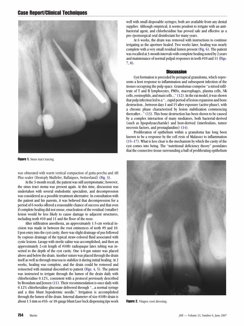
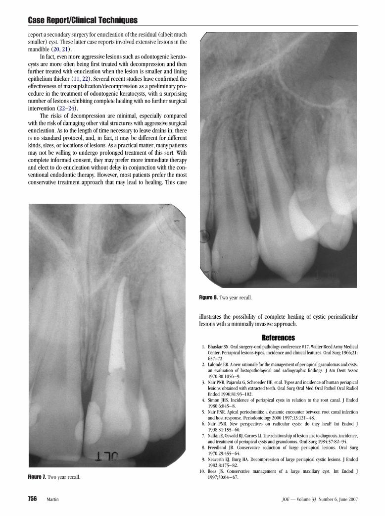
At 6 weeks, the drain was removed with instructions to continuerrigating as the aperture healed. Two weeks later, healing was nearlyomplete with a very small residual lumen present (Fig. 6). The patientas recalled at 3-month intervals with complete healing noted by 2 yearsnd maintenance of normal pulpal responses in teeth #10 and 11 (Figs., 8).
DiscussionCyst formation is preceded by periapical granuloma, which repre-
ents a host response to inflammation and subsequent infection of theissues occupying the pulp space. Granulomas comprise “a mixed infil-rate of T and B lymphocytes, PMNs, macrophages, plasma cells, NKells, eosinophils, and mast cells...” (12). In the rat model, it was shownhat pulp infection led to a “...rapid period of lesion expansion and boneestruction...between days 1 and 15 after exposure (active phase), withchronic phase characterized by lesion stabilization commencing
hereafter...” (13). This bone destruction has been shown to be causedy a complex interaction of many mediators, both bacterial-derivedsuch as lipopolysaccharide) and host-derived (interleukins, tumorecrosis factors, and prostaglandins) (14).
Proliferation of epithelium within a granuloma has long beennown to be a response by the cell rests of Malassez to inflammation15–17). What is less clear is the mechanism by which the cavity of theyst comes into being. The “nutritional deficiency theory” postulateshat the connective tissue surrounding a ball of proliferating epithelium
igure 2. Vitapex root dressing.
JOE — Volume 33, Number 6, June 2007

sclscnf(ea
ptatprtptplpbreu
cmcs
F
F
F
Case Report/Clinical Techniques
J
upplies its nutrition. If the epithelial proliferation proceeds too fast, theentral cells within this ball of epithelium will degenerate and die,eading to a fluid-filled cavity within the epithelial mass (16). The “ab-cess theory” supposes that proliferation of epithelium traps variousonnective tissue elements (including inflammatory cells) that thenecrose and lead to proteolytic activity, ultimately resulting in the fluid-illed cavity typical of a cyst (17). The fact that no more modern theoriesor definitive proof for either of the previously mentioned theories)xist 30 years later is a testament to the complexity of the cellular biologynd biochemistry relating to these lesions.
igure 3. Warm vertical compaction.
igure 4. Radiopaque latex drain. F
OE — Volume 33, Number 6, June 2007
Once the cystic cavity is formed, then the question is how the thirdhase (enlargement) progresses. Evidence for a “...molecular explana-
ion...” (5) is building as we add to our understanding of cytokines suchs interleukins (ILs), tumor necrosis factors, neuropeptides, and effec-or molecules such as matrix metalloproteinases. Although intracysticressure has long been postulated as a reason for cyst expansion, onlyecently have clues come to light about the possible mechanisms forhis. Very recent research suggests that IL-1 alpha expression might beartly regulated by intracystic pressure (18). IL-1 alpha has many func-
ions including induction of osteoclast formation and stimulation ofrostaglandin and collagenase production (19). Therefore, it is very
ikely that reduction of intracystic pressure is a key factor. It is alsolausible that reduction of the concentration of inflammatory mediatorsy irrigation of the cyst lumen could reduce epithelial proliferation andeverse bone resorption, leading to shrinkage of the cyst cavity. How-ver, the exact mechanisms of cyst expansion and shrinkage remainnknown at this time.
Although it is not known what percentage of radicular cyst casesan be expected to heal with only decompression, it is a viable treatmentodality that bears consideration when treating large, presumptively
ystic lesions. The literature is sparse on this topic. Some case reportshow complete healing with no subsequent enucleation (9, 10). Others
igure 5. Drain removed.
igure 6. Two weeks after drain removal.
Conventional Endodontic Therapy 755
rsm
cfeecni
weikmcavc
il
1F
F
Case Report/Clinical Techniques
7
eport a secondary surgery for enucleation of the residual (albeit muchmaller) cyst. These latter case reports involved extensive lesions in theandible (20, 21).
In fact, even more aggressive lesions such as odontogenic kerato-ysts are more often being first treated with decompression and thenurther treated with enucleation when the lesion is smaller and liningpithelium thicker (11, 22). Several recent studies have confirmed theffectiveness of marsupialization/decompression as a preliminary pro-edure in the treatment of odontogenic keratocysts, with a surprisingumber of lesions exhibiting complete healing with no further surgicalntervention (22–24).
The risks of decompression are minimal, especially comparedith the risk of damaging other vital structures with aggressive surgicalnucleation. As to the length of time necessary to leave drains in, theres no standard protocol, and, in fact, it may be different for differentinds, sizes, or locations of lesions. As a practical matter, many patientsay not be willing to undergo prolonged treatment of this sort. With
omplete informed consent, they may prefer more immediate therapynd elect to do enucleation without delay in conjunction with the con-entional endodontic therapy. However, most patients prefer the mostonservative treatment approach that may lead to healing. This case
igure 7. Two year recall.
56 Martin
llustrates the possibility of complete healing of cystic periradicularesions with a minimally invasive approach.
References1. Bhaskar SN. Oral surgery-oral pathology conference #17. Walter Reed Army Medical
Center. Periapical lesions-types, incidence and clinical features. Oral Surg 1966;21:657–72.
2. Lalonde ER. A new rationale for the management of periapical granulomas and cysts:an evaluation of histopathological and radiographic findings. J Am Dent Assoc1970;80:1056 –9.
3. Nair PNR, Pajarola G, Schroeder HE, et al. Types and incidence of human periapicallesions obtained with extracted teeth. Oral Surg Oral Med Oral Pathol Oral RadiolEndod 1996;81:93–102.
4. Simon JHS. Incidence of periapical cysts in relation to the root canal. J Endod1980;6:845– 8.
5. Nair PNR. Apical periodontitis: a dynamic encounter between root canal infectionand host response. Periodontology 2000 1997;13:121– 48.
6. Nair PNR. New perspectives on radicular cysts: do they heal? Int Endod J1998;31:155– 60.
7. Natkin E, Oswald RJ, Carnes LI. The relationship of lesion size to diagnosis, incidence,and treatment of periapical cysts and granulomas. Oral Surg 1984;57:82–94.
8. Freedland JB. Conservative reduction of large periapical lesions. Oral Surg1970;29:455– 64.
9. Neaverth EJ, Burg HA. Decompression of large periapical cystic lesions. J Endod1982;8:175– 82.
0. Rees JS. Conservative management of a large maxillary cyst. Int Endod J
igure 8. Two year recall.
1997;30:64 – 67.
JOE — Volume 33, Number 6, June 2007

1
1
1
1
1
11
1
1
2
2
2
2
2
Case Report/Clinical Techniques
J
1. Brondum N, Jensen VJ. Recurrence of keratocysts and decompression treatment.Oral Surg Oral Med Oral Pathol 1991;72:265–9.
2. Orstavik D, Pitt Ford TR. Essential Endodontology. Malden, PA: Blackwell Publishing,2002:47.
3. Stashenko P, et al. Kinetics of immune cell and bone resorptive responses to end-odontic infections. J Endod 1992;18:422– 6.
4. Wang CY, Stashenko P. Characterization of bone-resorbing activity in human peria-pical lesions. J Endod 1993;19:107–11.
5. Browne RM. The pathogenesis of odontogenic cysts: a review. J Oral Pathol1975;4:31– 46.
6. Shear M. The histogenesis of the dental cyst. Dent Practioner 1963;13:238 – 43.7. Summers L. The incidence of epithelium in periapical granulomas and the mech-
anism of cavitation in apical dental cysts in man. Arch Oral Biol1974;19:1177–9.
8. Kubota Y, Ninomiya T, Oka S, Takenoshita Y, Shirasuna K. Interleukin-1 alpha-
dependent regulation of matrix metalloproteinase-9 (MMP-9) secretion andOE — Volume 33, Number 6, June 2007
activation in the epithelial cells of odontogenic jaw cysts. J Dent Res2000;79:1423–30.
9. Motamedi MHK, Talesh KT. Management of extensive dentigerous cysts. Br Dent J2005;198:203– 6.
0. Tucker WM, Pleasants JE, MacComb WS. Decompression and secondary enucleationof a mandibular cyst: report of case. J Oral Surg 1972;30:669 –73.
1. Marker P, Brondum N. Treatment of large odontogenic keratocysts by decompres-sion and later cystectomy. Oral Surg Oral Med Oral Pathol 1996;82:122–31.
2. Nakamura N, Mitsuyasu T, Mitsuyasu Y, et al. Marsupialization for odontogenic ker-atocysts: long-term follow-up analysis of the effects and changes in growth charac-teristics. Oral Surg Oral Med Oral Path Oral Radiol Endod 2002;94:543–53.
3. Pogrel MA, Jordan RCK. Marsupialization as a definitive treatment for the odonto-genic keratocyst. J Oral Maxillofac Surg 2004;62:651–5.
4. Blanas N, Freund B, Schwartz M, Furst IM. Systematic review of the treatment andprognosis of the odontogenic keratocyst. Oral Surg Oral Med Oral Path Oral Radiol
Endod 2000;90:553– 8.Conventional Endodontic Therapy 757