Controlling the pyridinium–zwitterionic ligand ratio on ...
12
Controlling the pyridinium–zwitterionic ligand ratio on atomically precise gold nanoclusters allowing for eradicating Gram-positive drug- resistant bacteria and retaining biocompatibility† Zeyang Pang, ab Qizhen Li, a Yuexiao Jia, a Weixiao Yan, a Jie Qi, a Yuan Guo, c Fupin Hu, d Dejian Zhou * b and Xingyu Jiang * a Infections caused by multidrug-resistant (MDR) bacteria are an increasing global healthcare concern. In this study, we developed a dual-ligand-functionalised Au 25 (SR 1 ) x (SR 2 ) 18x -type gold nanocluster and determined its antibacterial activity against MDR bacterial strains. The pyridinium ligand (SR 1 ) provided bactericidal potency and the zwitterionic ligand (SR 2 ) enhanced the stability and biocompatibility. By optimising the ligand ratio, our gold nanocluster could effectively kill MDR Gram-positive bacteria via multiple antibacterial actions, including inducing bacterial aggregation, disrupting bacterial membrane integrity and potential, and generating reactive oxygen species. Moreover, combining the optimised gold nanocluster with common antibiotics could significantly enhance the antibacterial activity against MDR bacteria both in in vitro and animal models of skin infections. Furthermore, the fluorescence of the gold nanocluster at the second near-infrared (NIR-II) biological window allowed for the monitoring of its biodistribution and body clearance, which confirmed that the gold nanoclusters had good renal clearance and biocompatibility. This study provides a new strategy to combat the MDR challenge using multifunctional gold nanomaterials. Introduction Since the discovery of antibiotics in 1928, the unrestrained global use of antibiotics has imposed a highly selective pressure on all bacterial species, which has accelerated the acquisition and accumulation of drug-resistant genes via horizontal trans- mission. 1 Several multidrug-resistant (MDR) pathogens, or the so-called ‘superbugs,’ have emerged over the past 50 years. 2,3 Moreover, some drug-resistant bacteria that were previously considered less harmful, like Methicillin-resistant Staphylo- coccus epidermidis (MRSE), have gained much attention due to their role in the development of drug-resistant strains. 4,5 The widespread occurrence of MRSE inevitably increases the risk of infection and promotes intra- and inter-species horizontal transfer of the MDR gene. 6 MDR-related infections have imposed a signicant burden on the world economy and healthcare systems, yet the development of new antibiotics has largely stalled over the past 20 years. 7 Therefore, there is an urgent need to develop new strategies to address this signicant global health problem. Recently, gold nanomaterials, including gold nanoparticles (GNPs), 8–10 gold nanorods (GNRs) 11 and gold nanoclusters (GNCs), 12,13 have emerged as potentially effective antibacterial agents. They can exhibit several antibacterial actions, such as delivering antibiotics, producing reactive oxygen species (ROS) and offering photothermal treatments. 14,15 Among them, sub- 2 nm GNCs have a distinct advantage of rapid renal clearance, which can greatly reduce the potential long-term toxicity. 16 Recent studies have focused on synthesising atomically precise GNCs 17 to achieve better quality control, which is important for potential clinical approval. Among all formula-precise GNCs, the Au 25 (SR) 18 -type GNC is the most widely studied owing to its high stability, low toxicity, feasible preparation and stable near- infrared (NIR) uorescence, 18 making it a powerful tool for bioimaging, drug delivery and therapy. 19,20 However, most Au 25 (SR) 18 GNCs reported so far are capped with a single-type ligand, limiting the ability to tune their biocompatibility and antibacterial potency, as the requirements for the two are oen a Department of Biomedical Engineering, Southern University of Science and Technology, No 1088, Xueyuan Rd, Nanshan District, Shenzhen, Guangdong 518055, P. R. China. E-mail: [email protected] b School of Chemistry and Astbury Centre for Structural Molecular Biology, University of Leeds, Leeds LS2 9JT, UK. E-mail: [email protected] c School of Food Science and Nutrition and Astbury Centre for Structural Molecular Biology, University of Leeds, Leeds LS2 9JT, UK d Institute of Antibiotics, Huashan Hospital, Fudan University, Shanghai 200040, P. R. China † Electronic supplementary information (ESI) available. See DOI: 10.1039/d1sc03056f Cite this: Chem. Sci. , 2021, 12, 14871 All publication charges for this article have been paid for by the Royal Society of Chemistry Received 4th June 2021 Accepted 24th October 2021 DOI: 10.1039/d1sc03056f rsc.li/chemical-science © 2021 The Author(s). Published by the Royal Society of Chemistry Chem. Sci. , 2021, 12, 14871–14882 | 14871 Chemical Science EDGE ARTICLE Open Access Article. Published on 25 October 2021. Downloaded on 2/17/2022 9:11:43 PM. This article is licensed under a Creative Commons Attribution-NonCommercial 3.0 Unported Licence. View Article Online View Journal | View Issue
Transcript of Controlling the pyridinium–zwitterionic ligand ratio on ...
ChemicalScience
EDGE ARTICLE
Ope
n A
cces
s A
rtic
le. P
ublis
hed
on 2
5 O
ctob
er 2
021.
Dow
nloa
ded
on 2
/17/
2022
9:1
1:43
PM
. T
his
artic
le is
lice
nsed
und
er a
Cre
ativ
e C
omm
ons
Attr
ibut
ion-
Non
Com
mer
cial
3.0
Unp
orte
d L
icen
ce.
View Article OnlineView Journal | View Issue
Controlling the p
aDepartment of Biomedical Engineering,
Technology, No 1088, Xueyuan Rd, Na
518055, P. R. China. E-mail: jiang@sustechbSchool of Chemistry and Astbury Centre for
of Leeds, Leeds LS2 9JT, UK. E-mail: d.zhoucSchool of Food Science and Nutrition and
Biology, University of Leeds, Leeds LS2 9JT,dInstitute of Antibiotics, Huashan Hospital,
China
† Electronic supplementary informa10.1039/d1sc03056f
Cite this: Chem. Sci., 2021, 12, 14871
All publication charges for this articlehave been paid for by the Royal Societyof Chemistry
Received 4th June 2021Accepted 24th October 2021
DOI: 10.1039/d1sc03056f
rsc.li/chemical-science
© 2021 The Author(s). Published by
yridinium–zwitterionic ligandratio on atomically precise gold nanoclustersallowing for eradicating Gram-positive drug-resistant bacteria and retaining biocompatibility†
Zeyang Pang,ab Qizhen Li,a Yuexiao Jia,a Weixiao Yan,a Jie Qi,a Yuan Guo, c
Fupin Hu,d Dejian Zhou *b and Xingyu Jiang *a
Infections caused by multidrug-resistant (MDR) bacteria are an increasing global healthcare concern. In this
study, we developed a dual-ligand-functionalised Au25(SR1)x(SR2)18�x-type gold nanocluster and
determined its antibacterial activity against MDR bacterial strains. The pyridinium ligand (SR1) provided
bactericidal potency and the zwitterionic ligand (SR2) enhanced the stability and biocompatibility. By
optimising the ligand ratio, our gold nanocluster could effectively kill MDR Gram-positive bacteria via
multiple antibacterial actions, including inducing bacterial aggregation, disrupting bacterial membrane
integrity and potential, and generating reactive oxygen species. Moreover, combining the optimised gold
nanocluster with common antibiotics could significantly enhance the antibacterial activity against MDR
bacteria both in in vitro and animal models of skin infections. Furthermore, the fluorescence of the gold
nanocluster at the second near-infrared (NIR-II) biological window allowed for the monitoring of its
biodistribution and body clearance, which confirmed that the gold nanoclusters had good renal
clearance and biocompatibility. This study provides a new strategy to combat the MDR challenge using
multifunctional gold nanomaterials.
Introduction
Since the discovery of antibiotics in 1928, the unrestrainedglobal use of antibiotics has imposed a highly selective pressureon all bacterial species, which has accelerated the acquisitionand accumulation of drug-resistant genes via horizontal trans-mission.1 Several multidrug-resistant (MDR) pathogens, or theso-called ‘superbugs,’ have emerged over the past 50 years.2,3
Moreover, some drug-resistant bacteria that were previouslyconsidered less harmful, like Methicillin-resistant Staphylo-coccus epidermidis (MRSE), have gained much attention due totheir role in the development of drug-resistant strains.4,5 Thewidespread occurrence of MRSE inevitably increases the risk ofinfection and promotes intra- and inter-species horizontal
Southern University of Science and
nshan District, Shenzhen, Guangdong
.edu.cn
Structural Molecular Biology, University
@leeds.ac.uk
Astbury Centre for Structural Molecular
UK
Fudan University, Shanghai 200040, P. R.
tion (ESI) available. See DOI:
the Royal Society of Chemistry
transfer of the MDR gene.6 MDR-related infections haveimposed a signicant burden on the world economy andhealthcare systems, yet the development of new antibiotics haslargely stalled over the past 20 years.7 Therefore, there is anurgent need to develop new strategies to address this signicantglobal health problem.
Recently, gold nanomaterials, including gold nanoparticles(GNPs),8–10 gold nanorods (GNRs)11 and gold nanoclusters(GNCs),12,13 have emerged as potentially effective antibacterialagents. They can exhibit several antibacterial actions, such asdelivering antibiotics, producing reactive oxygen species (ROS)and offering photothermal treatments.14,15 Among them, sub-2 nm GNCs have a distinct advantage of rapid renal clearance,which can greatly reduce the potential long-term toxicity.16
Recent studies have focused on synthesising atomically preciseGNCs17 to achieve better quality control, which is important forpotential clinical approval. Among all formula-precise GNCs,the Au25(SR)18-type GNC is the most widely studied owing to itshigh stability, low toxicity, feasible preparation and stable near-infrared (NIR) uorescence,18 making it a powerful tool forbioimaging, drug delivery and therapy.19,20 However, mostAu25(SR)18 GNCs reported so far are capped with a single-typeligand, limiting the ability to tune their biocompatibility andantibacterial potency, as the requirements for the two are oen
Chem. Sci., 2021, 12, 14871–14882 | 14871

Chemical Science Edge Article
Ope
n A
cces
s A
rtic
le. P
ublis
hed
on 2
5 O
ctob
er 2
021.
Dow
nloa
ded
on 2
/17/
2022
9:1
1:43
PM
. T
his
artic
le is
lice
nsed
und
er a
Cre
ativ
e C
omm
ons
Attr
ibut
ion-
Non
Com
mer
cial
3.0
Unp
orte
d L
icen
ce.
View Article Online
incompatible. Consequently, most antibacterial nanomaterialshave only demonstrated effectiveness in vitro but not in vivo.12
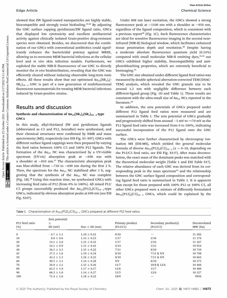
Herein, we synthesised a pyridinium–zwitterionic dual-ligand functionalised Au25(SR1)x(SR2)18�x GNC to addressMDR bacteria-induced infections, especially MRSE (Fig. 1A).The pyridinium ligand is derived from cetylpyridinium chloride
Fig. 1 (A) A schematic diagram of this study. GNCs with a specific P12/bacterial cell envelope while maintaining good biocompatibility. Localstructural deformation, which induce cell death. The distribution of GNlaser. Golden spheres indicate gold atoms, red spheres indicate the P12evolution of the UV-vis spectra of the 100% C5-capped GNC (represexcitation (black line, monitored with lEM ¼ 1100 nm) and emission (redspherical aberration-corrected TEM image (scale bar: 10 nm) of the 100copper grid. The UV-vis spectra, NIR fluorescence spectra and TEMdifferences from those of this representative species. (E) A plot of the Gindicates the theoretical output, assuming no ligand affinity difference, wdual-ligand GNCs prepared under different P12 feed ratios. The greatermaterial. A sudden change in haemolytic potency occurs as the feed raComparison of the cytotoxicity of the 45%- and 50%-P12 GNCs toward
14872 | Chem. Sci., 2021, 12, 14871–14882
(CPC), a commercial mouth mucosa aseptic additive approvedby the US Food and Drug Administration, which affords anti-bacterial ability although it suffers from poor prospects in invivo applications.21,22 The zwitterionic (ZW) ligand was intro-duced due to its biocompatibility, biosafety, and low biofoulingproperties.23,24 Previously, our group and other researchers
C5 composition aggregate planktonic bacteria by interacting with theinteractions of GNCs with bacteria cause cell content leakage and
Cs is tracked using NIR fluorescence upon excitation with an 808 nmligands, and blue spheres indicate the C5 ligands. (B) Time-dependententative species) during the synthesis process. (C) NIR fluorescenceline, lEX ¼ 810 nm) spectra of the 100% C5-capped GNC. (D) Double% C5-capped GNC prepared on an ultra-thin carbon film supported
images of GNCs with other P12/C5 compositions show no obviousNC surface P12 capping ratio versus the P12 feed ratio. The red line
hile the black dots represent the actual P12 ratio. (F) HD5/MIC values forthe HD5/MIC, the higher the biosafety and antibacterial ability of thetio approaches 50% (the MIC value is from anti-S. aureus results). (G)human umbilical vein endothelial cells (HUVECs) with 24 h incubation.
© 2021 The Author(s). Published by the Royal Society of Chemistry
Edge Article Chemical Science
Ope
n A
cces
s A
rtic
le. P
ublis
hed
on 2
5 O
ctob
er 2
021.
Dow
nloa
ded
on 2
/17/
2022
9:1
1:43
PM
. T
his
artic
le is
lice
nsed
und
er a
Cre
ativ
e C
omm
ons
Attr
ibut
ion-
Non
Com
mer
cial
3.0
Unp
orte
d L
icen
ce.
View Article Online
showed that ZW ligand-coated nanoparticles are highly stable,biocompatible and strongly resist biofouling.25,26 By adjustingthe GNC surface capping pyridinium/ZW ligand ratio, GNCsthat displayed low cytotoxicity and excellent antibacterialactivity against clinically isolated Gram-positive drug-resistantspecies were obtained. Besides, we discovered that the combi-nation of our GNCs with conventional antibiotics could signif-icantly enhance the bactericidal potency against MRSE,allowing us to overcomeMDR bacterial infections at the cellularlevel and in vivo skin infection models. Furthermore, weexploited the stable NIR-II uorescence of our GNC to directlymonitor the in vivo biodistribution, revealing that the GNC wasefficiently cleared without inducing observable long-term toxiceffects. All these results show that our optimised Au25(SR1)x(-SR2)18�x GNC is part of a new generation of multifunctionaluorescent nanomaterials for treating MDR bacterial infectionsinduced by Gram-positive strains.
Results and discussionSynthesis and characterisation of Au25(SR1)x(SR2)18�x-typeGNCs
In this study, alkyl-thiolated ZW and pyridinium ligands(abbreviated as C5 and P12, hereaer) were synthesised, andtheir chemical structures were conrmed by NMR and massspectrometry (MS), respectively (see ESI Fig. S1–S9†). GNCs withdifferent surface ligand cappings were then prepared by varyingthe feed ratios between 100% C5 and 100% P12 ligands. Theformation of the Au25 NC was characterised by a UV-visiblespectrum (UV-vis) absorption peak at �690 nm witha shoulder at �810 nm.27 The characteristic absorption peakwas blue-shied from �730 to �690 nm during the rst 2 h.Then, the spectrum for the Au25 NC stabilised aer 3 h, sug-gesting that the synthesis of the Au25 NC was complete(Fig. 1B).28 Using this reaction time, we synthesised GNCs withincreasing feed ratio of P12 (from 0% to 100%). All mixed P12/C5 groups successfully produced the Au25(P12)x(C5)18�x-typeGNCs, indicated by obvious absorption peaks at 690 nm (see ESIFig. S10†).
Table 1 Characterisation of Au25(P12)x(C5)18�x GNCs prepared at differe
P12 feed ratio(%)
Zeta potential�SD (mV) Size � SD (nm)
0 �4.7 � 1.1 1.18 � 0.2110 8.8 � 0.6 1.15 � 0.2320 14.1 � 2.0 1.21 � 0.3230 24.1 � 0.9 1.11 � 0.4140 36.3 � 2.3 1.15 � 0.2245 37.5 � 1.6 1.19 � 0.2450 43.3 � 1.5 1.16 � 0.2360 49.5 � 3.1 1.14 � 0.2870 56.9 � 3.1 1.15 � 0.2680 62.5 � 1.9 1.17 � 0.1790 66.5 � 1.0 1.14 � 0.27100 71.4 � 1.6 1.18 � 0.22
© 2021 The Author(s). Published by the Royal Society of Chemistry
Under 808 nm laser excitation, the GNCs showed a stronguorescence peak at �1100 nm with a shoulder at �950 nm,regardless of the ligand composition, which is consistent witha previous report29 (Fig. 1C). Such uorescence characteristicsare ideal for sensitive uorescence imaging in the second near-infrared (NIR-II) biological window, which facilitates enhancedtissue penetration depth and resolution.30 Despite havinga moderate absolute uorescence quantum yield (0.54%)compared with small molecular NIR-II emitting dyes,31,32 ourGNCs exhibited higher stability, biocompatibility and anti-photobleaching properties, which are extremely benecial tobioimaging.33
The GNC size obtained under different ligand feed ratios wasmeasured by double spherical aberration-corrected TEM (DSAC-TEM) analysis, which revealed the GNC granule size to bearound 1.2 nm with negligible difference between eachdifferent-ligand group (Fig. 1D and Table 1). These results areconsistent with the ultra-small size of Au25 NCs reported in theliterature.34
In addition, the zeta potentials of GNCs prepared underdifferent P12 ligand feed ratios were measured and aresummarized in Table 1. The zeta potential of GNCs graduallyand progressively shied from around�5 mV to >+70 mV as theP12 ligand feed ratio was increased from 0 to 100%, indicatingsuccessful incorporation of the P12 ligand onto the GNCsurface.
The GNCs were further characterised by electrospray ion-isation MS (ESI-MS), which yielded the general molecularformula of diverse Au25(P12)x(C5)18�x (x ¼ 0–18, depending onthe P12/C5 feed ratio, see ESI Fig. S11†). Aer mass deconvo-lution, the exact mass of the dominant peaks was matched withthe theoretical molecular weight (Table 1 and ESI Table S1†).The relative abundance of each GNC was derived from its cor-responding peak in the mass spectrum35 and the relationshipbetween the GNC surface ligand composition and correspond-ing ligand feed ratio is summarized in Table 1. It is apparentthat except for those prepared with 100% P12 or 100% C5, allother GNCs prepared were a mixture of differently formulatedAu25(P12)x(C5)18�x GNCs, which could be explained by the
nt P12 feed ratios
Primary product(P12/C5)
Secondary product(s)(P12/C5)
DeconvolutedMW (Da)
0/18 — 11 2821/17 2/16 11 1761/17 2/16 11 1674/14 5/13 10 9547/11 8/10 10 7108/10 7/11 & 9/9 10 7088/10 7/11 & 9/9 10 6629/9 8/10 10 57511/7 10/8 & 12/6 10 46612/6 11/7 10 40013/5 12/6 10 32718/0 — 9956
Chem. Sci., 2021, 12, 14871–14882 | 14873
Table 2 Antibacterial activity of different GNCs against five commonpathogens (the colour code represents the antibacterial activity: redfor high, yellow for medium, green for low and grey for negligible, andthe same below)
Chemical Science Edge Article
Ope
n A
cces
s A
rtic
le. P
ublis
hed
on 2
5 O
ctob
er 2
021.
Dow
nloa
ded
on 2
/17/
2022
9:1
1:43
PM
. T
his
artic
le is
lice
nsed
und
er a
Cre
ativ
e C
omm
ons
Attr
ibut
ion-
Non
Com
mer
cial
3.0
Unp
orte
d L
icen
ce.
View Article Online
putative forming mechanism of GNCs. Upon adding the dual-ligand solution to chloroauric acid, the thiol groups quicklyreduced the trivalent Au(III) ions to Au(I) to form Au(I)–ligandcomplexes.36 Due to the random reduction and combination ofAu(I) with different capping ligands, the complex units wouldhave multiple types. The subsequent addition of sodium boro-hydride to reduce Au(I) for cluster formation and sodiumhydroxide (for controlling ligand etching ability) allowed theprecise reduction of some Au(I)–ligand complexes to Au(0),forming a stochastic mixture of dual-ligand capped GNCs witha variety of ligand capping ratios.37 However, controlled mainlyby our ligand feed ratio, these synthesis conditions could allowfor some specic GNCs to be the main product. The relativeabundance of each GNC formulation could be derived from thecorresponding MS peak intensity.35
A plot of the average content of P12 in the GNCs against thefeed ratio (Fig. 1E) revealed that the product line was mostlybelow the feed line. Therefore, the C5 ligand appeared to bindmore strongly to the gold kernel than P12, possibly because ofthe lower electrostatic repulsion among the C5 (neutral overall)than P12 (positively charged) ligands. The only exception wasobserved at a feed ratio of 40–45%, where the product compo-sition (mainly Au25(P12)8(C5)10) matched the feed ratio, sug-gesting that the products of this ligand ratio were exceptionallystable.
a MIC—minimum inhibitory concentration. b Escherichia coli, Klebsiellapneumoniae, and Pseudomonas aeruginosa are Gram-negative bacteria.c Staphylococcus aureus and Methicillin-resistant Staphylococcus aureus(MRSA) are Gram-positive bacteria.
Antibacterial screening
The antibacterial activity of GNCs was assessed using a fewcommon Gram-positive (Staphylococcus aureus and MRSA) andGram-negative (Escherichia coli, Klebsiella pneumonia and Pseu-domonas aeruginosa) species (Table 2 [16 h] and ESI Table S2†[24 h]). The relationship between the antibacterial activity andthe P12 ligand feed ratio (i.e. surface positive charge density) ofGNCs was interesting: rst, the activity increased withincreasing P12 ligand feed ratio (up to 50%), then there wasa slight decrease (from 50% to 70%), and nally it increasedagain (from 70% to 100%). This trend was totally unexpected;since the antibacterial properties of GNCs were mainly derivedfrom the incorporated, positively charged P12 ligands, and notthe C5 ligands, we had anticipated the antibacterial activity tobe positively correlated with the P12 content (GNC surfacepositive charge density). The fact that the GNC with a 50% P12feed ratio exhibited comparable antibacterial activity to thatwith a 100% P12 ligand feed ratio suggests that there must bean optimal window to tune the GNC antibacterial propertiesand biocompatibility.
Table 2 reveals that the GNCs were more potent towardsGram-positive strains than towards Gram-negative ones. Thisphenomenon can be attributed to the differences in the surfacestructure between Gram-positive and -negative bacteria. Thesurface of Gram-positive bacteria is negatively charged;38 thus,they exhibit a strong electrostatic interaction with cationicmaterials. For example, wall teichoic acid (WTA), a distinct cellwall component in Gram-positive bacteria, consists of repeatingpoly(glycerol phosphate) units and a phosphodiester terminus.WTA constitutes the polyanionic network, making the cell
14874 | Chem. Sci., 2021, 12, 14871–14882
envelope highly negatively charged and susceptible to thebinding of cationic materials.39,40 Besides, anionic lipids, suchas phosphatidylglycerol (PG) and cardiolipin, constitute �80%of the total lipids in Gram-positive bacterial membranes butonly �30% in Gram-negative strains.41,42 Moreover, negativelycharged phospholipids, such as PGs, are present on both sidesof bacterial cell membranes rather than only the inside layerfound on mammalian cells. This difference can provide selec-tivity betweenmammalian cells and bacteria for some positivelycharged antibacterial agents.43
As shown in Table 2, the three best-performing GNCs (45%,50% and 100% P12 feed ratios) were selected for further testsusing more Gram-positive strains, including Staphylococcusepidermidis, Staphylococcus haemolyticus, Enterococcus faeciumand their corresponding drug-resistant strains (MRSE, MDR S.haemolyticus and vancomycin-resistant Enterococcus (VRE)).Table 3 reveals that although the GNCs with 100% P12 exhibitedthe best antibacterial potency among the three groups, espe-cially against MRSE, those with 45% and 50% P12 feed ratiosalso showed similar activity. These results further conrmedthat antibacterial potency is not linearly correlated with theactive P12 ligand content. Besides, during antibacterial tests, weobserved the aggregation of GNCs only with $60% P12 feedratios in LB culture media, suggesting signicant non-specicinteractions with negatively charged serum proteins in cellculture media, which could adversely affect their antibacterial
© 2021 The Author(s). Published by the Royal Society of Chemistry
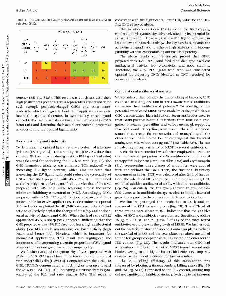
Table 3 The antibacterial activity toward Gram-positive bacteria ofselected GNCs
Edge Article Chemical Science
Ope
n A
cces
s A
rtic
le. P
ublis
hed
on 2
5 O
ctob
er 2
021.
Dow
nloa
ded
on 2
/17/
2022
9:1
1:43
PM
. T
his
artic
le is
lice
nsed
und
er a
Cre
ativ
e C
omm
ons
Attr
ibut
ion-
Non
Com
mer
cial
3.0
Unp
orte
d L
icen
ce.
View Article Online
potency (ESI Fig. S12†). This result was consistent with theirhigh positive zeta potentials. This represents a key drawback forsuch strongly positively-charged GNCs and other nano-materials, which can greatly limit their applications as anti-bacterial reagents. Therefore, in synthesising mixed-ligandcapped GNCs, we must balance the active/inert ligand (P12/C5here) ratio and determine their actual antibacterial propertiesin order to nd the optimal ligand ratio.
Biocompatibility and cytotoxicity
To determine the optimal ligand ratio, we performed a haemo-lytic test (ESI Fig. S13†). The resulting HD5 (the GNC dose thatcauses a 5% haemolysis value against the P12 ligand feed ratio)was calculated for optimising the P12 feed ratio (Fig. 1F). TheGNC haemolytic efficiency was enhanced (HD5 reduced) withincreasing P12 ligand content, which also indicated thatincreasing the ZW ligand ratio could reduce the cytotoxicity ofGNCs. The GNC prepared with 45% P12 still maintaineda relatively high HD5 of 34 mg mL�1, about twice that of the GNCprepared with 50% P12, while retaining almost the sameminimum inhibitory concentration (MIC). Accordingly, GNCsprepared with >45% P12 could be too cytotoxic, and thusunfavourable for in vivo applications. To determine the optimalP12 feed ratio, we plotted the HD5/MIC ratio versus the P12 feedratio to collectively depict the change of biosafety and antibac-terial activity of dual-ligand GNCs. When the feed ratio of P12approached 45%, a sharp peak appeared, indicating that theGNC prepared with a 45% P12 feed ratio has a high antibacterialability (low MIC) while maintaining low haemolyticity (highHD5), and hence high biosafety, which is important forbiomedical applications. This result also highlighted theimportance of incorporating a certain proportion of ZW ligandin order to maintain good overall biocompatibility.
We further evaluated the cytotoxicity of GNCs prepared with45% and 50% P12 ligand feed ratios toward human umbilicalvein endothelial cells (HUVECs). Compared with the 50%-P12GNC, HUVECs demonstrated a much higher tolerance towardthe 45%-P12 GNC (Fig. 1G), indicating a striking shi in cyto-toxicity as the P12 feed ratio reaches 50%. This result is
© 2021 The Author(s). Published by the Royal Society of Chemistry
consistent with the signicantly lower HD5 value for the 50%P12 GNC observed above.
The use of excess cationic P12 ligand on the GNC cappingcan lead to high cytotoxicity, adversely affecting its potential forin vivo application. However, too low P12 ligand content canlead to low antibacterial activity. The key here is to balance theactive/inert ligand ratio to achieve high stability and biocom-patibility without compromising antibacterial potency.
The above results comprehensively proved that GNCsprepared with 45% P12 ligand feed ratio displayed excellentantibacterial activity, low cytotoxicity, and good stability.Therefore, the 45% P12 ligand feed ratio was consideredoptimal for preparing GNCs (denoted as GNC hereaer) forsubsequent analyses.
Combinational antibacterial analyses
We considered that, besides the direct killing of bacteria, GNCcould sensitise drug-resistant bacteria toward varied antibioticsto restore their antibacterial potency.44 To investigate thispotential, we selected MRSE as the model microbe, to which ourGNC demonstrated high inhibition. Seven antibiotics used totreat Gram-positive bacterial infections from four main cate-gories: b-lactams (penicillins and carbapenem), glycopeptide,macrolides and tetracycline, were tested. The results demon-strated that, except for vancomycin and tetracycline, all theother antibiotics exhibited low efficacy against this bacterialstrain, with MIC values $32 mg mL�1 (ESI Table S3†). The testrevealed high drug resistance of MRSE to several antibiotics.
A checkerboard method was further employed to evaluatethe antibacterial properties of GNC–antibiotic combinationaltherapy.45,46 Imipenem (Imp), oxacillin (Oxa) and erythromycin(Ery), representing three classes of antibiotics, were appliedwith and without the GNC. Then, the fractional inhibitoryconcentration index (FICI) was calculated aer 24 h of incuba-tion. The calculated FICIs show that in joint applications, GNCexhibited additive antibacterial ability with all three antibiotics(Fig. 2A). Particularly, the Oxa group showed an exciting 128-fold decrease in antibiotic dosage needed to inhibit bacterialgrowth compared to the application of the antibiotic alone.
We further prolonged the incubation to 48 h and re-measured the FICI for each group (Fig. 2B). The FICIs of allthree groups were closer to 0.5, indicating that the additiveeffect of GNC and antibiotics was enhanced. Specically, adding16 mg mL�1 GNC and 2 mg mL�1 of any of the three testedantibiotics could prevent the growth of MRSE. We further tookout the bacterial mixture and spread it onto agar plates to checkthe survival of MRSE and the agar plates remained unstainedfor the test groups compared with innumerable colonies for thePBS control (Fig. 2C). The results indicated that GNC hada remarkable ability to re-sensitise MRSE toward several anti-biotics. Owing to the higher bactericidal efficiency, Imp wasselected as the model antibiotic for further studies.
The MRSE-killing efficiency of this combination wasmeasured by plotting a time-dependent killing graph (Fig. 2Dand ESI Fig. S14†). Compared to the PBS control, adding Impdid not signicantly inhibit bacterial growth due to the inherent
Chem. Sci., 2021, 12, 14871–14882 | 14875
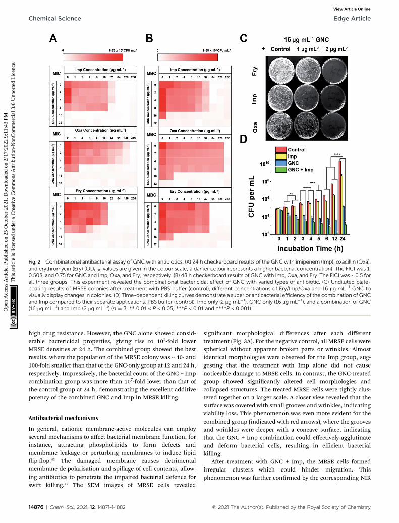
Fig. 2 Combinational antibacterial assay of GNC with antibiotics. (A) 24 h checkerboard results of the GNC with imipenem (Imp), oxacillin (Oxa),and erythromycin (Ery) (OD600 values are given in the colour scale; a darker colour represents a higher bacterial concentration). The FICI was 1,0.508, and 0.75 for GNC and Imp, Oxa, and Ery, respectively. (B) 48 h checkerboard results of GNCwith Imp, Oxa, and Ery. The FICI was�0.5 forall three groups. This experiment revealed the combinational bactericidal effect of GNC with varied types of antibiotic. (C) Undiluted plate-coating results of MRSE colonies after treatment with PBS buffer (control), different concentrations of Ery/Imp/Oxa and 16 mg mL�1 GNC tovisually display changes in colonies. (D) Time-dependent killing curves demonstrate a superior antibacterial efficiency of the combination of GNCand Imp compared to their separate applications. PBS buffer (control), Imp only (2 mg mL�1), GNC only (16 mg mL�1), and a combination of GNC(16 mg mL�1) and Imp (2 mg mL�1) (n ¼ 3, ** 0.01 < P < 0.05, ***P < 0.01 and ****P < 0.001).
Chemical Science Edge Article
Ope
n A
cces
s A
rtic
le. P
ublis
hed
on 2
5 O
ctob
er 2
021.
Dow
nloa
ded
on 2
/17/
2022
9:1
1:43
PM
. T
his
artic
le is
lice
nsed
und
er a
Cre
ativ
e C
omm
ons
Attr
ibut
ion-
Non
Com
mer
cial
3.0
Unp
orte
d L
icen
ce.
View Article Online
high drug resistance. However, the GNC alone showed consid-erable bactericidal properties, giving rise to 105-fold lowerMRSE densities at 24 h. The combined group showed the bestresults, where the population of the MRSE colony was �40- and100-fold smaller than that of the GNC-only group at 12 and 24 h,respectively. Impressively, the bacterial count of the GNC + Impcombination group was more than 107-fold lower than that ofthe control group at 24 h, demonstrating the excellent additivepotency of the combined GNC and Imp in MRSE killing.
Antibacterial mechanisms
In general, cationic membrane-active molecules can employseveral mechanisms to affect bacterial membrane function, forinstance, attracting phospholipids to form defects andmembrane leakage or perturbing membranes to induce lipidip-op.42 The damaged membrane causes detrimentalmembrane de-polarisation and spillage of cell contents, allow-ing antibiotics to penetrate the impaired bacterial defence forswi killing.47 The SEM images of MRSE cells revealed
14876 | Chem. Sci., 2021, 12, 14871–14882
signicant morphological differences aer each differenttreatment (Fig. 3A). For the negative control, all MRSE cells werespherical without apparent broken parts or wrinkles. Almostidentical morphologies were observed for the Imp group, sug-gesting that the treatment with Imp alone did not causenoticeable damage to MRSE cells. In contrast, the GNC-treatedgroup showed signicantly altered cell morphologies andcollapsed structures. The treated MRSE cells were tightly clus-tered together on a larger scale. A closer view revealed that thesurface was covered with small grooves and wrinkles, indicatingviability loss. This phenomenon was even more evident for thecombined group (indicated with red arrows), where the groovesand wrinkles were deeper with a concave surface, indicatingthat the GNC + Imp combination could effectively agglutinateand deform bacterial cells, resulting in efficient bacterialkilling.
Aer treatment with GNC + Imp, the MRSE cells formedirregular clusters which could hinder migration. Thisphenomenon was further conrmed by the corresponding NIR
© 2021 The Author(s). Published by the Royal Society of Chemistry

Fig. 3 GNC antibacterial mechanism investigation. (A) Typical SEM images of MRSE after a 4 h treatment with PBS buffer (control), 2 mgmL�1 Imp,16 mg mL�1 GNC, and a combination of Imp (2 mg mL�1) and GNC (16 mg mL�1). (B) Bright-field image (upper) and the corresponding NIRfluorescence image (lower) showing that the GNC + Imp treated MRSE bacteria were extensively aggregated. (C) TEM images of the PBS buffer-(control group, upper) and 16 mg mL�1 GNC + 2 mg mL�1 Imp-treated MRSE (lower).
Fig. 4 (A) Snapshot of the structure of an obtained Au25(P12)8(C5)10cluster with 45% P12 feed ratio: blue indicates C5; red indicates P12;yellow indicates thiol groups; pink indicates gold atoms. (B) Left:snapshot of the GNC interaction withWTA; right: scaled-up illustrationof the interacting part. (C) Interaction energy between GNC and WTA.Coul represents Coulomb force, while VDW denotes van der Waalsforce. (D) Number of hydrogen bond interactions between GNC andWTA units during a 5 ns simulation.
Edge Article Chemical Science
Ope
n A
cces
s A
rtic
le. P
ublis
hed
on 2
5 O
ctob
er 2
021.
Dow
nloa
ded
on 2
/17/
2022
9:1
1:43
PM
. T
his
artic
le is
lice
nsed
und
er a
Cre
ativ
e C
omm
ons
Attr
ibut
ion-
Non
Com
mer
cial
3.0
Unp
orte
d L
icen
ce.
View Article Online
uorescence image of the aggregated MRSE cells (Fig. 3B). Thisresult indicated that GNCs covered the envelope of MRSE cells,allowing them to aggregate by neutralising their surface nega-tive charges and form ‘bacterial clusters’ to limit their spreadand prolong the interaction.
TEM images (Fig. 3C) further conrmed the interaction ofGNC with MRSE cells, which demonstrated the adhesion andpenetration of GNCs within the cell envelope. Compared withthe intact shape and apparent division septa for bacterial cellsin the control group, the GNC + Imp treated group showed anabnormal cell division phenomenon: uneven division andempty cell walls were observed. Moreover, GNCs were mainlydistributed within particular spaces on the cell envelope, indi-cating specic binding (indicated with red arrows).48
The antibacterial mechanism of GNC was further investi-gated. Although GNC binding-induced bacterial aggregationwas already observed, it is still useful to investigate the specicbinding target. The high abundance of negatively charged WTAmay serve as a binding target for the positively charged GNCsvia electrostatic interactions. Isothermal titration calorimetry(ITC) was conducted between GNCs and WTA, which revealeda multiple-binding endothermic interaction (ESI Fig. S15A†),indicating that WTA could initiate bacteria–GNC interactions.49
To further verify this, a uorescence competition assay with TR-cadaverine was carried out. This dye can form a complex withWTA to quench its uorescence. If GNC can compete with TR-cadaverine in binding with WTA, adding GNC would result indye release and uorescence recovery. Fig. S15B (ESI†) showsthat the uorescence of TR-cadaverine signicantly increasedwith an increase in GNC concentration and was saturated at �8mg mL�1. Further increasing the GNC concentration couldquench the dye uorescence, possibly via dynamic quenching athigh concentrations.50 These results conrmed that WTA wasa binding target for the GNC.
The 45% P12 feed ratio yielded Au25(P12)8(C5)10 as theprimary product. Thus, we used this structure to simulate GNCbinding with WTA by molecular binding simulation.51 A surfacecoverage state was established by simulating the GNC congu-rations (Fig. 4A). Due to the electrostatic attraction between thecationic pyridinium and terminal anionic sulfonate groups, theligands on the GNC surface are bent rather than pointing
© 2021 The Author(s). Published by the Royal Society of Chemistry
outward and form a ‘cage-like’ cap to stabilise the entirestructure. This conguration explains the relatively highstability of GNC. Upon mixing with WTA, the electrostaticinteractions between the GNC surface pyridinium groups andWTA phosphonate groups appeared to support GNC invasion ofthe bacterial cell envelope (Fig. 4B). During this process, thecationic ligands interacted with the oppositely charged WTAlayer, while the C5 ligands mainly pointed away from thesurface. The initial electrostatic interactions further promoteextensive contact among the non-charged areas and strengthentheir interactions via van der Waals forces, which eventuallycontribute �57% of the overall interaction (Fig. 4C). Moreover,the weak hydrogen bonding between the WTA hydroxyl groupsand pyridinium p-ring can further enhance the interaction(Fig. 4D). WTA plays a vital role in drug resistance by providingattaching sites for other proteins that can replace synthasesinhibited by penicillin antibiotics.45 Thus, the binding of GNCsto WTA may compromise the bacterial drug resistance and
Chem. Sci., 2021, 12, 14871–14882 | 14877
Chemical Science Edge Article
Ope
n A
cces
s A
rtic
le. P
ublis
hed
on 2
5 O
ctob
er 2
021.
Dow
nloa
ded
on 2
/17/
2022
9:1
1:43
PM
. T
his
artic
le is
lice
nsed
und
er a
Cre
ativ
e C
omm
ons
Attr
ibut
ion-
Non
Com
mer
cial
3.0
Unp
orte
d L
icen
ce.
View Article Online
partially explain the sensitisation of MDR strains towardsantibiotics when co-treated with GNCs.
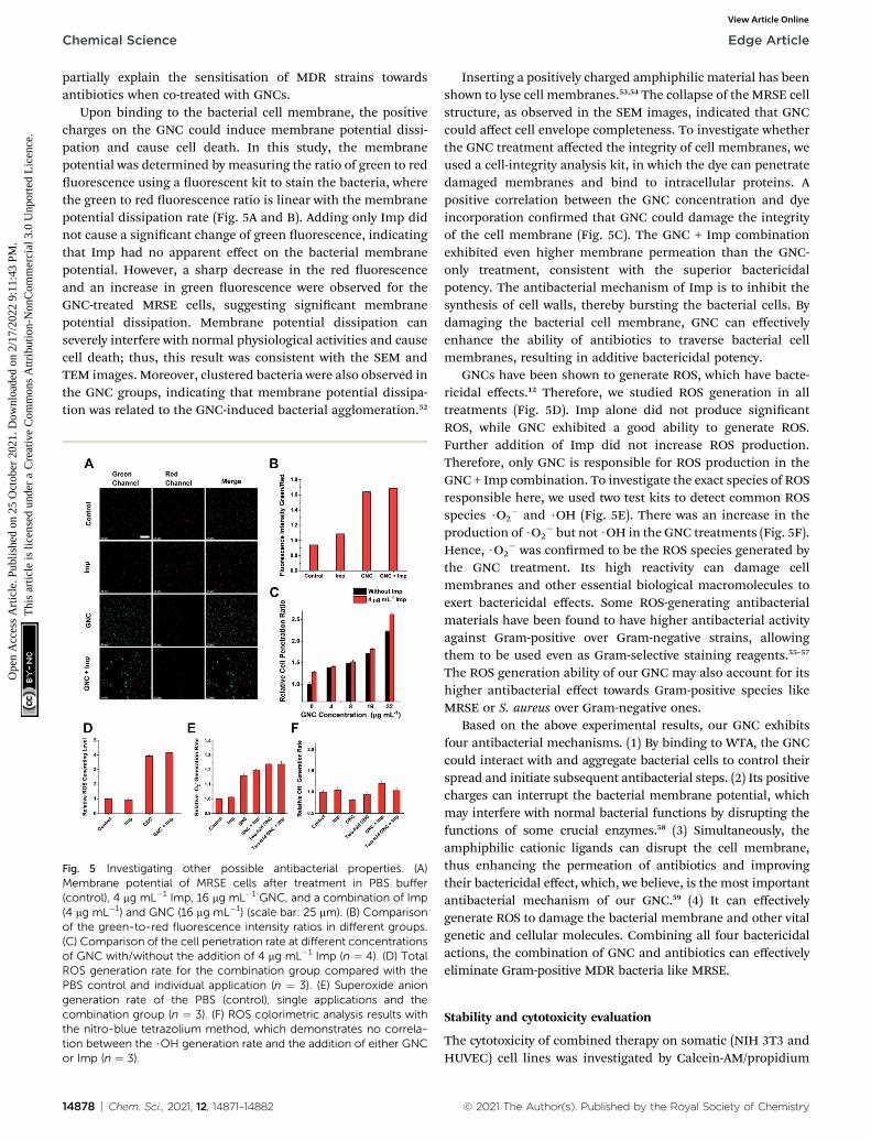
Upon binding to the bacterial cell membrane, the positivecharges on the GNC could induce membrane potential dissi-pation and cause cell death. In this study, the membranepotential was determined by measuring the ratio of green to reduorescence using a uorescent kit to stain the bacteria, wherethe green to red uorescence ratio is linear with the membranepotential dissipation rate (Fig. 5A and B). Adding only Imp didnot cause a signicant change of green uorescence, indicatingthat Imp had no apparent effect on the bacterial membranepotential. However, a sharp decrease in the red uorescenceand an increase in green uorescence were observed for theGNC-treated MRSE cells, suggesting signicant membranepotential dissipation. Membrane potential dissipation canseverely interfere with normal physiological activities and causecell death; thus, this result was consistent with the SEM andTEM images. Moreover, clustered bacteria were also observed inthe GNC groups, indicating that membrane potential dissipa-tion was related to the GNC-induced bacterial agglomeration.52
Fig. 5 Investigating other possible antibacterial properties. (A)Membrane potential of MRSE cells after treatment in PBS buffer(control), 4 mg mL�1 Imp, 16 mg mL�1 GNC, and a combination of Imp(4 mg mL�1) and GNC (16 mg mL�1) (scale bar: 25 mm). (B) Comparisonof the green-to-red fluorescence intensity ratios in different groups.(C) Comparison of the cell penetration rate at different concentrationsof GNC with/without the addition of 4 mg mL�1 Imp (n ¼ 4). (D) TotalROS generation rate for the combination group compared with thePBS control and individual application (n ¼ 3). (E) Superoxide aniongeneration rate of the PBS (control), single applications and thecombination group (n ¼ 3). (F) ROS colorimetric analysis results withthe nitro-blue tetrazolium method, which demonstrates no correla-tion between the $OH generation rate and the addition of either GNCor Imp (n ¼ 3).
14878 | Chem. Sci., 2021, 12, 14871–14882
Inserting a positively charged amphiphilic material has beenshown to lyse cell membranes.53,54 The collapse of the MRSE cellstructure, as observed in the SEM images, indicated that GNCcould affect cell envelope completeness. To investigate whetherthe GNC treatment affected the integrity of cell membranes, weused a cell-integrity analysis kit, in which the dye can penetratedamaged membranes and bind to intracellular proteins. Apositive correlation between the GNC concentration and dyeincorporation conrmed that GNC could damage the integrityof the cell membrane (Fig. 5C). The GNC + Imp combinationexhibited even higher membrane permeation than the GNC-only treatment, consistent with the superior bactericidalpotency. The antibacterial mechanism of Imp is to inhibit thesynthesis of cell walls, thereby bursting the bacterial cells. Bydamaging the bacterial cell membrane, GNC can effectivelyenhance the ability of antibiotics to traverse bacterial cellmembranes, resulting in additive bactericidal potency.
GNCs have been shown to generate ROS, which have bacte-ricidal effects.12 Therefore, we studied ROS generation in alltreatments (Fig. 5D). Imp alone did not produce signicantROS, while GNC exhibited a good ability to generate ROS.Further addition of Imp did not increase ROS production.Therefore, only GNC is responsible for ROS production in theGNC + Imp combination. To investigate the exact species of ROSresponsible here, we used two test kits to detect common ROSspecies $O2
� and $OH (Fig. 5E). There was an increase in theproduction of $O2
� but not $OH in the GNC treatments (Fig. 5F).Hence, $O2
� was conrmed to be the ROS species generated bythe GNC treatment. Its high reactivity can damage cellmembranes and other essential biological macromolecules toexert bactericidal effects. Some ROS-generating antibacterialmaterials have been found to have higher antibacterial activityagainst Gram-positive over Gram-negative strains, allowingthem to be used even as Gram-selective staining reagents.55–57
The ROS generation ability of our GNC may also account for itshigher antibacterial effect towards Gram-positive species likeMRSE or S. aureus over Gram-negative ones.
Based on the above experimental results, our GNC exhibitsfour antibacterial mechanisms. (1) By binding to WTA, the GNCcould interact with and aggregate bacterial cells to control theirspread and initiate subsequent antibacterial steps. (2) Its positivecharges can interrupt the bacterial membrane potential, whichmay interfere with normal bacterial functions by disrupting thefunctions of some crucial enzymes.58 (3) Simultaneously, theamphiphilic cationic ligands can disrupt the cell membrane,thus enhancing the permeation of antibiotics and improvingtheir bactericidal effect, which, we believe, is the most importantantibacterial mechanism of our GNC.59 (4) It can effectivelygenerate ROS to damage the bacterial membrane and other vitalgenetic and cellular molecules. Combining all four bactericidalactions, the combination of GNC and antibiotics can effectivelyeliminate Gram-positive MDR bacteria like MRSE.
Stability and cytotoxicity evaluation
The cytotoxicity of combined therapy on somatic (NIH 3T3 andHUVEC) cell lines was investigated by Calcein-AM/propidium
© 2021 The Author(s). Published by the Royal Society of Chemistry
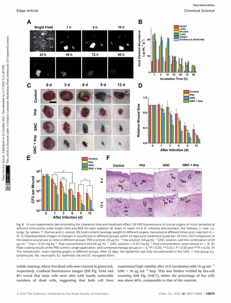
Fig. 6 In vivo experiments demonstrating the clearance time and treatment effect. (A) NIR fluorescence of crucial organs of mice harvested atdifferent time points under bright-field and 808 nm laser radiation (B: brain, H: heart, Int & St: intestine and stomach, Kid: kidneys, Li: liver, Lu:lungs, Sp: spleen, T: thymus and U: uterus). (B) Gold content (average weight) in different organs, harvested at different times post-injection (n ¼4). (C) Representative images of changes in wound size in different groups within 12 days post-treatment (scale bar: 10 mm). (D) Comparison ofthe relative wound size vs. time in different groups: PBS (control), 24 mgmL�1 Imp solution, 64 mg mL�1 GNC solution, and the combination of 24mg mL�1 Imp (�0.12 mg kg�1, final concentration) and 64 mg mL�1 GNC solution (�0.32 mg kg�1, final concentration, same below) (n ¼ 3). (E)Plate coating results of the PBS control, single application, and combined therapy groups (n¼ 3, *P > 0.05, ** 0.01 < P < 0.05 and ***P < 0.01). (F)The hematoxylin–eosin staining graphs in different groups. After 12 days, the epidermis was fully reconstructed in the GNC + Imp group (Ly:lymphocyte; Ne: neutrophil; Ec: epithelial cell and Ef: elongated fiber).
Edge Article Chemical Science
Ope
n A
cces
s A
rtic
le. P
ublis
hed
on 2
5 O
ctob
er 2
021.
Dow
nloa
ded
on 2
/17/
2022
9:1
1:43
PM
. T
his
artic
le is
lice
nsed
und
er a
Cre
ativ
e C
omm
ons
Attr
ibut
ion-
Non
Com
mer
cial
3.0
Unp
orte
d L
icen
ce.
View Article Online
iodide staining, where live/dead cells were stained in green/red,respectively. Confocal uorescence images (ESI Fig. S16A andB†) reveal that most cells were alive with hardly noticeablenumbers of dead cells, suggesting that both cell lines
© 2021 The Author(s). Published by the Royal Society of Chemistry
maintained high viability aer 24 h incubation with 70 mg mL�1
GNC + 30 mg mL�1 Imp. This was further veried by live-cellcounting (ESI Fig. S16C†), where the percentage of live cellswas above 80%, comparable to that of the controls.
Chem. Sci., 2021, 12, 14871–14882 | 14879

Chemical Science Edge Article
Ope
n A
cces
s A
rtic
le. P
ublis
hed
on 2
5 O
ctob
er 2
021.
Dow
nloa
ded
on 2
/17/
2022
9:1
1:43
PM
. T
his
artic
le is
lice
nsed
und
er a
Cre
ativ
e C
omm
ons
Attr
ibut
ion-
Non
Com
mer
cial
3.0
Unp
orte
d L
icen
ce.
View Article Online
The stability of nanomaterials during storage is crucial fortheir potential practical applications. We measured the UV-visspectrum of freshly prepared GNC solution and that aer 3month storage at 4 �C in a normal refrigerator (ESI Fig. S17†).We did not observe any deformation nor the emergence of newpeaks in the UV-vis spectrum. In addition, there were nochanges in the physical appearance, aggregation, or precipita-tion, conrming the good stability of the GNC.
NIR-II uorescence is an attractive imaging modality that iswell suited for in vivo applications. Owing to its attractive NIRuorescence, GNC can act as a uorescent probe for organdistribution tracking.60 We evaluated the GNC stability in vitrousing simulated biological uids. The GNC uorescence (128 mgmL�1) was highly stable and showed no observable changesaer 15 days incubation in PBS supplemented with up to 10%human serum albumin (HSA, see ESI Fig. S18†). Thus, its stableNIR uorescence can be used to directly evaluate the GNC organdistribution with a NIR animal imager using 808 nm laserirradiation (Fig. 6A). Upon intravenous injection of GNC + Imp,strong NIR uorescence was observed primarily in the liver withweak signals in the spleen and kidneys within the rst hour,suggesting preferential accumulation in these organs. The NIRuorescence in the main organs was maintained for 10 h, thenit gradually faded away aer 24 h and became almost invisibleat 96 h post-injection, indicating that most of the GNCs werecleared from the body. The gold content measured from criticalorgans harvested at different post-injection times was consis-tent with the NIR uorescence results (>80% of the Au contentwas cleared aer 96 h, Fig. 6B). These results indicate that theGNC has an adequate body clearance time and is suitable forintravenous administration. Moreover, the blood routine as wellas liver and kidney function indicators further conrmed thata high dosage of GNC + Imp did not induce any notable changewhen compared with the control group, suggesting minimal invivo toxicity (ESI Fig. S19A–F†).
Skin infection model experiments
As a coagulase-negative strain, MRSE can cause so tissue andskin infections and frequently induces infection in surgicalsites;5 thus, we established a skin infection model by creatingwounds with removal of epidermis and dermis on the back ofrats and infecting them with MRSE, where conventional woundtreatment methods nd it difficult to sustain antibacterialability. Different groups were treated with either the PBScontrol, Imp only, GNC only or GNC + Imp. The effect wasmonitored by measuring the wound size and bacterial countaer plate coating. Compared with the negative control, thewound healing rate for the rats treated with either Imp or GNCwas higher. Notably, the wound healing of the GNC + Impcombinational group was the fastest. The size of the wound wassignicantly smaller than that of other groups, and the normalskin was almost fully reconstructed in 9 days (Fig. 6C and D).Moreover, the scab size was signicantly smaller than in othergroups, indicating a markedly reduced bacterial infection.
To quantify the clearance of bacteria on the skin wound, weperformed plate coating to compare the number of colonies on
14880 | Chem. Sci., 2021, 12, 14871–14882
differently treated wounds (Fig. 6E). The MRSE counts of thenegative control and Imp groups were slowly reduced over timedue to autoimmunity, and the GNC-treated group showeda more signicant decrease. Treatment with GNC + Imp resul-ted in the most rapid reduction of the bacterial population, andby day 12, the bacterial count was >10-fold lower than that of theGNC-only group and almost two orders of magnitude lower thanthat of the negative control group. This in vivo result wasconsistent with the superior in vitro antibacterial potency ofGNC + Imp over that of GNC or Imp only. Pathological analysesfurther conrmed a complete repair of the wound tissue for theGNC + Imp group but not for the negative control and GNC- orImp-only groups. The reconstruction of well-stratied skinlayers and signicantly reduced immune cells indicated thesuccessful removal of the lesions in the GNC + Imp group(Fig. 6F).
Conclusion
In this study, we synthesised a series of formula-dened pyr-idinium–zwitterionic ligand-functionalised Au25(SR1)x(SR2)18�x
GNCs as antibacterial nanomaterials. By ne-tuning the ligandfeed ratios, we obtained GNCs that exhibited both excellentantibacterial ability and high stability, successfully addressinga major issue in antibacterial gold nanomaterial development.Besides, the GNCs employ multiple antibacterial mechanisms,giving rise to high potency against Gram-positive MDR bacteria.The optimized GNC can signicantly reduce the dosage ofantibiotics required to treat MDR bacterial infections, therebygreatly enhancing the efficacy of frontline antibiotics.Compared to other nanomaterials without dened chemicalformulae, which could cause difficulties in quality control andmechanism research, our Au25(SR1)x(SR2)18�x GNCs are poten-tially better suited for medical applications. Moreover,biocompatible Au25 GNCs capped with two or more ligands canincorporate more functions, thus widening the scope of theirbiomedical applications. We envisage that dual-/multi-ligand-functionalised GNCs will nd broad applications in chem-istry, physics, biology, and biomedical sciences.
Data availability
All other data have already been provided in the ESI.†
Author contributions
D. Z. and Z. P. conceived this study. Z. P. performed all theexperiments and analysed the data except those claried below.Q. L. and J. Q. took and analysed the TEM images. Y. J. and Y. G.contributed some of the bacterial experiments. W. Y. carried outthe animal experiments. F. H. contributed MDR bacteriastrains. Z. P. wrote the manuscript. D. Z. and X. J. co-supervisedthe project and obtained funding for this project.
Conflicts of interest
The authors declare no competing nancial interest.
© 2021 The Author(s). Published by the Royal Society of Chemistry
Edge Article Chemical Science
Ope
n A
cces
s A
rtic
le. P
ublis
hed
on 2
5 O
ctob
er 2
021.
Dow
nloa
ded
on 2
/17/
2022
9:1
1:43
PM
. T
his
artic
le is
lice
nsed
und
er a
Cre
ativ
e C
omm
ons
Attr
ibut
ion-
Non
Com
mer
cial
3.0
Unp
orte
d L
icen
ce.
View Article Online
Acknowledgements
We acknowledge the nancial support from the ShenzhenScience and Technology Programme(KQTD20190929172743294). This work was partly funded by theUK Biotechnology and Biological Sciences Research Council(grant no: BB/R007829/1) to DZ.
Notes and references
1 P. Fernandes, Nat. Biotechnol., 2006, 24, 1497–1503.2 W. Cassandra, Nature, 2017, 543, 15.3 L. Wang, C. Hu and L. Shao, Int. J. Nanomed., 2017, 12, 1227–1249.
4 M. Otto, Nat. Rev. Microbiol., 2009, 7, 555–567.5 L. M. Weiner-Lastinger, S. Abner, J. R. Edwards, A. J. Kallen,M. Karlsson, S. S. Magill, D. Pollock, I. See, M. M. Soe,M. S. Walters and M. A. Dudeck, Infect. Control Hosp.Epidemiol., 2020, 41, 1–18.
6 A. M. Hanssen, G. Kjeldsen and J. U. Ericson Sollid,Antimicrob. Agents Chemother., 2004, 48, 285–296.
7 U.S. Department of Health and Human Services, Centers Dis.Control Prev., 2019, 1–113.
8 Z. Jiang, A. Sahar, X. Li, S. M. Robinson, V. M. Rotello,K. Saha, M. A. Riley, D. F. Moyano and A. Gupta, ACS Nano,2014, 8, 10682–10686.
9 A. Gupta, S. Mumtaz, C. H. Li, I. Hussain and V. M. Rotello,Chem. Soc. Rev., 2019, 48, 415–427.
10 H. T. T. Duong, N. N. M. Adnan, N. Barraud, J. S. Basuki,S. K. Kutty, K. Jung, N. Kumar, T. P. Davis and C. Boyer, J.Mater. Chem. B, 2014, 2, 5003–5011.
11 K. Ma, Y. Li, Z. Wang, Y. Chen, X. Zhang, C. Chen, H. Yu,J. Huang, Z. Yang, X. Wang and Z. Wang, ACS Appl. Mater.Interfaces, 2019, 11, 29630–29640.
12 K. Zheng, M. I. Setyawati, D. T. Leong and J. Xie, ACS Nano,2017, 11, 6904–6910.
13 R. R. Nasaruddin, T. Chen, N. Yan and J. Xie, Coord. Chem.Rev., 2018, 368, 60–79.
14 Y. Xie, Y. Liu, J. Yang, Y. Liu, F. Hu, K. Zhu and X. Jiang,Angew. Chem., Int. Ed., 2018, 57, 3958–3962.
15 Y. Xie, W. Zheng and X. Jiang, ACS Appl. Mater. Interfaces,2020, 12, 9041–9049.
16 M. Yu, J. Xu and J. Zheng, Angew. Chem., Int. Ed., 2019, 131,4156–4172.
17 R. Jin, Nanoscale, 2015, 7, 1549–1565.18 X. Kang, H. Chong and M. Zhu, Nanoscale, 2018, 10, 10758–
10834.19 Z. Yu, H. Xiao, X. Zhang, Y. Yang, Y. Yu, H. Chen, X. Meng,
W. Ma, M. Yu, Z. Li, C. Li and H. Liu, ACS Nano, 2020, 14,13536–13547.
20 D. Li, Q. Liu, Q. Qi, H. Shi, E. C. Hsu, W. Chen, W. Yuan,Y. Wu, S. Lin, Y. Zeng, Z. Xiao, L. Xu, Y. Zhang,T. Stoyanova, W. Jia and Z. Cheng, Small, 2020, 16, 1–9.
21 M. Petkovic, K. R. Seddon, L. P. N. Rebelo and C. S. Pereira,Chem. Soc. Rev., 2011, 40, 1383–1403.
22 P. K. Sreenivasan, V. I. Haraszthy and J. J. Zambon, Lett. Appl.Microbiol., 2013, 56, 14–20.
© 2021 The Author(s). Published by the Royal Society of Chemistry
23 S. Chen, L. Li, C. Zhao and J. Zheng, Polymer, 2010, 51, 5283–5293.
24 J. Shaoyi and C. Zhiqiang, Adv. Mater., 2009, 22, 920–932.25 A. K. Murthy, R. J. Stover, W. G. Hardin, R. Schramm,
G. D. Nie, S. Gourisankar, T. M. Truskett, K. V. Sokolovand K. P. Johnston, J. Am. Chem. Soc., 2013, 135, 7799–7802.
26 Y. Guo, C. Sakonsinsiri, I. Nehlmeier, M. A. Fascione,H. Zhang, W. Wang, S. Pohlmann, W. B. Turnbull andD. Zhou, Angew. Chem., Int. Ed., 2016, 128, 4816–4820.
27 Z. Luo, V. Nachammai, B. Zhang, N. Yan, D. T. Leong,D. E. Jiang and J. Xie, J. Am. Chem. Soc., 2014, 136, 10577–10580.
28 R. Jin, H. Qian, Z. Wu, Y. Zhu, M. Zhu, A. Mohanty andN. Garg, J. Phys. Chem. Lett., 2010, 1, 2903–2910.
29 H. Liu, G. Hong, Z. Luo, J. Chen, J. Chang, M. Gong, H. He,J. Yang, X. Yuan, L. Li, X. Mu, J. Wang, W. Mi, J. Luo, J. Xieand X. D. Zhang, Adv. Mater., 2019, 31, 1–9.
30 G. Hong, A. L. Antaris and H. Dai, Nat. Biomed. Eng., 2017, 1,1–22.
31 A. L. Antaris, H. Chen, S. Diao, Z. Ma, Z. Zhang, S. Zhu,J. Wang, A. X. Lozano, Q. Fan, L. Chew, M. Zhu, K. Cheng,X. Hong, H. Dai and Z. Cheng, Nat. Commun., 2017, 8, 1–11.
32 Y. Sun, M. Ding, X. Zeng, Y. Xiao, H. Wu, H. Zhou, B. Ding,C. Qu, W. Hou, A. G. A. Er-bu, Y. Zhang, Z. Cheng andX. Hong, Chem. Sci., 2017, 8, 3489–3493.
33 F. Aldeek, M. A. H. Muhammed, G. Palui, N. Zhan andH. Mattoussi, ACS Nano, 2013, 7, 2509–2521.
34 M. Song, G. Zhou, N. Lu, J. Lee, E. Nakouzi, H. Wang andD. Li, Science, 2020, 367, 40–45.
35 Y. Ishida, K. Narita, T. Yonezawa and R. L. Whetten, J. Phys.Chem. Lett., 2016, 7, 3718–3722.
36 X. Yuan, B. Zhang, Z. Luo, Q. Yao, D. T. Leong, N. Yan andJ. Xie, Angew. Chem., Int. Ed., 2014, 53, 4623–4627.
37 T. Chen, V. Fung, Q. Yao, Z. Luo, D. E. Jiang and J. Xie, J. Am.Chem. Soc., 2018, 140, 11370–11377.
38 N. Malanovic and K. Lohner, Biochim. Biophys. Acta,Biomembr., 2016, 1858, 936–946.
39 L. Pasquina-Lemonche, J. Burns, R. D. Turner, S. Kumar,R. Tank, N. Mullin, J. S. Wilson, B. Chakrabarti,P. A. Bullough, S. J. Foster and J. K. Hobbs, Nature, 2020,582, 294–297.
40 Z. V. Feng, I. L. Gunsolus, T. A. Qiu, K. R. Hurley, L. H. Nyberg,H. Frew, K. P. Johnson, A. M. Vartanian, L. M. Jacob,S. E. Lohse, M. D. Torelli, R. J. Hamers, C. J. Murphy andC. L. Haynes, Chem. Sci., 2015, 6, 5186–5196.
41 P. Kumar, J. N. Kizhakkedathu and S. K. Straus, Biomolecules,2018, 8, 4.
42 R. M. R. F. Epand, C. Walker, R. M. R. F. Epand andN. A. Magarvey, Biochim. Biophys. Acta, Biomembr., 2016,1858, 980–987.
43 M. Zasloff, Nature, 2002, 415, 389–395.44 Y. Zhao, Z. Chen, Y. Chen, J. Xu, J. Li and X. Jiang, J. Am.
Chem. Soc., 2013, 135, 12940–12943.45 M. A. Foxley, S. N. Wright, A. K. Lam, A. W. Friedline,
S. J. Strange, M. T. Xiao, E. L. Moen and C. V. Rice, ACSMed. Chem. Lett., 2017, 8, 1083–1088.
Chem. Sci., 2021, 12, 14871–14882 | 14881
Chemical Science Edge Article
Ope
n A
cces
s A
rtic
le. P
ublis
hed
on 2
5 O
ctob
er 2
021.
Dow
nloa
ded
on 2
/17/
2022
9:1
1:43
PM
. T
his
artic
le is
lice
nsed
und
er a
Cre
ativ
e C
omm
ons
Attr
ibut
ion-
Non
Com
mer
cial
3.0
Unp
orte
d L
icen
ce.
View Article Online
46 J. Campbell, A. Singh, J. Santa Maria, K. Younghoon,S. Brown, J. G. Swoboda, E. Mylonakis, B. J. Wilkinson andS. Walker, ACS Chem. Biol., 2010, 6, 106–116.
47 N. Zhang and S. Ma, Eur. J. Med. Chem., 2019, 184, 111743.48 S. C. Hayden, G. Zhao, K. Saha, R. L. Phillips, X. Li,
O. R. Miranda, V. M. Rotello, M. A. El-Sayed, I. Schmidt-Krey and U. H. F. Bunz, J. Am. Chem. Soc., 2012, 134, 6920–6923.
49 B. Wang, L. Zhang, C. B. Sung and S. Granick, Proc. Natl.Acad. Sci. U. S. A., 2008, 105, 18171–18175.
50 B. Dubertret, M. Calame and A. J. Libchaber, Nat. Biotechnol.,2001, 19, 365–370.
51 E. R. Caudill, R. T. Hernandez, K. P. Johnson, J. T. O'Rourke,L. Zhu, C. L. Haynes, Z. V. Feng and J. A. Pedersen, Chem.Sci., 2020, 11, 4106–4118.
52 Z. Huang, Y. Liu, L. Wang, A. Ali, Q. Yao, X. Jiang and Y. Gao,Biomaterials, 2020, 253, 120124.
53 J. N. Pendleton and B. F. Gilmore, Int. J. Antimicrob. Agents,2015, 46, 131–139.
14882 | Chem. Sci., 2021, 12, 14871–14882
54 A. Chompoosor, K. Saha, P. S. Ghosh, D. J. Macarthy,O. R. Miranda, Z.-J. Zhu, K. F. Arcaro and V. M. Rotello,Small, 2010, 6, 2246–2249.
55 M. Kang, C. Zhou, S. Wu, B. Yu, Z. Zhang, N. Song,M. M. S. Lee, W. Xu, F. J. Xu, D. Wang, L. Wang andB. Z. Tang, J. Am. Chem. Soc., 2019, 141, 16781–16789.
56 J. Li, H. Zhou, J. Wang, D. Wang, R. Shen, X. Zhang, P. Jinand X. Liu, Nanoscale, 2016, 8, 11907–11923.
57 R. J. Barnes, R. Molina, J. Xu, P. J. Dobson andI. P. Thompson, J. Nanopart. Res., 2013, 15, 1432.
58 Y. Cui, Y. Zhao, Y. Tian, W. Zhang, X. Lu and X. Jiang,Biomaterials, 2012, 33, 2327–2333.
59 M. Godoy-Gallardo, U. Eckhard, L. M. Delgado, Y. J. D. deRoo Puente, M. Hoyos-Nogues, F. J. Gil and R. A. Perez,Bioact. Mater., 2021, 6, 4470–4490.
60 Y. Kong, J. Chen, H. Fang, G. Heath, Y. Wo, W. Wang, Y. Li,Y. Guo, S. D. Evans, S. Chen and D. Zhou, Chem. Mater.,2016, 28, 3041–3050.
© 2021 The Author(s). Published by the Royal Society of Chemistry
![Bis[4-(dimethylamino)pyridinium] octaaquachloridolanthanum ...journals.iucr.org/e/issues/2012/11/00/su2504/su2504.pdfBis[4-(dimethylamino)pyridinium] octaaquachloridolanthanum(III)](https://static.fdocuments.in/doc/165x107/5e0610443af6f93e3057972f/bis4-dimethylaminopyridinium-octaaquachloridolanthanum-4-dimethylaminopyridinium.jpg)