Controlled Drug-Release from Mesoporous...
31
Department of Chemical and Biological Engineering CHALMERS UNIVERSITY OF TECHNOLOGY Gothenburg, Sweden 2015 Controlled Drug-Release from Mesoporous Hydrogels Master of Science Thesis in the Master Degree Program, Materials Chemistry and Nanotechnology ANNA PEKKARI
Transcript of Controlled Drug-Release from Mesoporous...

Department of Chemical and Biological Engineering CHALMERS UNIVERSITY OF TECHNOLOGY Gothenburg, Sweden 2015
Controlled Drug-Release from Mesoporous Hydrogels
Master of Science Thesis in the Master Degree Program, Materials Chemistry
and Nanotechnology
ANNA PEKKARI

Controlled Drug-Release from Mesoporous Hydrogels
ANNA PEKKARI
Supervised by Professor Martin Andersson Department of Chemical and Biological Engineering CHALMERS UNIVERSITY OF TECHNOLOGY
Gothenburg, Sweden 2015

Controlled Drug-Release from Mesoporous Hydrogels
Anna Pekkari
© Anna Pekkari, 2015
Department of Chemical and Biological Engineering
CHALMERS UNIVERSITY OF TECHNOLOGY
SE-412 96 Göteborg
Sweden
Telephone + 46 (0)31-772 1000
Printed by: Chalmers Reproservice Gothenburg, Sweden 2015

Abstract Hydrogels are an interesting group of materials used in biomedical applications. The cross-linked
polymeric network enables large water absorption and physical properties similar to soft tissue giving
it excellent biocompatibility. Hydrogels with a controlled nanostructure are especially interesting for
biomedical applications such as controlled drug-delivery systems since the meso-ordered structure
enables loading of drugs followed by a local sustained release. This may minimize systemic side effect
and give a safer and more effective delivery of drugs.
The aim of this thesis was to form meso-ordered hydrogels and hydrogel particles by the use of
Lyotropic Liquid Crystals (LLC), and to evaluate them as controlled drug-release systems using the
drugs Ibuprofen and 14C radiolabelled Alendronate. LLC phases are formed when amphiphilic
molecules mixed with water at certain concentration starts to form ordered mesostructures. A
polymerizable amphiphile, a triblock copolymer with trade name Pluronic ® F127 was used to form
meso-ordered bulk hydrogels by photopolymerization of LLCs with cubic and hexagonal geometries.
Small-Angle X-ray Scattering measurements revealed structure retention after crosslinking of the
LLC gel. Meso-ordered Poly(ethylene glycol) diacrylate (PEG-DA) hydrogel particles were formed
by photopolymerization in presence of surfactants in a water-in-oil emulsion. Particles with a size of
209-242 nm were formed, measured with Dynamic Light Scattering and presence of long-range
order was revealed with Polarized Light Microscopy.
Ibuprofen and 14C Alendronate were successfully loaded into F127 hydrogels and PEG-DA hydrogel
particles. Ibuprofen incorporated into F127 hydrogels existed in an amorphous form confirmed with
X-Ray Diffraction. Both hydrogels showed a controlled release of drugs, with an initial burst
followed by a sustained release. There was a clear difference in release rate between Ibuprofen and
Alendronate, with a more rapid release of the more hydrophilic Alendronate. Addition of the
surfactant SDS in the release media resulted in an increased release rate and solubility of Ibuprofen
from F127 hydrogels. Meso-ordered hydrogel particles based on PEG-DA presented significantly
slower release behavior compared to F127 hydrogels. Mathematic modelling of the drug-release
kinetics for the hydrogels corresponded best to the first-order model and showed good correlation to
data.
Keywords: Mesoporous hydrogel, drug delivery, Lyotropic Liquid Crystal (LLC), release kinetics

List of Abbreviations
LLC Lyotropic Liquid Crystal
PPO Poly(propylene) oxide
PEO Poly(ethylene) oxide
DA Diacrylate
PEG Poly(ethylene) glycol
MEC Minimum Effective Concentration
MTC Minimum Toxic Concentration
SDS Sodium Dodecyl Sulfate
Span-80 Sorbitan monooleate
SAXS Small Angle X-ray Scattering
XRD X-Ray Diffraction
PLM Polarized Light Microscopy
DLS Dynamic Light Scattering
PdI Polydispersity Index
UV-VIS Ultraviolet-Visible
LSC Liquid Scintillation Counting

1. INTRODUCTION 1
1.1 Objective of this study 2
2. THEORY AND BACKGROUND 3
2.1 Lyotropic liquid crystals (LLCs) 3
2.2 Meso-ordered hydrogels from LLCs 3
2.3 Meso-ordered hydrogel particles from LLCs 4
2.4 Controlled drug-release 5
2.5 Drug-release kinetics 6
3. MATERIALS AND METHODS 7
3.1 Surfactants and drugs used in this project 7
3.2 Synthesis of diacrylate modified triblock copolymer 8
3.3 The LLC system 8
3.4 Polymerized LLCs: formation of meso-ordered hydrogel 9
3.5 Formation of meso-ordered PEG-particles 9
3.6 Loading and release of drugs 10
3.7 Analytical methods 11
4. RESULTS AND DISCUSSION 14
4.1 Material characterization of MF127 14
4.2 Material characterization of PEG-DA hydrogel particles 16
4.3 Drug-loading and release 17
5. CONCLUSIONS 22
6. FUTURE WORK 22
ACKNOWLEDGEMENTS 23
REFERENCES 24

1
Introduction 1.Hydrogels, consisting of a cross-linked hydrophilic polymer network represent an important class of
materials used in biomedical and pharmaceutical applications [1, 2]. The cross-linked polymeric
structure enables large water absorption giving physical properties similar to soft tissue, and the
hydrogel also have high permeability and diffusivity for water and other nutrients[1, 2]. These
characteristics together with a high biocompatibility have made hydrogels interesting for biomedical
applications including contact lenses, tissue engineering, biosensors, and drug-delivery [2].
In recent years there has been a growing interest in the formation of hydrogels with a controlled
nanostructure. Advantages of these ordered hydrogels compared to conventional hydrogels are
increased compressive strength, swelling, permeability and biological compatibility, which has
sparked the interest for use in biomedical applications[3]. Meso-ordered hydrogels are interesting to
use as controlled release systems since their high porosity enables loading of drug molecules into the
gel matrix and subsequent sustained release of drugs, thus maintaining a high local concentration of
drugs in the surrounding tissue over an extended time period[4]. This might minimize systemic
effects and permit lower drug dosage and safer delivery of therapeutics[5].
Introduction of nanostructures into hydrogels can be achieved by using surfactant templates, where
monomers can adsorb and crosslink. However, issues associated to the thermodynamically driven
phase separation when monomers are converted to polymers, which leads to formation of hydrogels
with poorly defined nanostructures, is often an problem[3]. A solution to this phase separation
problem has been proposed by Guymon et al. where rapid crosslinking of monomers utilizing
photo-polymerization results in a highly ordered hydrogel polymer[6]. In this present study, meso-
ordered PEG-based hydrogel particles have been synthesized by photo-polymerization in the
presence of liquid crystalline phases formed by surfactants. Nanostructures can also be incorporated
in hydrogels by the use of polymerizable amphiphiles, and by rapid crosslinking of ordered liquid
crystalline phases creating meso-ordered hydrogels. In the present study the amphiphilic triblock
copolymer Pluronic® F127 has been utilized for the formation of meso-ordered hydrogels.

2
1.1 Objective of this study This study aimed at forming new types of meso-ordered hydrogels and hydrogel particles by the use
of Lyotropic Liquid Crystals (LLCs) either with the use of templates or with polymerizable
amphiphiles and to evaluate them as controlled drug-delivery systems. The objectives can be divided
into three main parts:
Form meso-ordered hydrogels using diacrylate modified triblock copolymers (Pluronic® F127)
Form meso-ordered hydrogel particles using diacrylate modified Poly (ethylene) glycol.
Evaluate loading and release of the drugs Ibuprofen and 14C radiolabeled Alendronate from the hydrogels
3
Theory and background 2.
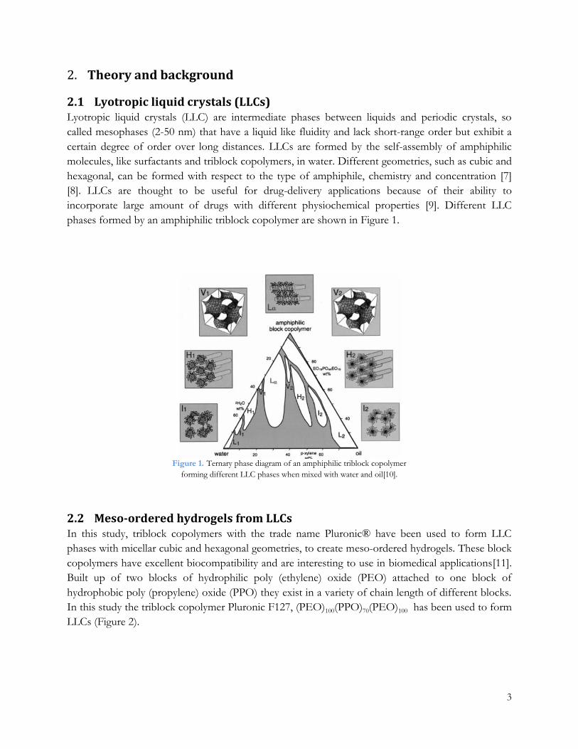
2.1 Lyotropic liquid crystals (LLCs) Lyotropic liquid crystals (LLC) are intermediate phases between liquids and periodic crystals, so
called mesophases (2-50 nm) that have a liquid like fluidity and lack short-range order but exhibit a
certain degree of order over long distances. LLCs are formed by the self-assembly of amphiphilic
molecules, like surfactants and triblock copolymers, in water. Different geometries, such as cubic and
hexagonal, can be formed with respect to the type of amphiphile, chemistry and concentration [7]
[8]. LLCs are thought to be useful for drug-delivery applications because of their ability to
incorporate large amount of drugs with different physiochemical properties [9]. Different LLC
phases formed by an amphiphilic triblock copolymer are shown in Figure 1.
Figure 1. Ternary phase diagram of an amphiphilic triblock copolymer
forming different LLC phases when mixed with water and oil[10].
2.2 Meso-ordered hydrogels from LLCs In this study, triblock copolymers with the trade name Pluronic® have been used to form LLC
phases with micellar cubic and hexagonal geometries, to create meso-ordered hydrogels. These block
copolymers have excellent biocompatibility and are interesting to use in biomedical applications[11].
Built up of two blocks of hydrophilic poly (ethylene) oxide (PEO) attached to one block of
hydrophobic poly (propylene) oxide (PPO) they exist in a variety of chain length of different blocks.
In this study the triblock copolymer Pluronic F127, (PEO)100(PPO)70(PEO)100 has been used to form
LLCs (Figure 2).

4
By modifying the hydrophilic endgroups of the triblock copolymer with cross-linkable groups a
polymerizable amphiphile can be formed. Mixing the modified polymer with solvent at a certain
concentration followed by rapid crosslinking then creates meso-ordered hydrogels. [12, 13].
2.3 Meso-ordered hydrogel particles from LLCs An alternative method for introducing nanostructure into hydrogels is the use of templates, directing
the formation of polymer networks into structures with different architecture. LLCs have extensively
been used as templates in the formation of meso-ordered silica and titania [14, 15]. The use of LLCs
as soft templates provides control over both pore size and morphology, which is dictated by the
choice of amphiphile and its concentration. This enables formation of hydrogel networks with
mesostructures ranging from lamellar, hexagonal and bicontinuous structures [16]. In the present
study, diacrylate-modified (DA) Polyethylene glycol (PEG) based hydrogel particles have been
formed in a water-in-oil emulsion to form meso-ordered particles. Figure 3 shows a templating route
using surfactants for the formation of meso-ordered hydrogels based on PEG-DA.
Figure 3. Schematic illustration of the LLC templating process. Clockwise rotation: Hexagonal
LLCs mesophases formed by surfactants, 2D illustration of the hexagonal phase, 2D illustration
of how PEG-DA monomers adsorb onto LLCs, photopolymerization and surfactant removal
results in a meso-ordered PEG-DA hydrogel[6].
100 100 70
PEO PEO PPO
Figure 2. Chemical structure of the amphiphilic triblock copolymer F127 (trade name Pluronic ®) used in this
study showing hydrophilic Polyethylene oxide (PEO) blocks in blue and hydrophobic Polypropylene oxide (PPO)
blocks in red.

5
2.4 Controlled drug-release Controlled drug release represents a rapidly advancing area in pharmaceutical science and aims to
improve the effectiveness of drug therapy by controlling drug exposure over time, overcome
physiological barriers and prevent premature degradation of the drug. A controlled release of drugs
minimizes the patients’ compliance by reducing the frequency of administration. Conventional
administration routes where drugs enter through the systemic circulation often suffer from drug
toxicity and side effects related to absorption of non-target tissue. Local drug-delivery will permit
direct release to the target tissue and thus provide lower drug dosage to reach the desired effect and
less exposure to other tissue [17, 18].
Controlled drug release over an extended duration is often beneficial, especially for drugs that are
rapidly released and eliminated. The release should result in a concentration between the minimum
effective concentration (MEC) and the minimum toxic concentration (MTC), as shown in Figure 4.
A controlled release system may maintain the drug concentration within the therapeutic window for
a longer time and thus avoid toxic side effect or underexposure and enable fewer administrations [18,
19].
Figure 4. Plasma drug concentration obtained by different dosage forms, single dosing (black line), multiple dosing (dotted
line), zero-order controlled release (solid line). The range between MTC and MEC represents the therapeutic window [20].

6
2.5 Drug-release kinetics In-vitro drug delivery studies often involve mathematical modelling of the release behavior for the
prediction of the release kinetics of the drug delivery system. Several kinetic models have been
developed for different dosage forms, such as tablets, polymers etc.[21, 22]. I this study, the zero-
order model and the first-order model were used to interpret the data obtained from the release
studies.
2.5.1 Zero-order model
Pharmaceutical dosage forms that do not disaggregate and release the drug slowly can be represented
by the zero-order model. Here, the drug release is only dependent on time
𝑄0 − 𝑄𝑡 = 𝐾𝑡 (1)
Where Q0 is the initial amount of drug in the dosage form, Qt is the amount of drug in the dosage
form at time t and K is the proportionality constant. Dividing the equation by Q0 and simplifying
leads to:
𝐹𝑡 = 𝐾0𝑡 (2)
Where 𝐹𝑡 = 100(1 − (𝑄𝑡
𝑄0)) and Ft represent the percentage of drug released at time t. K0 is the
zero-order release constant.
2.5.2 First- order model
This model is typically used to describe absorption and/ or elimination of drugs. The first-order
model, derived from first-order kinetics states that the concentration change with time is only
dependent on concentration.
𝑑𝐶
𝑑𝑡= −𝐾𝐶 (3)
Where K is a first-order proportionality constant and C is the concentration of drug. Equation 3 can
be expressed as:
𝑄𝑡 = 𝑄0𝑒−𝐾𝑡 (4)
Where Qt is the concentration of drug in the dosage form at time t and Q0 is the initial concentration
of drug. Equation 4 can be rewritten to:
𝐹𝑡 = 100(1 − 𝑒−𝐾1𝑡) (5)
Where 𝐹𝑡 = 100(1 − (𝑄𝑡
𝑄0)) and represents the percentage of drug released at time t, and K1 is the
first-order release constant expressed in t-1.

7
Materials and methods 3.14C radiolabeled Alendronate was purchased from Moravek Biochemicals. All other chemicals were
purchased from Sigma-Aldrich and used as received. Experiments were performed in room
temperature (23 ±2°C).
3.1 Surfactants and drugs used in this project
3.1.1 Ibuprofen
Ibuprofen is a hydrophobic, non-steroidal anti-inflammatory drug derived from propionic acid,
commonly used to treat inflammation, relieve pain and reduce fever. It exists in two isomers, R- and
S, where the S-isomer is the most biologically active [23]. The structure of Ibuprofen is shown in
Figure 5.
3.1.2 Alendronate
Alendronate is a bisphosphonate and an osteoporosis drug that has shown to improve bone mass
density and reduce the risk of bone fractions[24]. For this study, Alendronate (Figure 5) with a 14C-
isotope was evaluated in drug release studies from the micellar cubic (I1B) F127-hydrogel.
3.1.3 Sodium dodecyl sulfate
Sodium Dodecyl Sulfate (SDS) (Figure 6) is an anionic surfactant used in various cleaning and
hygiene products. In this project, it was used to enhance the solubility of the drug Ibuprofen in the
release studies from the F127-hydrogels.
Figure 6. Molecular structure of the surfactant sodium dodecyl sulfate.
3.1.4 Sorbitan monooleate
Sorbitan monooleate (Span-80) (Figure 7) is a nonionic surfactant often used as an emulsifier. In the
present study it was used as a template and emulsifier in the formation of PEG-DA hydrogel
particles.
Figure 5. Molecular structures of the two drugs used in this work,
Alendronate (left) and Ibuprofen (right).

8
3.2 Synthesis of diacrylate modified triblock copolymer The acrylate derivate of Pluronic® F127 was synthesized by reacting the triblock copolymer with
acryloyl chloride (Figure 8). To a solution of F127 in chloroform and with twice the molar amount of
triethylamine, acryloyl chloride dissolved in chloroform was added drop-wise under N2 atmosphere
and magnetic stirring. After 24h reaction the product was washed three times with Na2CO3 (5 %),
dried over anhydrous magnesium sulfate (MgSO4), vacuum filtrated followed by solvent removal at
reduced pressure. The diacrylate derivative of F127 was synthesized with an end product yield of 85-
90%.
3.3 The LLC system The phase behavior of F127 has extensively been studied by Alexandridis et. al (25). In Figure 9 the
ternary phase diagram for F127/water/butanol is shown. In this study, only the F127/water system
has been studied and the phases of interest have been marked in the figure: the micellar cubic (I1)
and hexagonal (H1) phase. Table 1 shows the composition by weight of the components forming the
LLCs.
Figure 9. Ternary phase diagram for F127/water/butanol system with the phases of interest marked with red[25].
N2
2TEA
4 100 100 70 70 100 100
Figure 8. Reaction scheme of the synthesis route of the diacrylate modified triblock copolymer Pluronic F127.
Figure 7. Molecular structure of the emulsifier sorbitan monooleate (Span-80).

9
Table 1. LLC phases studied for the DA-modified F127 copolymer and water with the relative weight composition of each
component.
3.4 Polymerized LLCs: formation of meso-ordered hydrogel Hexagonal and micellar cubic liquid crystal gels were prepared by mixing of acrylate modified F127,
water and photoinitiator, 2-Hydroxy-2-methylpropiophenone, at different ratios presented in Table
1. The initiator had a concentration of 1 wt. % of the amphiphile. The components were manually
mixed with a spatula in a vial forming a thick homogenous gel. The gel was then applied onto glass
slides (gel thickness ~2 mm) and cross-linked under UV-light (90 W lamp, λ= 252 nm) for 10 min
creating a rubbery polymerized liquid crystal (hydrogel).
3.5 Formation of meso-ordered PEG-DA particles The formation procedure of meso-ordered PEG-DA particles was directly adopted from Wallin et. al
and performed as described before[26]. Diacrylate modified poly(ethylene) glycol (PEG-DA) (Figure
10) (1500 g/mol) had previously been synthesized and was used as received.
Formation of PEG-DA hydrogel particles was performed in a water-in-oil emulsion. A nonpolar
solution was prepared by mixing 1,12 g emulsifier (Span-80) with 20 ml of hexane. The solution was
rapidly stirred using a homogenizer (Silent Crusher M, Heidolph, Schwabach, Germany) followed by
slow addition (~1 min) of PEG-DA mixture containing 5 wt. % PEG-DA (1500 g/mol), Milli-Q
water and photoinitiator (2-Hydroxy-2-methylpropiophenone) with a concentration of 1 wt. % in
respect to PEG. The formed water-in-oil emulsion was under constant stirring put under UV-light
for 1h to ensure complete crosslinking of PEG-DA. The surfactant template was removed by liquid-
liquid extraction (Milli-Q water: hexane, 1:3) four times, followed by water removal by Freeze-drying
24h.
Triblock
copolymer Phase Designation % copolymer % water
Pluronic F127 Micellar cubic
I1 I1A 25 75
Pluronic F127 Micellar cubic
I1 I1B 30 70
Pluronic F127 Hexagonal
H1 H1 75 25
Figure 9. Chemical structure of diacrylate modified
poly(ethylene) glycol (PEG-DA).

10
3.6 Loading and release of drugs
3.6.1 Determination of Ibuprofen concentration in F127 hydrogel
The total amount of drug absorbed in the hydrogel was determined by dissolution, using triplicate of
samples. After polymerization of LLCs the hydrogel was freeze-dried for 24h and cut into
appropriate sizes (0.015 g). The dried hydrogel was soaked in a 20 mg/ml Ibuprofen: ethanol
solution for 24h, then taken out of solution to let the ethanol evaporate. The loaded hydrogel was
put in 1.5 ml sodium hydroxide (NaOH) solution (1M) for 24h until complete dissolution was
observed. Samples were then taken out, properly diluted and analyzed with UV/VIS Spectroscopy to
determine the total drug uptake in the hydrogel.
3.6.2 Release of Ibuprofen from F127 hydrogel Meso-ordered hydrogels of micellar cubic (I1A) and hexagonal phase (H1) were investigated for
Ibuprofen drug-release. Freeze-dried hydrogels were loaded with Ibuprofen as described in 3.6.1.
The release was performed with 6 replicas in Milli-Q water or SDS (1 wt. %) as release media, with
constant stirring using a shaker -plate (Yellow Line OS2 basic). Samples were taken out at specific
time intervals and the amount of drug released was measured with UV/VIS spectroscopy.
3.6.3 Determination of Ibuprofen concentration in PEG-DA particles
Freeze-dried PEG-DA particles were soaked in a 20 mg/ml Ibuprofen/ethanol solution for 24h.
The solution was centrifuged at 3500 rpm for 30 min, the supernatant removed with a plastic pipette
and soaked particles were put on glass slides to dry for 24h. The loaded particles (0.01 5g) were put
in 1.5 ml sodium hydroxide solution (1M) for 24h to allow complete dissolution. After 24h, particles
had agglomerated and the solution was therefore filtered through 0.8 µm filters prior to UV-VIS
Spectroscopy analysis to determine total amount of drug uptake in the particles.
3.6.4 Release of Ibuprofen from PEG-DA particles
Freeze-dried PEG-DA particles were loaded with Ibuprofen as described in 3.6.3. Ibuprofen loaded
particles were transferred to a dialysis membrane (Spectra/Por) with 1ml Milli-Q water. The
membrane was put in Milli-Q water for release measurements under magnetic stirring.
3.6.5 Determination of 14C-Alendronate concentration in F127 hydrogel
The polymerized F127 hydrogel was oven-dried at 40°C for 24h and was then cut into pieces (0.045
g) and put in 14C Alendronate water solution to soak for 24h. Dissolution of the hydrogel was
performed as described in 3.6.1. Samples were collected and analyzed with Liquid Scintillation
Counting.
3.6.6 Release of 14C-Alendronate from F127 hydrogel
Meso-ordered hydrogels of micellar cubic (I1B) phase were studied as drug-delivery systems for
Alendronate. The soaked 14C Alendronate loaded hydrogel was then placed in Milli-Q water for
release measurements. Samples were taken out at specific time periods and the β-radiation released
from the hydrogel was measured using Liquid Scintillation Counting.

11
3.7 Analytical methods
3.7.1 Small Angle X-ray Scattering
Small angle x-ray scattering (SAXS) is a technique where the inelastic scattering of X-rays (1-2 Å) at
low angles (1-10°) provides structural information of a material at a length scale of 1-100 nm.
Evaluation of the out-coming scattering patterns can provide morphological information like particle
size, pore size distribution and more. The structure in LLC phases can be studied by SAXS by
looking at relative distance between peaks to detect e.g. hexagonal and cubic phases. For instance H1
phases can be distinguished by peak position ratios of 1, √3, 2, √7, and the micellar cubic phase (I1)
with primitive (P) cubic structure by relative distances of 1, √2, √3, √4. In this study, SAXS was used
to study the meso-ordered hydrogels to confirm that the ordered LLC phases were retained after
polymerization. Synchrotron SAXS measurements were performed on beamline I911 at the Max Lab
synchrotron facility in Lund, Sweden.
3.7.2 X-ray diffraction
X-ray diffraction (XRD) is a tool for determining the molecular structure and crystallinity of a
material by obtaining information about lattice parameters. The principle is to bombard the sample
with an X-ray beam with different incoming angles generating a diffraction pattern. Constructive
interference is observed when Bragg’s law (eq.6) is fulfilled resulting in peaks in the diffraction
pattern.
2𝑑 sin 𝜃 = 𝑛𝜆 (6)
Where n is any integer, θ is the scattering angle, λ is the wavelength of the X-rays. The obtained data
can be compared with the Joint Committee on Powder Diffraction Standards (JCPDS) registry to
determine the crystal structure of the material. In the present study, a Bruker D8 Advance X-ray
diffractometer (Cu-K 1 radiation and = 1.54056Å) with a 2θ range of 20-60°, step size 0.050° and
data acquisition time of 30 min was utilized. XRD was used to determine the crystallinity of
Ibuprofen absorbed in the F127 hydrogel.
3.7.3 Polarized Light Microscopy
Polarized Light Microscopy (PLM) is a technique that uses a transmission light microscope equipped
with two polarizing filters placed perpendicular to each other and is normally used to detect
birefringence in samples, like LLC phases. Placing a LLC sample having an isotropic structure like
the cubic phase between the filters will result in a dark image since the two polarizers will block the
light. For an anisotropic sample, the polarized light will interact with the sample and change direction
resulting in structure induced birefringence patterns characteristic for different phases; the hexagonal
phase shows rod-like streaks while the lamellar phase shows fan-like patterns. PLM measurements
were performed on fully swollen PEG-DA particles with a Zeiss microscope (Axio Scope A.1, Carl
Zeiss Microscopy, Germany) equipped with an AxioCam ICc5 camera (40x objective) and polarized
filters.

12
3.7.4 Dynamic Light Scattering
Dynamic Light Scattering (DLS) is technique that can be used to measure size distribution of small
particles in suspensions, typically emulsions, micelles, polymers and nanoparticles. The principle of
the technique is the illumination of the sample by a laser beam and the fluctuations of the scattering
is detected at a known scattering angle and collected by the detector. Scattering of light occurs if the
particles are smaller compared to the wavelength of the light (<250 nm). The scattering fluctuations
due to Brownian motion of the particles yields information of hydrodynamic radius or diameter of
the particles calculated via the Stokes-Einstein relation. In the present study, PEG-DA particles (1
mg/ml) were filtered through 0.8 µm filters and the size distribution measured with DLS using a
Zetasizer Nano-ZS instrument Malvern instruments (Worcestershire, UK).
3.7.5 Ultraviolet- Visible spectroscopy
Ultraviolet –Visible (UV-VIS) Spectroscopy involves absorption of radiation in the Ultraviolet (180-
400 nm) and the visible (400-800 nm) region in the electromagnetic spectrum, where the absorption
induces excitation of electrons from ground state to a higher energy state. This technique has
applications mainly in quantitative measurements of transition metals, conjugated organic
compounds and biological macromolecules. Lambert-Beer’s law (eq. 7) expresses the proportionality
between absorption, concentration in solution and path length.
𝐴 = 𝑙𝑜𝑔10 (𝐼0
𝐼) = 𝜀𝑐𝑙 (7)
Where A is the Absorbance, I0 is the intensity of incident radiation, I is the intensity of the
transmitted radiation, ε the molar absorptivity or molar extinction coefficient, c concentration of
solution (mol/l), and l the path length of the sample. For an organic compound the molar
absorptivity is constant at a certain wavelength, thus a calibration curve with different concentrations
can be constructed, and the concentration of a sample can be determined. In the present study, UV-
VIS Spectroscopy was used to measure the release of Ibuprofen from the F127 hydrogels and the
amount of drug absorbed in F127 hydrogels. Measurements were performed with 1 ml quartz
cuvettes, using an Agilent 8453 UV-VIS Spectrophotometer.

13
3.7.6 Liquid Scintillation counting
Liquid scintillation counting (LSC) is a technique measuring low energy emitting radiation such as β-
emitting isotopes. The principle of LSC is that energy is emitted from radioactive decay to a
scintillation cocktail consisting of an organic solvent and scintillators (fluors). The energy is absorbed
by the solvent and transferred to the fluor molecules, which upon de-excitation emit photons of
visible light. These flashes of light are then detected by a photomultiplier tube and converted into a
flow of electrons and measured as an electric pulse (Figure 10). [27]. In this study, LSC was used to
measure drug-release of 14C labeled Alendronate by detecting emitting β-radiation. Samples were
taken out (0.1 or 0.2 ml) and added to scintillation vials filled with 12 ml Emulsifier-Safe liquid
scintillation cocktail (PerkinElmer) and shaken to obtain a homogenous solution. The measurements
were performed using a Wallac Guardian 1414 Liquid Scintillation Counter (PerkinElmer).
Figure 10. Basic sketch of the scintillation process[28].
14
Results and discussion 4.The aim of this study can be divided into two parts, where the first part aimed to synthesize two
types of meso-ordered hydrogels; based on crosslinking of LLC phases formed by a triblock
copolymer, and formation of PEG-based hydrogel particles using LLC templates. The second part of
the project involved drug-release studies from the hydrogels, examining the release behavior of the
drugs Ibuprofen and 14C radiolabeled Alendronate.
4.1 Material characterization of MF127
4.1.1 LLC gel and hydrogel formation
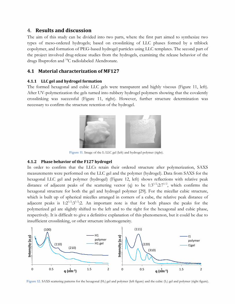
The formed hexagonal and cubic LLC gels were transparent and highly viscous (Figure 11, left).
After UV-polymerization the gels turned into rubbery hydrogel polymers showing that the covalently
crosslinking was successful (Figure 11, right). However, further structure determination was
necessary to confirm the structure retention of the hydrogel.
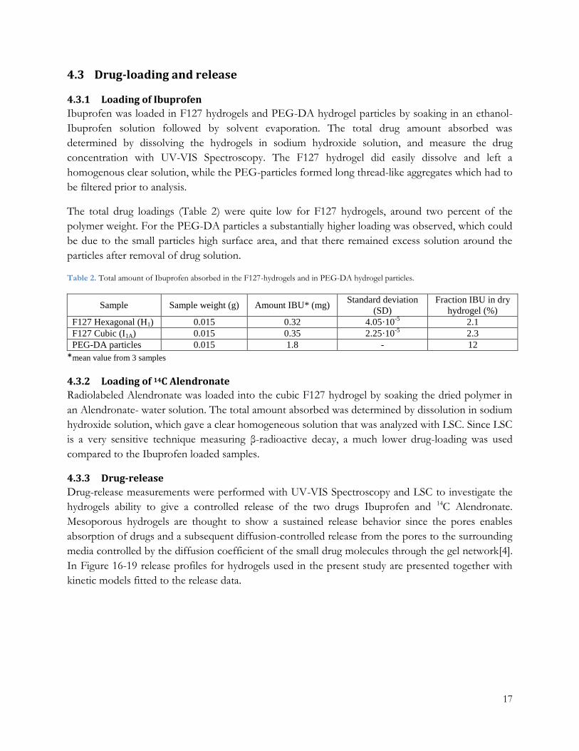
4.1.2 Phase behavior of the F127 hydrogel
In order to confirm that the LLCs retain their ordered structure after polymerization, SAXS
measurements were performed on the LLC gel and the polymer (hydrogel). Data from SAXS for the
hexagonal LLC gel and polymer (hydrogel) (Figure 12, left) shows reflections with relative peak
distance of adjacent peaks of the scattering vector (q) to be 1:31/2:2:71/2, which confirms the
hexagonal structure for both the gel and hydrogel polymer [29]. For the micellar cubic structure,
which is built up of spherical micelles arranged in corners of a cube, the relative peak distance of
adjacent peaks is 1:21/2:31/2:2. An important note is that for both phases the peaks for the
polymerized gel are slightly shifted to the left and to the right for the hexagonal and cubic phase,
respectively. It is difficult to give a definitive explanation of this phenomenon, but it could be due to
insufficient crosslinking, or other structure inhomogeneity.
1
Figure 11. Image of the I1 LLC gel (left) and hydrogel polymer (right).
0 0.5 1 1.5 2
Inte
nsi
ty (
a.u
)
q (nm-1)
H1polymerH1 gel
0 0.5 1 1.5 2
Inte
nsi
ty (
a.u
)
q (nm-1)
I1polymer
I1gel
Figure 12. SAXS scattering patterns for the hexagonal (H1) gel and polymer (left figure) and the cubic (I1) gel and polymer (right figure).
figure).
(111)
(220)
(310)
(100)
(110) (210)

15
4.1.3 Determination of drug crystallinity
XRD was used to determine the crystallinity of the drug Ibuprofen, both as dry powder and when
incorporated into F127 hydrogels with cubic and hexagonal phase. Most drugs form crystals at room
temperature, existing in multiple crystal forms (polymorphs) or as crystal hydrates. The type of
polymorph affects drug dissolution which plays an important role in determining the release
behavior[18]. For hydrophobic drugs, like Ibuprofen poor water solubility is an issue in
pharmaceutical applications since it lowers the bioavailability. Converting the drug to a more soluble
sodium salt or specific polymorphic form can increase solubility and thus improve the
bioavailability[30].
In Figure 13 diffraction patterns for the cubic and hexagonal phase for the soft and freeze-dried
hydrogel with and without Ibuprofen, and Ibuprofen powder are shown. The Ibuprofen powder
shows diffraction peaks corresponding to high crystallinity. The dry hydrogel shows two distinct
peaks due to crystallization after water removal, also seen in the hexagonal soft hydrogel. However,
when incorporating Ibuprofen in the freeze-dried hydrogel matrixes no diffraction peaks from
Ibuprofen could be identified, which imply that that the hydrogel has disrupted the crystalline
structure of the drug.
16.5 21.5 26.5 31.5
Inte
nsi
ty (
a.u
)
two theta
Figure 13. XRD patterns for the F127 hydrogel for soft polymer, freeze-dried polymer and freeze-dried
polymer with Ibuprofen, and pure Ibuprofen. Cubic phase (I1) (left) and hexagonal phase (H1) (right).
16.5 21.5 26.5 31.5
Inte
nsi
ty (
a.u
)
two theta
Pure IBU
F127 I1 f.d hydrogel
IBU
F127 I1 f.d hydrogel
F127 I1 hydrogel
Pure IBU
F127 H1 f.d hydrogel
IBU
F127 H1 f.d hydrogel
F127 H1 hydrogel

16
4.2 Material characterization of PEG-DA hydrogel particles
4.2.1 Formation of meso-ordered PEG-DA particles
The formed PEG-DA particles appeared as a white fluffy powder, and when re-dispersed in water
the particles swelled, confirmed that the crosslinking was successful. However, further structure
determination was necessary to confirm the order and phase of the material.
4.2.2 Phase behavior of PEG-DA hydrogel particles
In order to detect presence of anisotropy in synthesized PEG-DA, PLM measurements were
performed. Figure 14 shows a micrograph of the hydrogel particles showing clear birefringence
patterns, and thereby confirming long-range order in the sample. It is however difficult to tell
whether the patterns correspond to hexagonal or lamellar phases and to further determine the exact
phase SAXS scattering measurements have to be performed.
4.2.3 Size determination of PEG-DA hydrogel particles
The size distribution of the PEG-DA particles was measured with DLS, presented in Figure 15. The
intensity distribution of particle size corresponds to the scattering intensity of each particle fraction,
and hence larger particles give a larger contribution. Therefore, also the number average distribution
is presented, where smaller particles contribute more. The polydispersity index (PdI) is a
dimensionless value between 0 and 1 estimating the size distribution. As can be seen in Figure 15,
the size distribution for the PEG-particles is quite narrow, with a PdI of 0.35 and particle size
estimated to 242 and 209 nm for intensity and number average, respectively.
Figure 14. PLM micrograph of the hydrogel particles showing birefringence
confirming that the particles have an anisotropic long-range order.
0
5
10
15
20
25
0 200 400 600
Inte
nsi
ty/
Nu
mb
er
%
Size d (nm)
Intensityaverage
Numberaverage
Figure 15. Intensity- and number average size distribution for
formed PEG-DA hydrogel particles.
17
4.3 Drug-loading and release
4.3.1 Loading of Ibuprofen
Ibuprofen was loaded in F127 hydrogels and PEG-DA hydrogel particles by soaking in an ethanol-
Ibuprofen solution followed by solvent evaporation. The total drug amount absorbed was
determined by dissolving the hydrogels in sodium hydroxide solution, and measure the drug
concentration with UV-VIS Spectroscopy. The F127 hydrogel did easily dissolve and left a
homogenous clear solution, while the PEG-particles formed long thread-like aggregates which had to
be filtered prior to analysis.
The total drug loadings (Table 2) were quite low for F127 hydrogels, around two percent of the
polymer weight. For the PEG-DA particles a substantially higher loading was observed, which could
be due to the small particles high surface area, and that there remained excess solution around the
particles after removal of drug solution.
Table 2. Total amount of Ibuprofen absorbed in the F127-hydrogels and in PEG-DA hydrogel particles.
Sample Sample weight (g) Amount IBU* (mg) Standard deviation
(SD)
Fraction IBU in dry
hydrogel (%)
F127 Hexagonal (H1) 0.015 0.32 4.05·10-5
2.1
F127 Cubic (I1A) 0.015 0.35 2.25·10-5
2.3
PEG-DA particles 0.015 1.8 - 12
*mean value from 3 samples
4.3.2 Loading of 14C Alendronate
Radiolabeled Alendronate was loaded into the cubic F127 hydrogel by soaking the dried polymer in
an Alendronate- water solution. The total amount absorbed was determined by dissolution in sodium
hydroxide solution, which gave a clear homogeneous solution that was analyzed with LSC. Since LSC
is a very sensitive technique measuring β-radioactive decay, a much lower drug-loading was used
compared to the Ibuprofen loaded samples.
4.3.3 Drug-release
Drug-release measurements were performed with UV-VIS Spectroscopy and LSC to investigate the
hydrogels ability to give a controlled release of the two drugs Ibuprofen and 14C Alendronate.
Mesoporous hydrogels are thought to show a sustained release behavior since the pores enables
absorption of drugs and a subsequent diffusion-controlled release from the pores to the surrounding
media controlled by the diffusion coefficient of the small drug molecules through the gel network[4].
In Figure 16-19 release profiles for hydrogels used in the present study are presented together with
kinetic models fitted to the release data.
18
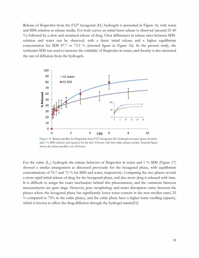
Release of Ibuprofen from the F127 hexagonal (H1) hydrogels is presented in Figure 16, with water
and SDS-solution as release media. For both curves an initial burst release is observed (around 25-40
%) followed by a slow and sustained release of drug. Clear differences in release rates between SDS-
solution and water can be observed, with a faster initial release and a higher equilibrium
concentration for SDS 87.7 vs 73.3 % (inserted figure in Figure 16). In the present study, the
surfactant SDS was used to increase the solubility of Ibuprofen in water, and thereby it also increased
the rate of diffusion from the hydrogels.
For the cubic (I1A) hydrogels the release behavior of Ibuprofen in water and 1 % SDS (Figure 17)
showed a similar arrangement as discussed previously for the hexagonal phase, with equilibrium
concentrations of 76.7 and 71 % for SDS and water, respectively. Comparing the two phases reveals
a more rapid initial release of drug for the hexagonal phase, and also more drug is released with time.
It is difficult to assign the exact mechanism behind this phenomenon, and the variations between
measurements are quite large. However, pore morphology and water absorption varies between the
phases where the hexagonal phase has significantly lower water content in the non-swollen state( 25
% compared to 75% in the cubic phase), and the cubic phase have a higher water swelling capacity,
which is known to affect the drug-diffusion through the hydrogel matrix[31]
0
10
20
30
40
50
60
70
80
90
100
0 2 4 6 8 10
% R
ele
ase
d
t (h)
H1 water
H1 SDS
0
50
100
0 5 10 15 20 25
Figure 16. Release profiles for Ibuprofen from F127 hexagonal (H1) hydrogels in water (green rhombs)
and 1 % SDS solution (red squares) for the first 10 hours with first-order release models. Inserted figure
shows the release profiles over 24 hours.

19
0
10
20
30
40
50
60
70
80
90
100
0 2 4 6 8 10
% R
ele
ase
d
t (h)
I1 water
I1 SDS
0
50
100
0 5 10 15 20 25
Figure 17. Release profiles for Ibuprofen from F127 Cubic (I1A) hydrogels in water (green rhombs) and 1
% SDS solution (red squares) for the first 10 hours with first-order release models. Inserted figure shows
the release profiles over 24 hours.
Figure 18 demonstrates the release behaviour of Ibuprofen from PEG-DA particles, showing a small
burst release (10 %) followed by a controlled release reaching equilibrium concentration of 74 %
after 20 hours. The release rate of Ibuprofen from PEG-DA particles are considerably slower than
for the F127 hydrogels, probably because the small size of the particles gives a large surface area and
by so slower diffusion through the pores.
0
10
20
30
40
50
60
70
80
90
100
0 5 10 15 20
% R
ele
ase
d
t (h)
0
20
40
60
80
100
0 20 40 60 80
Figure 18. Release profile for Ibuprofen from PEG-DA hydrogel particles for the first 20 hours, with
first-order release model. Inserted figure shows the release profile for 72 hours.

20
The release profile of 14C Alendronate from the cubic (I1B) F127 hydrogel is presented in Figure 19.
An initial burst release of 30 % is observed followed by a sustained release with equilibrium
concentration of 92 % reached after one hour. The magnitude of the burst could be explained by the
hydrophilic nature of ALN and by presence of drug solution at the surface of the hydrogel.
For all release profiles shown in the Figures above, an initial burst-release could be observed (around
10-35 %), which is a rapid initial release of drug that commonly exist in controlled release systems
and is probably due to drug molecules weakly adsorbed on the surface of the hydrogel[32]. Following
the burst- release was a slow sustained release of drug, implying a drug-release from the pores of the
hydrogels where the interaction between drug and gel is stronger and the release mainly is diffusion
controlled.
Kinetic modeling was applied to the drug- release data to evaluate release mechanisms and kinetics.
Models investigated were the zero-order model and the first-order model, earlier described in Section
2.5. For the existing release data, the initial burst was not taken into account in the modeling. The
model that gave the best fitting to existing data was the first-order model, and in order to give the
best fit, two parameters were added to the equation resulting in the following expression:
𝐹𝑡 = 100(1 − 𝐴 ∙ 𝑒−𝐾1𝑡) + 𝐶 (8)
Where parameters A, C and K1 were fitted to respective release data using MatLab. A summary of
values obtained from the kinetic modelling is presented in Table 3.
0
10
20
30
40
50
60
70
80
90
100
0 0.2 0.4 0.6 0.8 1 1.2 1.4
% R
ele
ase
d
t (h)
0
20
40
60
80
100
0 2 4 6 8 10
Figure 19. Release profile for 14C-Alendronate from the F127 Cubic (I1B) hydrogel for the
first 1, 4 hours, with first-order release model. Inserted figure shows the release profile for 10
hours release.
21
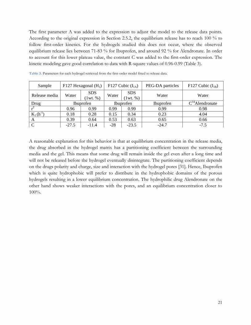
The first parameter A was added to the expression to adjust the model to the release data points.
According to the original expression in Section 2.5.2, the equilibrium release has to reach 100 % to
follow first-order kinetics. For the hydrogels studied this does not occur, where the observed
equilibrium release lies between 71-83 % for Ibuprofen, and around 92 % for Alendronate. In order
to account for this lower plateau value, the constant C was added to the first-order expression. The
kinetic modeling gave good correlation to data with R-square values of 0.96-0.99 (Table 3).
Table 3. Parameters for each hydrogel retrieved from the first-order model fitted to release data.
A reasonable explanation for this behavior is that at equilibrium concentration in the release media,
the drug absorbed in the hydrogel matrix has a partitioning coefficient between the surrounding
media and the gel. This means that some drug will remain inside the gel even after a long time and
will not be released before the hydrogel eventually disintegrate. The partitioning coefficient depends
on the drugs polarity and charge, size and interaction with the hydrogel pores [31]. Hence, Ibuprofen
which is quite hydrophobic will prefer to distribute in the hydrophobic domains of the porous
hydrogels resulting in a lower equilibrium concentration. The hydrophilic drug Alendronate on the
other hand shows weaker interactions with the pores, and an equilibrium concentration closer to
100%.
Sample F127 Hexagonal (H1) F127 Cubic (I1A) PEG-DA particles F127 Cubic (I1B)
Release media Water SDS
(1wt. %) Water
SDS
(1wt. %) Water Water
Drug Ibuprofen Ibuprofen Ibuprofen C14
Alendronate
r2 0.96 0.99 0.99 0.99 0.99 0.98
K1 (h-1
) 0.18 0.28 0.15 0.34 0.23 4.04
A 0.39 0.64 0.53 0.63 0.65 0.66
C -27.5 -11.4 -28 -23.5 -24.7 -7.5

22
Conclusions 5.This project aimed at forming two novel meso-ordered hydrogels and to evaluate them as controlled
release systems. Meso-ordered hydrogels based on LLCs formed by F127 triblock copolymers with
hexagonal and cubic phases were successfully formed, and retention of the structure after
polymerization was detected with SAXS. Ordered mesoporous PEG-DA hydrogel particles with a
size of 209-242 nm were formed using a w/o emulsion based technique.
Ibuprofen and 14C labeled Alendronate were successfully loaded into the hydrogels. For F127
hydrogels loaded with Ibuprofen, XRD measurements revealed a disruption of the drugs crystallinity,
implying that when absorbed in the hydrogel the drug exist in an amorphous phase.
The F127 hydrogels served as controlled release systems with an initial burst effect followed by
sustained drug release. Clear differences in drug release rates were observed between Ibuprofen and
Alendronate, which were explained by difference in polarity, where the more hydrophilic drug
Alendronate was released faster. Altering the release media by addition of SDS increased the release
rate and final concentration of Ibuprofen by enhancing its solubility. PEG-DA hydrogel particles
presented a controlled release behavior with significantly slower release compared to F127 hydrogels.
Drug-release kinetics for the hydrogels best corresponded to the first-order model with good
correlation with data with r2-values varying between 0. 96-0. 99.
Future work 6.This project has shown possibilities to use mesoporous hydrogels in applications as controlled
release systems.
It would be of interest to further study the release properties from the PEG-DA hydrogel particles,
the effect of the small particles size and its high porosity and surface area.
When it comes to release behavior it would be interesting to examine release performance of drugs
with different chemical properties. Also, it would be interesting to study the effect of altering the
surrounding release media by studying drug-release in simulated body fluid.
In vivo studies would be a great complement to this work to see how the different hydrogels work as
drug-delivery systems in a more complex environment and to evaluate the therapeutic response.

23
Acknowledgements I would like to express my gratitude to the following people:
My supervisor Martin Andersson for the opportunity to be involved in this research project and for
you guidance, support and inspiration throughout this year.
Anand Kumar Rajasekharan for your advice, guidance and help with lab work.
Stefan Allard for assistance and guiding during the radioactive experiments.
Maria Wallin for helping with the synthesis of PEG-particles.
Jonatan Bergek for help with the UV/VIS and discussions of release results
Simon Isaksson for assisting with DLS measurements and the implementation of release models.
All the other people of M.A Research group: Johan Karlsson, Wenxiao ”Chlor” He, Saba Atefyekta,
Mats Hulander, Emma Westas, Maria Pihl, Ali Alinezi , Maya Arvidsson and Vijayakumar.
Dr. Tomás Plivelic and Dr. Christopher Söderberg of MAX-II, Lund
Ann Jakobsson for all help with administrate aspects
My boyfriend Christoffer and to my family for your encouragement and support

24
References
1. Peppas, N.A., Hydrogels and drug delivery. Current Opinion in Colloid & Interface Science, 1997. 2(5): p. 531-537.
2. Nguyen, K.T. and J.L. West, Photopolymerizable hydrogels for tissue engineering applications. Biomaterials, 2002. 23(22): p. 4307-4314.
3. Forney, B.S. and C.A. Guymon, Nanostructure Evolution during Photopolymerization in Lyotropic Liquid Crystal Templates. Macromolecules, 2010. 43(20): p. 8502-8510.
4. Hoare, T.R. and D.S. Kohane, Hydrogels in drug delivery: Progress and challenges. Polymer, 2008. 49(8): p. 1993-2007.
5. Langer, R., Drugs on Target. Science, 2001. 293(5527): p. 58-59. 6. Clapper, J.D. and C.A. Guymon, Physical Behavior of Cross-Linked PEG Hydrogels
Photopolymerized within Nanostructured Lyotropic Liquid Crystalline Templates. Macromolecules, 2007. 40(4): p. 1101-1107.
7. Holmberg, K., et al., Phase Behaviour of Concentrated Surfactant Systems, in Surfactants and Polymers in Aqueous Solution. 2003, John Wiley & Sons, Ltd. p. 67-96.
8. Alexandridis, P., Amphiphilic copolymers and their applications. Current Opinion in Colloid & Interface Science, 1996. 1(4): p. 490-501.
9. Malmsten, M., Soft drug delivery systems. Soft Matter, 2006. 2(9): p. 760-769. 10. Alexandridis, P., U. Olsson, and B. Lindman, A Record Nine Different Phases (Four Cubic, Two
Hexagonal, and One Lamellar Lyotropic Liquid Crystalline and Two Micellar Solutions) in a Ternary Isothermal System of an Amphiphilic Block Copolymer and Selective Solvents (Water and Oil). Langmuir, 1998. 14(10): p. 2627-2638.
11. Xiong, X.Y., K.C. Tam, and L.H. Gan, Polymeric Nanostructures for Drug Delivery Applications Based on Pluronic Copolymer Systems. Journal of Nanoscience and Nanotechnology, 2006. 6(9): p. 2638-2650.
12. Hentze, H.P., et al., Lyotropic Mesophases of Poly(ethylene oxide)-b-poly(butadiene) Diblock Copolymers and Their Cross-Linking To Generate Ordered Gels. Macromolecules, 1999. 32(18): p. 5803-5809.
13. Gin, D.L., et al., Polymerized Lyotropic Liquid Crystal Assemblies for Membrane Applications. Macromolecular Rapid Communications, 2008. 29(5): p. 367-389.
14. Harmankaya, N., et al., Raloxifene and alendronate containing thin mesoporous titanium oxide films improve implant fixation to bone. Acta Biomater, 2013. 9(6): p. 7064-73.
15. Yang, H., N. Coombs, and G.A. Ozin, Morphogenesis of shapes and surface patterns in mesoporous silica. Nature, 1997. 386(6626): p. 692-695.
16. Claesson, M., et al., Meso-ordered soft hydrogels. Soft Matter, 2012. 8(31): p. 8149-8156. 17. Weiser, J.R. and W.M. Saltzman, Controlled release for local delivery of drugs: barriers and models.
Journal of Controlled Release, 2014. 190(0): p. 664-673. 18. Siegel, R. and M. Rathbone, Overview of Controlled Release Mechanisms, in Fundamentals and
Applications of Controlled Release Drug Delivery, J. Siepmann, R.A. Siegel, and M.J. Rathbone, Editors. 2012, Springer US. p. 19-43.
19. Uhrich, K.E., et al., Polymeric Systems for Controlled Drug Release. Chemical Reviews, 1999. 99(11): p. 3181-3198.
20. Lee, J.H. and Y. Yeo, Controlled drug release from pharmaceutical nanocarriers. Chemical Engineering Science, (0).
21. Costa, P. and J.M. Sousa Lobo, Modeling and comparison of dissolution profiles. European Journal of Pharmaceutical Sciences, 2001. 13(2): p. 123-133.

25
22. Ritger, P.L. and N.A. Peppas, A simple equation for description of solute release I. Fickian and non-fickian release from non-swellable devices in the form of slabs, spheres, cylinders or discs. Journal of Controlled Release, 1987. 5(1): p. 23-36.
23. Davies, N.M., Clinical pharmacokinetics of ibuprofen. The first 30 years. Clinical pharmacokinetics, 1998. 34(2): p. 101-154.
24. Sambrook, P.N., et al., Alendronate produces greater effects than raloxifene on bone density and bone turnover in postmenopausal women with low bone density: results of EFFECT (EFficacy of FOSAMAX® versus EVISTA®Comparison Trial) International1. Journal of Internal Medicine, 2004. 255(4): p. 503-511.
25. Holmqvist, P., P. Alexandridis, and B. Lindman, Modification of the Microstructure in Block
Copolymer−Water−“Oil” Systems by Varying the Copolymer Composition and the “Oil” Type: Small-Angle X-ray Scattering and Deuterium-NMR Investigation. The Journal of Physical Chemistry B, 1998. 102(7): p. 1149-1158.
26. Wallin M., A.A., Nordstierna L., Andersson M., PEG-based hexosomes, 2014: Manuscript submitted for publication.
27. L'Annunziata, M.F. and M.J. Kessler, 5 - Liquid Scintillation Analysis: Principles and Practice, in Handbook of Radioactivity Analysis (Second Edition), M.F. L'Annunziata, Editor. 2004, Academic Press: San Diego. p. 347-535.
28. Liquid scintillation counting. [cited 2014 1124]; Available from: http://www.perkinelmer.com/resources/technicalresources/applicationsupportknowledgebase/radiometric/liquid_scint.xhtml.
29. Holmqvist, P., P. Alexandridis, and B. Lindman, Phase Behavior and Structure of Ternary
Amphiphilic Block Copolymer−Alkanol−Water Systems: Comparison of Poly(ethylene oxide)/Poly(propylene oxide) to Poly(ethylene oxide)/Poly(tetrahydrofuran) Copolymers. Langmuir, 1997. 13(9): p. 2471-2479.
30. Rawlinson, C.F., et al., Polymer-mediated disruption of drug crystallinity. International Journal of Pharmaceutics, 2007. 336(1): p. 42-48.
31. Hoffman, A.S., Hydrogels for biomedical applications. Advanced Drug Delivery Reviews, 2002. 54(1): p. 3-12.
32. Huang, X. and C.S. Brazel, On the importance and mechanisms of burst release in matrix-controlled drug delivery systems. Journal of Controlled Release, 2001. 73(2–3): p. 121-136.