Control of Wilt and Rot Pathogens of Tomato by ... · cereus PPB-1 inoculated tomato plants, when...
11
ORIGINAL RESEARCH published: 07 November 2016 doi: 10.3389/fpls.2016.01626 Frontiers in Plant Science | www.frontiersin.org 1 November 2016 | Volume 7 | Article 1626 Edited by: Anton Hartmann, Helmholtz Zentrum München, Germany Reviewed by: Oswaldo Valdes-Lopez, National Autonomous University of Mexico, Mexico Wubei Dong, Huazhong Agricultural University, China *Correspondence: Rangasamy Anandham [email protected] Tongmin Sa [email protected] Specialty section: This article was submitted to Plant Biotic Interactions, a section of the journal Frontiers in Plant Science Received: 14 July 2016 Accepted: 14 October 2016 Published: 07 November 2016 Citation: Janahiraman V, Anandham R, Kwon SW, Sundaram S, Karthik Pandi V, Krishnamoorthy R, Kim K, Samaddar S and Sa T (2016) Control of Wilt and Rot Pathogens of Tomato by Antagonistic Pink Pigmented Facultative Methylotrophic Delftia lacustris and Bacillus spp. Front. Plant Sci. 7:1626. doi: 10.3389/fpls.2016.01626 Control of Wilt and Rot Pathogens of Tomato by Antagonistic Pink Pigmented Facultative Methylotrophic Delftia lacustris and Bacillus spp. Veeranan Janahiraman 1 , Rangasamy Anandham 1 *, Soon W. Kwon 2 , Subbiah Sundaram 1, 3 , Veeranan Karthik Pandi 4 , Ramasamy Krishnamoorthy 1 , Kiyoon Kim 3 , Sandipan Samaddar 3 and Tongmin Sa 3 * 1 Department of Agricultural Microbiology, Agricultural College and Research Institute, Tamil Nadu Agricultural University, Madurai, India, 2 Korean Agricultural Culture Collection, National Academy of Agricultural Science, Rural Development Administration, Jeonju, South Korea, 3 Department of Environmental and Biological Chemistry, Chungbuk National University, Cheongju, South Korea, 4 Department of Plant Pathology, Agricultural College and Research Institute, Tamil Nadu Agricultural University, Coimbatore, India The studies on the biocontrol potential of pink pigmented facultative methylotrophic (PPFM) bacteria other than the genus Methylobacterium are scarce. In the present study, we report three facultative methylotrophic isolates; PPO-1, PPT-1, and PPB- 1, respectively, identified as Delftia lacustris, Bacillus subtilis, and Bacillus cereus by 16S rRNA gene sequence analysis. Hemolytic activity was tested to investigate the potential pathogenicity of isolates to plants and humans, the results indicates that the isolates PPO-1, PPT-1, and PPB-1 are not pathogenic strains. Under in vitro conditions, D. lacustris PPO-1, B. subtilis PPT-1, and B. cereus PPB-1 showed direct antagonistic effect by inhibiting the mycelial growth of fungal pathogens; Fusarium oxysporum f. sp. lycopersici (2.15, 2.05, and 1.95 cm), Sclerotium rolfsii (2.14, 2.04, and 1.94 cm), Pythium ultimum (2.12, 2.02, and 1.92 cm), and Rhizoctonia solani (2.18, 2.08, and 1.98 cm) and also produced volatile inhibitory compounds. Under plant growth chamber condition methylotrophic bacterial isolates; D. lacustris PPO-1, B. subtilis PPT-1, and B. cereus PPB-1 significantly reduced the disease incidence of tomato. Under greenhouse condition, D. lacustris PPO-1, B. subtilis PPT-1, and B. cereus PPB-1 inoculated tomato plants, when challenged with F. oxysporum f. sp. lycopersici, S. rolfsii, P. ultimum, and R. solani, increased the pathogenesis related proteins (β-1,3-glucanase and chitinase) and defense enzymes (phenylalanine ammonia lyase, peroxidase, polyphenol oxidase, and catalase) on day 5 after inoculation. In the current study, we first report the facultative methylotrophy in pink pigmented D. lacustris, B. subtilis, and B. cereus and their antagonistic potential against fungal pathogens. Direct antagonistic and ISR effects of these isolates against fungal pathogens of tomato evidenced their possible use as a biocontrol agent. Keywords: methylotrophs, induced systemic resistance, pathogenesis-related proteins, biological control, tomato, antagonism
Transcript of Control of Wilt and Rot Pathogens of Tomato by ... · cereus PPB-1 inoculated tomato plants, when...

ORIGINAL RESEARCHpublished: 07 November 2016doi: 10.3389/fpls.2016.01626
Frontiers in Plant Science | www.frontiersin.org 1 November 2016 | Volume 7 | Article 1626
Edited by:
Anton Hartmann,
Helmholtz Zentrum München,
Germany
Reviewed by:
Oswaldo Valdes-Lopez,
National Autonomous University of
Mexico, Mexico
Wubei Dong,
Huazhong Agricultural University,
China
*Correspondence:
Rangasamy Anandham
Tongmin Sa
Specialty section:
This article was submitted to
Plant Biotic Interactions,
a section of the journal
Frontiers in Plant Science
Received: 14 July 2016
Accepted: 14 October 2016
Published: 07 November 2016
Citation:
Janahiraman V, Anandham R,
Kwon SW, Sundaram S, Karthik
Pandi V, Krishnamoorthy R, Kim K,
Samaddar S and Sa T (2016) Control
of Wilt and Rot Pathogens of Tomato
by Antagonistic Pink Pigmented
Facultative Methylotrophic Delftia
lacustris and Bacillus spp.
Front. Plant Sci. 7:1626.
doi: 10.3389/fpls.2016.01626
Control of Wilt and Rot Pathogens ofTomato by Antagonistic PinkPigmented FacultativeMethylotrophic Delftia lacustris andBacillus spp.Veeranan Janahiraman 1, Rangasamy Anandham 1*, Soon W. Kwon 2,
Subbiah Sundaram 1, 3, Veeranan Karthik Pandi 4, Ramasamy Krishnamoorthy 1,
Kiyoon Kim 3, Sandipan Samaddar 3 and Tongmin Sa 3*
1Department of Agricultural Microbiology, Agricultural College and Research Institute, Tamil Nadu Agricultural University,
Madurai, India, 2 Korean Agricultural Culture Collection, National Academy of Agricultural Science, Rural Development
Administration, Jeonju, South Korea, 3Department of Environmental and Biological Chemistry, Chungbuk National University,
Cheongju, South Korea, 4Department of Plant Pathology, Agricultural College and Research Institute, Tamil Nadu Agricultural
University, Coimbatore, India
The studies on the biocontrol potential of pink pigmented facultative methylotrophic
(PPFM) bacteria other than the genus Methylobacterium are scarce. In the present
study, we report three facultative methylotrophic isolates; PPO-1, PPT-1, and PPB-
1, respectively, identified as Delftia lacustris, Bacillus subtilis, and Bacillus cereus by
16S rRNA gene sequence analysis. Hemolytic activity was tested to investigate the
potential pathogenicity of isolates to plants and humans, the results indicates that
the isolates PPO-1, PPT-1, and PPB-1 are not pathogenic strains. Under in vitro
conditions, D. lacustris PPO-1, B. subtilis PPT-1, and B. cereus PPB-1 showed direct
antagonistic effect by inhibiting the mycelial growth of fungal pathogens; Fusarium
oxysporum f. sp. lycopersici (2.15, 2.05, and 1.95 cm), Sclerotium rolfsii (2.14, 2.04,
and 1.94 cm), Pythium ultimum (2.12, 2.02, and 1.92 cm), and Rhizoctonia solani
(2.18, 2.08, and 1.98 cm) and also produced volatile inhibitory compounds. Under
plant growth chamber condition methylotrophic bacterial isolates; D. lacustris PPO-1,
B. subtilis PPT-1, and B. cereus PPB-1 significantly reduced the disease incidence of
tomato. Under greenhouse condition, D. lacustris PPO-1, B. subtilis PPT-1, and B.
cereus PPB-1 inoculated tomato plants, when challenged with F. oxysporum f. sp.
lycopersici, S. rolfsii, P. ultimum, and R. solani, increased the pathogenesis related
proteins (β-1,3-glucanase and chitinase) and defense enzymes (phenylalanine ammonia
lyase, peroxidase, polyphenol oxidase, and catalase) on day 5 after inoculation. In the
current study, we first report the facultative methylotrophy in pink pigmented D. lacustris,
B. subtilis, and B. cereus and their antagonistic potential against fungal pathogens.
Direct antagonistic and ISR effects of these isolates against fungal pathogens of tomato
evidenced their possible use as a biocontrol agent.
Keywords: methylotrophs, induced systemic resistance, pathogenesis-related proteins, biological control,
tomato, antagonism
Janahiraman et al. Antagonistic Activity of PPFM against Phytopathogens
INTRODUCTION
Tomato (Lycopersicon esculentum) is one of the most popularcommercial vegetable crops. In India, it occupies an area of0.54 million ha with a production of 7.60 million tons (Kumaret al., 2012). Among the pathogens that affect the tomatocrop, soil borne fungal pathogens, including Fusarium, Pythium,Rhizoctonia, and Verticillium causing the root rot or damping-off and wilt affect the quality with yield reduction (Lucas et al.,1997). Among these pathogens, Fusarium oxysporum f. sp.lycopersici is a highly destructive pathogen on both greenhouseand field grown tomatoes. In spite of the promising results shownby chemical treatments in controlling the fungal pathogens,phytotoxicity, and chemical residues are the major problemsleading to environmental pollution and human health hazards.Alternatively in the present study, we have tested the possibilityof using facultativemethylotrophic bacteria which is ubiquitouslyoccurring with intimate association with plants, as a biocontrolagent in controlling wilt and root rot pathogens of tomato.
Methylotrophs consists of a subpopulation of the bacteriawhich are able to use single carbon compounds (methanol andother C1 carbon compounds) as a sole carbon and energy source.Methylotropic bacteria can take up greenhouse gases and reduceglobal warming (Iguchi et al., 2015). Methylotrophs are foundin both phyllosphere and rhizosphere of the plants and utilizethe plant waste methanol as carbon source. Recently, therehave been extensive studies on methylotrophs and their abilityto promote plant growth were carried out (Madhaiyan et al.,2004; Poonguzhali et al., 2008; Yim et al., 2012). In addition,methylotrophic activity was reported in different bacterialfamilies (Eyice and Schäfer, 2016). Among the facultativemethylotrophic (FM) bacteria belonging to Alpha-, Beta-, andGamma-subclasses of Proteobacteria and Firmicutes, the genusMethylobacterium has been widely studied. These FM bacteriaubiquitously occurring in plants can be isolated using selectivemedia containing methanol as the sole carbon source andidentified by their characteristic pink color (Corpe and Basile,1982; Lidstrom and Chistoserdova, 2002). They are commonlyreferred as pink pigmented facultative methylotrophic (PPFM)bacteria. They are non-pathogenic and distributed widely inthe plant phyllosphere, and have been isolated from more than100 species of plants, ranging from liverworts and mosses toangiosperms and gymnosperms (Corpe and Basile, 1982).
Among the PPFM, the genus Methylobacterium is one ofthe dominant genera, which act as a symbiont with plantsby consuming methanol, a plant metabolic waste and inturn providing vitamin B12, auxins, cytokinins useful for theplant growth (Madhaiyan et al., 2006). Although the genusMethylobacterium has also been well-documented for theirinduction of systemic resistance (ISR) in plants against plantpathogens (Madhaiyan et al., 2004, 2006; Indiragandhi et al.,2008), till date to the best of our knowledge there is no reportavailable on the direct antagonistic effect of FMbacteria. Hence inthe current study, we tested both direct antagonistic effect of FMbacteria against Fusarium oxysporum f. sp. lycopersici, Sclerotiumrolfsii, Pythium ultimum, and Rhizoctonia solani causing tomatoroot rot and wilt diseases and indirect effect by the induction
of ISR and pathogenesis related (PR) proteins in the pathogenchallenged tomato plants grown under greenhouse condition.
MATERIALS AND METHODS
Source of Bacterial Strains, FungalPathogens, and Tomato SeedsPPFM bacteria were isolated from the phylloplane of crop plantslisted in Table 1 through leaf imprinting technique on solidammoniummineral salts (AMS)medium (pH 6.8) supplementedwith 0.5% methanol and cyclohexamide (30 µg ml−1) andincubated at 28◦C for 3–5 days (Whittenbury et al., 1970). Thesingle colonies with reddish pink pigmentation were pickedand purified. The PPFM isolates were grown for 72 h onAMS medium. Gram reaction and other biochemical tests werecarried out as described previously (Gerhardt et al., 1981). Plantpathogens (Fusarium oxysporum f. sp. lycopersici MTCC 4356,S. rolfsii KACC 43068, R. solani MTCC 4633, and P. ultimumKACC 40705) were obtained from Microbial Type CultureCollection, Chandigarh, India and Korean Agricultural CultureCollection, Jeonju, Republic of Korea. Fungal pathogens weregrown and maintained in potato dextrose agar (PDA) beforefurther use. Tomato cultivar CO4, which is susceptible to bothseedling wilt and root rot, was obtained from the Department ofvegetables at Horticultural College and Research Institute, TamilNadu Agricultural University, Coimbatore, India.
Antagonistic Assay on Solid MediumAll PPFM bacterial isolates were screened for their ability toinhibit the growth of fungal pathogens on PDA plates by dualculturing technique with three replications (Yoshida et al., 2001).After 120 h incubation at 30 ± 2◦C, the distance between thebacterial growth and fungal mycelial growth was measured tofind out the antagonistic effect of PPFM bacterial isolates on testpathogenic fungi. The antagonistic effect of volatile antifungalcompounds produced by PPFM bacterial isolates against testfungal pathogens was evaluated on PDA medium with threereplications after 120 h of incubation at 30 ± 2◦C as describedby Trivedi et al. (2008). Briefly, PPFM isolates antagonism dueto volatile compounds was evaluated by preparing a bacteriallawn on agar plates. After incubation for 24 h, the lid of theplate was replaced by a plate containing an agar block of thetest fungus grown on PDA. The two plates were sealed togetherwith parafilm. Control sets were prepared in a similar manner,without PPFM in the bottom plate. Then the Petri dishes wereincubated at 28◦C, and the observations were recorded after 5days. Growth inhibition of the test fungus was calculated in %using the formula: (r1 − r2/r1) ∗ 100, where r1 (a control value)represents the radial growth of the fungus in control sets without,and r2 with bacteria.
Antagonistic Assay in Liquid MediumA volume of 1 ml of bacterial culture grown in potato dextrosebroth for 72 h (109 cfu ml−1) and a disc of test fungus (10 mm)from a well-grown fungal colony on PDA plates were inoculatedinto 50 ml potato dextrose broth in 250 ml conical flasks andincubated at 30 ± 2◦C on a rotary shaker for 120 h. Potato
Frontiers in Plant Science | www.frontiersin.org 2 November 2016 | Volume 7 | Article 1626
Janahiraman et al. Antagonistic Activity of PPFM against Phytopathogens
TABLE1|In
vitro
antagonisticpotentialofpinkpigmentedfacultativemethylotrophic
(PPFM)bacteriaagainstplantpathogenic
fungiandproductionofantimicrobialcompounds.
Isolates
Source
Inhibitionzone(cm)
Siderophore
(µgml−
1)
Salicylicacid
(µgml−
1)
β-1,3-g
lucanase(µgof
glucosereleasedmin
−1mgof
protein
−1)
Chitinase(1
µmolofNAG
releasedh
−1mgofprotein
−1)
F.oxysporum
f.sp.
lycopersici
S.rolfsii
P.ultim
um
R.solani
PPT-1
Tomato
2.05±
0.04b
2.04±
0.04b
2.02±
0.04b
2.08±
0.04b
27.45±
0.55a
88±
1.76a
278±
5.34b
4.95±
0.014a
PPN-1
Neem
1.85±
0.04d
1.64±
0.03f
1.12±
0.02k
1.38±
0.03i
20.54±
0.41d
71±
1.42de
118±
1.17i
3.89±
0.038d
PPB-1
Bhendi
1.95±
0.04c
1.94±
0.04c
1.92±
0.04c
1.98±
0.04c
25.44±
0.51b
87±
1.74a
265±
4.28c
4.80±
0.042a
PPBJ-1
Brin
jal
1.75±
0.04e
1.14±
0.02k
1.42±
0.03h
1.68±
0.03f
23.19±
0.46c
77±
1.54b
178±
1.30f
3.39±
0.058e
PPG-1
Guava
1.25±
0.02j
1.54±
0.03g
1.82±
0.04d
1.48±
0.03h
18.45±
0.37f
61±
1.22i
140±
2.40h
3.86±
0.012d
PPL-1
Lim
e1.65±
0.03f
1.24±
0.02j
1.52±
0.03g
1.18±
0.02k
20.02±
0.40de
70±
1.40ef
135±
2.18h
2.78±
0.038g
PPP-1
Proso
phis
1.1
±0.02k
1.74±
0.03e
1.72±
0.03e
1.88±
0.04d
12.45±
0.25i
65±
1.30h
218±
4.54e
2.56±
0.052h
PPGR-1
Groundnut
1.55±
0.03g
1.44±
0.03h
1.22±
0.02j
1.58±
0.03g
17.32±
0.35g
73±
1.46cd
168±
3.15g
3.1
±0.023f
PPO-1
Onion
2.15±
0.04a
2.14±
0.04a
2.12±
0.04a
2.18±
0.04a
27.94±
0.56a
89±
1.78a
292±
3.65a
5.09±
0.111a
PPBO-1
Bougainvillea
1.45±
0.03h
1.84±
0.04d
1.32±
0.03i
1.78±
0.04e
17.45±
0.35g
68±
1.36g
186±
1.36f
3.50±
0.026e
PPPA-1
Parthenium
1.05±
0.02l
1.34±
0.03i
1.62±
0.03f
1.08±
0.02l
19.47±
0.39e
75±
1.50bc
234±
2.07d
4.2
±0.026c
PPGS-1
Grass
1.35±
0.03i
1.04±
0.02l
1.02±
0.02l
1.28±
0.03j
15.32±
0.31h
6767±
1.34gh
224±
3.03e
4.5
±0.080b
LSD(P
≤0.05)
0.06
0.06
0.05
0.06
3.11
0.71
8.64
0.17
NoneoftheisolatesproducedHCNundertestedcondition;valuesaremeanofthreereplications±SD(standard
deviation).Valuesineachcolumnfollowedbysameletter(s)arenotstatisticallydifferentbyLSD(P
≤0.05).
dextrose broth inoculated only with fungi served as positivecontrol. The broth with fungal mat in various treatments waspassed through a pre-weighed Whatman No. 1 filter paper. Thefilter papers were dried for 24 h at 70 ± 1◦C to obtain a constantweight. The percentage reduction in fungal mycelial weight wascalculated as described previously by Trivedi et al. (2008).
Other Antimicrobial Traits AssaySiderophore, hydrocyanic acid (HCN), and salicylicacid production, β-1,3-glucanase activity and chitinaseactivity of the isolates were measured as mentioned inSupporting Information File Materials and Method I.
16S rRNA Gene Sequence AnalysisBacterial isolates were grown in AMS medium for 72 h andDNA was extracted according to Sambrook et al. (1989). Thegene encoding bacterial 16S rRNA was amplified through PCRwith forward primer 27f: 5′-AGAGTTTGATCCTGGCTCAG-3′ and reverse primer 1492r: 5′-GGTTACCTTGTTACGACTT-3′. Nearly, complete 16S rRNA gene sequences of PPFMbacterial isolates from the automatic sequencer were alignedusing the integrated SINA alignment tool from the ARB-SILVAwebsite (Pruesse et al., 2007) and bacterial identities deduced byusing the EzTaxon-e server (http://eztaxon-e.ezbiocloud.net/)to ascertain their closest relatives (Kim et al., 2012).Bacterial gyrA gene is a potential chromosomal markerfor the phylogenetic identification of Bacillus genera. PCRamplification and detection of gyr A gene was given in theSupporting Information File Materials and Method II.
Methanol Utilization and Detection ofMethanol Dehydrogenase Gene (mxaF)Methanol utilization by the isolates was checked according toSy et al. (2001) in M72 medium. The bacterial suspensionswere diluted in M72 medium [optical density (OD) = 0.05],and added with one of the following compounds; methanol(MeOH) (10, 50, 100, or 500 mM), pyruvate (10 mM), orsuccinate (10 mM). Growth was monitored by measuringOD at 620 nm. Detection of mxaF gene was given inSupporting Information File Materials and Method III.
Hemolytic Activity and Response toAntibioticsThis experiment was conducted to test the production ofexotoxins called hemolysin able to destroy the Red Blood Cells(RBC) and hemoglobin by the methylotrophic isolates Delftialacustris PPO-1, Bacillus subtilis PPT-1, Bacillus cereus PPB-1 insheep blood agar plates (catalog no. MP1301 Himedia, India).The plates were incubated for 48–72 h at 30◦C and observedfor presence of clear zone, greenish brown, and no zone whichindicates the complete (β hemolysis), partial (α hemolysis),and no hemolytic (È) activity, respectively. Susceptibilityto the antibiotics rifampicin, ampicillin, oleandomycin,chloramphenicol, tetracycline, novobiocin, streptomycin,spectinomycin, kanamycin, trimethoprim, hygromycin,ceftriaxone, gentamycin, cefepime, and amikacin (each at
Frontiers in Plant Science | www.frontiersin.org 3 November 2016 | Volume 7 | Article 1626

Janahiraman et al. Antagonistic Activity of PPFM against Phytopathogens
50 µg ml−1) was tested for D. lacutris PPO-1 on R2A agar platesas per Bauer et al. (1966).
Plant Growth Chamber AssayPlant growth chamber experiment was conducted to assess thebiocontrol potential of PPFM bacterial isolates on wilt androt diseases of tomato. Bacterial cultures grown for 72 h wereharvested by centrifugation at 10,000 × g for 10 min at 4◦C,washed twice with sterile distilled water and suspended in 0.03 MMgSO4 solution. Tomato seeds were surface sterilized with 70%ethanol for 1 min, 0.5% NaOCl for 2 min, and washed fourtimes with sterilized distilled water. The surface-sterilized seedswere imbibed in the 5 ml of bacterial suspension (109 cfu ml−1)for 2 h, and single seed was sown in a 400 ml plastic pot filledwith mixture of sterilized soil and farm yard manure in 3:1ratio. The pots were subjected to following treatments; T1—B.subtilis PPT-1 + F. oxysporum f. sp. lycopersici, T2—B. cereusPPB-1 + F. oxysporum f. sp. lycopersici, T3—D. lacustris PPO-1 + F. oxysporum f. sp. lycopersici, T4—B. subtilis PPT-1 +
S. rolfsii, T5—B. cereus PPB-1 + S. rolfsii, T6—D. lacustrisPPO-1 + S. rolfsii, T7—B. subtilis PPT-1 + R. solani, T8—B. cereus PPB-1 + R. solani, T9—D. lacustris PPO-1 + R.solani, T10—B. subtilis PPT-1 + P. ultimum, T11—B. cereusPPB-1 + P. ultimum, T12—D. lacustris PPO-1 + P. ultimum,T13—F. oxysporum f. sp. Lycopersici, T14—S. rolfsii, T15—R.solani, T16—P. ultimum (Figure S1). One milliliter of fungalspore suspension (106 spore ml−1) was applied to the soil on30th day after sowing. Ten potted plants were maintained foreach treatment. The experiment was conducted in a completelyrandomized block design with three replications. Pots wereplaced in growth chambers at 20 ± 1◦C, photoperiod startingwith 12 h dark followed by 12 h lights (18µmol m−2 s−2). Plantswere watered with 1 X Hoagland solution. The disease incidencewas observed by recording the wilting of leaves leading to plantdrying. Randomly 15 plants were selected from each treatmentand the number of wilted plants was recorded. The mean wiltdisease incidence was expressed in percentage. Disease incidencewas recorded up to 30 d after challenge inoculation with fungalpathogens. The percent disease incidence (PDI) was calculated,by using the formula. PDI = Number of plants infected/Totalnumber of plants observed× 100.
Greenhouse AssayTomato seeds were surface sterilized and imbibed in the PPFMbacterial suspension as described earlier. Four seeds were sown ina 20 L earthen pot containing 10 Kg of unsterilized soil. Controlseeds were imbibed with 0.03 M MgSO4 for 2 h. All treatmentsfollowed in the plant growth chamber assay were imposed. Theexperiment was conducted in a completely randomized blockdesign with three replications. Twenty-one days after sowing,PPFM bacterial cells suspended in 0.03 M MgSO4 (109 cfuml−1) were sprayed over tomato plants with handheld pneumaticsprayer till the plants were completely wetted. Thirty days aftersowing, 20 ml of fungal spore suspension (106 spore ml−1) wasapplied into the soil. Initially, F. oxysporum f. sp. lycopersici (1.2× 10² cfu), S. rolfsii (1.5× 10² cfu), R. solani (2.1× 10² cfu), andP. ultimum (1 × 10² cfu) were present in 1 g of experimental dry
soil. Germination percentage was recorded on 7 days after sowing(DAS). Seedling vigor index was calculated as % germination ×
seedling length (shoot length + root length) in cm on 15 DAS(Baki and Anderson, 1973). Leaf samples were collected on 0, 3, 5,7, and 9 days after challenge inoculation of pathogen and frozenimmediately at −20◦C for analyses of PR proteins and defenseenzymes. The plant height was recorded on 30, 60, and 90 DAS.The crop was harvested at physiological maturity and yield wasrecorded.
PR Proteins AnalysisFrozen leaves were ground at 4◦C in an ice-chilled mortar withliquid nitrogen, and the resulting powder was suspended in 100mM potassium phosphate buffer, pH 7.0 (2:2.5, w/v). Crudehomogenates were centrifuged at 8000 × g for 10 min at 4◦C,and the supernatants were kept frozen at −20◦C until use.β-1,3-glucanase was assayed based on the release of reducingsugars from laminarin, as described by Liang et al. (1995). Oneunit of β-1,3-glucanase activity was determined as the amountof enzyme required to liberate 1 ng of glucose equivalent at37◦C in 1 h. Chitinase activity was assayed by measuring therelease of N-acetyl-glucosamine from prepared colloidal chitin,as described by Singh et al. (1999). One unit of chitinase activitywas determined as the amount of enzyme required to liberate 1n mol of N-acetyl-glucosamine equivalent at 37◦C in 1 h. PALactivity was measured using the procedure described by El-shora(2002). One unit represented the conversion of 1 µ mol of L-phenylalanine to cinnamic acid per min. Oxidative enzymes likePO and PPO were estimated as described previously (Compantet al., 2005). The activity of PO and PPO were expressed as thechange in unit of absorbance at 420 nm/g of fresh weight per min.Catalase activity was assayed spectrophotometrically as describedby Stern (1937). The activity was measured by monitoring thedegradation of H2O2 using a spectrophotometer at 240 nm over1min. expressed in mmol min g−1 of leaf tissue−1.
Statistical AnalysisThe data were analyzed by an analysis of variance (ANOVA)using the general linear model version 9.1; SAS Institute Inc.,Cary, NC, USA. Means were compared using the least significantdifference (LSD). The significance levels were within confidencelimits of 0.05 or less.
RESULTS
Antagonistic Potential of PPFM BacterialIsolates against Fungal Pathogens ofTomatoTwenty PPFM bacteria were isolated from phylloplane of variouscrop plants (Table S1). Among the PPFM bacterial isolates, 12isolates showed direct antagonism toward tested pathogens. Theisolates PPO-1, PPT-1, and PPB-1 exhibited mycelial growthinhibition of Fusarium oxysporum f. sp. lycopersici (2.15, 2.05,and 1.95 cm), S. rolfsii (2.14, 2.04, and 1.94 cm), P. ultimum(2.12, 2.02, and 1.92 cm), R. solani (2.18, 2.08, and 1.98 cm),respectively, in a dual plate assay on day 5 (Table 1). All the testedmethylotrophic isolates were able to produce siderophores and
Frontiers in Plant Science | www.frontiersin.org 4 November 2016 | Volume 7 | Article 1626
Janahiraman et al. Antagonistic Activity of PPFM against Phytopathogens
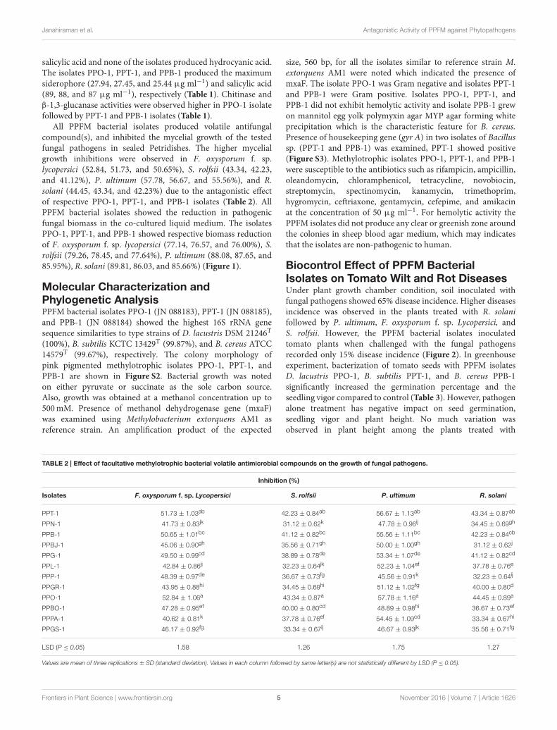
salicylic acid and none of the isolates produced hydrocyanic acid.The isolates PPO-1, PPT-1, and PPB-1 produced the maximumsiderophore (27.94, 27.45, and 25.44 µg ml−1) and salicylic acid(89, 88, and 87 µg ml−1), respectively (Table 1). Chitinase andβ-1,3-glucanase activities were observed higher in PPO-1 isolatefollowed by PPT-1 and PPB-1 isolates (Table 1).
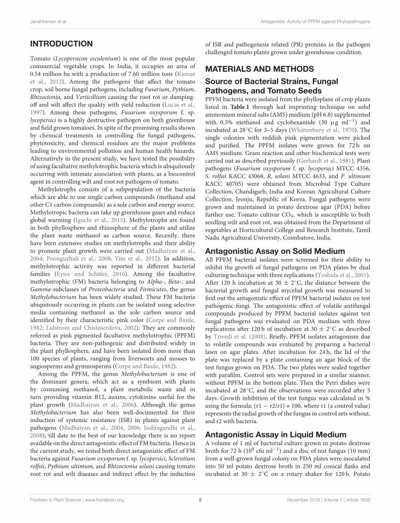
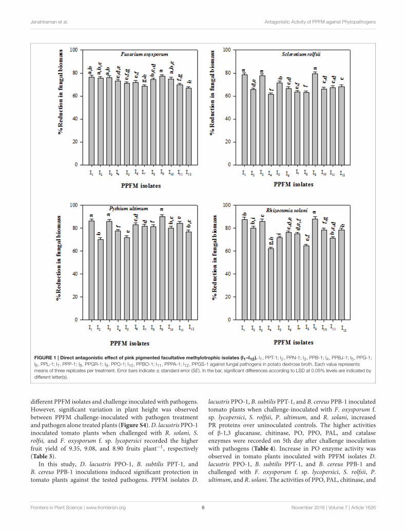
All PPFM bacterial isolates produced volatile antifungalcompound(s), and inhibited the mycelial growth of the testedfungal pathogens in sealed Petridishes. The higher mycelialgrowth inhibitions were observed in F. oxysporum f. sp.lycopersici (52.84, 51.73, and 50.65%), S. rolfsii (43.34, 42.23,and 41.12%), P. ultimum (57.78, 56.67, and 55.56%), and R.solani (44.45, 43.34, and 42.23%) due to the antagonistic effectof respective PPO-1, PPT-1, and PPB-1 isolates (Table 2). AllPPFM bacterial isolates showed the reduction in pathogenicfungal biomass in the co-cultured liquid medium. The isolatesPPO-1, PPT-1, and PPB-1 showed respective biomass reductionof F. oxysporum f. sp. lycopersici (77.14, 76.57, and 76.00%), S.rolfsii (79.26, 78.45, and 77.64%), P. ultimum (88.08, 87.65, and85.95%), R. solani (89.81, 86.03, and 85.66%) (Figure 1).
Molecular Characterization andPhylogenetic AnalysisPPFM bacterial isolates PPO-1 (JN 088183), PPT-1 (JN 088185),and PPB-1 (JN 088184) showed the highest 16S rRNA genesequence similarities to type strains of D. lacustris DSM 21246T
(100%), B. subtilis KCTC 13429T (99.87%), and B. cereus ATCC14579T (99.67%), respectively. The colony morphology ofpink pigmented methylotrophic isolates PPO-1, PPT-1, andPPB-1 are shown in Figure S2. Bacterial growth was notedon either pyruvate or succinate as the sole carbon source.Also, growth was obtained at a methanol concentration up to500mM. Presence of methanol dehydrogenase gene (mxaF)was examined using Methylobacterium extorquens AM1 asreference strain. An amplification product of the expected
size, 560 bp, for all the isolates similar to reference strain M.extorquens AM1 were noted which indicated the presence ofmxaF. The isolate PPO-1 was Gram negative and isolates PPT-1and PPB-1 were Gram positive. Isolates PPO-1, PPT-1, andPPB-1 did not exhibit hemolytic activity and isolate PPB-1 grewon mannitol egg yolk polymyxin agar MYP agar forming whiteprecipitation which is the characteristic feature for B. cereus.Presence of housekeeping gene (gyr A) in two isolates of Bacillussp. (PPT-1 and PPB-1) was examined, PPT-1 showed positive(Figure S3). Methylotrophic isolates PPO-1, PPT-1, and PPB-1were susceptible to the antibiotics such as rifampicin, ampicillin,oleandomycin, chloramphenicol, tetracycline, novobiocin,streptomycin, spectinomycin, kanamycin, trimethoprim,hygromycin, ceftriaxone, gentamycin, cefepime, and amikacinat the concentration of 50 µg ml−1. For hemolytic activity thePPFM isolates did not produce any clear or greenish zone aroundthe colonies in sheep blood agar medium, which may indicatesthat the isolates are non-pathogenic to human.
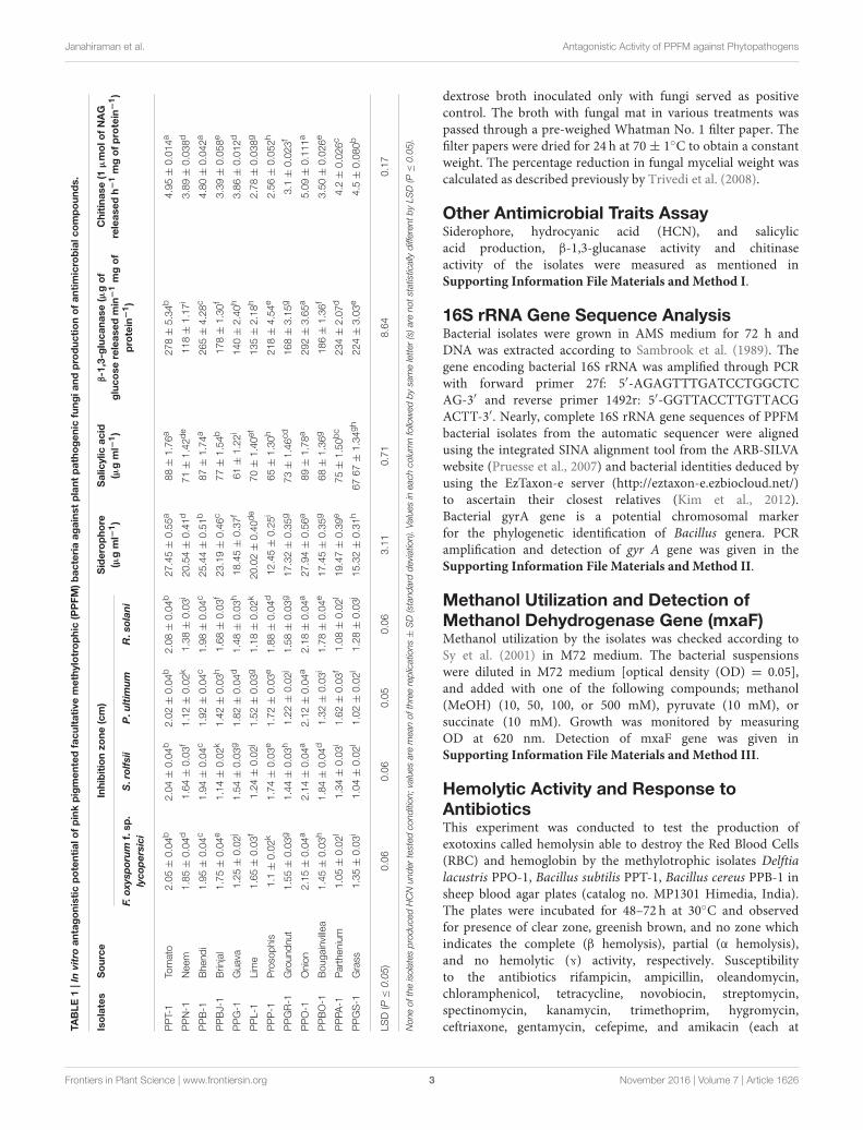
Biocontrol Effect of PPFM BacterialIsolates on Tomato Wilt and Rot DiseasesUnder plant growth chamber condition, soil inoculated withfungal pathogens showed 65% disease incidence. Higher diseasesincidence was observed in the plants treated with R. solanifollowed by P. ultimum, F. oxysporum f. sp. Lycopersici, andS. rolfsii. However, the PPFM bacterial isolates inoculatedtomato plants when challenged with the fungal pathogensrecorded only 15% disease incidence (Figure 2). In greenhouseexperiment, bacterization of tomato seeds with PPFM isolatesD. lacustris PPO-1, B. subtilis PPT-1, and B. cereus PPB-1significantly increased the germination percentage and theseedling vigor compared to control (Table 3). However, pathogenalone treatment has negative impact on seed germination,seedling vigor and plant height. No much variation wasobserved in plant height among the plants treated with
TABLE 2 | Effect of facultative methylotrophic bacterial volatile antimicrobial compounds on the growth of fungal pathogens.
Inhibition (%)
Isolates F. oxysporum f. sp. Lycopersici S. rolfsii P. ultimum R. solani
PPT-1 51.73 ± 1.03ab 42.23 ± 0.84ab 56.67 ± 1.13ab 43.34 ± 0.87ab
PPN-1 41.73 ± 0.83jk 31.12 ± 0.62k 47.78 ± 0.96ij 34.45 ± 0.69gh
PPB-1 50.65 ± 1.01bc 41.12 ± 0.82bc 55.56 ± 1.11bc 42.23 ± 0.84cb
PPBJ-1 45.06 ± 0.90gh 35.56 ± 0.71gh 50.00 ± 1.00gh 31.12 ± 0.62j
PPG-1 49.50 ± 0.99cd 38.89 ± 0.78de 53.34 ± 1.07de 41.12 ± 0.82cd
PPL-1 42.84 ± 0.86ij 32.23 ± 0.64jk 52.23 ± 1.04ef 37.78 ± 0.76e
PPP-1 48.39 ± 0.97de 36.67 ± 0.73fg 45.56 ± 0.91k 32.23 ± 0.64ij
PPGR-1 43.95 ± 0.88hi 34.45 ± 0.69hi 51.12 ± 1.02fg 40.00 ± 0.80d
PPO-1 52.84 ± 1.06a 43.34 ± 0.87a 57.78 ± 1.16a 44.45 ± 0.89a
PPBO-1 47.28 ± 0.95ef 40.00 ± 0.80cd 48.89 ± 0.98hi 36.67 ± 0.73ef
PPPA-1 40.62 ± 0.81k 37.78 ± 0.76ef 54.45 ± 1.09cd 33.34 ± 0.67hi
PPGS-1 46.17 ± 0.92fg 33.34 ± 0.67ij 46.67 ± 0.93jk 35.56 ± 0.71fg
LSD (P ≤ 0.05) 1.58 1.26 1.75 1.27
Values are mean of three replications ± SD (standard deviation). Values in each column followed by same letter(s) are not statistically different by LSD (P ≤ 0.05).
Frontiers in Plant Science | www.frontiersin.org 5 November 2016 | Volume 7 | Article 1626
Janahiraman et al. Antagonistic Activity of PPFM against Phytopathogens
FIGURE 1 | Direct antagonistic effect of pink pigmented facultative methylotrophic isolates (I1–I12). I1, PPT-1; I2, PPN-1; I3, PPB-1; I4, PPBJ-1; I5, PPG-1;
I6, PPL-1; I7, PPP-1; I8, PPGR-1; I9, PPO-1; I10, PPBO-1; I11, PPPA-1; I12, PPGS-1 against fungal pathogens in potato dextrose broth. Each value represents
means of three replicates per treatment. Error bars indicate ± standard error (SE). In the bar, significant differences according to LSD at 0.05% levels are indicated by
different letter(s).
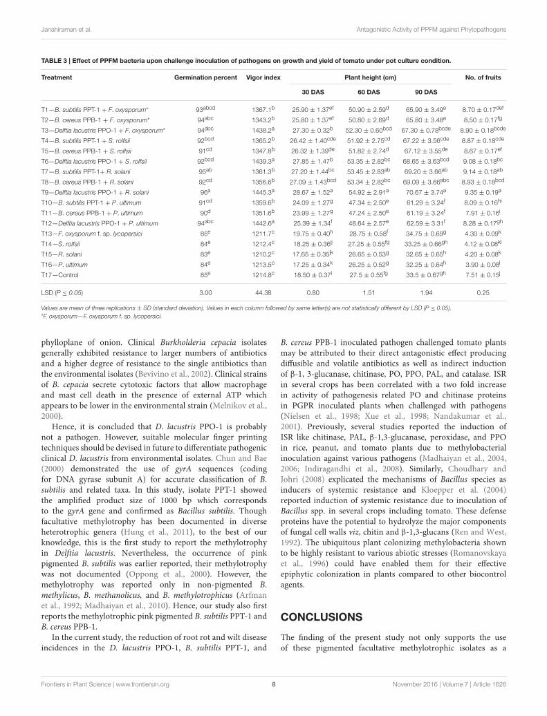
different PPFM isolates and challenge inoculated with pathogens.However, significant variation in plant height was observedbetween PPFM challenge-inoculated with pathogen treatmentand pathogen alone treated plants (Figure S4).D. lacustris PPO-1inoculated tomato plants when challenged with R. solani, S.rolfsi, and F. oxysporum f. sp. lycopersici recorded the higherfruit yield of 9.35, 9.08, and 8.90 fruits plant−1, respectively(Table 3).
In this study, D. lacustris PPO-1, B. subtilis PPT-1, andB. cereus PPB-1 inoculations induced significant protection intomato plants against the tested pathogens. PPFM isolates D.
lacustris PPO-1, B. subtilis PPT-1, and B. cereus PPB-1 inoculatedtomato plants when challenge-inoculated with F. oxysporum f.sp. lycopersici, S. rolfsii, P. ultimum, and R. solani, increasedPR proteins over uninoculated controls. The higher activitiesof β-1,3 glucanase, chitinase, PO, PPO, PAL, and catalaseenzymes were recorded on 5th day after challenge inoculationwith pathogens (Table 4). Increase in PO enzyme activity wasobserved in tomato plants inoculated with PPFM isolates D.lacustris PPO-1, B. subtilis PPT-1, and B. cereus PPB-1 andchallenged with F. oxysporum f. sp. lycopersici, S. rolfsii, P.ultimum, and R. solani. The activities of PPO, PAL, chitinase, and
Frontiers in Plant Science | www.frontiersin.org 6 November 2016 | Volume 7 | Article 1626

Janahiraman et al. Antagonistic Activity of PPFM against Phytopathogens
FIGURE 2 | Facultative methylotrophic bacterial inoculation effect on
percent disease incidence in pathogen challenged tomato plants
grown in growth chamber. T1—PPT-1 + F. oxysporum f. sp. lycopersici,
T2—PPB-1 + F. oxysporum f. sp. lycopersici, T3—PPO-1 + F. oxysporum f.
sp. lycopersici, T4—PPT-1 + S. rolfsii, T5—PPB-1 + S. rolfsii, T6—PPO-1 +
S. rolfsii, T7—PPT-1 + R. solani, T8—PPB-1 + R. solani, T9—PPO-1 + R.
solani, T10—PPT-1 + P. ultimum, T11—PPB-1 + P. ultimum, T12—PPO-1 +
P. ultimum, T13—F. oxysporum f. sp. Lycopersici, T14—S. rolfsii, T15—R.
solani, T16—P. ultimum. Each value represents means of three replicates per
treatment. Error bars indicate ± standard error (SE). In the bar, significant
differences according to LSD at 0.05% levels are indicated by different letter(s).
catalase enzymes also had a similar trend (Table 4). In particular,D. lacustris PPO-1 inoculated tomato plants when challengeinoculated with R. solani, P. ultimum, S. rolfsii, and F. oxysporumf. sp. lycopersici exhibited significant increase in β-1,3-glucanaseactivity (Table 4). Delftia lacustris PPO-1 challenge inoculatedwith pathogen exhibited higher chitinase and catalase activitycompared to other treatments.
DISCUSSION
Members of pink-pigmented facultative methylotrophs occupiesdifferent habitats such as leaf surface, soil, water, and air. Theplant-methylotroph association could be attributed to the uniqueability of these bacteria to grow at the expense of methanol, acell wall pectin degradative product from plants. PPFM has theability to oxidize methanol using the methanol dehydrogenaseenzyme. ThemxaF gene encodes the large subunit of this enzyme,which is key in methylotrophic metabolism. In recent years,interaction study have shown that PPFM bacteria can increaseplant protection against phytopathogen attack (Benhamou et al.,2000). In the current study, PPFM bacteria were isolated fromvarious crop plants and used as a bio-control agent againsttomato phytopathogens.
Bio-control ability of the PPFM isolates againstphytopathogens were tested using three methods. One isby dual culture assay in which the PPFM isolate and pathogenswere grown in same plate. In that the isolates PPO-1, PPT-1,and PPB-1, exhibited higher antagonistic potential against F.
oxysporum f. sp. lycopersici, S. rolfsii, P. ultimum, and R. solani.The inhibitory effect of fungal growth observed in the currentstudy in the dual culturing assay may be attributed to certaindiffusible antifungal metabolites of the methylotrophic isolates(Montealegre et al., 2003). In the second method, the effect ofPPFM volatile compound against phytopathogen was tested.The results revealed that PPFM bacterial isolates producedvolatile antifungal compound which inhibited the growth oftest fungal pathogens. In third method, both PPFM isolatesand phytopathogens were simultaneously grown in liquidmedium as co-cultures. The reduction in fungal biomass due toinhibitory effect of antagonistic PPFMwas compared with fungalcultures grown in medium without antagonistic PPFM. Earliera non-methylotrophic Delftia lacustris possessing nitrogenfixing trait capable of suppressing the growth of rice pathogens(Xanthomonas oryzae pv. oryzae, R. solani, and Pyriculariaoryzae) was reported by Han et al. (2005).
The PPFM isolates capable of producing siderophopres provestheir competitive advantage in the natural ecosystem by limitingthe supply of iron and essential trace elements to the fugalpathogens as previously suggested by Indiragandhi et al. (2008).The inhibitory effect on the fungal pathogens tested mayalso be attributed to the salicylic acid production capabilityof PPFM isolates, as already evidenced in Methylobacteriumoryzae CBMB20 inoculated Pseudomonas syringae pv. tomatochallenged tomato plants (Indiragandhi et al., 2008). However,PPO-1, PPT-1, and PPB-1 methylotrophic bacterial isolatesnot producing HCN, eliminated the possibility of blocking thecytochrome oxidase system in fungal pathogen (Trivedi et al.,2008). Presence of chitinase and β-1,3-glucanase activities werepreviously reported in Delftia sp., B. subtilis and B. cereus (Chenet al., 2004; Jørgensen et al., 2009; Liang et al., 2014). Chitinand glucans those are responsible for the rigidity of fungal cellwalls, thereby destroying cell wall integrity due to secretionof chitinase and β-1,3-glucanase limiting the growth of thepathogen (Chen et al., 2004; Jørgensen et al., 2009; Liang et al.,2014).
Methylotrophs oxidize methanol to formaldehyde via a keyenzyme methanol dehydrogenase and the use of structural genemxaF as a functional probe for methylotrophs is well-explained(Sy et al., 2001). Therefore, to evaluate the presence of methanoloxidation genes PCR amplification was performed with non-degenerate primers corresponding to highly conserved regions ofmxaF gene sequences. Presence of mxaF gene and utilization ofmethanol clearly indicated the methylotrophic nature of isolatesPPO-1, PPT-1, and PPB-1.
In the current study, none of the tested methylotrophicisolates exhibited hemolytic activity. This test was appliedto investigate the potential pathogenicities of clinical andenvironmental isolates for plants and humans (Bevivino et al.,2002). In a previous study, Shin et al. (2012) observed thehemolytic behavior and resistance to antibiotics such as cefepime,amikacin, and gentamycin in opportunistic pathogenic D.lacustris. In the present study D. lacustris PPO-1 did not exhibithemolytic activity and susceptibility toward various antibiotics.Shin et al. (2012) isolated D. lacustris from blood and bilefluids of human whereas D. lacustris PPO-1 was isolated from
Frontiers in Plant Science | www.frontiersin.org 7 November 2016 | Volume 7 | Article 1626
Janahiraman et al. Antagonistic Activity of PPFM against Phytopathogens
TABLE 3 | Effect of PPFM bacteria upon challenge inoculation of pathogens on growth and yield of tomato under pot culture condition.
Treatment Germination percent Vigor index Plant height (cm) No. of fruits
30 DAS 60 DAS 90 DAS
T1—B. subtilis PPT-1 + F. oxysporum* 93abcd 1367.1b 25.90 ± 1.37ef 50.90 ± 2.59d 65.90 ± 3.49e 8.70 ± 0.17def
T2—B. cereus PPB-1 + F. oxysporum* 94abc 1343.2b 25.80 ± 1.37ef 50.80 ± 2.69d 65.80 ± 3.48e 8.50 ± 0.17fg
T3—Delftia lacustris PPO-1 + F. oxysporum* 94abc 1438.2a 27.30 ± 0.32b 52.30 ± 0.60bcd 67.30 ± 0.78bcde 8.90 ± 0.18bcde
T4—B. subtilis PPT-1 + S. rolfsii 92bcd 1365.2b 26.42 ± 1.40cde 51.92 ± 2.75cd 67.22 ± 3.56cde 8.87 ± 0.18cde
T5—B. cereus PPB-1 + S. rolfsii 91cd 1347.8b 26.32 ± 1.39de 51.82 ± 2.74d 67.12 ± 3.55de 8.67 ± 0.17ef
T6—Delftia lacustris PPO-1 + S. rolfsii 92bcd 1439.3a 27.85 ± 1.47b 53.35 ± 2.82bc 68.65 ± 3.63bcd 9.08 ± 0.18bc
T7—B. subtilis PPT-1+ R. solani 95ab 1361.3b 27.20 ± 1.44bc 53.45 ± 2.83ab 69.20 ± 3.66ab 9.14 ± 0.18ab
T8—B. cereus PPB-1 + R. solani 92cd 1356.6b 27.09 ± 1.43bcd 53.34 ± 2.82bc 69.09 ± 3.66abc 8.93 ± 0.18bcd
T9—Delftia lacustris PPO-1 + R. solani 96a 1445.3a 28.67 ± 1.52a 54.92 ± 2.91a 70.67 ± 3.74a 9.35 ± 0.19a
T10—B. subtilis PPT-1 + P. ultimum 91cd 1359.6b 24.09 ± 1.27g 47.34 ± 2.50e 61.29 ± 3.24f 8.09 ± 0.16hi
T11—B. cereus PPB-1 + P. ultimum 90d 1351.6b 23.99 ± 1.27g 47.24 ± 2.50e 61.19 ± 3.24f 7.91 ± 0.16i
T12—Delftia lacustris PPO-1 + P. ultimum 94abc 1442.6a 25.39 ± 1.34f 48.64 ± 2.57e 62.59 ± 3.31f 8.28 ± 0.17gh
T13—F. oxysporum f. sp. lycopersici 85e 1211.7c 19.75 ± 0.40h 28.75 ± 0.58f 34.75 ± 0.69g 4.30 ± 0.09k
T14—S. rolfsii 84e 1212.4c 18.25 ± 0.36ij 27.25 ± 0.55fg 33.25 ± 0.66gh 4.12 ± 0.08kl
T15—R. solani 83e 1210.2c 17.65 ± 0.35jk 26.65 ± 0.53g 32.65 ± 0.65h 4.20 ± 0.08k
T16—P. ultimum 84e 1213.5c 17.25 ± 0.34k 26.25 ± 0.52g 32.25 ± 0.64h 3.90 ± 0.08l
T17—Control 85e 1214.8c 18.50 ± 0.37i 27.5 ± 0.55fg 33.5 ± 0.67gh 7.51 ± 0.15j
LSD (P ≤ 0.05) 3.00 44.38 0.80 1.51 1.94 0.25
Values are mean of three replications ± SD (standard deviation). Values in each column followed by same letter(s) are not statistically different by LSD (P ≤ 0.05).
*F. oxysporum—F. oxysporum f. sp. lycopersici.
phylloplane of onion. Clinical Burkholderia cepacia isolatesgenerally exhibited resistance to larger numbers of antibioticsand a higher degree of resistance to the single antibiotics thanthe environmental isolates (Bevivino et al., 2002). Clinical strainsof B. cepacia secrete cytotoxic factors that allow macrophageand mast cell death in the presence of external ATP whichappears to be lower in the environmental strain (Melnikov et al.,2000).
Hence, it is concluded that D. lacustris PPO-1 is probablynot a pathogen. However, suitable molecular finger printingtechniques should be devised in future to differentiate pathogenicclinical D. lacustris from environmental isolates. Chun and Bae(2000) demonstrated the use of gyrA sequences (codingfor DNA gyrase subunit A) for accurate classification of B.subtilis and related taxa. In this study, isolate PPT-1 showedthe amplified product size of 1000 bp which correspondsto the gyrA gene and confirmed as Bacillus subtilis. Thoughfacultative methylotrophy has been documented in diverseheterotrophic genera (Hung et al., 2011), to the best of ourknowledge, this is the first study to report the methylotrophyin Delftia lacustris. Nevertheless, the occurrence of pinkpigmented B. subtilis was earlier reported, their methylotrophywas not documented (Oppong et al., 2000). However, themethylotrophy was reported only in non-pigmented B.methylicus, B. methanolicus, and B. methylotrophicus (Arfmanet al., 1992; Madhaiyan et al., 2010). Hence, our study also firstreports the methylotrophic pink pigmented B. subtilis PPT-1 andB. cereus PPB-1.
In the current study, the reduction of root rot and wilt diseaseincidences in the D. lacustris PPO-1, B. subtilis PPT-1, and
B. cereus PPB-1 inoculated pathogen challenged tomato plantsmay be attributed to their direct antagonistic effect producingdiffusible and volatile antibiotics as well as indirect inductionof β-1, 3-glucanase, chitinase, PO, PPO, PAL, and catalase. ISRin several crops has been correlated with a two fold increasein activity of pathogenesis related PO and chitinase proteinsin PGPR inoculated plants when challenged with pathogens(Nielsen et al., 1998; Xue et al., 1998; Nandakumar et al.,2001). Previously, several studies reported the induction ofISR like chitinase, PAL, β-1,3-glucanase, peroxidase, and PPOin rice, peanut, and tomato plants due to methylobacterialinoculation against various pathogens (Madhaiyan et al., 2004,2006; Indiragandhi et al., 2008). Similarly, Choudhary andJohri (2008) explicated the mechanisms of Bacillus species asinducers of systemic resistance and Kloepper et al. (2004)reported induction of systemic resistance due to inoculation ofBacillus spp. in several crops including tomato. These defenseproteins have the potential to hydrolyze the major componentsof fungal cell walls viz, chitin and β-1,3-glucans (Ren and West,1992). The ubiquitous plant colonizing methylobacteria shownto be highly resistant to various abiotic stresses (Romanovskayaet al., 1996) could have enabled them for their effectiveepiphytic colonization in plants compared to other biocontrolagents.
CONCLUSIONS
The finding of the present study not only supports the useof these pigmented facultative methylotrophic isolates as a
Frontiers in Plant Science | www.frontiersin.org 8 November 2016 | Volume 7 | Article 1626

Janahiraman et al. Antagonistic Activity of PPFM against Phytopathogens
TABLE4|Inductionofdefenseenzymesin
tomato
againstwiltandrotpathogenselicitedbymethylotrophic
isolatesunderpotculture
conditiononday5afterchallengeinoculationofpathogens.
Treatm
ent
Peroxidaseactivity(PO)
Polyphenoloxidase(PPO)
Phenylalanineammonia
lyase(PAL)
β-1,3
glucanaseactivity
Chitinaseactivity
Catalaseactivity
(Changesin
absorbancemin
−1gof
leaftissues−1)
(Changesin
absorbance
min
−1gofleaftissues−1)
(µmoloftrans-cinnamic
acid
min
−1
gofleaftissues−1)
(ngofglucosemin
−1gof
leaftissues−1)
(nmolofGlc
NAcmin
−1
gofleaftissues−1)
(µmolmin
−1gofleaf
tissues−1)
T1—B.subtilis
PPT-1+
F.oxysporum*
1.23bc
0.94efg
0.84d
60.37a
124.77b
1.06b
T2—B.cereusPPB-1
+F.oxysporum*
1.22c
0.91h
0.84d
56.45b
122.08b
0.91c
T3—Delftialacustris
PPO-1
+
F.oxysporum*
1.33a
0.97bcd
0.86bcd
58.77a
129.49a
1.16a
T4—B.subtilis
PPT-1+
S.rolfsii
1.24bc
0.95def
0.85cd
60.39a
124.79b
1.07b
T5—B.cereusPPB-1
+S.rolfsii
1.23bc
0.92gh
0.85cd
56.47b
122.10b
0.92c
T6—Delftialacustris
PPO-1
+S.rolfsii
1.34a
0.98abc
0.87abc
58.79a
129.51a
1.17a
T7—B.subtilis
PPT-1+
R.solani
1.26b
0.97bcd
0.87abc
60.40a
124.80b
1.04b
T8—B.cereusPPB-1
+R.solani
1.25bc
0.94efg
0.87abc
56.50b
122.13b
0.89c
T9—Delftialacustris
PPO-1
+R.solani
1.36a
1.00a
0.89a
58.80a
129.54a
1.14a
T10—B.subtilis
PPT-1+P.ultimum
1.25bc
0.96cde
0.86bcd
60.41a
124.81b
1.05b
T11—B.cereusPPB-1
+P.ultimum
1.24bc
0.93fgh
0.86bcd
56.49b
122.12b
0.90c
T12—Delftialacustris
PPO-1
+P.ultimum
1.35a
0.99ab
0.88ab
58.81a
129.53a
1.15a
T13—F.oxysporum
f.
sp.lycopersici
0.76d
0.57j
0.68f
39.67c
102.40c
0.61d
T14—S.rolfsii
0.77d
0.58ij
0.69ef
36.69d
102.42c
0.62d
T15—R.solani
0.79d
0.60i
0.71e
39.70c
102.45c
0.59d
T16—P.ultimum
0.78d
0.59ij
0.70ef
39.71c
102.42c
0.60d
LSD(P
≤0.05)
0.039
0.029
0.027
1.807
3.996
0.031
Valuesaremeanofthreereplications±SD(standard
deviation).Valuesineachcolumnfollowedbysameletter(s)arenotstatisticallydifferentbyLSD(P
≤0.05).*F.oxysporum—F.oxysporumf.sp.Lycopersici.
Frontiers in Plant Science | www.frontiersin.org 9 November 2016 | Volume 7 | Article 1626

Janahiraman et al. Antagonistic Activity of PPFM against Phytopathogens
potential biocontrol agents against tomato root pathogensbut also expands our current knowledge on the taxonomyof facultative methylotrophic bacteria. Further field levelperformance testing of these methylotrophic isolates willconfirm their biocontrol efficacy against root pathogens oftomato.
AUTHOR CONTRIBUTIONS
VJ and VK—Isolated bacterial culture and conducted pot cultureexperiment; RA—conceived and designed the experiments; VJand RA—analyzed the data; SK—Sequenced the bacterial culture;SUSU, TS, RK, KK, and SS—Manuscript preparation andediting.
ACKNOWLEDGMENTS
This study was supported by Basic Science Research Programthrough the National Research Foundation (NRF) fundedby the Ministry of Education, Science and Technology(2015R1A2A1A05001885), Republic of Korea.
SUPPLEMENTARY MATERIAL
The Supplementary Material for this article can be foundonline at: http://journal.frontiersin.org/article/10.3389/fpls.2016.01626/full#supplementary-material
Figure S1 | Treatment details used in this experiments. Filled arrow indicates
that the respective column contains the bacterial treatment. Dotted arrows
indicates the challenge inoculation of pathogen.
Figure S2 | Colony morphology of pink pigmented facultative
methylotrophic isolates in Ammonium mineral salts medium
supplemented with 0.5% methanol incubated at 28 ± 2◦ C for 5 days. (A)
Bacillus subtilis PPT-1; (B) Bacillus cereus PPB-1; (C) Delftia lacustris PPO-1.
Figure S3 | Amplification of gyr A gene. L1, Negative control; L2,
Methylotrophic isolate PPB-1 L3, Methylotrophic isolate PPT-1; L4, Positive
control (Bacillus subtilis MTCC 121); M, Marker (0.5–10 kb DNA Ladder).
Figure S4 | Influences on methylotrophs on plant growth and diseases
control in pot culture experiment. T1—PPT-1 + F. oxysporum f. sp.
lycopersici, T4—PPT-1 + S. rolfsii, T7—PPT-1 + R. solani, T12—PPO-1 +
P. ultimum, T13—F. oxysporum f. sp. lycopersici T14—S. rolfsii, T15—R. solani,
T16—P. ultimum.
Table S1 | Screening for antagonistic facultative methylotrophs against
plant pathogens.
REFERENCES
Arfman, N., Dijkhuizen, L., Kirchhof, G., and Ludwig, W. (1992). Bacillus
methanolicus sp. nov., a new species of thermotolerant, methanol-utilizing,
endospore-forming bacteria. Int. J. Syst. Bacteriol. 42, 439–445. doi: 10.1099/
00207713-42-3-439
Baki, A. A., and Anderson, J. D. (1973). Vigour determination in
soybean seed by multiple criteria. Crop Sci. 13, 630–632. doi: 10.2135/
cropsci1973.0011183X001300060013x
Bauer, A. W., Kirby, W. M., Sherris, J. C., and Turek, M. (1966). Antibiotic
susceptibility testing by a standardized single disk method. Am. J. Clin. Pathol.
45, 493–496.
Benhamou, N., Gagné, S., Le Quéré, D., and Dehbi, L. (2000). Bacterial-mediated
induced resistance in cucumber: beneficial effect of the endophytic bacterium
Serratia plymuthica on the protection against infection by Pythium ultimum.
Phytopathology 90, 45–56. doi: 10.1094/PHYTO.2000.90.1.45
Bevivino, A., Dalmastri, C., Tabacchioni, S., Chiarini, L., Belli, M. L., Piana, S. et al.
(2002). Burkholderia cepacia complex bacteria from clinical and environmental
sources in Italy: genomovar status and distribution of traits related to virulence
and transmissibility. J. Clin. Microbiol. 40, 846–851. doi: 10.1128/JCM.40.3.846-
851.2002
Chen, C. Y., Wang, Y. H., and Huang, C. J. (2004). Enhancement of the antifungal
activity of Bacillus subtilis F29-3 by the chitinase encoded by Bacillus circulans
chiA gene. Can. J. Microbiol. 50, 451–454. doi: 10.1139/w04-027
Choudhary, D. K., and Johri, B. N. (2008). Interactions of Bacillus spp. and plants–
with special reference to induced systemic resistance (ISR).Microbiol. Res. 164,
493–513. doi: 10.1016/j.micres.2008.08.007
Chun, J., and Bae, K. S. (2000). Phylogenetic analysis of Bacillus subtilis and
related taxa based on partial gyrA gene sequences. Antonie Van Leeuwenhoek
78, 123–127. doi: 10.1023/A:1026555830014
Compant, S., Duffy, B., Nowak, J., and Clément, C. (2005). Use of plant growth
promoting bacteria for biocontrol of plant diseases: principles, mechanisms
of action, and future prospects. Appl. Environ. Microbiol. 71, 4951–4959. doi:
10.1128/AEM.71.9.4951-4959.2005
Corpe, W. A., and Basile, D. V. (1982). Methanol-utilizing bacteria associated with
green plants. Dev. Indust. Microbiol. 23, 483–493.
El-shora, H. M. (2002). Properties of phenylalanine ammonia-lyase from marrow
cotyledons. Plant Sci. 162, 1–7. doi: 10.1016/S0168-9452(01)00471-X
Eyice, O., and Schäfer, H. (2016). Culture-dependent and culture-independent
methods reveal diverse methylotrophic communities in terrestrial
environments. Arch. Microbiol. 198, 17–26. doi: 10.1007/s00203-015-1160-x
Gerhardt, P., Murry, R. G. E., Costilow, R. N., and Nester, E. W. (1981). Manual
of Methods for General Bacteriology. Washington, DC: American Society for
Microbiology.
Han, J., Sun, L., Dong, X., and Cai, Z. (2005). Characterization of a novel
plant growth-promoting bacteria strain Delftia tsuruhatensis HR4 both as a
diazotroph and a potential biocontrol agent against various plant pathogens.
Syst. Appl. Microbiol. 28, 66–76. doi: 10.1016/j.syapm.2004.09.003
Hung, W. L., Wade, W. G., Boden, R., and Kelly, D. P. (2011). Facultative
methylotrophs from the human oral cavity and methylotrophy in strains of
Gordonia, Leifsonia, and Microbacterium. Arch. Microbiol. 193, 407–417. doi:
10.1007/s00203-011-0689-6
Iguchi, H., Yurimoto, H., and Sakai, Y. (2015). Interactions of methylotrophs
with plants and other heterotrophic bacteria. Microorganisms 3, 137–151. doi:
10.3390/microorganisms3020137
Indiragandhi, P., Anandham, R., Kim, K. Y., and Yim, W. J. (2008). Induction
of systemic resistance by modulating ethylene biosynthesis pathway by ACC
deaminase containing Methylobacterium oryzae against Pseudomonas syringae
in tomato.World J. Microbiol. Biotechnol. 24, 1037–1045. doi: 10.1007/s11274-
007-9572-7
Jørgensen, N. O. G., Brandt, K. K., Nybroe, O., and Hansen, M. (2009). Delftia
lacustris sp. nov., a peptidoglycandegrading bacterium from fresh water, and
emended description of Delftia tsuruhatensis as a peptidoglycan-degrading
bacterium. Int. J. Syst. Evol. Microbiol. 59, 2195–2199. doi: 10.1099/ijs.0.
008375-0
Kim, O. S., Cho, Y. J., Lee, K., and Yoon, S. H. (2012). Introducing EzTaxon-
e: a prokaryotic 16S rRNA Gene sequence database with phylotypes that
represent uncultured species. Int. J. Syst. Evol. Microbiol. 62, 716–721. doi:
10.1099/ijs.0.038075-0
Kloepper, J. W., Ryu, C. M., and Zhang, S. (2004). Induced systemic resistance and
promotion of plant growth by Bacillus spp. Phytopathology 94, 1259–1266. doi:
10.1094/PHYTO.2004.94.11.1259
Kumar, P., Anupama, D., Singh, R. K., and Thenmozhi, R. (2012). Performance
studies of free-living tomato (Lycopersicon exculentum L.) rhizospheric Bacillus
for their multiple plant growth promoting activity. J. Soil Sci. Environ. Manage.
3, 142–153. doi: 10.5897/JSSEM12.005
Frontiers in Plant Science | www.frontiersin.org 10 November 2016 | Volume 7 | Article 1626

Janahiraman et al. Antagonistic Activity of PPFM against Phytopathogens
Liang, T. W., Chen, Y. Y., Pan, P. S., and Wang, S. L. (2014). Purification
of chitinase/chitosanase from Bacillus cereus and discovery of an enzyme
inhibitor. Int. J. Biol. Macromol. 63, 8–14. doi: 10.1016/j.ijbiomac.2013.10.027
Liang, Z. C., Hseu, R. S., and Wang, H. H. (1995). Partial purification and
characterization of a 1,3-β-D-glucanase from Ganoderma tsugae. J. Indust.
Microbiol. 14, 5–9. doi: 10.1007/BF01570058
Lidstrom, M. E., and Chistoserdova, L. (2002). Plants in the pink:
cytokinin production by Methylobacterium. J. Bacteriol. 184, 1818. doi:
10.1128/JB.184.7.1818.2002
Lucas, G. B., Campbell, C. L., and Lucas, L. T. (1997). Introduction to Plant Disease
Identification and Management. New Delhi: CBS Pub. and Distributors. 364.
Madhaiyan, M., Poonguzhali, S., Kwon, S. W., and Sa, T. (2010). Bacillus
methylotrophicus. sp. nov., a methanol-utilizing, plant growth promoting
bacterium isolated from rice rhizosphere soil. Int. J. Syst. Evol. Microbiol. 60,
2490–2495. doi: 10.1099/ijs.0.015487-0
Madhaiyan, M., Poonguzhali, S., Senthilkumar, M., Seshadri, S., Chung, H. Y.,
Sundaram, S. P., et al. (2004). Growth promotion and induction of systemic
resistance in rice cultivar Co-47 (Oryza sativa L.) byMethylobacterium spp. Bot.
Bull. Acad. Sin. 45, 315–324.
Madhaiyan, M., Suresh Reddy, B. V., Anandham, R., Senthilkumar, M.,
Poonguzhali, S., Sundaram, S. P., et al. (2006). Plant growth promoting
Methylobacterium induces defense responses in groundnut (Arachis hypogaea
L.) compared with rot pathogens. Curr. Microbiol. 53, 270–276. doi:
10.1007/s00284-005-0452-9
Melnikov, A., Zaborina, O., Dhiman, N., Prabhakar, B. S., Chakrabarty, A.
M., and Hendrickson, W. (2000). Clinical and environmental isolates of
Burkholderia cepacia exhibit differential cytotoxicity towards macrophages
and mast cells. Mol. Microbiol. 36, 1481–1493. doi: 10.1046/j.1365-2958.2000.
01976.x
Montealegre, R. J., Reyes, R., Pérez, M. L., and Herrera, R. (2003). Selection of
bioantagonistic bacteria to be used in biological control of Rhizoctonia solani in
tomato. Electron J. Biotechnol. 6, 115–127. doi: 10.2225/vol6-issue2-fulltext-8
Nandakumar, R., Babu, S., Viswanathan, R., and Raguchander, T. (2001). Induction
of systemic resistance in rice against sheath blight disease by plant growth
promoting rhizobacteria. Soil Biol. Biochem. 33, 603–612. doi: 10.1016/S0038-
0717(00)00202-9
Nielsen, M. N., Sorenson, J., Fels, J., and Pedersen, H. C. (1998). Secondary
metabolite and endo-chitinase dependent antagonism towards plant-
pathogenic micro fungi of pseudomonas fluorescence isolates from sugar
beet rhizospher. Appl. Environ. Microbiol. 64, 3563–3569.
Oppong, D., King, V. M., Zhou, X., and Bowen, J. A. (2000). Cultural and
biochemical diversity of pink pigmented bacteria isolated from paper slimes.
J. Ind. Microbiol. Biotechnol. 25, 74–80. doi: 10.1038/sj.jim.7000036
Poonguzhali, S., Madhaiyan, M., Yim, W. J., Kim, K. A., and Sa, T. M. (2008).
Colonization pattern of plant root and leaf surfaces visualized by use of green-
fluorescent-marked strain ofMethylobacterium suomiense and its persistence in
rhizosphere. Appl. Microbiol. Biotechnol. 78, 1033–1043. doi: 10.1007/s00253-
008-1398-1
Pruesse, E., Quast, C., Knittel, K., and Fuchs, B. M. (2007). SILVA: a
comprehensive online resource for quality checked and aligned ribosomal RNA
sequence data compatible with ARB. Nucleic Acids Res. 35, 7188–7196. doi:
10.1093/nar/gkm864
Ren, Y. Y., and West, C. A. (1992). Elicitation of diterpene bio-synthesis
in rice (Oryzae sativa L.) by chitin. Plant Physiol. 9, 1169–1178. doi:
10.1104/pp.99.3.1169
Romanovskaya, V. A., Stoylar, S. M., and Malashenko, Y. R. (1996). Distribution
of bacteria of the genus Methylobacterium in different ecosystems of Ukraine.
Mikrobiol. Zn (Kiev) 58, 3–10.
Sambrook, J., Fritsch, E. F., and Maniatis, T. (1989). “Precipitation of DNA with
Isopropanol,” inMolecular cloning: Laboratory Manual, eds N. Ford, C. Nolan,
M. Ferguson, and M. Ockler (New York, NY: Cold Spring Harbor Laboratory
Press), 26–28.
Shin, S. Y., Choi, J. Y., and Ko, K. S. (2012). Four cases of possible human infections
with Delftia lacustris. Infection 40, 709–712. doi: 10.1007/s15010-012-0
339-1
Singh, P. P., Shin, Y. C., Park, C. S., and Chung, Y. R. (1999). Biological control of
Fusarium wilt of cucumber by chitinolytic bacteria. Phytopathology 89, 92–99.
doi: 10.1094/PHYTO.1999.89.1.92
Stern, K. G. (1937). On the absorbtion spectrum of catalase. J. Biol. Chem. 121, 561.
Sy, A., Giraud, E., Jourand, P., Garcia, N., Willems, A., de Lajudie, P., et al.
(2001). MethylotrophicMethylobacterium bacteria nodulate and fix nitrogen in
symbiosis with legumes. J. Bacteriol. 183, 214–220. doi: 10.1128/JB.183.1.214-
220.2001
Trivedi, P., Pandey, A., and Palani, L. S. (2008). In vitro evaluation of antagonistic
properties of Pseudomonas corrugata. Microbiol. Res. 163, 329–336. doi:
10.1016/j.micres.2006.06.007
Whittenbury, R., Phillips K. C., and Wilkinson, J. F. (1970). Enrichment, isolation
and some properties of methane-utilizing bacteria. J. Gen. Microbiol. 61,
205–218. doi: 10.1099/00221287-61-2-205
Xue, L., Charest, P. M., and Jabaji-hare, S. H. (1998). Systemic induction
of peroxidases, β-1,3-glucanases, chitinases and resistance in bean plants
by binucleate Rhizoctonia species. Phytopathology 88, 359–365. doi:
10.1094/PHYTO.1998.88.4.359
Yim, W., Woo, S., Kim, K., and Sa, T. (2012). Regulation of ethylene
emission in tomato (Lycopersicon esculentumMill.) and red pepper (Capsicum
annuum L.) inoculated with ACC deaminase producing Methylobacterium
spp. Korean J. Soil Sci. Fert. 45, 37–42. doi: 10.7745/KJSSF.2012.45.
1.037
Yoshida, S., Hiradate, S., Tsukamoto, T., and Hatakeda, K. (2001).
Antimicrobial activity of culture filtrate of Bacillus amyloliquefaciens
RC-2 isolated from mulberry leaves. Phytopathology 91, 181–187. doi:
10.1094/PHYTO.2001.91.2.181
Conflict of Interest Statement: The authors declare that the research was
conducted in the absence of any commercial or financial relationships that could
be construed as a potential conflict of interest.
Copyright © 2016 Janahiraman, Anandham, Kwon, Sundaram, Karthik Pandi,
Krishnamoorthy, Kim, Samaddar and Sa. This is an open-access article distributed
under the terms of the Creative Commons Attribution License (CC BY). The use,
distribution or reproduction in other forums is permitted, provided the original
author(s) or licensor are credited and that the original publication in this journal
is cited, in accordance with accepted academic practice. No use, distribution or
reproduction is permitted which does not comply with these terms.
Frontiers in Plant Science | www.frontiersin.org 11 November 2016 | Volume 7 | Article 1626