
Contribution of Lipoproteins and Lipoprotein Processing to ...pxu/image/2009_Contribution of...
14
JOURNAL OF BACTERIOLOGY, July 2009, p. 4166–4179 Vol. 191, No. 13 0021-9193/09/$08.000 doi:10.1128/JB.01739-08 Copyright © 2009, American Society for Microbiology. All Rights Reserved. Contribution of Lipoproteins and Lipoprotein Processing to Endocarditis Virulence in Streptococcus sanguinis § Sankar Das, 1 Taisei Kanamoto, 1 † Xiuchun Ge, 1 Ping Xu, 1,2,3 Takeshi Unoki, 4 ‡ Cindy L. Munro, 4 and Todd Kitten 1,2,3 * The Philips Institute of Oral and Craniofacial Molecular Biology, 1 Department of Microbiology and Immunology, 2 Center for the Study of Biological Complexity, 3 and Department of Adult Health Nursing, 4 Virginia Commonwealth University, Richmond, Virginia 23298 Received 11 December 2008/Accepted 15 April 2009 Streptococcus sanguinis is an important cause of infective endocarditis. Previous studies have identified lipoproteins as virulence determinants in other streptococcal species. Using a bioinformatic approach, we identified 52 putative lipoprotein genes in S. sanguinis strain SK36 as well as genes encoding the lipoprotein- processing enzymes prolipoprotein diacylglyceryl transferase (lgt) and signal peptidase II (lspA). We employed a directed signature-tagged mutagenesis approach to systematically disrupt these genes and screen each mutant for the loss of virulence in an animal model of endocarditis. All mutants were viable. In competitive index assays, mutation of a putative phosphate transporter reduced in vivo competitiveness by 14-fold but also reduced in vitro viability by more than 20-fold. Mutations in lgt, lspA, or an uncharacterized lipoprotein gene reduced competitiveness by two- to threefold in the animal model and in broth culture. Mutation of ssaB, encoding a putative metal transporter, produced a similar effect in culture but reduced in vivo competiveness by >1,000-fold. [ 3 H]palmitate labeling and Western blot analysis confirmed that the lgt mutant failed to acylate lipoproteins, that the lspA mutant had a general defect in lipoprotein cleavage, and that SsaB was processed differently in both mutants. These results indicate that the loss of a single lipoprotein, SsaB, dramatically reduces endocarditis virulence, whereas the loss of most other lipoproteins or of normal lipopro- tein processing has no more than a minor effect on virulence. Streptococcus sanguinis is a member of the viridans group of streptococci and is a primary colonizer of teeth (8). The viri- dans species and, in particular, S. sanguinis (15, 18) are a leading cause of infective endocarditis, a serious infection of the valves or lining of the heart (48). Damage to the heart resulting from rheumatic fever or certain congenital heart de- fects dramatically increases the risk of developing endocarditis (48, 71). The damage is thought to result in the formation of sterile cardiac “vegetations” composed of platelets and fibrin (48) that can be colonized by certain bacteria during periods of bacteremia. This view is supported by animal studies in which formation of sterile vegetation by cardiac catheterization is required for the efficient establishment of streptococcal endo- carditis (17). Prevention of infective endocarditis currently re- lies upon prophylactic administration of antibiotics prior to dental or other surgical procedures that are likely to produce bacteremia. The growing realization that oral bacteria such as S. sanguinis can enter the bloodstream through routine daily activities such as eating has led the American Heart Associa- tion (71) and others (57) to question the value of using anti- biotic prophylaxis for dental procedures. Clearly, a better un- derstanding of the bacterial virulence factors that contribute to endocarditis could lead to better preventive measures, such as a vaccine that could potentially afford continuous protection to high-risk patients (71). In a previous study, we used the signature-tagged mutagen- esis (STM) technique to search for endocarditis virulence fac- tors of S. sanguinis in a rabbit model (53). This study identified a number of housekeeping enzymes that contribute to endo- carditis. Because these proteins are not likely to be surface localized, they hold little promise as vaccine candidates. One class of streptococcal surface proteins that is rich in both vir- ulence factors (4, 7, 25, 33, 38, 60) and promising vaccine candidates (6, 39, 42, 51, 70) is the lipoproteins. Lipoprotein activities that have been suggested to contribute to streptococ- cal virulence include adhesion (4, 7, 63), posttranslational modification (25, 29, 51), and ATP-binding cassette (ABC)- mediated transport (33, 52, 60). In the last instance, lipopro- teins anchored to the cell membrane by their lipid tails appear to serve the same transport function as the periplasmic sub- strate-binding proteins of gram-negative bacteria (66). STM studies performed with Streptococcus pneumoniae (26, 41, 55) and Streptococcus agalactiae (34) have identified multiple li- poprotein mutants among collections of reduced virulence mu- tants. In an attempt to determine the cumulative contribution of streptococcal lipoproteins to virulence, some investigators have created mutations in the lgt or lspA genes, encoding li- poprotein-processing enzymes (12, 25, 27, 36). The lgt gene encodes prolipoprotein diacylglyceryl transferase, which cata- * Corresponding author. Mailing address: The Philips Institute of Oral and Craniofacial Molecular Biology, Virginia Commonwealth University, 521 North 11th Street, Richmond, VA 23298-0566. Phone: (804) 628-7010. Fax: (804) 828-0150. E-mail: [email protected]. † Present address: Department of Microbiology, St. Marianna Uni- versity School of Medicine, 2-16-1 Sugao Miyamae-ku, Kawasaki 216- 8511, Japan. ‡ Present address: Department of Adult Nursing, St. Luke’s College of Nursing, Akashicho 10-1, Chuo-ku, Tokyo 104-0044, Japan. § Supplemental material for this article may be found at http://jb .asm.org/. Published ahead of print on 24 April 2009. 4166 at VIRGINIA COMMONWEALTH UNIV on September 16, 2009 jb.asm.org Downloaded from
Transcript of Contribution of Lipoproteins and Lipoprotein Processing to ...pxu/image/2009_Contribution of...
JOURNAL OF BACTERIOLOGY, July 2009, p. 4166–4179 Vol. 191, No. 130021-9193/09/$08.00�0 doi:10.1128/JB.01739-08Copyright © 2009, American Society for Microbiology. All Rights Reserved.
Contribution of Lipoproteins and Lipoprotein Processing toEndocarditis Virulence in Streptococcus sanguinis�§
Sankar Das,1 Taisei Kanamoto,1† Xiuchun Ge,1 Ping Xu,1,2,3 Takeshi Unoki,4‡Cindy L. Munro,4 and Todd Kitten1,2,3*
The Philips Institute of Oral and Craniofacial Molecular Biology,1 Department of Microbiology and Immunology,2
Center for the Study of Biological Complexity,3 and Department of Adult Health Nursing,4
Virginia Commonwealth University, Richmond, Virginia 23298
Received 11 December 2008/Accepted 15 April 2009
Streptococcus sanguinis is an important cause of infective endocarditis. Previous studies have identifiedlipoproteins as virulence determinants in other streptococcal species. Using a bioinformatic approach, weidentified 52 putative lipoprotein genes in S. sanguinis strain SK36 as well as genes encoding the lipoprotein-processing enzymes prolipoprotein diacylglyceryl transferase (lgt) and signal peptidase II (lspA). We employeda directed signature-tagged mutagenesis approach to systematically disrupt these genes and screen eachmutant for the loss of virulence in an animal model of endocarditis. All mutants were viable. In competitiveindex assays, mutation of a putative phosphate transporter reduced in vivo competitiveness by 14-fold but alsoreduced in vitro viability by more than 20-fold. Mutations in lgt, lspA, or an uncharacterized lipoprotein genereduced competitiveness by two- to threefold in the animal model and in broth culture. Mutation of ssaB,encoding a putative metal transporter, produced a similar effect in culture but reduced in vivo competivenessby >1,000-fold. [3H]palmitate labeling and Western blot analysis confirmed that the lgt mutant failed toacylate lipoproteins, that the lspA mutant had a general defect in lipoprotein cleavage, and that SsaB wasprocessed differently in both mutants. These results indicate that the loss of a single lipoprotein, SsaB,dramatically reduces endocarditis virulence, whereas the loss of most other lipoproteins or of normal lipopro-tein processing has no more than a minor effect on virulence.
Streptococcus sanguinis is a member of the viridans group ofstreptococci and is a primary colonizer of teeth (8). The viri-dans species and, in particular, S. sanguinis (15, 18) are aleading cause of infective endocarditis, a serious infection ofthe valves or lining of the heart (48). Damage to the heartresulting from rheumatic fever or certain congenital heart de-fects dramatically increases the risk of developing endocarditis(48, 71). The damage is thought to result in the formation ofsterile cardiac “vegetations” composed of platelets and fibrin(48) that can be colonized by certain bacteria during periods ofbacteremia. This view is supported by animal studies in whichformation of sterile vegetation by cardiac catheterization isrequired for the efficient establishment of streptococcal endo-carditis (17). Prevention of infective endocarditis currently re-lies upon prophylactic administration of antibiotics prior todental or other surgical procedures that are likely to producebacteremia. The growing realization that oral bacteria such asS. sanguinis can enter the bloodstream through routine dailyactivities such as eating has led the American Heart Associa-
tion (71) and others (57) to question the value of using anti-biotic prophylaxis for dental procedures. Clearly, a better un-derstanding of the bacterial virulence factors that contribute toendocarditis could lead to better preventive measures, such asa vaccine that could potentially afford continuous protection tohigh-risk patients (71).
In a previous study, we used the signature-tagged mutagen-esis (STM) technique to search for endocarditis virulence fac-tors of S. sanguinis in a rabbit model (53). This study identifieda number of housekeeping enzymes that contribute to endo-carditis. Because these proteins are not likely to be surfacelocalized, they hold little promise as vaccine candidates. Oneclass of streptococcal surface proteins that is rich in both vir-ulence factors (4, 7, 25, 33, 38, 60) and promising vaccinecandidates (6, 39, 42, 51, 70) is the lipoproteins. Lipoproteinactivities that have been suggested to contribute to streptococ-cal virulence include adhesion (4, 7, 63), posttranslationalmodification (25, 29, 51), and ATP-binding cassette (ABC)-mediated transport (33, 52, 60). In the last instance, lipopro-teins anchored to the cell membrane by their lipid tails appearto serve the same transport function as the periplasmic sub-strate-binding proteins of gram-negative bacteria (66). STMstudies performed with Streptococcus pneumoniae (26, 41, 55)and Streptococcus agalactiae (34) have identified multiple li-poprotein mutants among collections of reduced virulence mu-tants. In an attempt to determine the cumulative contributionof streptococcal lipoproteins to virulence, some investigatorshave created mutations in the lgt or lspA genes, encoding li-poprotein-processing enzymes (12, 25, 27, 36). The lgt geneencodes prolipoprotein diacylglyceryl transferase, which cata-
* Corresponding author. Mailing address: The Philips Institute ofOral and Craniofacial Molecular Biology, Virginia CommonwealthUniversity, 521 North 11th Street, Richmond, VA 23298-0566. Phone:(804) 628-7010. Fax: (804) 828-0150. E-mail: [email protected].
† Present address: Department of Microbiology, St. Marianna Uni-versity School of Medicine, 2-16-1 Sugao Miyamae-ku, Kawasaki 216-8511, Japan.
‡ Present address: Department of Adult Nursing, St. Luke’s Collegeof Nursing, Akashicho 10-1, Chuo-ku, Tokyo 104-0044, Japan.
§ Supplemental material for this article may be found at http://jb.asm.org/.
� Published ahead of print on 24 April 2009.
4166
at VIR
GIN
IA C
OM
MO
NW
EA
LTH
UN
IV on S
eptember 16, 2009
jb.asm.org
Dow
nloaded from
lyzes the transfer of a diacylglycerol lipid unit to a cysteine inthe conserved N-terminal “lipobox” of lipoproteins, while lspAencodes the signal peptidase II enzyme that cleaves the signalpeptide of the prolipoprotein just prior to the conserved cys-teine (59, 65). While mutation of these genes has been shownto be lethal in gram-negative bacteria (21, 73), many gram-positive bacterial species have been shown to tolerate suchmutations, often with only minor effects on growth (3, 12, 13,25, 27, 36, 54). Some of these studies indicated a deleteriouseffect on the virulence of the lgt (25, 54) or lspA (36) mutation,but others found no effect (12) or an enhancement of virulence(27). It is clear from these and other studies (3, 13) that neitherthe loss of acylation due to lgt inactivation nor the loss of signalpeptidase II-mediated cleavage completely eliminates lipopro-tein function, necessitating alternative approaches for assess-ing the global contribution of lipoproteins to virulence.
We have used bioinformatic approaches to identify everyputative lipoprotein encoded by S. sanguinis strain SK36. Todetermine the contribution of these lipoproteins to the endo-carditis virulence of S. sanguinis, we have systematically mu-tagenized each of these genes, as well as the lgt and lspA genes,and evaluated these mutants for virulence by using STM in ananimal model. Selected mutants were further examined forvirulence in competitive index (CI) assays. A strain with adisrupted ssaB gene, which encodes a putative metal transportprotein, was found to exhibit a profound defect in virulencethat was far greater than that of any other strain tested, in-cluding the lgt or lspA mutant.
MATERIALS AND METHODS
Bacterial strains and growth conditions. The bacterial strains and plasmidsused in this study are listed in Table 1. S. sanguinis strain SK36 (37) and itsderivatives were routinely grown in brain heart infusion (BHI) broth (Difco Inc.,Detroit, MI) at 37°C with 6% O2 in an Anoxomat jar (Spiral Biotech), unlessotherwise stated. Antibiotics employed for selection in streptococci were chlor-amphenicol (Cm), erythromycin (Em), spectinomycin (Spc), tetracycline (Tet),and kanamycin (Kan) at concentrations of 5 �g/ml, 10 �g/ml, 200 �g/ml, 5 �g/ml,and 500 �g/ml, respectively. For transformation, S. sanguinis cells were grown inTodd-Hewitt broth (Difco Inc.) adjusted to pH 7.6, containing 2.5% heat-inac-tivated horse serum. For the selection of plasmids in Escherichia coli, Em, Spc,Tet, and Kan at concentrations of 300, 100, 2.5, and 50 �g/ml, respectively, wereadded to Luria-Bertani (LB) medium.
Bioinformatics. Genes encoding putative lipoproteins in S. sanguinis wereidentified by two methods. Conceptual translations of the unfinished genomesequence of S. sanguinis strain SK36 (www.sanguinis.mic.vcu.edu/) were searched
for matches to the gram-positive bacterial lipoprotein pattern from Sutcliffe andHarrington (65), modified to include an “L” two positions upstream from thefinal “C,” using the program fuzzpro (56). This search was repeated on the openreading frames (ORFs) from the final genome sequence (72). The final list ofORFs was also examined for putative lipoproteins using the LipoP web server(35). Putative Lgt and LspA ORFs were identified in the S. sanguinis sequenceby BLASTp searches of SK36 proteins using ORFs annotated as such in pub-lished streptococcal genome sequences. These results were confirmed by per-forming hidden Markov model analyses using the hmmsearch program (http://hmmer.janelia.org/) (19), with the Pfam profiles PF01252 for LspA andPF01790 for Lgt (62).
Creation of signature-tagged mutants by in vitro transposition. The collectionof 41 tagged mariner-derived magellan2 transposons used previously for randommutagenesis of S. sanguinis SK36 (53) was employed in the present study tocreate directed mutations in lipoprotein-related genes. Each gene to be assessedwithin the same pool was mutagenized with a differently tagged transposon.Oligonucleotide primers were designed to amplify a fragment approximately 2.5kb in length containing each gene to be mutagenized and flanking upstream anddownstream DNA (see Table S1 in the supplemental material). Amplificationswere performed using purified SK36 genomic DNA as a template and a high-fidelity polymerase mix (Platinum PCR supermix high fidelity; Invitrogen). Invitro transposition reactions were performed with differently tagged transposons,as described by Paik et al. (53). One-fourth of each reaction mixture was used totransform S. sanguinis SK36, as described previously (53). Cells were plated onBHI agar with Cm. In most cases, at least eight transformants were selected andscreened for insertion site location by PCR using primer M-out (Table 2), whichbinds to the inverted repeat at either end of the transposon facing outward, incombination with each of the two primers used to create the 2.5-kb targetamplicon (see Table S1 in the supplemental material). A single transformantwith the transposon in the desired location within the gene of interest wasselected. The exact location and orientation of each insertion were subsequentlydetermined by amplifying each locus using the same primers used to create theoriginal amplicon and then performing sequencing reactions with transposon-specific primers M110L14 and M1163U18 (Table 2) (11). Insertion locations areindicated in Table S1 in the supplemental material and Fig. 1. Differently taggedmutants were assembled into pools of 40 and 17 mutants each.
Screening of mutants in a rabbit model of endocarditis. Signature-taggedmutants were screened for virulence in a rabbit model of infective endocarditis,exactly as described previously (53). Selected strains were also examined in therabbit model via a CI assay, as described previously (53), with the followingmodifications. First, mutant strains were competed against derivatives of SK36that were resistant to Em (JFP36) or Tet (JFP76) rather than against SK36.Second, competing strains were grown separately rather than together for 3hours prior to inoculation. Third, the layer plating technique described previ-ously (53) employed BHI medium rather than tryptic soy broth. Antibioticconcentrations in the top layer were 10 �g/ml (Cm or Tet) or 20 �g/ml (Em). Theexperiments received IACUC approval and complied with all applicable federalguidelines and institutional policies.
Creation of allelic exchange mutants. The SSA_0588k mutant was created byoverlap extension PCR (30). A Kan cassette was PCR amplified using primersDAM 303 and DAM 347 (64). Primers 1U20Lp33 and 796L41Lp33 (Table 2)were used to amplify the first 45 bp of SSA_0588 along with 768 bp of upstream
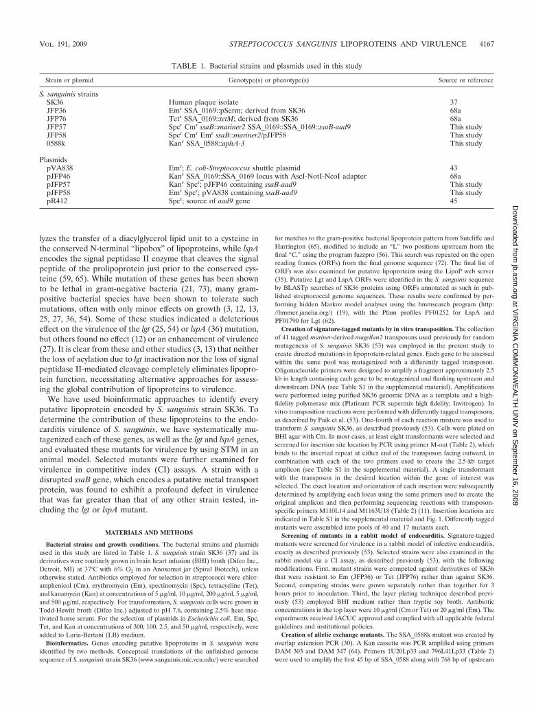
TABLE 1. Bacterial strains and plasmids used in this study
Strain or plasmid Genotype(s) or phenotype(s) Source or reference
S. sanguinis strainsSK36 Human plaque isolate 37JFP36 Emr SSA_0169::pSerm; derived from SK36 68aJFP76 Tetr SSA_0169::tetM; derived from SK36 68aJFP57 Spcr Cmr ssaB::mariner2 SSA_0169::SSA_0169::ssaB-aad9 This studyJFP58 Spcr Cmr Emr ssaB::mariner2/pJFP58 This study0588k Kanr SSA_0588::aphA-3 This study
PlasmidspVA838 Emr; E. coli-Streptococcus shuttle plasmid 43pJFP46 Kanr SSA_0169::SSA_0169 locus with AscI-NotI-NcoI adapter 68apJFP57 Kanr Spcr; pJFP46 containing ssaB-aad9 This studypJFP58 Emr Spcr; pVA838 containing ssaB-aad9 This studypR412 Spcr; source of aad9 gene 45
VOL. 191, 2009 STREPTOCOCCUS SANGUINIS LIPOPROTEINS AND VIRULENCE 4167
at VIR
GIN
IA C
OM
MO
NW
EA
LTH
UN
IV on S
eptember 16, 2009
jb.asm.org
Dow
nloaded from
DNA. Primers1163U55Lp33 and 2678L19Lp33 were used to amplify the last 193bp of the same gene along with 806 bp of downstream DNA. Both internalprimers had 5� tails complementary to the Kan cassette. All products werepurified with MinElute PCR purification columns (Qiagen Inc., Valencia, CA).The final fused PCR product was used to transform SK36, selecting for Kanresistance. A transformant with the expected DNA sequence was selected forfurther study. A similar homologous recombination method was used for allelicexchange mutagenesis of SSA_0941, SSA_0753, and SSA_1667 (X. Ge and P.Xu, unpublished data). The primers employed are listed in Table 2.
Creation of constructs for complementation of ssaB. Overlap extension PCRwas used to create a tripartite fusion of the ssaA promoter region, the ssaB gene,and a Spc cassette. The ssaB gene was PCR amplified from SK36 genomic DNAusing primers SsaBF1 and SsaBR1 (Table 2). The purified product was fusedwith a 71-bp sequence upstream from the ssaA gene using primers AscI-ssaB-prom and SsaBR1 in a second PCR. A Spc cassette was PCR amplified from theplasmid pR412 (45) using the primers Spc-F1 and NcoI-Spc-R1. The purified Spccassette was fused to the ssaA-ssaB fusion sequence using the AscI-SsaB-promand NcoI-Spc primers. This product was ligated into the SSA_0169 gene con-tained in the insertion vector pJFP46 (68a). The plasmid isolated from a trans-formant, called pJFP57, was confirmed to have the expected sequence. Trans-formation of the ssaB (SSA_0260) mutant with pJFP57 created strain JFP57,possessing the ssaA promoter-ssaB gene-Spc cassette fusion within the chromo-somal SSA_0169 locus. This locus was amplified from the chromosome of JFP57with primers SsaB-Spc-F EcoRI and Spc-EcoRI. The purified product was li-gated into the EcoRI site of the shuttle vector pVA838 (43). The purifiedplasmid pJFP58 was confirmed to have the correct sequence and desired insertorientation before introduction into the ssaB mutant to create strain JFP58.
In vitro growth studies. Competitive growth of selected strains was measuredusing in vitro CI assays. JFP36 and a competitor strain were grown in BHImedium and harvested as in the in vivo CI assay described above. Equal volumesof strains at the same optical density (OD) were combined, and 10 �l of themixed culture was diluted into 10 ml of preincubated BHI medium for growth at37°C for 20 h. The inoculum and 20-h cultures were serially diluted and subjectedto layer plating with selective antibiotics, as described earlier.
Sodium dodecyl sulfate-polyacrylamide gel electrophoresis (SDS-PAGE) andWestern blotting. Cultures were grown overnight with the appropriate antibiot-ics, diluted tenfold, and grown for 4 h to exponential phase. Cultures werecentrifuged, washed with phosphate-buffered saline, resuspended in TEP buffer
(10 mM Tris, 1 mM EDTA, 1 mM phenylmethylsulfonyl fluoride; pH 7.5), andtransferred to a tube containing an equal volume of Lysing Matrix B (MPBiomedicals, Solon, OH). Cells were lysed in a bead beater (FastPrep FP120; MPBiomedicals). Protein concentrations of supernatants were determined using theBCA protein assay kit (Pierce, Rockford, IL). Samples were mixed with samplebuffer, heated for 10 min at 70°C, and electrophoresed on a 12% polyacrylamidegel, along with 5 ml of kaleidoscope marker (Bio-Rad), and transferred to anitrocellulose membrane by electroblotting. The membrane was blocked with 5%nonfat dry milk in TBST (10 mM Tris-HCl [pH 7.4], 100 mM NaCl, and 0.1%Tween 20) for 1 h and then incubated with rabbit polyclonal anti-FimA antibodyat a dilution of 1:4,000 for 2 h before being washed and incubated with anti-rabbitimmunoglobulin G horseradish peroxidase-conjugated secondary antibody (Am-ersham Biosciences, Pittsburgh, PA) (1:5,000 dilution) for 1 h at room temper-ature. The polyclonal antiserum was generated by the vaccination of rabbits withrecombinant FimA protein, as described previously (70). Blots were developedusing a chemiluminescence system (ECL Plus; Amersham Biosciences) andexposed to X-ray film (Blue Devil Film, San Diego, CA). Band intensities werequantified using a FluorChem imager.
Labeling of lipoproteins with [3H]palmitate. S. sanguinis lipoproteins werelabeled with [3H]palmitate, essentially as described by McNab and Jenkinson(47), except that BHI medium was used in place of BHI medium containing 0.5%yeast extract. After electrophoresis of lysates, the gel was fixed with 40% meth-anol, 10% glycerol, and 7.5% acetic acid for 1 h and then incubated in 10 volumesof Amplify (GE Healthcare, Piscataway, NJ) for 45 min with gentle shaking. Thefluor-impregnated gel was vacuum dried onto Whatman 3MM filter paper at 60to 70°C and exposed to X-ray film for up to 7 days at �80°C.
Statistics. In vivo and in vitro CI values were calculated as the ratio ofmutant strain colony numbers/virulent strain colony numbers in the outputsample divided by the mutant/virulent strain ratio of the inoculum. Geometricmean CI values were calculated from log-transformed individual CI values.Mutant strains were determined to be significantly more or less competitivethan their virulent competitors by comparing the log-transformed CI valuesto 0 (log1) in paired Student’s t test, with � of 0.05. Log-transformed CI valuesfrom different strains or from the same strain assessed in vitro and in vivowere compared by unpaired t test with Welch correction for unequal standarddeviations, with � of 0.05.
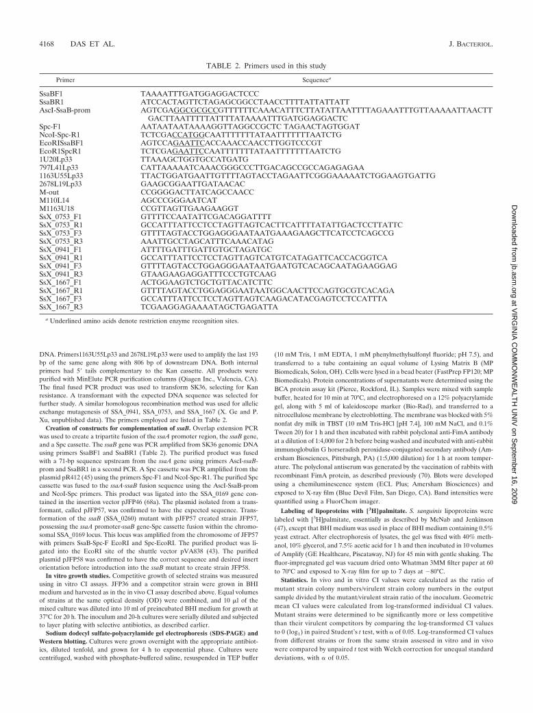
TABLE 2. Primers used in this study
Primer Sequencea
SsaBF1 TAAAATTTGATGGAGGACTCCCSsaBR1 ATCCACTAGTTCTAGAGCGGCCTAACCTTTTATTATTATTAscI-SsaB-prom AGTCGAGGCGCGCCGTTTTTTCAAACATTTCTTATATTAATTTTAGAAATTTGTTAAAAATTAACTT
GACTTAATTTTTATTTTATAAAATTTGATGGAGGACTCSpc-F1 AATAATAATAAAAGGTTAGGCCGCTC TAGAACTAGTGGATNcoI-Spc-R1 TCTCGACCATGGCAATTTTTTTATAATTTTTTTAATCTGEcoRISsaBF1 AGTCCAGAATTCACCAAACCAACCTTGGTCCCGTEcoR1SpcR1 TCTCGAGAATTCCAATTTTTTTATAATTTTTTTAATCTG1U20Lp33 TTAAAGCTGGTGCCATGATG797L41Lp33 CATTAAAAATCAAACGGGCCCTTGACAGCCGCCAGAGAGAA1163U55Lp33 TTACTGGATGAATTGTTTTAGTACCTAGAATTCGGGAAAAATCTGGAAGTGATTG2678L19Lp33 GAAGCGGAATTGATAACACM-out CCGGGGACTTATCAGCCAACCM110L14 AGCCCGGGAATCATM1163U18 CCGTTAGTTGAAGAAGGTSsX_0753_F1 GTTTTCCAATATTCGACAGGATTTTSsX_0753_R1 GCCATTTATTCCTCCTAGTTAGTCACTTCATTTTATATTGACTCCTTATTCSsX_0753_F3 GTTTTAGTACCTGGAGGGAATAATGAAAGAAGCTTCATCCTCAGCCGSsX_0753_R3 AAATTGCCTAGCATTTCAAACATAGSsX_0941_F1 ATTTTGATTTGATTGTGCTAGATGCSsX_0941_R1 GCCATTTATTCCTCCTAGTTAGTCATGTCATAGATTCACCACGGTCASsX_0941_F3 GTTTTAGTACCTGGAGGGAATAATGAATGTCACAGCAATAGAAGGAGSsX_0941_R3 GTAAGAAGAGGATTTCCCTGTCAAGSsX_1667_F1 ACTGGAAGTCTGCTGTTACATCTTCSsX_1667_R1 GTTTTAGTACCTGGAGGGAATAATGGCAACTTCCAGTGCGTCACAGASsX_1667_F3 GCCATTTATTCCTCCTAGTTAGTCAAGACATACGAGTCCTCCATTTASsX_1667_R3 TCGAAGGAGAAAATAGCTGAGATTA
a Underlined amino acids denote restriction enzyme recognition sites.
4168 DAS ET AL. J. BACTERIOL.
at VIR
GIN
IA C
OM
MO
NW
EA
LTH
UN
IV on S
eptember 16, 2009
jb.asm.org
Dow
nloaded from
RESULTS
Identification of putative lipoprotein-related genes in S. san-guinis. The genome sequence of S. sanguinis strain SK36 (72)was examined for putative lipoprotein genes, as described inMaterials and Methods. Use of the LipoP web server (35)resulted in the identification of 59 putative lipoprotein ORFs.This number was greater than that predicted by the samemethod for several other streptococcal genomes, includingStreptococcus mutans UA159, with 29 predicted lipoproteins;Streptococcus pyogenes M1, with 30 predicted lipoproteins; andS. pneumoniae strains D39, R6, and TIGR4, with 43, 39, and 40predicted lipoproteins, respectively. Streptococcus gordonii
strain CH1, however, had a greater number, with 63 predictedlipoproteins. Although the hidden Markov model employed bythe LipoP server (35) was developed for gram-negative bacte-ria, the results of the S. sanguinis analysis were similar to thoseobtained from a regular-expression pattern search created forgram-positive bacterial lipoproteins. The modified patternidentified 52 of the ORFs identified in the LipoP analysis andnone that were not identified by LipoP. The seven ORFs iden-tified solely by the LipoP analysis were examined further. Fiveof these (SSA_0493, -1038, -1315, -1340, and -2143) possessedonly one mismatch to the pattern and had at least one orthologin the NCBI nonredundant protein sequence database (with an
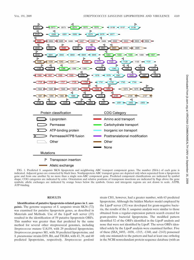
FIG. 1. Predicted S. sanguinis SK36 lipoprotein and neighboring ABC transport component genes. The number (SSA-) of each gene isindicated. Adjacent genes are connected by black lines. Nonlipoprotein ABC transport genes are depicted only when separated from a lipoproteingene and from one another by no more than a single non-ABC component gene. Predicted component classifications are indicated by symbolshape; COG categories are indicated by color. Orientation and relative positions of transposon insertions are indicated by flags above the genesymbols; allelic exchanges are indicated by orange boxes below the symbols. Genes and intergenic regions are not drawn to scale. ATPB,ATP-binding.
VOL. 191, 2009 STREPTOCOCCUS SANGUINIS LIPOPROTEINS AND VIRULENCE 4169
at VIR
GIN
IA C
OM
MO
NW
EA
LTH
UN
IV on S
eptember 16, 2009
jb.asm.org
Dow
nloaded from
E value of �10�5 by BLASTp) that was also classified as alipoprotein by both LipoP and the pattern search. These ORFswere therefore retained. The sixth ORF, SSA_0245, possessedno orthologs in the NCBI database, with an E value of less than10�5. It was paralogous to the ORFs encoded by its flankinggenes, SSA_0244 and SSA_0246, neither of which was recog-nized as encoding a lipoprotein by either algorithm. This ORFwas therefore not retained. The seventh ORF, encoded bySSA_2192, possessed no orthologs in NCBI and had a pre-dicted signal sequence that was 49 amino acids (aa) in lengthcompared to lengths of 17 to 29 aa for the remaining candi-dates. This ORF was also rejected, leaving 57 candidates.
We next considered the possibility that some of the ORFsidentified by both analyses might have matched by chance. Asone test of this possibility, the remaining candidates were usedas query sequences in BLASTp searches of the NCBI database,as described above, to identify candidates that either had noorthologs or had orthologs that were not annotated as lipopro-teins. Six candidates were dropped from the list based on thisanalysis, as follows: SSA_0384, -0850, -1116, -1792, -1860, and-2139.
Finally, we considered the possibility that some potentiallipoproteins may have been missed due to mistakes in thegenomic sequence or to selection of an incorrect start codon inORFs possessing more than one potential start site. A numberof genes were examined that had homology to proteins anno-tated as lipoproteins or as substrate-binding proteins in theNCBI database. SSA_0941 was the first gene in an apparentfive-gene operon putatively encoding an ABC-type phosphatetransport system and had homology to the substrate-bindingcomponent. An in-frame potential TTG start site was discov-ered 32 aa upstream from the annotated ATG start site. If thatsite were used, the ORF would have the same start site as its10 closest relatives in the NCBI protein database and would beidentified as a lipoprotein by both LipoP and the pattern anal-ysis applied above. SSA_0941 was therefore added to the list ofputative lipoproteins, and the modified start site was added tothe GenBank record. This left the final count of candidatelipoproteins at 52. Features of these proteins are listed inTable 3.
The genome was next examined for genes encoding thelipoprotein-processing enzymes lipoprotein signal peptidase(LspA) and prolipoprotein diacylglyceryl transferase (Lgt).Both BLAST and hidden Markov model analyses of the S.sanguinis genome (see Materials and Methods) identified asingle candidate for each ORF, SSA_1069 for LspA andSSA_1546 for Lgt.
Properties of putative S. sanguinis lipoproteins. Predictedsignal sequences of the 52 putative lipoproteins ranged in sizefrom 17 to 26 aa in length (Table 3). The total protein lengthranged from 59 to 661 aa, with the smallest proteins annotatedas hypothetical proteins and the largest as oligopeptide trans-porters. The most common function evident in Table 3 istransport, as could be expected for proteins that are oftencomponents of ABC transport systems. As another indicationof putative function, the clusters of orthologous group (COG)category (67) for each putative lipoprotein was extracted fromthe published genome annotation (72). In addition, wheneverother apparent ABC transport system component genes wereseparated by no more than one gene from a putative lipopro-
tein gene or one another, the COG classifications of thesegenes were also extracted. The result is shown in Fig. 1. Inagreement with expectations, most of the lipoproteins anno-tated as transporters (16 of 21) were adjacent to other appar-ent ABC transport component genes annotated with the sametransport function. Four of the five remaining genes were notlocated in apparent transport operons, and in only one case(SSA_1990) was the putative lipoprotein assigned to a COGcategory different from that of its neighboring genes. Also, inagreement with expectations, the four proteins assigned to thecategory of posttranslational modification were not locatedwithin ABC transport operons. Almost half of the lipoproteins(25 of 52) were not assigned to any COG category.
Examination of the contribution of lipoprotein-relatedgenes to endocarditis virulence by STM. To determine the roleof lipoproteins in virulence for endocarditis, we sought to sys-tematically mutagenize each of the 52 genes identified as en-coding putative lipoproteins. As described in Materials andMethods, each gene was mutagenized by in vitro transpositionwith one of 40 magellan2-derived minitransposons (26), eachcontaining a Cm resistance gene and a unique 40-bp signaturetag (53). Mutants were successfully created for 51 of the 52genes. As described below, the final gene (SSA_0941) wassuccessfully mutagenized by allelic exchange, indicating thatnone of the genes were essential. An insertion was also createdin SSA_0499, encoding a predicted protein with homology topeptide transport lipoproteins in other bacteria and which islocated within an apparent peptide transport operon (Fig. 1).The predicted protein lacks an N-terminal cysteine; however,introduction of a single A to a run of seven A’s in the upstreamnucleotide sequence would have created an ORF predicted tobe a lipoprotein by the LipoP software. Additional sequencingreactions confirmed that the published sequence was correct,indicating that SSA_0499 does not encode a conventional li-poprotein. Nevertheless, the mutant was retained for virulencetesting. Finally, signature-tagged transposons were also intro-duced into the putative lspA and lgt genes SSA_1069 andSSA_1546. As with the lipoprotein mutants, the successfulcreation of these mutants indicated that neither enzyme wasrequired for growth in BHI medium.
We next wanted to assess the ability of each of the insertionmutants to cause endocarditis in the rabbit model (17). Giventhe relative complexity of the model and the large number ofmutants, it was desirable to examine the mutants in poolsrather than individually. The STM technique allows for suchtesting (28) and has been used previously by us with the sametransposons and the same animal model to test pools of ran-domly generated mutants of the same S. sanguinis strain (53).With this technique, the signature tags from the pooled inoc-ulum and from vegetation-derived cells are amplified and la-beled. Any tag that produces a strong signal in a dot blotprobed with the inoculum probe but a weak signal with thevegetation-derived probe indicates a strain with a potentialdefect in virulence (28, 53).
Forty mutants, each with a different signature tag, wereassembled into a pool and screened for virulence reduction bySTM. The results from one study performed with two rabbitsare shown in Fig. 2, top. Strains with mutations in two genes, S.sanguinis adhesin B (ssaB) (SSA_0260) and lspA (SSA_1069),were detected in the inoculum but were essentially undetect-
4170 DAS ET AL. J. BACTERIOL.
at VIR
GIN
IA C
OM
MO
NW
EA
LTH
UN
IV on S
eptember 16, 2009
jb.asm.org
Dow
nloaded from
able in the population recovered �20 h postinoculation fromthe infected heart of either rabbit. SsaB is an LraI familylipoprotein (22, 32), homologs of which have been shown to beimportant for endocarditis virulence in other viridans groupstreptococci (7, 52). The lspA gene (SSA_1069) encodes thelipoprotein-processing enzyme signal peptidase II. Four addi-
tional rabbits from two additional experiments also exhibitedcomplete (ssaB) or partial (lspA) loss of detection for bothmutants (data not shown).
The remaining 14 lipoprotein mutants and the lgt mutantwere assembled into a second pool for STM screening (pool 2)(Fig. 2). Strains with mutations in SSA_0004, SSA_1546, and
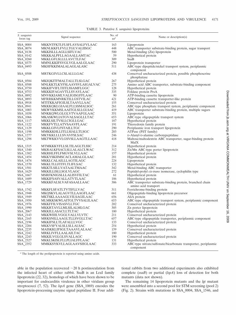
TABLE 3. Putative S. sanguinis lipoproteins
S. sanguinislocus tag Signal sequence No. of
aaa Name or description(s)
SSA_0004 MKKNTFKTLFLSFLAVSALFVLAAC 163 LipoproteinSSA_0074 MKNLKKFLFVLLTGLVALIISGC 448 ABC transporter substrate-binding protein, sugar transportSSA_0138 MKKISLLLAGLLSIFLVAC 500 Metal-binding (Zn) lipoproteinSSA_0142 MKKKALPFLLAGAALLAMTAC 59 Hypothetical proteinSSA_0260 MKKLGFLSLLLLAVCTLFAC 309 SsaBSSA_0375 MMNLKKIFSVGLVGLAALGLAAC 290 Lipoprotein transporterSSA_0493 MKHFKKIMALALAGLALASC 511 ABC-type dipeptide/nickel transport system, periplasmic
componentSSA_0508 MRTKGIVLLCSLALLLGAC 438 Conserved uncharacterized protein, possible phosphoserine
phosphataseSSA_0584 MKIQKITFMALTALLTLSLGAC 267 Hypothetical proteinSSA_0588 MNLKKTLKYFSLAAVSVLAIGALVAC 275 Amino acid ABC transporter, substrate-binding componentSSA_0750 MKKIFVIFLTIITLSSAMFLGGC 128 Hypothetical proteinSSA_0753 MKKKIFAGAVTLLSVAVLAAC 335 Foldase protein PrsASSA_0892 MNVKKIAMLVALIGISGFFLAAC 90 ATP-binding cassette lipoproteinSSA_0893 MFRRKKMNRKTILLGSTVILAC 421 ATP-binding cassette transporter-like proteinSSA_0918 MTITKKAFIGILSLTAAVLLAAC 155 Conserved uncharacterized proteinSSA_0941 MKKKQKLGIAALFLLSSMALSGC 261 ABC-type phosphate transport system, periplasmic componentSSA_1003 MKWYKKMSLAAITGLSLLGLSAC 419 ABC transporter substrate-binding protein, multiple sugarsSSA_1038 MNKKQWLGLGLVTVAAFGLAAC 351 LipoproteinSSA_1066 MKASKWLIATGVALSAGLLLTAC 653 ABC-type oligopeptide transport systemSSA_1117 MKKLSILTVSLLCIGLLGAC 187 Hypothetical proteinSSA_1122 MKKFVTLLATVSAAVFLAAC 191 Thioredoxin family proteinSSA_1129 MKKLGIVLFSTAILLTGC 290 Periplasmic iron transport lipoproteinSSA_1198 MMKKKHLLITLLSIALLTLSGC 203 ATPase (PilT family)SSA_1277 MKYKKLLLLSVAVFSLSAC 246 D-Alanyl-D-alanine carboxypeptidaseSSA_1298 MKTWKKVVLGSVSLLAAGTLLAAC 418 Maltose/maltodextrin ABC transporter, sugar-binding protein
MalXSSA_1315 MTMKKKYFLLSLTILAGLTLSSC 214 Hypothetical proteinSSA_1340 MKRALKFSACLSLLALALCLWAC 312 Zn/Mn ABC-type porter lipoproteinSSA_1382 MKRHLFFLFMGVSLVLLAAC 157 Hypothetical proteinSSA_1454 MKKYSKIIMSCACLAMALGLAAC 281 Hypothetical proteinSSA_1474 MKKLCALAILLLAGTILAGC 224 LipoproteinSSA_1570 MKKLTLLFITFLTLIFLSAC 132 Hypothetical proteinSSA_1581 MKKTLSILLVATAALTMAAC 342 Metal-binding ABC transporterSSA_1629 MKKILLISLLSGLVLAGC 272 Peptidyl-prolyl cis-trans isomerase, cyclophilin typeSSA_1667 MSRFKNIGSLLLALISVFILTAC 61 Hypothetical proteinSSA_1689 MKKIFSASVALLATVTLAAC 186 Hypothetical proteinSSA_1729 MKKKFALSLVAFASAALLAAC 386 ABC transporter substrate-binding protein, branched chain
amino acid transportSSA_1742 MKKFLSFATLTVTIFLLVAC 311 Ferrichrome-binding proteinSSA_1948 MKQSKVLALAGVTLLAAGFLAAC 661 Oligopeptide-binding lipoprotein precursorSSA_1949 MKTSKLAAAAGLVILSAGILAAC 656 AliA proteinSSA_1950 MLMKKSKWLAITGLTVVSALILAAC 653 ABC-type oligopeptide transport system, periplasmic componentSSA_1976 MKKFFILVISASVLLTGC 202 Conserved uncharacterized proteinSSA_1990 MKKRTAVLLMLSILALMLGAC 305 Zn porter lipoproteinSSA_2067 MKKILLASACLLTLTAC 180 Hypothetical proteinSSA_2143 MKKWIIILVGGLVALLVLVTC 211 Conserved uncharacterized proteinSSA_2165 MRKSNILLAAGLTLLSVGLLTAC 657 ABC-type oligopeptide transporter, periplasmic componentSSA_2196 MKKIFSLLTLAFALLLVGC 133 Conserved uncharacterized proteinSSA_2204 MKKVSFYALSLLSLLALSAC 254 Hypothetical proteinSSA_2235 MAISKKLIFSGLTAAAVLALAAC 159 Conserved uncharacterized proteinSSA_2242 MRKLIVFLLAALAILTAC 180 Hypothetical proteinSSA_2243 MKKILVGLGLSVALLAGC 190 Conserved uncharacterized proteinSSA_2327 MKKLSKISLFLLPLIALFFLAAC 131 Hypothetical proteinSSA_2352 MMKKSYKVLLAGLAAVSIIGLAAC 333 ABC-type nitrate/sulfonate/bicarbonate transporter, periplasmic
component
a The length of the prolipoprotein is reported using amino acids.
VOL. 191, 2009 STREPTOCOCCUS SANGUINIS LIPOPROTEINS AND VIRULENCE 4171
at VIR
GIN
IA C
OM
MO
NW
EA
LTH
UN
IV on S
eptember 16, 2009
jb.asm.org
Dow
nloaded from
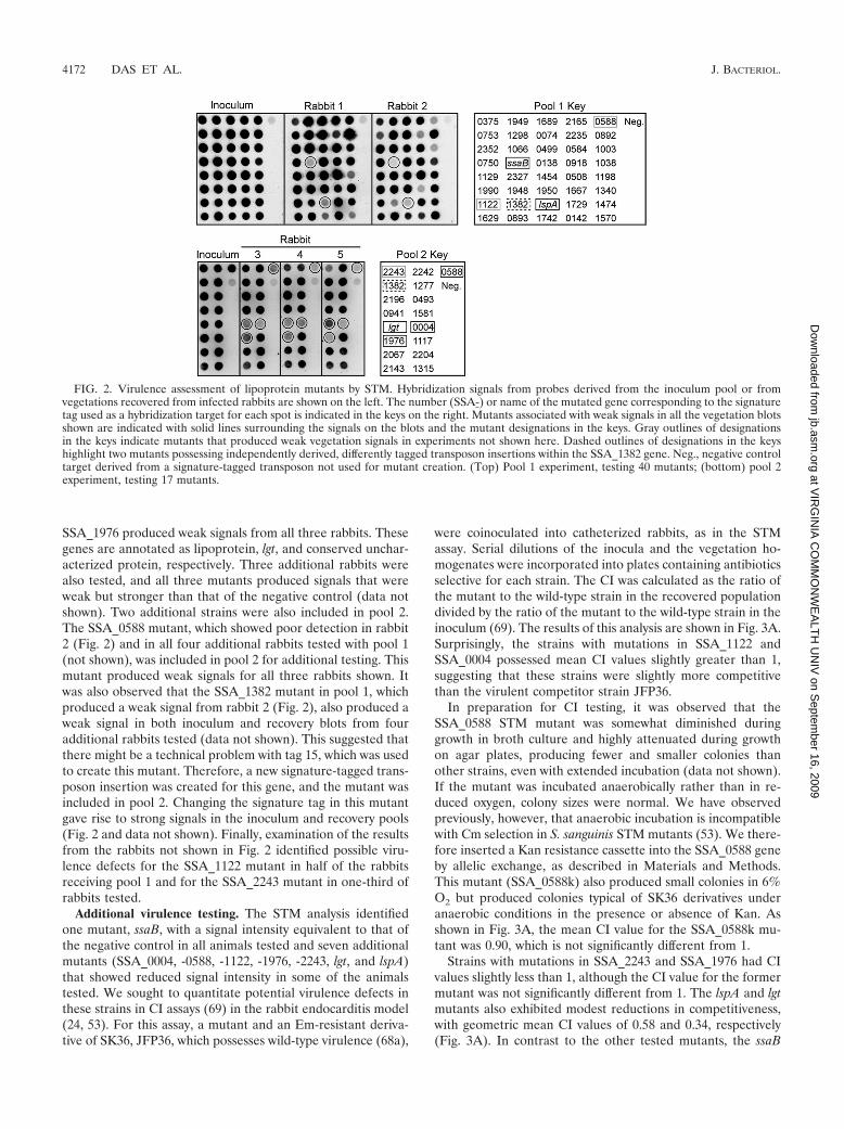
SSA_1976 produced weak signals from all three rabbits. Thesegenes are annotated as lipoprotein, lgt, and conserved unchar-acterized protein, respectively. Three additional rabbits werealso tested, and all three mutants produced signals that wereweak but stronger than that of the negative control (data notshown). Two additional strains were also included in pool 2.The SSA_0588 mutant, which showed poor detection in rabbit2 (Fig. 2) and in all four additional rabbits tested with pool 1(not shown), was included in pool 2 for additional testing. Thismutant produced weak signals for all three rabbits shown. Itwas also observed that the SSA_1382 mutant in pool 1, whichproduced a weak signal from rabbit 2 (Fig. 2), also produced aweak signal in both inoculum and recovery blots from fouradditional rabbits tested (data not shown). This suggested thatthere might be a technical problem with tag 15, which was usedto create this mutant. Therefore, a new signature-tagged trans-poson insertion was created for this gene, and the mutant wasincluded in pool 2. Changing the signature tag in this mutantgave rise to strong signals in the inoculum and recovery pools(Fig. 2 and data not shown). Finally, examination of the resultsfrom the rabbits not shown in Fig. 2 identified possible viru-lence defects for the SSA_1122 mutant in half of the rabbitsreceiving pool 1 and for the SSA_2243 mutant in one-third ofrabbits tested.
Additional virulence testing. The STM analysis identifiedone mutant, ssaB, with a signal intensity equivalent to that ofthe negative control in all animals tested and seven additionalmutants (SSA_0004, -0588, -1122, -1976, -2243, lgt, and lspA)that showed reduced signal intensity in some of the animalstested. We sought to quantitate potential virulence defects inthese strains in CI assays (69) in the rabbit endocarditis model(24, 53). For this assay, a mutant and an Em-resistant deriva-tive of SK36, JFP36, which possesses wild-type virulence (68a),
were coinoculated into catheterized rabbits, as in the STMassay. Serial dilutions of the inocula and the vegetation ho-mogenates were incorporated into plates containing antibioticsselective for each strain. The CI was calculated as the ratio ofthe mutant to the wild-type strain in the recovered populationdivided by the ratio of the mutant to the wild-type strain in theinoculum (69). The results of this analysis are shown in Fig. 3A.Surprisingly, the strains with mutations in SSA_1122 andSSA_0004 possessed mean CI values slightly greater than 1,suggesting that these strains were slightly more competitivethan the virulent competitor strain JFP36.
In preparation for CI testing, it was observed that theSSA_0588 STM mutant was somewhat diminished duringgrowth in broth culture and highly attenuated during growthon agar plates, producing fewer and smaller colonies thanother strains, even with extended incubation (data not shown).If the mutant was incubated anaerobically rather than in re-duced oxygen, colony sizes were normal. We have observedpreviously, however, that anaerobic incubation is incompatiblewith Cm selection in S. sanguinis STM mutants (53). We there-fore inserted a Kan resistance cassette into the SSA_0588 geneby allelic exchange, as described in Materials and Methods.This mutant (SSA_0588k) also produced small colonies in 6%O2 but produced colonies typical of SK36 derivatives underanaerobic conditions in the presence or absence of Kan. Asshown in Fig. 3A, the mean CI value for the SSA_0588k mu-tant was 0.90, which is not significantly different from 1.
Strains with mutations in SSA_2243 and SSA_1976 had CIvalues slightly less than 1, although the CI value for the formermutant was not significantly different from 1. The lspA and lgtmutants also exhibited modest reductions in competitiveness,with geometric mean CI values of 0.58 and 0.34, respectively(Fig. 3A). In contrast to the other tested mutants, the ssaB
FIG. 2. Virulence assessment of lipoprotein mutants by STM. Hybridization signals from probes derived from the inoculum pool or fromvegetations recovered from infected rabbits are shown on the left. The number (SSA-) or name of the mutated gene corresponding to the signaturetag used as a hybridization target for each spot is indicated in the keys on the right. Mutants associated with weak signals in all the vegetation blotsshown are indicated with solid lines surrounding the signals on the blots and the mutant designations in the keys. Gray outlines of designationsin the keys indicate mutants that produced weak vegetation signals in experiments not shown here. Dashed outlines of designations in the keyshighlight two mutants possessing independently derived, differently tagged transposon insertions within the SSA_1382 gene. Neg., negative controltarget derived from a signature-tagged transposon not used for mutant creation. (Top) Pool 1 experiment, testing 40 mutants; (bottom) pool 2experiment, testing 17 mutants.
4172 DAS ET AL. J. BACTERIOL.
at VIR
GIN
IA C
OM
MO
NW
EA
LTH
UN
IV on S
eptember 16, 2009
jb.asm.org
Dow
nloaded from
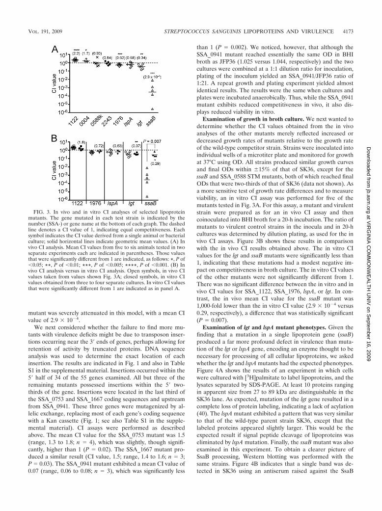
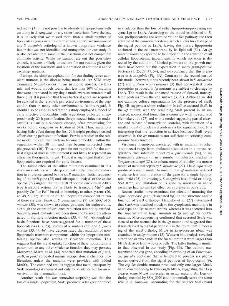
mutant was severely attenuated in this model, with a mean CIvalue of 2.9 � 10�4.
We next considered whether the failure to find more mu-tants with virulence deficits might be due to transposon inser-tions occurring near the 3� ends of genes, perhaps allowing forretention of activity by truncated proteins. DNA sequenceanalysis was used to determine the exact location of eachinsertion. The results are indicated in Fig. 1 and also in TableS1 in the supplemental material. Insertions occurred within the5� half of 34 of the 55 genes examined. All but three of theremaining mutants possessed insertions within the 5� two-thirds of the gene. Insertions were located in the last third ofthe SSA_0753 and SSA_1667 coding sequences and upstreamfrom SSA_0941. These three genes were mutagenized by al-lelic exchange, replacing most of each gene’s coding sequencewith a Kan cassette (Fig. 1; see also Table S1 in the supple-mental material). CI assays were performed as describedabove. The mean CI value for the SSA_0753 mutant was 1.5(range, 1.3 to 1.8; n 4), which was slightly, though signifi-cantly, higher than 1 (P 0.02). The SSA_1667 mutant pro-duced a similar result (CI value, 1.5; range, 1.4 to 1.6; n 3;P 0.03). The SSA_0941 mutant exhibited a mean CI value of0.07 (range, 0.06 to 0.08; n 3), which was significantly less
than 1 (P 0.002). We noticed, however, that although theSSA_0941 mutant reached essentially the same OD in BHIbroth as JFP36 (1.025 versus 1.044, respectively) and the twocultures were combined at a 1:1 dilution ratio for inoculation,plating of the inoculum yielded an SSA_0941/JFP36 ratio of1:21. A repeat growth and plating experiment yielded almostidentical results. The results were the same when cultures andplates were incubated anaerobically. Thus, while the SSA_0941mutant exhibits reduced competitiveness in vivo, it also dis-plays reduced viability in vitro.
Examination of growth in broth culture. We next wanted todetermine whether the CI values obtained from the in vivoanalyses of the other mutants merely reflected increased ordecreased growth rates of mutants relative to the growth rateof the wild-type competitor strain. Strains were inoculated intoindividual wells of a microtiter plate and monitored for growthat 37°C using OD. All strains produced similar growth curvesand final ODs within 15% of that of SK36, except for thessaB and SSA_0588 STM mutants, both of which reached finalODs that were two-thirds of that of SK36 (data not shown). Asa more sensitive test of growth rate differences and to measureviability, an in vitro CI assay was performed for five of themutants tested in Fig. 3A. For this assay, a mutant and virulentstrain were prepared as for an in vivo CI assay and thencoinoculated into BHI broth for a 20-h incubation. The ratio ofmutants to virulent control strains in the inocula and in 20-hcultures was determined by dilution plating, as used for the invivo CI assays. Figure 3B shows these results in comparisonwith the in vivo CI results obtained above. The in vitro CIvalues for the lgt and ssaB mutants were significantly less than1, indicating that these mutations had a modest negative im-pact on competitiveness in broth culture. The in vitro CI valuesof the other mutants were not significantly different from 1.There was no significant difference between the in vitro and invivo CI values for SSA_1122, SSA_1976, lspA, or lgt. In con-trast, the in vivo mean CI value for the ssaB mutant was1,000-fold lower than the in vitro CI value (2.9 � 10�4 versus0.29, respectively), a difference that was statistically significant(P 0.007).
Examination of lgt and lspA mutant phenotypes. Given thefinding that a mutation in a single lipoprotein gene (ssaB)produced a far more profound defect in virulence than muta-tion of the lgt or lspA gene, encoding an enzyme thought to benecessary for processing of all cellular lipoproteins, we askedwhether the lgt and lspA mutants had the expected phenotypes.Figure 4A shows the results of an experiment in which cellswere cultured with [3H]palmitate to label lipoproteins, and thelysates separated by SDS-PAGE. At least 10 proteins rangingin apparent size from 27 to 89 kDa are distinguishable in theSK36 lane. As expected, mutation of the lgt gene resulted in acomplete loss of protein labeling, indicating a lack of acylation(40). The lspA mutant exhibited a pattern that was very similarto that of the wild-type parent strain SK36, except that thelabeled proteins appeared slightly larger. This would be theexpected result if signal peptide cleavage of lipoproteins waseliminated by lspA mutation. Finally, the ssaB mutant was alsoexamined in this experiment. To obtain a clearer picture ofSsaB processing, Western blotting was performed with thesame strains. Figure 4B indicates that a single band was de-tected in SK36 using an antiserum raised against the SsaB
FIG. 3. In vivo and in vitro CI analyses of selected lipoproteinmutants. The gene mutated in each test strain is indicated by thenumber (SSA-) or gene name at the bottom of each graph. The dashedline denotes a CI value of 1, indicating equal competitiveness. Eachsymbol indicates the CI value derived from a single animal or bacterialculture; solid horizontal lines indicate geometric mean values. (A) Invivo CI analysis. Mean CI values from five to six animals tested in twoseparate experiments each are indicated in parentheses. Those valuesthat were significantly different from 1 are indicated, as follows: *, P of�0.05; **, P of �0.01; ***, P of �0.005; ****, P of �0.001. (B) Invivo CI analysis versus in vitro CI analysis. Open symbols, in vivo CIvalues taken from values shown Fig. 3A; closed symbols, in vitro CIvalues obtained from three to four separate cultures. In vitro CI valuesthat were significantly different from 1 are indicated as in panel A.
VOL. 191, 2009 STREPTOCOCCUS SANGUINIS LIPOPROTEINS AND VIRULENCE 4173
at VIR
GIN
IA C
OM
MO
NW
EA
LTH
UN
IV on S
eptember 16, 2009
jb.asm.org
Dow
nloaded from
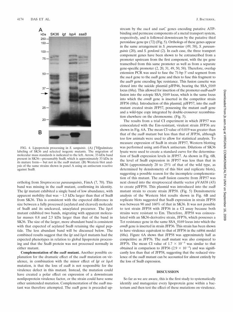
ortholog from Streptococcus parasanguinis, FimA (7, 70). Thisband was missing in the ssaB mutant, confirming its identity.The lgt mutant exhibited a single band of low abundance, withapparent mobility that was �1.5 kDa larger than that of SsaBfrom SK36. This is consistent with the expected difference insize between a fully processed (acylated and cleaved) moleculeof SsaB and its uncleaved, unacylated precursor. The lspAmutant exhibited two bands, migrating with apparent molecu-lar masses 0.8 and 2.5 kDa larger than that of the band inSK36. The size of the larger, more abundant band is consistentwith that expected of acylated SsaB retaining the signal pep-tide. The less abundant band will be discussed below. Thecombined results suggest that the lgt and lspA mutants had theexpected phenotypes in relation to global lipoprotein process-ing and that the SsaB protein was not processed normally ineither mutant.
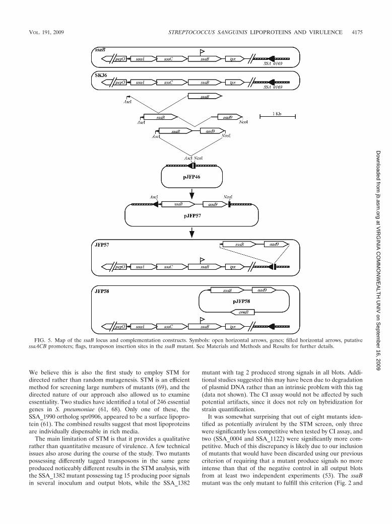
Complementation of the ssaB mutant. Another possible ex-planation for the dramatic effect of the ssaB mutation on vir-ulence, in combination with the minor effect of lgt or lspAmutation, is that the loss of ssaB is not responsible for thevirulence defect in this mutant. Instead, the mutation couldhave created a polar effect on expression of a downstreamnonlipoprotein virulence factor, or the strain could have someother unintended mutation. Complementation of the ssaB mu-tant was therefore attempted. The ssaB gene is preceded up-
stream by the ssaA and ssaC genes encoding putative ATP-binding and permease components of a metal transport system,respectively, and is followed downstream by the putative thiolperoxidase gene tpx (72) (Fig. 5). Orthologs of these genes appearin the same arrangement in S. pneumoniae (49, 58), S. parasan-guinis (20), and S. gordonii (2). In each case, the three transportcomponent genes have been shown to be cotranscribed from apromoter upstream from the first component, with the tpx genetranscribed from this same promoter as well as from a separategene-specific promoter (2, 20, 31, 49, 50, 58). Therefore, overlapextension PCR was used to fuse the 71-bp 5�-end segment fromthe ssaA gene to the ssaB gene and then to fuse this fragment tothe aad9 gene encoding Spc resistance. This fusion cassette wascloned into the suicide plasmid pJFP46, bearing the SSA_0169locus (68a). This allowed for insertion of the promoter-ssaB-aad9fusion into the ectopic SSA_0169 locus, which is the same locusinto which the ermB gene is inserted in the competitor strainJFP36 (68a). Introduction of this plasmid, pJFP57, into the ssaBmutant created strain JFP57, possessing the mutant ssaB geneand a wild-type copy integrated by double-crossover recombina-tion elsewhere on the chromosome. (Fig. 5).
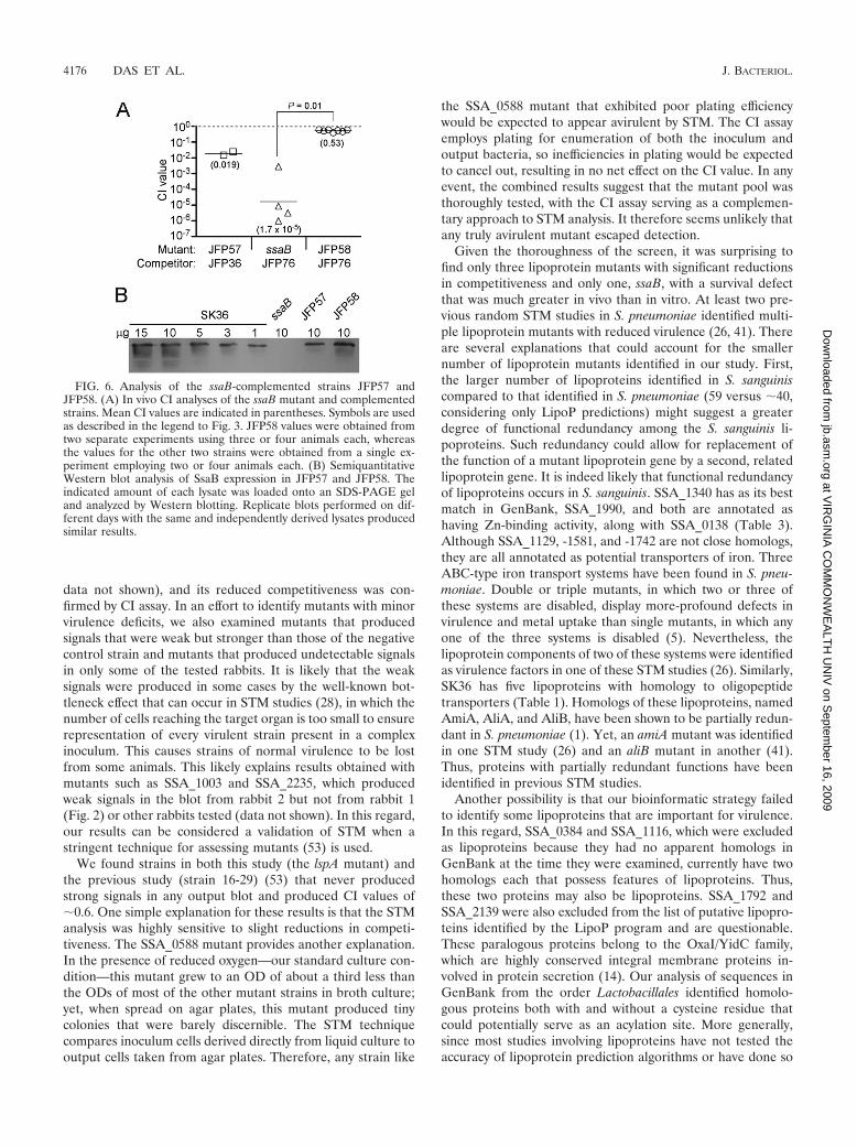
The results from a trial CI experiment in which JFP57 wascoinoculated with the Em-resistant, virulent strain JFP36 areshown in Fig. 6A. The mean CI value of 0.019 was greater thanthat of the ssaB mutant but less than that of JFP36, althoughtoo few animals were used to allow for statistical analysis. Tomeasure expression of SsaB in strain JFP57, Western blottingwas performed using anti-FimA antiserum. Dilutions of SK36lysate were used to create a standard curve for the determina-tion of SsaB expression levels in JFP57. As shown in Fig. 6B,the level of SsaB expression in JFP57 was less than that inSK36 (approximately 20 to 25% of that of the wild type, asdetermined by densitometry of this blot and replicate blots),suggesting a possible reason for the incomplete complementa-tion of this mutant. The ssaB fusion cassette from JFP57 wasnext cloned into the streptococcal shuttle vector pVA838 (43)to create pJFP58. This plasmid was introduced into the ssaBmutant strain to create strain JFP58. (Fig. 5) Densitometricanalysis of the Western blot results shown in Fig. 6B andreplicate blots suggested that SsaB expression in strain JFP58was between 90 and 160% of that in SK36. It was not possibleto test strain JFP58 with JFP36 in a CI assay because bothstrains were resistant to Em. Therefore, JFP58 was coinocu-lated with an SK36-derivative strain, JFP76, which possesses aTet resistance gene in the same SSA_0169 locus into which theermB gene is inserted in strain JFP36. This strain has been shownto have virulence equivalent to that of JFP36 in the rabbit model(68a). Figure 6A shows that JFP58 was approximately half ascompetitive as JFP76. The ssaB mutant was also compared toJFP76. The mean CI value of 1.7 � 10�5 was similar to thatobtained in comparison to JFP36 (2.9 � 10�4) and was signifi-cantly less than that of JFP58, suggesting that the reduced viru-lence of the ssaB mutant can be accounted for almost entirely bythe loss of SsaB expression.
DISCUSSION
So far as we are aware, this is the first study to systematicallyidentify and mutagenize every lipoprotein gene within a bac-terium and then test the effect of these mutations on virulence.
FIG. 4. Lipoprotein processing in S. sanguinis. (A) [3H]palmitatelabeling of SK36 and selected isogenic mutants. The migration ofmolecular mass standards is indicated to the left. Arrow, 33-kDa bandpresent in SK36—presumably SsaB, which is approximately 33 kDa inits mature form— but not in the ssaB mutant. (B) Western blot anal-ysis of the same strains shown in panel A using an antiserum reactiveagainst SsaB.
4174 DAS ET AL. J. BACTERIOL.
at VIR
GIN
IA C
OM
MO
NW
EA
LTH
UN
IV on S
eptember 16, 2009
jb.asm.org
Dow
nloaded from
We believe this is also the first study to employ STM fordirected rather than random mutagenesis. STM is an efficientmethod for screening large numbers of mutants (69), and thedirected nature of our approach also allowed us to examineessentiality. Two studies have identified a total of 246 essentialgenes in S. pneumoniae (61, 68). Only one of these, theSSA_1990 ortholog spr0906, appeared to be a surface lipopro-tein (61). The combined results suggest that most lipoproteinsare individually dispensable in rich media.
The main limitation of STM is that it provides a qualitativerather than quantitative measure of virulence. A few technicalissues also arose during the course of the study. Two mutantspossessing differently tagged transposons in the same geneproduced noticeably different results in the STM analysis, withthe SSA_1382 mutant possessing tag 15 producing poor signalsin several inoculum and output blots, while the SSA_1382
mutant with tag 2 produced strong signals in all blots. Addi-tional studies suggested this may have been due to degradationof plasmid DNA rather than an intrinsic problem with this tag(data not shown). The CI assay would not be affected by suchpotential artifacts, since it does not rely on hybridization forstrain quantification.
It was somewhat surprising that out of eight mutants iden-tified as potentially avirulent by the STM screen, only threewere significantly less competitive when tested by CI assay, andtwo (SSA_0004 and SSA_1122) were significantly more com-petitive. Much of this discrepancy is likely due to our inclusionof mutants that would have been discarded using our previouscriterion of requiring that a mutant produce signals no moreintense than that of the negative control in all output blotsfrom at least two independent experiments (53). The ssaBmutant was the only mutant to fulfill this criterion (Fig. 2 and
FIG. 5. Map of the ssaB locus and complementation constructs. Symbols: open horizontal arrows, genes; filled horizontal arrows, putativessaACB promoters; flags, transposon insertion sites in the ssaB mutant. See Materials and Methods and Results for further details.
VOL. 191, 2009 STREPTOCOCCUS SANGUINIS LIPOPROTEINS AND VIRULENCE 4175
at VIR
GIN
IA C
OM
MO
NW
EA
LTH
UN
IV on S
eptember 16, 2009
jb.asm.org
Dow
nloaded from
data not shown), and its reduced competitiveness was con-firmed by CI assay. In an effort to identify mutants with minorvirulence deficits, we also examined mutants that producedsignals that were weak but stronger than those of the negativecontrol strain and mutants that produced undetectable signalsin only some of the tested rabbits. It is likely that the weaksignals were produced in some cases by the well-known bot-tleneck effect that can occur in STM studies (28), in which thenumber of cells reaching the target organ is too small to ensurerepresentation of every virulent strain present in a complexinoculum. This causes strains of normal virulence to be lostfrom some animals. This likely explains results obtained withmutants such as SSA_1003 and SSA_2235, which producedweak signals in the blot from rabbit 2 but not from rabbit 1(Fig. 2) or other rabbits tested (data not shown). In this regard,our results can be considered a validation of STM when astringent technique for assessing mutants (53) is used.
We found strains in both this study (the lspA mutant) andthe previous study (strain 16-29) (53) that never producedstrong signals in any output blot and produced CI values of�0.6. One simple explanation for these results is that the STManalysis was highly sensitive to slight reductions in competi-tiveness. The SSA_0588 mutant provides another explanation.In the presence of reduced oxygen—our standard culture con-dition—this mutant grew to an OD of about a third less thanthe ODs of most of the other mutant strains in broth culture;yet, when spread on agar plates, this mutant produced tinycolonies that were barely discernible. The STM techniquecompares inoculum cells derived directly from liquid culture tooutput cells taken from agar plates. Therefore, any strain like
the SSA_0588 mutant that exhibited poor plating efficiencywould be expected to appear avirulent by STM. The CI assayemploys plating for enumeration of both the inoculum andoutput bacteria, so inefficiencies in plating would be expectedto cancel out, resulting in no net effect on the CI value. In anyevent, the combined results suggest that the mutant pool wasthoroughly tested, with the CI assay serving as a complemen-tary approach to STM analysis. It therefore seems unlikely thatany truly avirulent mutant escaped detection.
Given the thoroughness of the screen, it was surprising tofind only three lipoprotein mutants with significant reductionsin competitiveness and only one, ssaB, with a survival defectthat was much greater in vivo than in vitro. At least two pre-vious random STM studies in S. pneumoniae identified multi-ple lipoprotein mutants with reduced virulence (26, 41). Thereare several explanations that could account for the smallernumber of lipoprotein mutants identified in our study. First,the larger number of lipoproteins identified in S. sanguiniscompared to that identified in S. pneumoniae (59 versus �40,considering only LipoP predictions) might suggest a greaterdegree of functional redundancy among the S. sanguinis li-poproteins. Such redundancy could allow for replacement ofthe function of a mutant lipoprotein gene by a second, relatedlipoprotein gene. It is indeed likely that functional redundancyof lipoproteins occurs in S. sanguinis. SSA_1340 has as its bestmatch in GenBank, SSA_1990, and both are annotated ashaving Zn-binding activity, along with SSA_0138 (Table 3).Although SSA_1129, -1581, and -1742 are not close homologs,they are all annotated as potential transporters of iron. ThreeABC-type iron transport systems have been found in S. pneu-moniae. Double or triple mutants, in which two or three ofthese systems are disabled, display more-profound defects invirulence and metal uptake than single mutants, in which anyone of the three systems is disabled (5). Nevertheless, thelipoprotein components of two of these systems were identifiedas virulence factors in one of these STM studies (26). Similarly,SK36 has five lipoproteins with homology to oligopeptidetransporters (Table 1). Homologs of these lipoproteins, namedAmiA, AliA, and AliB, have been shown to be partially redun-dant in S. pneumoniae (1). Yet, an amiA mutant was identifiedin one STM study (26) and an aliB mutant in another (41).Thus, proteins with partially redundant functions have beenidentified in previous STM studies.
Another possibility is that our bioinformatic strategy failedto identify some lipoproteins that are important for virulence.In this regard, SSA_0384 and SSA_1116, which were excludedas lipoproteins because they had no apparent homologs inGenBank at the time they were examined, currently have twohomologs each that possess features of lipoproteins. Thus,these two proteins may also be lipoproteins. SSA_1792 andSSA_2139 were also excluded from the list of putative lipopro-teins identified by the LipoP program and are questionable.These paralogous proteins belong to the OxaI/YidC family,which are highly conserved integral membrane proteins in-volved in protein secretion (14). Our analysis of sequences inGenBank from the order Lactobacillales identified homolo-gous proteins both with and without a cysteine residue thatcould potentially serve as an acylation site. More generally,since most studies involving lipoproteins have not tested theaccuracy of lipoprotein prediction algorithms or have done so
FIG. 6. Analysis of the ssaB-complemented strains JFP57 andJFP58. (A) In vivo CI analyses of the ssaB mutant and complementedstrains. Mean CI values are indicated in parentheses. Symbols are usedas described in the legend to Fig. 3. JFP58 values were obtained fromtwo separate experiments using three or four animals each, whereasthe values for the other two strains were obtained from a single ex-periment employing two or four animals each. (B) SemiquantitativeWestern blot analysis of SsaB expression in JFP57 and JFP58. Theindicated amount of each lysate was loaded onto an SDS-PAGE geland analyzed by Western blotting. Replicate blots performed on dif-ferent days with the same and independently derived lysates producedsimilar results.
4176 DAS ET AL. J. BACTERIOL.
at VIR
GIN
IA C
OM
MO
NW
EA
LTH
UN
IV on S
eptember 16, 2009
jb.asm.org
Dow
nloaded from
indirectly (3), it is not possible to identify all lipoproteins withcertainty in S. sanguinis or any other bacterium. Nevertheless,it is unlikely that we missed more than a small number oflipoprotein genes in our mutant screen, and we are unaware ofany S. sanguinis ortholog of a known lipoprotein virulencefactor that was not identified and mutagenized in our study. Itis also possible that some of the insertions did not completelyeliminate activity. While we cannot rule out this possibilityentirely, it seems unlikely to account for our results, given thelocations of the insertions and our creation of additional allelicexchange mutants.
Perhaps the simplest explanation for our finding fewer avir-ulent mutants is the disease being modeled. An STM studyexamining Staphylococcus aureus in mouse abscess, bactere-mia, and wound models found that less than 10% of mutantsthat were attenuated in any single model were attenuated in allthree (10). It is possible that fewer bacterial genes are requiredfor survival in the relatively protected environment of the veg-etation than in many other environments. In this regard, itshould also be emphasized that this study employed a model ofearly infective endocarditis, with vegetations collected at ap-proximately 20 h postinfection. Streptococcal infective endo-carditis is usually a subacute disease, often progressing forweeks before diagnosis or treatment (48). Thus, mutationshaving little effect during the first 20 h might produce markedeffects during persistent infections. Previous studies in the rab-bit model indicate that bacteria become embedded within thevegetation within 30 min and then become protected fromphagocytosis (16). Thus, any protein not required for the ear-liest stages of disease development is not likely to represent anattractive therapeutic target. Thus, it is significant that so fewlipoproteins are required for early disease.
The minor effect of the other mutations examined in thisstudy on virulence is in sharp contrast to the dramatic reduc-tion in virulence caused by the ssaB mutation. Initial sequenc-ing of the ssaB gene (22) and subsequent analysis of the entireoperon (72) suggest that the ssaACB operon encodes an ABC-type transport system that is likely to transport Mn2� andpossibly Zn2�or Fe2� based on homology to other systems (20,46, 49, 50, 52). Mutation of the lipoprotein component in twoof these systems, FimA of S. parasanguinis (7) and SloC of S.mutans (38), was shown to reduce virulence for endocarditis,although the extent of virulence reduction was not quantified.Similarly, psaA mutants have been shown to be severely atten-uated in multiple infection models (33, 44, 46). Although ad-hesin functions have been suggested for a number of theselipoproteins (4, 7, 23), studies of S. mutans (52) and S. pneu-moniae (33, 44, 46) have demonstrated that mutation of non-lipoprotein transport components within the lipoprotein-con-taining operon also results in virulence reduction. Thissuggests that the metal uptake function of these lipoproteins isparamount to any other virulence function they may possess.Moreover, Marra et al. (44) reported that mutation of psaA,psaB, or psaC abrogated murine intraperitoneal chamber pro-liferation, unless the mutants were provided with addedMnSO4. The combined results suggest that metal transport bySsaB homologs is required not only for virulence but for meresurvival in the mammalian host.
Another result that was somewhat surprising was that theloss of a single lipoprotein, SsaB, produced a far greater defect
in virulence than the loss of either lipoprotein-processing en-zyme Lgt or LspA. According to the model established in E.coli, prelipoproteins are secreted via the Sec pathway and thenacylated at the conserved cysteine, which allows for cleavage ofthe signal peptide by LspA, leaving the mature lipoproteinanchored in the cell membrane by its lipid tail (59). An lgtmutant would be expected to be deficient in the acylation of allcellular lipoproteins. Experiments in which acylation is de-tected by the addition of labeled palmitate to the growth me-dium have borne out this expectation in many gram-positivebacteria (3, 25, 27, 47, 54), and we confirmed that this is alsotrue in S. sanguinis (Fig. 4A). Contrary to the second part ofthis model, however, it has recently been shown in S. agalactiae(27) and Listeria monocytogenes (3) that nonacylated proli-poproteins produced in lgt mutants are subject to cleavage byLspA. The result is the enhanced release of cleaved, nonacy-lated proteins from the cell surface (3, 27). Although we didnot examine culture supernatants for the presence of SsaB,Fig. 4B suggests a sharp reduction in cell-associated SsaB inthe lgt mutant, with the remaining SsaB present in an un-cleaved, nonacylated form. This is consistent with the results ofHenneke et al. (27) and with a model suggesting partial cleav-age and release of nonacylated proteins, with retention of asmall amount of uncleaved protein via the signal peptide. It isinteresting that the reduction in surface-localized SsaB levelsobserved in the lgt mutant is not sufficient to seriously com-promise SsaB function.
Virulence phenotypes associated with lgt mutation in otherstreptococci range from profound attenuation in a mouse re-spiratory tract infection model by S. pneumoniae (54), to in-termediate attenuation in a number of infection studies byStreptococcus equi (25), to enhancement of lethality in a mousemodel of neonatal sepsis by S. agalactiae (27). The S. equi studyproduced a result similar to ours, in that lgt mutation reducedvirulence less than mutation of the gene for a single lipopro-tein, PrtM (25). Interestingly, S. sanguinis has a PrtM ortholog,SSA_0753, and mutation of its gene by insertion or allelicexchange had no marked effect on virulence in our study.
Recent studies have examined the effects of mutating thesignal peptidase gene (designated lsp) on the structure and/orfunction of SsaB orthologs. Henneke et al. (27) determinedthat ScaA was localized mostly to the cytoplasmic membrane inwild-type and lsp mutant strains, whereas it was secreted intothe supernatant in large amounts in lgt and lgt lsp doublemutants. Microsequencing confirmed that secreted ScaA wascleaved at the normal site in the lgt mutant and suggested thatit was cleaved by signal peptidase I in the lsp mutant. Process-ing of the SsaB ortholog MtuA in Streptococcus uberis wasexamined in an lsp mutant (13). Western blot analysis revealedeither one or two bands in the lsp mutant that were larger thanMtuA derived from wild-type cells. The latter finding is similarto that observed in our study (Fig. 4B). The authors mu-tagenized the eep gene, encoding an ortholog of an Enterococ-cus faecalis peptidase that is believed to process sex phero-mones derived from the signal peptides of lipoproteins (9).The eep lsp double mutant produced only one larger MtuAband, corresponding to full-length MtuA, suggesting that Eepcleaves some MtuA molecules in an lsp mutant. An Eep or-tholog encoded by SSA_2070 could potentially serve the samerole in S. sanguinis, accounting for the smaller SsaB band
VOL. 191, 2009 STREPTOCOCCUS SANGUINIS LIPOPROTEINS AND VIRULENCE 4177
at VIR
GIN
IA C
OM
MO
NW
EA
LTH
UN
IV on S
eptember 16, 2009
jb.asm.org
Dow
nloaded from
observed in the lspA mutant (Fig. 4B). Finally, Khandavilli etal. (36) described the effect of the lsp mutation on PsaA struc-ture and function and on virulence in S. pneumoniae. The lspmutant possessed phenotypes typical of a psaA mutant andexhibited reduced competitiveness in disease models. Al-though individual lipoprotein mutants were not tested, thereduced competitiveness phenotype appeared less severe thanthat expected for psaA mutants (44, 46).
The overall picture that emerges from the lgt and lsp mutantanalyses described above in combination with our own is thatthere is far less of an effect on growth or virulence than wouldbe expected if all lipoprotein function were lost. This suggeststhat many lipoproteins retain at least partial activity in theabsence of acylation or Lsp-mediated cleavage. This couldexplain the presence of proteins such as SSA_0499 in S. san-guinis, which does not possess an N-terminal cysteine for acy-lation but is encoded within an apparent ABC transportoperon and has extensive homology to the substrate-bindingcomponent of ABC transport systems, including SSA_0493.
In conclusion, our systematic analysis of lipoproteins in S.sanguinis has identified a single lipoprotein, SsaB, that is crit-ical for early endocarditis virulence. Although our data suggestthat interference with acylation or signal peptide cleavage haslittle effect on its function, SsaB appears to be a singularlypromising target for new inhibitors or for its renewed exami-nation as a vaccine candidate (39, 70).
ACKNOWLEDGMENTS
We thank Nicai Zollar for technical assistance and Lauren SentyTurner for useful discussions.
This work was supported by National Institutes of Health grantsR01AI47841 and K02AI05490 (T.K.) and R01DE18138 (P.X.).
REFERENCES
1. Alloing, G., P. de Philip, and J. P. Claverys. 1994. Three highly homologousmembrane-bound lipoproteins participate in oligopeptide transport by theAmi system of the gram-positive Streptococcus pneumoniae. J. Mol. Biol.241:44–58.
2. Andersen, R. N., R. D. Lunsford, and P. E. Kolenbrander. 1997. Determi-nation of the transcript size and start site of the putative sca operon ofStreptococcus gordonii ATCC 51656 (formerly strain PK488), p. 657–660. InT. Horaud, A. Bouvet, R. Leclercq, H. de Montclos, and M. Sicard (ed.),Streptococci and the host, vol. 418. Plenum Press, New York, NY.
3. Baumgartner, M., U. Karst, B. Gerstel, M. Loessner, J. Wehland, and L.Jansch. 2007. Inactivation of Lgt allows systematic characterization of li-poproteins from Listeria monocytogenes. J. Bacteriol. 189:313–324.
4. Berry, A. M., and J. C. Paton. 1996. Sequence heterogeneity of PsaA, a37-kilodalton putative adhesin essential for virulence of Streptococcus pneu-moniae. Infect. Immun. 64:5255–5262.
5. Brown, J. S., S. M. Gilliland, J. Ruiz-Albert, and D. W. Holden. 2002.Characterization of Pit, a Streptococcus pneumoniae iron uptake ABC trans-porter. Infect. Immun. 70:4389–4398.
6. Brown, J. S., A. D. Ogunniyi, M. C. Woodrow, D. W. Holden, and J. C. Paton.2001. Immunization with components of two iron uptake ABC transportersprotects mice against systemic Streptococcus pneumoniae infection. Infect.Immun. 69:6702–6706.
7. Burnette-Curley, D., V. Wells, H. Viscount, C. L. Munro, J. C. Fenno, P.Fives-Taylor, and F. L. Macrina. 1995. FimA, a major virulence factorassociated with Streptococcus parasanguis endocarditis. Infect. Immun. 63:4669–4674.
8. Carlsson, J. 1965. Zooglea-forming streptococci, resembling Streptococcussanguis, isolated from dental plaque in man. Odontol. Revy 16:348–358.
9. Chandler, J. R., and G. M. Dunny. 2008. Characterization of the sequencespecificity determinants required for processing and control of sex phero-mone by the intramembrane protease Eep and the plasmid-encoded proteinPrgY. J. Bacteriol. 190:1172–1183.
10. Coulter, S. N., W. R. Schwan, E. Y. Ng, M. H. Langhorne, H. D. Ritchie, S.Westbrock-Wadman, W. O. Hufnagle, K. R. Folger, A. S. Bayer, and C. K.Stover. 1998. Staphylococcus aureus genetic loci impacting growth and sur-vival in multiple infection environments. Mol. Microbiol. 30:393–404.
11. Das, S., J. C. Noe, S. Paik, and T. Kitten. 2005. An improved arbitraryprimed PCR method for rapid characterization of transposon insertion sites.J. Microbiol. Methods 63:89–94.
12. de Greeff, A., A. Hamilton, I. C. Sutcliffe, H. Buys, L. van Alphen, and H. E.Smith. 2003. Lipoprotein signal peptidase of Streptococcus suis serotype 2.Microbiology 149:1399–1407.
13. Denham, E. L., P. N. Ward, and J. A. Leigh. 2008. Lipoprotein signalpeptides are processed by Lsp and Eep of Streptococcus uberis. J. Bacteriol.190:4641–4647.
14. Dong, Y., S. R. Palmer, A. Hasona, S. Nagamori, H. R. Kaback, R. E. Dalbey,and L. J. Brady. 2008. Functional overlap but lack of complete cross-comple-mentation of Streptococcus mutans and Escherichia coli YidC orthologs. J.Bacteriol. 190:2458–2469.
15. Douglas, C. W., J. Heath, K. K. Hampton, and F. E. Preston. 1993. Identityof viridans streptococci isolated from cases of infective endocarditis. J. Med.Microbiol. 39:179–182.
16. Durack, D. T. 1975. Experimental bacterial endocarditis. IV. Structure andevolution of very early lesions. J. Pathol. 115:81–89.
17. Durack, D. T., P. B. Beeson, and R. G. Petersdorf. 1973. Experimentalbacterial endocarditis. 3. Production and progress of the disease in rabbits.Br. J. Exp. Pathol. 54:142–151.
18. Dyson, C., R. A. Barnes, and G. A. J. Harrison. 1999. Infective endocarditis:an epidemiological review of 128 episodes. J. Infect. 38:87–93.
19. Eddy, S. R. 1998. Profile hidden Markov models. Bioinformatics 14:755–763.20. Fenno, J. C., A. Shaikh, G. Spatafora, and P. Fives-Taylor. 1995. The fimA
locus of Streptococcus parasanguis encodes an ATP-binding membrane trans-port system. Mol. Microbiol. 15:849–863.
21. Gan, K., S. D. Gupta, K. Sankaran, M. B. Schmid, and H. C. Wu. 1993.Isolation and characterization of a temperature-sensitive mutant of Salmo-nella typhimurium defective in prolipoprotein modification. J. Biol. Chem.268:16544–16550.
22. Ganeshkumar, N., P. M. Hannam, P. E. Kolenbrander, and B. C. McBride.1991. Nucleotide sequence of a gene coding for a saliva-binding protein(SsaB) from Streptococcus sanguis 12 and possible role of the protein incoaggregation with actinomyces. Infect. Immun. 59:1093–1099.
23. Ganeshkumar, N., M. Song, and B. C. McBride. 1988. Cloning of a Strepto-coccus sanguis adhesin which mediates binding to saliva-coated hydroxyap-atite. Infect. Immun. 56:1150–1157.
24. Ge, X., T. Kitten, Z. Chen, S. P. Lee, C. L. Munro, and P. Xu. 2008.Identification of Streptococcus sanguinis genes required for biofilm formationand examination of their role in endocarditis virulence. Infect. Immun.76:2551–2559.
25. Hamilton, A., C. Robinson, I. C. Sutcliffe, J. Slater, D. J. Maskell, N.Davis-Poynter, K. Smith, A. Waller, and D. J. Harrington. 2006. Mutation ofthe maturase lipoprotein attenuates the virulence of Streptococcus equi to agreater extent than does loss of general lipoprotein lipidation. Infect. Im-mun. 74:6907–6919.
26. Hava, D. L., and A. Camilli. 2002. Large-scale identification of serotype 4Streptococcus pneumoniae virulence factors. Mol. Microbiol. 45:1389–1406.
27. Henneke, P., S. Dramsi, G. Mancuso, K. Chraibi, E. Pellegrini, C. Thei-lacker, J. Hubner, S. Santos-Sierra, G. Teti, D. T. Golenbock, C. Poyart, andP. Trieu-Cuot. 2008. Lipoproteins are critical TLR2 activating toxins ingroup B streptococcal sepsis. J. Immunol. 180:6149–6158.
28. Hensel, M. 1998. Whole genome scan for habitat-specific genes by signature-tagged mutagenesis. Electrophoresis 19:608–612.
29. Hermans, P. W. M., P. V. Adrian, C. Albert, S. Estevao, T. Hoogenboezem,I. H. T. Luijendijk, T. Kamphausen, and S. Hammerschmidt. 2006. Thestreptococcal lipoprotein rotamase A (SlrA) is a functional peptidyl-prolylisomerase involved in pneumococcal colonization. J. Biol. Chem. 281:968–976.
30. Horton, R. M. 1995. PCR-mediated recombination and mutagenesis. SOE-ing together tailor-made genes. Mol. Biotechnol. 3:93–99.
31. Jakubovics, N. S., A. W. Smith, and H. F. Jenkinson. 2000. Expression of thevirulence-related Sca (Mn2�) permease in Streptococcus gordonii is regu-lated by a diphtheria toxin metallorepressor-like protein ScaR. Mol. Micro-biol. 38:140–153.
32. Jenkinson, H. F. 1994. Cell surface protein receptors in oral streptococci.FEMS Microbiol. Lett. 121:133–140.
33. Johnston, J. W., L. E. Myers, M. M. Ochs, W. H. Benjamin, Jr., D. E. Briles,and S. K. Hollingshead. 2004. Lipoprotein PsaA in virulence of Streptococcuspneumoniae: surface accessibility and role in protection from superoxide.Infect. Immun. 72:5858–5867.
34. Jones, A. L., K. M. Knoll, and C. E. Rubens. 2000. Identification of Strep-tococcus agalactiae virulence genes in the neonatal rat sepsis model usingsignature-tagged mutagenesis. Mol. Microbiol. 37:1444–1455.
35. Juncker, A. S., H. Willenbrock, G. von Heijne, S. Brunak, H. Nielsen, and A.Krogh. 2003. Prediction of lipoprotein signal peptides in Gram-negativebacteria. Protein Sci. 12:1652–1662.
36. Khandavilli, S., K. A. Homer, J. Yuste, S. Basavanna, T. Mitchell, and J. S.Brown. 2008. Maturation of Streptococcus pneumoniae lipoproteins by a typeII signal peptidase is required for ABC transporter function and full viru-lence. Mol. Microbiol. 67:541–557.
4178 DAS ET AL. J. BACTERIOL.
at VIR
GIN
IA C
OM
MO
NW
EA
LTH
UN
IV on S
eptember 16, 2009
jb.asm.org
Dow
nloaded from
37. Kilian, M., L. Mikkelsen, and J. Henrichsen. 1989. Taxonomic study ofviridans streptococci: description of Streptococcus gordonii sp. nov. andemended descriptions of Streptococcus sanguis (White and Niven 1946),Streptococcus oralis (Bridge and Sneath 1982), and Streptococcus mitis (An-drewes and Horder 1906). Int. J. Syst. Bacteriol. 39:471–484.
38. Kitten, T., C. L. Munro, S. M. Michalek, and F. L. Macrina. 2000. Geneticcharacterization of a Streptococcus mutans LraI family operon and role invirulence. Infect. Immun. 68:4441–4451.
39. Kitten, T., C. L. Munro, A. Wang, and F. L. Macrina. 2002. Vaccination withFimA from Streptococcus parasanguis protects rats from endocarditis causedby other viridans streptococci. Infect. Immun. 70:422–425.
40. Lai, S. H., W. M. Philbrick, and H. C. Wu. 1980. Acyl moieties in phospho-lipids are the precursors for the fatty acids in murein lipoprotein of Esche-richia coli. J. Biol. Chem. 255:5384–5387.
41. Lau, G. W., S. Haataja, M. Lonetto, S. E. Kensit, A. Marra, A. P. Bryant, D.McDevitt, D. A. Morrison, and D. W. Holden. 2001. A functional genomicanalysis of type 3 Streptococcus pneumoniae virulence. Mol. Microbiol. 40:555–571.
42. Lei, B., M. Liu, G. L. Chesney, and J. M. Musser. 2004. Identification of newcandidate vaccine antigens made by Streptococcus pyogenes: purification andcharacterization of 16 putative extracellular lipoproteins. J. Infect. Dis. 189:79–89.
43. Macrina, F. L., J. A. Tobian, K. R. Jones, R. P. Evans, and D. B. Clewell.1982. A cloning vector able to replicate in Escherichia coli and Streptococcussanguis. Gene 19:345–353.
44. Marra, A., S. Lawson, J. S. Asundi, D. Brigham, and A. E. Hromockyj. 2002.In vivo characterization of the psa genes from Streptococcus pneumoniae inmultiple models of infection. Microbiology 148:1483–1491.
45. Martin, B., M. Prudhomme, G. Alloing, C. Granadel, and J. P. Claverys.2000. Cross-regulation of competence pheromone production and export inthe early control of transformation in Streptococcus pneumoniae. Mol. Mi-crobiol. 38:867–878.
46. McAllister, L. J., H.-J. Tseng, A. D. Ogunniyi, M. P. Jennings, A. G. McEwan,and J. C. Paton. 2004. Molecular analysis of the psa permease complex ofStreptococcus pneumoniae. Mol. Microbiol. 53:889–901.
47. McNab, R., and H. F. Jenkinson. 1998. Lipoproteins and other cell-surfaceassociated proteins in streptococci. Methods Cell Sci. 20:209–216.
48. Moreillon, P., and Y. A. Que. 2004. Infective endocarditis. Lancet 363:139–149.
49. Novak, R., J. S. Braun, E. Charpentier, and E. Tuomanen. 1998. Penicillintolerance genes of Streptococcus pneumoniae: the ABC-type manganese per-mease complex Psa. Mol. Microbiol. 29:1285–1296.
50. Oetjen, J., P. Fives-Taylor, and E. H. Froeliger. 2002. The divergently tran-scribed Streptococcus parasanguis virulence-associated fimA operon encodingan Mn2�-responsive metal transporter and pepO encoding a zinc metal-lopeptidase are not coordinately regulated. Infect. Immun. 70:5706–5714.
51. Overweg, K., A. Kerr, M. Sluijter, M. H. Jackson, T. J. Mitchell, A. P. deJong, R. de Groot, and P. W. Hermans. 2000. The putative proteinasematuration protein A of Streptococcus pneumoniae is a conserved surfaceprotein with potential to elicit protective immune responses. Infect. Immun.68:4180–4188.
52. Paik, S., A. Brown, C. L. Munro, C. N. Cornelissen, and T. Kitten. 2003. ThesloABCR operon of Streptococcus mutans encodes an Mn and Fe transportsystem required for endocarditis virulence and its Mn-dependent repressor.J. Bacteriol. 185:5967–5975.
53. Paik, S., L. Senty, S. Das, J. C. Noe, C. L. Munro, and T. Kitten. 2005.Identification of virulence determinants for endocarditis in Streptococcussanguinis by signature-tagged mutagenesis. Infect. Immun. 73:6064–6074.
54. Petit, C. M., J. R. Brown, K. Ingraham, A. P. Bryant, and D. J. Holmes. 2001.Lipid modification of prelipoproteins is dispensable for growth in vitro butessential for virulence in Streptococcus pneumoniae. FEMS Microbiol. Lett.200:229–233.
55. Polissi, A., A. Pontiggia, G. Feger, M. Altieri, H. Mottl, L. Ferrari, and D.Simon. 1998. Large-scale identification of virulence genes from Streptococcuspneumoniae. Infect. Immun. 66:5620–5629.
56. Rice, P., I. Longden, and A. Bleasby. 2000. EMBOSS: the European Molec-ular Biology Open Software Suite. Trends Genet. 16:276–277.
57. Roberts, G. J. 1999. Dentists are innocent! “Everyday” bacteremia is the real
culprit: a review and assessment of the evidence that dental surgical proce-dures are a principal cause of bacterial endocarditis in children. Pediatr.Cardiol. 20:317–325.
58. Sampson, J. S., S. P. O’Connor, A. R. Stinson, J. A. Tharpe, and H. Russell.1994. Cloning and nucleotide sequence analysis of psaA, the Streptococcuspneumoniae gene encoding a 37-kilodalton protein homologous to previouslyreported Streptococcus sp. adhesins. Infect. Immun. 62:319–324.
59. Sankaran, K., and H. C. Wu. 1994. Lipid modification of bacterial proli-poprotein. Transfer of diacylglyceryl moiety from phosphatidylglycerol.J. Biol. Chem. 269:19701–19706.
60. Smith, A. J., P. N. Ward, T. R. Field, C. L. Jones, R. A. Lincoln, and J. A.Leigh. 2003. MtuA, a lipoprotein receptor antigen from Streptococcus uberis,is responsible for acquisition of manganese during growth in milk and isessential for infection of the lactating bovine mammary gland. Infect. Im-mun. 71:4842–4849.
61. Song, J. H., and K. S. Ko. 2008. Detection of essential genes in Streptococcuspneumoniae using bioinformatics and allelic replacement mutagenesis.Methods Mol. Biol. 416:401–408.
62. Sonnhammer, E. L. L., S. R. Eddy, E. Birney, A. Bateman, and R. Durbin.1998. Pfam: multiple sequence alignments and HMM-profiles of proteindomains. Nucleic Acids Res. 26:320–322.
63. Spellerberg, B., E. Rozdzinski, S. Martin, J. Weber-Heynemann, N. Schnit-zler, R. Lutticken, and A. Podbielski. 1999. Lmb, a protein with similaritiesto the LraI adhesin family, mediates attachment of Streptococcus agalactiaeto human laminin. Infect. Immun. 67:871–878.
64. Sung, C. K., H. Li, J. P. Claverys, and D. A. Morrison. 2001. An rpsLcassette, Janus, for gene replacement through negative selection in Strepto-coccus pneumoniae. Appl. Environ. Microbiol. 67:5190–5196.
65. Sutcliffe, I. C., and D. J. Harrington. 2002. Pattern searches for the identi-fication of putative lipoprotein genes in Gram-positive bacterial genomes.Microbiology 148:2065–2077.
66. Sutcliffe, I. C., and R. R. Russell. 1995. Lipoproteins of gram-positive bac-teria. J. Bacteriol. 177:1123–1128.
67. Tatusov, R. L., D. A. Natale, I. V. Garkavtsev, T. A. Tatusova, U. T. Shanka-varam, B. S. Rao, B. Kiryutin, M. Y. Galperin, N. D. Fedorova, and E. V.Koonin. 2001. The COG database: new developments in phylogenetic clas-sification of proteins from complete genomes. Nucleic Acids Res. 29:22–28.
68. Thanassi, J. A., S. L. Hartman-Neumann, T. J. Dougherty, B. A. Dougherty,and M. J. Pucci. 2002. Identification of 113 conserved essential genes usinga high-throughput gene disruption system in Streptococcus pneumoniae. Nu-cleic Acids Res. 30:3152–3162.
68a.Turner, L. S., S. Das, T. Kanamoto, C. L. Munro, and T. Kitten. Develop-ment of genetic tools for in vivo virulence analysis of Streptococcus sanguinis.Microbiology, in press.
69. Unsworth, K. E., and D. W. Holden. 2000. Identification and analysis ofbacterial virulence genes in vivo. Philos. Trans. R. Soc. Lond. B 355:613–622.
70. Viscount, H. B., C. L. Munro, D. Burnette-Curley, D. L. Peterson, and F. L.Macrina. 1997. Immunization with FimA protects against Streptococcusparasanguis endocarditis in rats. Infect. Immun. 65:994–1002.
71. Wilson, W., K. A. Taubert, M. Gewitz, P. B. Lockhart, L. M. Baddour, M.Levison, A. Bolger, C. H. Cabell, M. Takahashi, R. S. Baltimore, J. W.Newburger, B. L. Strom, L. Y. Tani, M. Gerber, R. O. Bonow, T. Pallasch,S. T. Shulman, A. H. Rowley, J. C. Burns, P. Ferrieri, T. Gardner, D. Goff,and D. T. Durack. 2007. Prevention of infective endocarditis: guidelines fromthe American Heart Association: a guideline from the American HeartAssociation Rheumatic Fever, Endocarditis and Kawasaki Disease Commit-tee, Council on Cardiovascular Disease in the Young, and the Council onClinical Cardiology, Council on Cardiovascular Surgery and Anesthesia, andthe Quality of Care and Outcomes Research Interdisciplinary WorkingGroup. Circulation 116:1736–1754.
72. Xu, P., J. M. Alves, T. Kitten, A. Brown, Z. Chen, L. S. Ozaki, P. Manque,X. Ge, M. G. Serrano, D. Puiu, S. Hendricks, Y. Wang, M. D. Chaplin, D.Akan, S. Paik, D. L. Peterson, F. L. Macrina, and G. A. Buck. 2007. Genomeof the opportunistic pathogen Streptococcus sanguinis. J. Bacteriol. 189:3166–3175.
73. Yamagata, H., N. Taguchi, K. Daishima, and S. Mizushima. 1983. Geneticcharacterization of a gene for prolipoprotein signal peptidase in Escherichiacoli. Mol. Gen. Genet. 192:10–14.
VOL. 191, 2009 STREPTOCOCCUS SANGUINIS LIPOPROTEINS AND VIRULENCE 4179
at VIR
GIN
IA C
OM
MO
NW
EA
LTH
UN
IV on S
eptember 16, 2009
jb.asm.org
Dow
nloaded from


![Journal of Diabetes & Metabolism Open Access...after apo B containing lipoproteins precipitation. LDL-C was calculated by the Friedewald equation [26]. Lipoprotein particle size and](https://static.fdocuments.in/doc/165x107/611ae5facd57260cac021e7c/journal-of-diabetes-metabolism-open-access-after-apo-b-containing-lipoproteins.jpg)


