Molecular And Biochemical Steps In Biosynthesis Of Ethylene In Plant
Contribution of Ethylene Biosynthesis for Resistanceto Blast Fungus Infection in Young Rice Plants1[OA]
Takayoshi Iwai, Atsushi Miyasaka, Shigemi Seo, and Yuko Ohashi*
National Institute of Agrobiological Sciences, Tsukuba, Ibaraki 305–8602, Japan (T.I., S.S., Y.O.);Miyagi Prefectural Agriculture and Horticulture Research Center, Takadate-kawakami, Natori,Miyagi 981–1243, Japan (T.I.); and National Agriculture and Food Research Organization, Tsukuba,Ibaraki 305–8518, Japan (A.M.)
The role of ethylene (ET) in resistance to infection with blast fungus (Magnaporthe grisea) in rice (Oryza sativa) is poorlyunderstood. To study it, we quantified ET levels after inoculation, using young rice plants at the four-leaf stage of rice cvNipponbare (wild type) and its isogenic plant (IL7), which contains the Pi-i resistance gene to blast fungus race 003. Smallnecrotic lesions by hypersensitive reaction (HR) were formed at 42 to 72 h postinoculation (hpi) in resistant IL7 leaves, andwhitish expanding lesions at 96 hpi in susceptible wild-type leaves. Notable was the enhanced ET emission at 48 hpiaccompanied by increased 1-aminocyclopropane-1-carboxylic acid (ACC) levels and highly elevated ACC oxidase (ACO)activity in IL7 leaves, whereas only an enhanced ACC increase at 96 hpi in wild-type leaves. Among six ACC synthase (ACS)and seven ACO genes found in the rice genome, OsACS2 was transiently expressed at 48 hpi in IL7 and at 96 hpi in wild type,and OsACO7 was expressed at 48 hpi in IL7. Treatment with an inhibitor for ACS, aminooxyacetic acid, suppressed enhancedET emission at 48 hpi in IL7, resulting in expanding lesions instead of HR lesions. Exogenously supplied ACC compromised theaminooxyacetic acid-induced breakdown of resistance in IL7, and treatment with 1-methylcyclopropene and silver thiosulfate,inhibitors of ET action, did not suppress resistance. These findings suggest the importance of ET biosynthesis and,consequently, the coproduct, cyanide, for HR-accompanied resistance to blast fungus in young rice plants and the contributionof induced OsACS2 and OsACO7 gene expression to it.
In monocot plants, the mechanism of disease resis-tance, including the roles of defense signal compoundsfor resistance (R)-gene-mediated resistance, such assalicylic acid (SA), ethylene (ET), and jasmonic acid(JA), has not been well elucidated. To study the mech-anism, the Japonica rice (Oryza sativa) cv Nipponbareis an attractive model because of recent developmentsin genomic and molecular information, such as the RiceGenome Research Program (http//rgp.dna.affrc.go.jp;International Rice Genome Sequencing Project, 2005).Rice blast fungus (Magnaporthe grisea) is an extensivelystudied pathogen whose infection seriously affects riceyields worldwide. Genetic studies have identified 13major R genes to blast fungi in rice plants, and standardrice cultivars with individual R genes and correspond-ing standard blast fungal races have been prepared toidentify the race of blast fungus and R genes in a ricecultivar (Yamada et al., 1976; Kiyosawa, 1984). In Japan,attempts have been made to generate individual iso-
genic lines that contain a specific R gene with the samegenetic background as practical rice cultivars, such as cvNipponbare (Ise and Horisue, 1988). Actually, multilinescontaining compatible and incompatible lines werereported to be effective for disease control in the field(Browning and Frey, 1969) and, to our knowledge, thereis no information to date on the breakdown of diseaseresistance by blast fungus races that have newly ac-quired the ability to infect all lines composing a multiline.
To analyze the resistant mechanism of rice plants toblast fungus infection, we used an isogenic line con-taining the R gene Pi-i to blast fungus race 003 in thebackground of cv Nipponbare and first focused on thelevels of defense signal compounds after blast fungusinfection. Koga (1994) reported that R-gene-mediatedresistance to blast fungus infection was accompaniedby the hypersensitive reaction or response (HR).
On resistance with the HR in dicot plants such asTobacco mosaic virus (TMV) in tobacco (Nicotiana taba-cum; De Laat and van Loon, 1983; Malamy et al., 1990)and Cladosporium fulvum in tomato (Lycopersicon escu-lentum; Hammond-Kosack et al., 1996), SA accumula-tion and transient ET emission were accompanied bythe formation of HR lesions (HRLs). Many reportsindicated the importance of SA on HR-mediated re-sistance against pathogens such as TMV and Perono-spora parasitica (Gaffney et al., 1993; Delaney et al.,1994); however, SA is reportedly not required for Cf-2-and Cf-9-dependent resistance of tomato to C. fulvum(Brading et al., 2000). In rice plants, SA levels did notincrease in the upper leaves in which the resistance to
1 This work was partially supported by the Program for Promo-tion of Basic Research Activities for Innovative Biosciences(PROBRAIN).
* Corresponding author; e-mail [email protected]; fax 81–298–38–7469.
The author responsible for distribution of materials integral to thefindings presented in this article in accordance with the policydescribed in the Instructions for Authors (www.plantphysiol.org) is:Yuko Ohashi ([email protected]).
[OA] The online version of this article contains Web-only data.www.plantphysiol.org/cgi/doi/10.1104/pp.106.085258
1202 Plant Physiology, November 2006, Vol. 142, pp. 1202–1215, www.plantphysiol.org � 2006 American Society of Plant Biologists www.plantphysiol.orgon May 25, 2018 - Published by Downloaded from
Copyright © 2006 American Society of Plant Biologists. All rights reserved. www.plantphysiol.orgon May 25, 2018 - Published by Downloaded from
Copyright © 2006 American Society of Plant Biologists. All rights reserved. www.plantphysiol.orgon May 25, 2018 - Published by Downloaded from
Copyright © 2006 American Society of Plant Biologists. All rights reserved.
blast fungus was induced by Pseudomonas syringae D20preinoculation to the under leaves (Silverman et al.,1995). We also detected no enhanced SA accumulationon Pi-i R-gene-mediated resistance to blast fungusin young rice plants (T. Iwai, S. Seo, I. Mitsuhara, andY. Ohashi, unpublished data). Therefore, SA may notbe the critical defense signal for induced or R-gene-mediated resistance in young rice plants, whereas SAmay play an important role in modulating the redoxbalance protecting rice plants from oxidative stress(Yang et al., 2004). Although JA is known as anotherimportant defense signal compound, blast fungusinfection did not alter the endogenous JA level andpretreatment with exogenous JA did not induce localresistance to blast fungus infection in a compatibleinteraction (Schweizer et al., 1997). Our preliminaryexperiment also showed that spraying JA solutionbefore blast fungus inoculation did not induce a de-fensive response in a compatible host, resulting in asimilar number of growing lesions in both JA-treatedand -untreated leaves. Although JA pretreatment beforefungal inoculation was reported to induce systemicresistance to blast fungus infection (Schweizer et al.,1998), JA likely does not contribute to local resistance.
On the other hand, the role of ET emission onresistance with the HR was poorly understood. There-fore, we were especially interested in the ET level afterblast fungus inoculation in both susceptible and resis-tant rice lines. We studied the role of ET emission usingsusceptible wild-type Nipponbare and its isogenicresistant line, IL7, which contains R gene Pi-i to blastfungus race 003 (Ise and Horisue, 1988). Because thelevel of pathogen resistance in adult rice plants islikely different from that in young rice plants (Kim
et al., 1987; Yeh et al., 1989; Century et al., 1999), weused the fully developed fourth leaf of young riceplants at the four-leaf stage (about 16 d old) as thematerial. Our data presented here suggest the involve-ment of enhanced ET biosynthesis, which accom-panies the production of not only ET, but alsocyanide, for R-gene-mediated resistance to blast fun-gus and the possible contribution of specific types ofgenes for 1-aminocyclopropane-1-carboxylic acid (ACC)synthase (ACS) and ACC oxidase (ACO) to increasedET biosynthesis after blast fungus infection at a tran-scriptional level.
RESULTS
Transient Increase in ET Emission during theFormation of HRLs in IL7
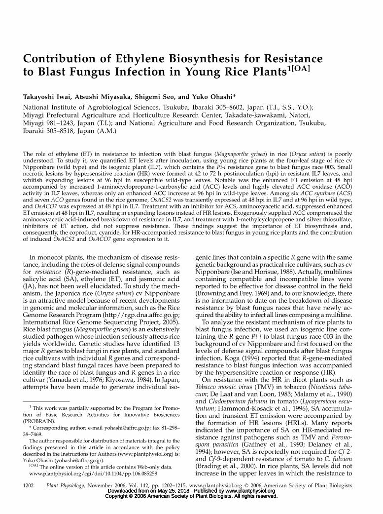
First, we established an experimental system toanalyze the mechanism of resistance to pathogeninfection in rice plants. We used young rice plants atthe four-leaf stage of wild-type Nipponbare and itsisogenic line, IL7, which contains the R gene Pi-i (Iseet al., 1988). Wild type is susceptible and IL7 is resis-tant to infection by blast fungus race 003 (isolateKyu89-241). In the fully expanded fourth leaf of IL7plants, small HRLs were found at 42 h postinoculation(hpi) and gradually developed into dark-brown le-sions up to 0.5 mm in diameter by 63 hpi, with noremarkable increase in size thereafter (Fig. 1), exhibit-ing a resistant response to race 003. In wild-typeleaves, no detectable phenotype was found within 63hpi, and whitish expanding lesions (ELs) 0.5 mm indiameter were first observed at 96 hpi, and the ELs
Figure 1. Phenotypes of lesions in young rice leaves inoculated with blast fungus. Photographs of blast fungus-inoculated fourthleaves of IL7 and wild-type (WT) plants at the four-leaf stage. A conidial suspension of blast fungus race 003 was sprayed on anincompatible cultivar IL7 (top), which contains the R gene Pi-i against blast fungus race 003 in the Nipponbare background, andon wild-type Nipponbare, which is a compatible cultivar (bottom). In inoculated IL7 leaves, HRLs (black arrowhead) appeared at42 hpi and matured by 63 hpi, turning dark brown. In inoculated wild-type leaves, whitish ELs (white arrowhead) were found at96 hpi. Bar 5 1 mm.
Ethylene Biosynthesis for Resistance to Blast Fungus
Plant Physiol. Vol. 142, 2006 1203 www.plantphysiol.orgon May 25, 2018 - Published by Downloaded from
Copyright © 2006 American Society of Plant Biologists. All rights reserved.
rapidly grew in size thereafter and infected leaveswilted by 144 hpi when inoculated with 1 3 105
conidia mL21. At lower concentrations, such as 1 3 104
conidia mL21, ELs grew to about 2 3 1.5 mm in size,developing conidia at the center of each lesion by144 hpi (Fig. 1), indicating susceptibility of wild typeto infection.
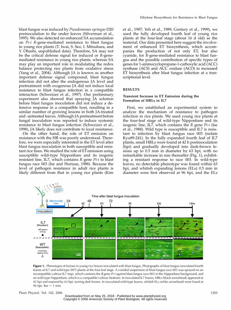
We determined the level of ET emitted from thefourth leaves after blast fungus inoculation, using 16fourth leaves as one sample (Fig. 2A). One gram ofhealthy leaf from both IL7 and wild type emitted about0.9 nL of ET per hour just after detaching. In bothmock- and fungus-inoculated IL7 plants, the first peakin ET emission was found at 24 hpi, which was also thecase in wild type. This peak by mock inoculation maybe caused by the inoculation itself, which involved
incubation of plants under high humidity in the darkfor 20 h just after spraying the inoculum. The peak at24 hpi was higher after blast fungus inoculation thanmock inoculation in both IL7 and wild type (Fig. 2A),indicating an additive enhancement of ET emission byfungal infection. The second peak of ET emission wasdetected only in fungus-inoculated IL7 plants at 48hpi. The enhanced rate of ET emission by fungalinoculation at 48 hpi was 6.4 nL g21 h21 fresh weightin inoculated IL7 and 0.36 nL g21 h21 fresh weight ininoculated wild type, respectively. Because primaryHRLs found at 42 hpi (black arrow) were mature by 63hpi, the significant enhancement of ET emission foundin IL7 was thought to be related to the formation ofHRLs. During 63 to 96 hpi, emitted ET from inoculatedIL7 was maintained at a high level (4 nL g21 h21 fresh
Figure 2. Profiles of ET emission from young rice leaves inoculated with blast fungus. A, ET emission in resistant IL7 leaves andsusceptible wild-type leaves determined using 16 fourth leaves at the four-leaf stage for one sample. Solid lines show the level ofET emission after inoculation. Broken lines show the level after mock inoculation. B, ACC content determined using 12 fourthleaves at the four-leaf stage for one sample. Inoculated leaves were homogenized and the ACC in the extract was chemicallyconverted to ET. An expansion at 48 to 72 hpi is shown in frame. C, ACO activity determined as the capacity for converting ACCto ET using 12 fourth leaves at the four-leaf stage for one sample. Values in A, B, and C are shown as the means 6 SD based onthree independent experiments. The experiment was repeated two times with similar results. The symbol without visible errorbars indicates that bars are present inside the symbol.
Iwai et al.
1204 Plant Physiol. Vol. 142, 2006 www.plantphysiol.orgon May 25, 2018 - Published by Downloaded from
Copyright © 2006 American Society of Plant Biologists. All rights reserved.

weight). In inoculated wild-type leaves, the level of ETemission was clearly lower than that in inoculated IL7and similar to that in mock-inoculated wild-typeleaves from 36 to 62 hpi. In wild type, a slight increasewas detected at 72 to 96 hpi at which time the devel-opment of ELs had started (white arrow). Independenttriplicate experiments with similar results confirmedthat the data obtained here were reproducible.
The major pathway of ET biosynthesis contains twocatalytic steps involving ACS and ACO, producingequal moles of ET and cyanide (Peiser et al., 1984). Tostudy the mechanism of enhanced ET biosynthesis inthe resistant response, we determined the levels ofemitted ET (Fig. 2A), ACC (Fig. 2B), and ACO activity(Fig. 2C) in blast fungus-inoculated rice leaves atthe same time. High levels of ACC were accumulatedin both IL7 and wild type at 24 to 36 h after both fun-gus and mock inoculation. ACC content was 1.5-foldhigher after fungus inoculation than mock inocula-tion, indicating that the fungal infection and/or spray-ing of the conidial suspension further enhanced ACCaccumulation in both rice plants. The peaks at 24 hpiseem to be related to the first peaks in ET emission inFigure 2A. At 48 to 72 hpi during which HRLs wereformed and completed, the ACC content of inoculatedIL7 leaves was about 1.5- to 1.7-fold that of mock-inoculated leaves, whereas no significant difference inACC levels was found between mock- and fungus-inoculated wild-type leaves (Fig. 2B). These resultssuggested that enhanced ACC synthesis and/or sup-pressed ACC degradation were more strongly in-duced in resistant IL7 than in susceptible wild typeafter blast fungus inoculation. In wild type, the ACClevel was increased at 96 hpi (Fig. 2B) when ELformation was started. These results indicated thatACC synthesis was accompanied by the formation ofboth HRLs and ELs.
In inoculated IL7 leaves, ACO activity was dramat-ically increased at 48 to 63 hpi and remained at aconsiderable level at 72 to 96 hpi, about 4 nL g21 h21
fresh weight, which was 2.5-fold higher than that inmock-inoculated leaves. In wild-type plants, the pro-file of ACO activity was quite different from that inIL7. The level was only slightly higher than in mock-inoculated leaves at and after 48 hpi with no clearpeak. The time-course profile of ACO activity ininoculated IL7 leaves resembled that of the ET emis-sion profile at 36 to 96 hpi (Fig. 2A), suggesting thatincreased ACO activity leads to enhanced ET emissionin incompatible interaction at and after 48 hpi. At 63hpi, ACO activity in inoculated IL7 leaves had a peakinstead of a rapid decrease in ET emission, probablybecause of decreased ACC content and/or possiblenegative regulation by elevated ET.
Characterization of the Rice ACS Gene Family
What kinds of ACS and ACO contribute to ETbiosynthesis in blast fungus-infected rice plants? Wesearched for ACS and ACO genes from expressed
sequence tags, full-length cDNAs, and genome data-bases of rice cv Nipponbare (http://riceblast.dna.affrc.go.jp/; http://cdna01.dna.affrc.go.jp/cDNA).
At least five rice ACS genes are reported to exist inthe rice genome (Zarembinski and Theologis, 1993); cor-responding cDNAs are OsACS1 (AK071011), OsACS2(AK064250), OsACS3 (P0617H07.9 in AC135427),OsACS4 (OSJNBb0006B22.3 in AC136224), andOsACS5 (D46839). OsACS6 (AK065212) was newlyidentified as the ortholog of ACS1 (U35779) fromwheat (Triticum aestivum). The homology in aminoacid sequence between OsACS1 and OsACS2 toOsACS6 is 54%, 59%, 56%, 56%, and 46%, respectively.An alignment of the six putative OsACS polypeptidesis shown in Figure 3A. All ACS isoforms contain theseven conserved domains (Fig. 3A, boxes), which werefound in ACSs from other plant species (Yamagamiet al., 2003). The 11 invariant amino acid residuesbetween ACS and aminotransferases in Arabidopsis(Arabidopsis thaliana; Yamagami et al., 2003) and to-mato (Rottmann et al., 1991), which are shaded inFigure 3A, were also found in the members of the riceACS gene family. In addition, the Tyr residue at 245 inOsACS1 (black inverted triangle), which is a part ofthe pyridoxal-5#-P-binding site, was conserved amongOsACS1 to OsACS5, and it was replaced by Phe inOsACS6.
A phylogenetic analysis with ACS proteins fromrice, Arabidopsis (Yamagami et al., 2003), tobacco (Liuand Zhang, 2004), and wheat (Subramaniam et al.,1996) revealed that the genes fall into three groups(Fig. 3B). ACSs from Arabidopsis in groups I and IIexhibited ACS activity, but not in group III, whichcontains AtACS10 and 12 similar to Ala or Asp ami-notransferases in Arabidopsis (Yamagami et al., 2003).Thus, OsACS1 to OsACS5 in groups I or II, but not III,might work in rice.
To elucidate the genomic organization of ACS inrice, genomic DNA from rice cv Nipponbare (wildtype) was subjected to Southern-blot analysis withmixed probes containing the catalytic domain of ACS,which were prepared by PCR amplification usingOsACS1, OsACS2, and OsACS5 as templates (Fig. 3C).Because the highest homology based on nucleotidesequence between the probe and OsACS1 to OsACS6 is100%, 100%, 76%, 73%, 100%, and 51%, respectively,the mixed probes would detect five signals corre-sponding to OsACS1 to OsACS5 in the digests by EcoRIwhose internal site was not found in OsACSs. Theseresults suggested that five rice ACSs in groups I and IIcompose a functional ACS gene family.
Characterization of the Rice ACO Gene Family
As an ACO gene in deepwater rice, OS-ACO1(X85747) has been reported in relation to submergence(Mekhedov and Kende, 1996) and OS-ACO2 (AF049888)and OS-ACO3 (AF049889) in relation to hormonal crosstalk (Chae et al., 2000). We searched for their orthologs
Ethylene Biosynthesis for Resistance to Blast Fungus
Plant Physiol. Vol. 142, 2006 1205 www.plantphysiol.orgon May 25, 2018 - Published by Downloaded from
Copyright © 2006 American Society of Plant Biologists. All rights reserved.
and homologs in expressed sequence tags, full-lengthcDNAs, and genome databases of rice, and foundseven possible ACO genes, designated OsACO1(AK058296), OsACO2 (AK071557), OsACO3 (AK065039),OsACO4 (AK105491), OsACO5 (AK061064), OsACO6(OJ1504_G04.8 in AC105772), and OsACO7 (AK102472),respectively. OsACO6 was mapped next to OsACO5 inchromosome 5 and we found that OsACO6 is a pseu-dogene encoding a truncated ACO peptide. The ho-mology in amino acid sequence between OsACO1 andOsACO2 to OsACO5 and OsACO7 is 93%, 73%, 47%,48%, and 43%, respectively. An alignment of the sixputative ACO polypeptides is shown in Figure 4A.ACO is a member of the Fe(II) ascorbate family ofdioxygenases in which the nine amino acid residuesshaded in Figure 4A are conserved (Lasserre et al.,1996). Three of these nine residues with an arrowheadcontribute to the binding of Fe(II), namely, His at 183,Asp at 185, and His at 240 in OsACO1. OsACO4 lackstwo and OsACO5 lacks one of the nine conservedamino acid residues with white and black circles,respectively. Therefore, OsACO4 and OsACO5 mightnot actually function.
OsACO genes were classified into three major groupsin a phylogenic tree based on putative amino acidsequences (Fig. 4B). OsACO1, OsACO2, and OsACO3are classified in group I, which contains NtACO1 andNtACO2, whose gene expression was accompanied bythe formation of HRLs in tobacco (Liu and Zhang,2004). OsACO7 is classified in group II, which containsStACO3, which was induced in the potato (Solanumtuberosum) tuber by inoculation with Fusarium eumartiiand treatment of SA and indole acetic acid (Zanetti et al.,2002), and LEACO5, whose expression was anaerobi-cally induced (Sell and Hehl, 2005).
Genomic Southern-blot analysis with mixed probescontaining the catalytic domain of ACO, which wereprepared by PCR amplification using OsACO1 toOsACO5 and OsACO7 as templates, detected six sig-nals in both EcoRI and HindIII digests and sevensignals in NcoI digests, indicating that seven OsACOgenes, including the OsACO6 pseudogene, compose agene family in rice (Fig. 4C).
Specific ACS and ACO Genes Are Induced Transientlyat 48 hpi in IL7
As described, our results suggested the importanceof ET biosynthesis for resistance to blast fungus infec-tion. When the data on ACC content and ACO activity
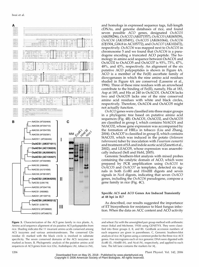
Figure 3. Characterization of the ACS gene family in rice plants. A,Amino acid sequence alignment of six putative ACS polypeptides fromrice. Shading indicates the 11 invariant amino acids conserved amongACS isozymes and various aminotransferases. The conserved Glnresidue (E) marked with the black circle is involved in substratespecificity. The seven conserved domains of the ACS isozymes aremarked as boxes. B, Phylogenetic analysis of the putative amino acidsequences of ACS genes from rice (Os), Arabidopsis (At), tobacco (Nt),
and wheat (Ta) with the unweighted pair group method with arithmeticmean (Sokal and Michener, 1958) using GENETYX. They were classi-fied into three groups (I, II, and III). GenBank accession numbers ofeach sequence are given in parentheses. C, Genomic Southern-blotanalysis of rice ACS genes using a common probe for the three rice ACSgenes. Five micrograms each of rice genomic DNA were digested withEcoRI (E), HindIII (H), and NcoI (N), respectively, and applied to eachlane. The left lane contains the markers for Mr .
Iwai et al.
1206 Plant Physiol. Vol. 142, 2006 www.plantphysiol.orgon May 25, 2018 - Published by Downloaded from
Copyright © 2006 American Society of Plant Biologists. All rights reserved.
after blast fungus inoculation in Figure 2 were recon-structed in Figure 5A, it became clearer that the levelsbefore and during the formation of HRLs (36–63 hpi)were significantly higher in IL7 than those in wildtype, suggesting an additive effect on ET biosynthesisin IL7 leaves. Thus, we studied the expression profilesof ACS and ACO genes in inoculated IL7 and wild-type plants.
The time-course expression profiles of six ACS geneswere studied by one-step reverse transcription (RT)-
PCR with specific primers for each ACS gene (Fig. 5B).Transient OsACS1 expression was found in mock-inoculated leaves at a low level and in blast fungus-inoculated leaves at a high level at 24 hpi in bothplants, indicating OsACS1 may function for the firstpeak of ET emission at 24 hpi (Fig. 2A). OsACS2 wastransiently induced at 48 hpi in fungus-inoculated IL7plants, which is likely related to increased ACC con-tent and dramatic ET emission at 48 hpi (Fig. 2A). Aconsiderable level of OsACS2 transcript was found at
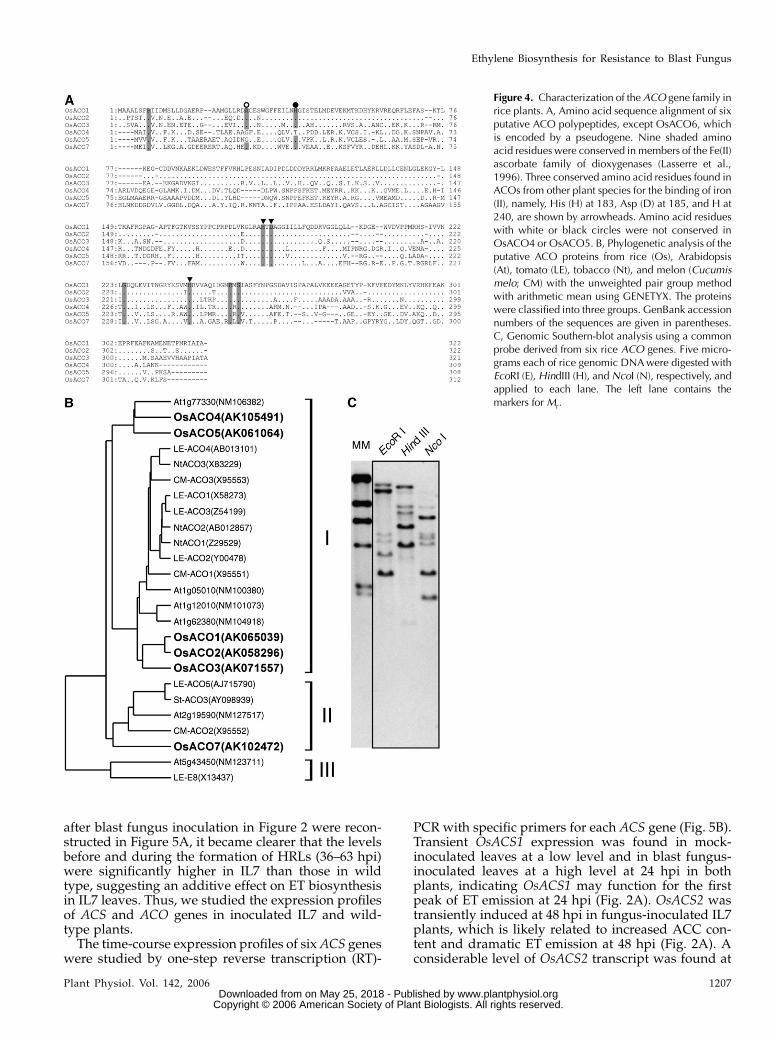
Figure 4. Characterization of the ACO gene family inrice plants. A, Amino acid sequence alignment of sixputative ACO polypeptides, except OsACO6, whichis encoded by a pseudogene. Nine shaded aminoacid residues were conserved in members of the Fe(II)ascorbate family of dioxygenases (Lasserre et al.,1996). Three conserved amino acid residues found inACOs from other plant species for the binding of iron(II), namely, His (H) at 183, Asp (D) at 185, and H at240, are shown by arrowheads. Amino acid residueswith white or black circles were not conserved inOsACO4 or OsACO5. B, Phylogenetic analysis of theputative ACO proteins from rice (Os), Arabidopsis(At), tomato (LE), tobacco (Nt), and melon (Cucumismelo; CM) with the unweighted pair group methodwith arithmetic mean using GENETYX. The proteinswere classified into three groups. GenBank accessionnumbers of the sequences are given in parentheses.C, Genomic Southern-blot analysis using a commonprobe derived from six rice ACO genes. Five micro-grams each of rice genomic DNA were digested withEcoRI (E), HindIII (H), and NcoI (N), respectively, andapplied to each lane. The left lane contains themarkers for Mr.
Ethylene Biosynthesis for Resistance to Blast Fungus
Plant Physiol. Vol. 142, 2006 1207 www.plantphysiol.orgon May 25, 2018 - Published by Downloaded from
Copyright © 2006 American Society of Plant Biologists. All rights reserved.
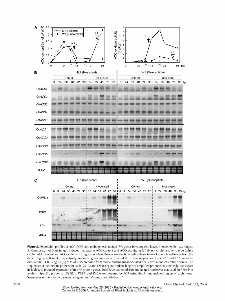
Figure 5. Expression profiles of ACS, ACO, and pathogenesis-related (PR) genes in young rice leaves infected with blast fungus.A, Comparison of blast fungus-induced increase in ACC content and ACO activity in IL7 (black circle) and wild type (whitecircle). ACC content and ACO activity of fungus-inoculated leaves were subtracted by those in mock-inoculated leaves from thedata in Figure 2, B and C, respectively, and new figures were reconstructed. B, Expression profiles of rice ACS and ACO genes byone-step RT-PCR using 0.5 mg of total RNA prepared from mock- and fungus-inoculated rice leaves at indicated time points. Thesequences of the specific primers for each OsACS and OsACO gene and the length of amplified products, respectively, are shownin Table I. C, Induced expression of rice PR protein genes. Total RNA extracted from inoculated rice leaves was used for RNA-blotanalysis. Specific probes for OsPR1a, PBZ1, and PAL were prepared by PCR using the 3#-untranslated region of each clone.Sequences of the specific primers are given in ‘‘Materials and Methods.’’
1208 Plant Physiol. Vol. 142, 2006 www.plantphysiol.orgon May 25, 2018 - Published by Downloaded from
Copyright © 2006 American Society of Plant Biologists. All rights reserved.
96 hpi in wild type, but not IL7. It may be accompaniedby the formation of ELs in wild-type plants. OsACS3,OsACS4, and OsACS6 were almost constitutively ex-pressed in mock- and blast fungus-inoculated IL7 andwild-type leaves. No signal for the OsACS5 transcriptwas found in fourth leaves under the same conditions(data not shown). These results suggest that OsACS1contributes to dark- and high humidity-induced ETemission found at 24 hpi, and OsACS2 mainly acts toincrease the ACC level during HRL formation at 48 hpiin IL7 and EL formation at 96 hpi in wild type.
Next, expression profiles of the six ACO genes,except for the pseudogene OsACO6, were determinedusing one-step RT-PCR with specific primers for eachgene (Fig. 5B). OsACO1 was detected at 24 to 48 hpiin mock- and fungus-inoculated IL7 and wild-typeplants. OsACO1 may be expressed under dark and highhumidity conditions during inoculation, probably con-tributing to the first peak of ET emission at 24 hpi(Fig. 2A). OsACO2 was constitutively expressed in bothplants, but down-regulated in blast fungus-inoculatedIL7 at 72 and 96 hpi. OsACO3 was constitutivelyexpressed in both plants not affected by the treat-ments. No signal for the OsACO4 transcript was foundin either plant (data not shown). Expression of bothOsACO5 and OsACO7 was inducible and enhancedby infection in both plants. Notably, expression ofOsACO7 was very transient and strong at 48 hpi in IL7,but not wild-type plants. Whereas OsACO5 was alsotransiently enhanced to express at 48 hpi in IL7, thetranscript was found in wild type at 48 to 96 hpi as well.From these results, transient expression of OsACO7 isthought to be most important for the increase of ACOactivity at 48 to 72 hpi in infected IL7 leaves, possiblyin cooperation with OsACO5 expression.
Blast fungus-induced OsACSs and OsACOs expres-sion profiles were compared with control defense-related genes, such as OsPR1a (AJ278436; Agrawalet al., 2000) and PBZ1 (D38170; Midoh and Iwata, 1996)using each specific probe (Fig. 5C). The transcript forOsPR1a, but not PBZ1, was transiently and slightlyaccumulated at 24 h after mock and blast fungusinoculation in both host plants. In addition, in IL7, thetranscript of OsPR1a was expressed again at 48 hpi,increasing to greater amounts thereafter, at which timethe formation of HRLs was completed. In wild type,the OsPR1a transcript accumulated at 72 and 96 hpiaccompanied by the formation of ELs. PBZ1 expres-sion was found to be accompanied by the formation oflesions, but not mock inoculation. The transcript ac-cumulated at 36 hpi and was almost saturated at 72 hpiin IL7. In wild type, it was found at 48 hpi, increasingat 96 hpi. Thus, expression of the two defense markergenes was found during and after the formation oflesions in both blast fungus-infected IL7 and wild-typeplants. In IL7, these expression levels were remarkablyhigher after lesions had formed (72–96 hpi) thanduring their formation (48 hpi), indicating the down-stream genes of defense signaling in rice. These ex-pression profiles were clearly different from those of
OsACS2 and OsACO7, which are very transient at 48 dpostinfection in IL7. Expression of the Phe ammonialyase (PAL) gene (X16099; Minami et al., 1989) wasslightly down-regulated by the inoculation procedureand the level of expression induced by fungus inocu-lation was higher in wild type than in IL7.
Inhibition of ET Biosynthesis, But Not ET Signaling,
Results in Suppressed Resistance to Fungal Infection
Enhanced ET emission during the formation ofHRLs in IL7 depends on ET biosynthesis. To confirmthe contribution of ET biosynthesis to resistance, theeffect of aminooxyacetic acid (AOA), which is aninhibitor of pyridoxal-5#-P-related enzymes, such asACS (Yu et al., 1979; Yang and Hoffman, 1984), wasstudied. The fourth leaves of 16 independent blastfungus-inoculated rice plants were detached at 24 hpiand a solution of AOA at 0.1, 0.5, or 1.0 mM was fedthrough the base of the detached leaf blades for 24 h,respectively. Then the leaves were put in an airtightvial and the level of ET released from the leaf wasdetermined for 3 h (Fig. 6, A and B). Mock-inoculatedleaves from IL7 and wild-type plants, which had beendetached at 24 hpi and fed with water for 24 h, emittedET at the rate of 7.5 and 8.5 nL g21 h21 fresh weight,respectively, whereas those from intact IL7 and wild-type plants were 1.7 and 3.1 nL g21 h21 fresh weight,respectively. These results indicate that detachingleaves, which is a kind of wounding, enhanced ETemission from the leaf blade. When mock-inoculatedleaves were treated with 0.1 mM AOA solution at24 hpi, ET emission was diminished to about 1 nL g21
h21 fresh weight in both IL7 and wild type, which issimilar to that at time 0 (see Fig. 2B), indicating thatwound-induced ET emission was completely sup-pressed by 0.1 mM AOA. In fungus-inoculated leavesfed water at 24 hpi, the highest increase in ET emission(i.e. 14.8 nL g21 h21 fresh weight in IL7 and 10.6 nL g21
h21 fresh weight in wild type) was observed. When fedwith 0.1, 0.5, or 1.0 mM AOA at 24 hpi, fungus-inoculated IL7 leaves emitted ET at the rate of 5.4, 1.1,and 1.0 nL g21 h21 fresh weight, which correspondedto a 64%, 92%, and 93% decrease of that emitted fromwater-treated control leaves, respectively. In fungus-inoculated wild type, AOA treatment at 0.1, 0.5, or1.0 mM resulted in an 86%, 91%, and 92% decrease inthe level of ETcompared with the control, respectively.These results indicate that, for strong inhibition offungus-induced ET emission, 0.1 mM AOA was notenough and 0.5 mM was required for IL7, whereas0.1 mM was enough for wild type. Therefore, a higherlevel of ACS activity, which could not be inhibited by0.1 mM AOA, seems to be induced in fungus-inoculatedIL7, but not in wild type.
To confirm the effect of AOA on resistance to blastfungus in rice, infected leaf pieces were treated at96 hpi with lactophenol-trypan blue, which stains themycelium blue. In 0.1 mM AOA-treated IL7 leaf pieces,HRLs remained dark brown and were not stained blue
Ethylene Biosynthesis for Resistance to Blast Fungus
Plant Physiol. Vol. 142, 2006 1209 www.plantphysiol.orgon May 25, 2018 - Published by Downloaded from
Copyright © 2006 American Society of Plant Biologists. All rights reserved.
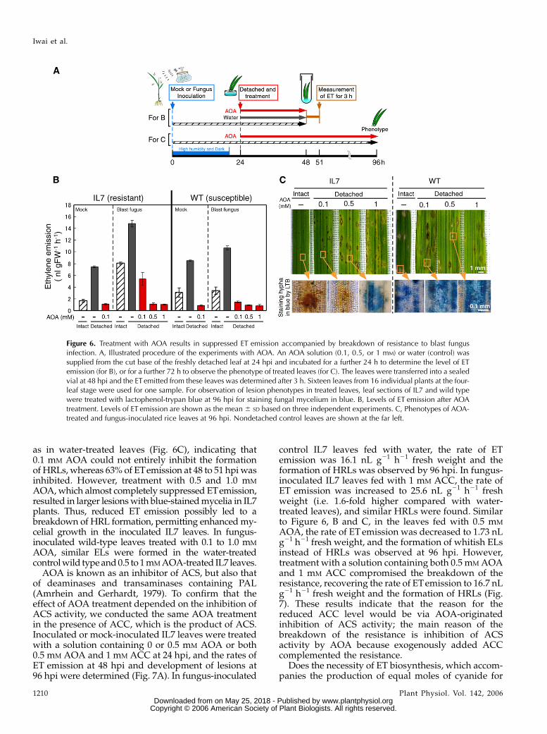
as in water-treated leaves (Fig. 6C), indicating that0.1 mM AOA could not entirely inhibit the formationof HRLs, whereas 63% of ETemission at 48 to 51 hpi wasinhibited. However, treatment with 0.5 and 1.0 mM
AOA, which almost completely suppressed ETemission,resulted in larger lesions with blue-stained mycelia in IL7plants. Thus, reduced ET emission possibly led to abreakdown of HRL formation, permitting enhanced my-celial growth in the inoculated IL7 leaves. In fungus-inoculated wild-type leaves treated with 0.1 to 1.0 mM
AOA, similar ELs were formed in the water-treatedcontrol wild type and 0.5 to 1 mM AOA-treated IL7 leaves.
AOA is known as an inhibitor of ACS, but also thatof deaminases and transaminases containing PAL(Amrhein and Gerhardt, 1979). To confirm that theeffect of AOA treatment depended on the inhibition ofACS activity, we conducted the same AOA treatmentin the presence of ACC, which is the product of ACS.Inoculated or mock-inoculated IL7 leaves were treatedwith a solution containing 0 or 0.5 mM AOA or both0.5 mM AOA and 1 mM ACC at 24 hpi, and the rates ofET emission at 48 hpi and development of lesions at96 hpi were determined (Fig. 7A). In fungus-inoculated
control IL7 leaves fed with water, the rate of ETemission was 16.1 nL g21 h21 fresh weight and theformation of HRLs was observed by 96 hpi. In fungus-inoculated IL7 leaves fed with 1 mM ACC, the rate ofET emission was increased to 25.6 nL g21 h21 freshweight (i.e. 1.6-fold higher compared with water-treated leaves), and similar HRLs were found. Similarto Figure 6, B and C, in the leaves fed with 0.5 mM
AOA, the rate of ET emission was decreased to 1.73 nLg21 h21 fresh weight, and the formation of whitish ELsinstead of HRLs was observed at 96 hpi. However,treatment with a solution containing both 0.5 mM AOAand 1 mM ACC compromised the breakdown of theresistance, recovering the rate of ETemission to 16.7 nLg21 h21 fresh weight and the formation of HRLs (Fig.7). These results indicate that the reason for thereduced ACC level would be via AOA-originatedinhibition of ACS activity; the main reason of thebreakdown of the resistance is inhibition of ACSactivity by AOA because exogenously added ACCcomplemented the resistance.
Does the necessity of ET biosynthesis, which accom-panies the production of equal moles of cyanide for
Figure 6. Treatment with AOA results in suppressed ET emission accompanied by breakdown of resistance to blast fungusinfection. A, Illustrated procedure of the experiments with AOA. An AOA solution (0.1, 0.5, or 1 mM) or water (control) wassupplied from the cut base of the freshly detached leaf at 24 hpi and incubated for a further 24 h to determine the level of ETemission (for B), or for a further 72 h to observe the phenotype of treated leaves (for C). The leaves were transferred into a sealedvial at 48 hpi and the ET emitted from these leaves was determined after 3 h. Sixteen leaves from 16 individual plants at the four-leaf stage were used for one sample. For observation of lesion phenotypes in treated leaves, leaf sections of IL7 and wild typewere treated with lactophenol-trypan blue at 96 hpi for staining fungal mycelium in blue. B, Levels of ET emission after AOAtreatment. Levels of ET emission are shown as the mean 6 SD based on three independent experiments. C, Phenotypes of AOA-treated and fungus-inoculated rice leaves at 96 hpi. Nondetached control leaves are shown at the far left.
Iwai et al.
1210 Plant Physiol. Vol. 142, 2006 www.plantphysiol.orgon May 25, 2018 - Published by Downloaded from
Copyright © 2006 American Society of Plant Biologists. All rights reserved.
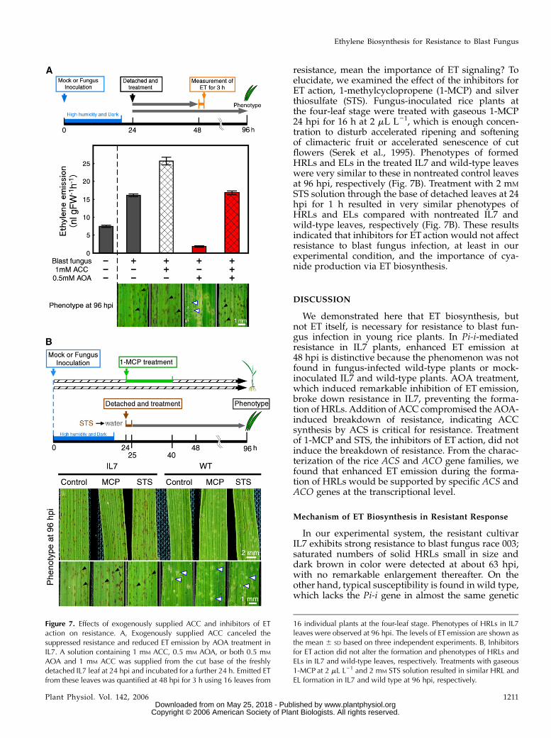
resistance, mean the importance of ET signaling? Toelucidate, we examined the effect of the inhibitors forET action, 1-methylcyclopropene (1-MCP) and silverthiosulfate (STS). Fungus-inoculated rice plants atthe four-leaf stage were treated with gaseous 1-MCP24 hpi for 16 h at 2 mL L21, which is enough concen-tration to disturb accelerated ripening and softeningof climacteric fruit or accelerated senescence of cutflowers (Serek et al., 1995). Phenotypes of formedHRLs and ELs in the treated IL7 and wild-type leaveswere very similar to these in nontreated control leavesat 96 hpi, respectively (Fig. 7B). Treatment with 2 mM
STS solution through the base of detached leaves at 24hpi for 1 h resulted in very similar phenotypes ofHRLs and ELs compared with nontreated IL7 andwild-type leaves, respectively (Fig. 7B). These resultsindicated that inhibitors for ET action would not affectresistance to blast fungus infection, at least in ourexperimental condition, and the importance of cya-nide production via ET biosynthesis.
DISCUSSION
We demonstrated here that ET biosynthesis, butnot ET itself, is necessary for resistance to blast fun-gus infection in young rice plants. In Pi-i-mediatedresistance in IL7 plants, enhanced ET emission at48 hpi is distinctive because the phenomenon was notfound in fungus-infected wild-type plants or mock-inoculated IL7 and wild-type plants. AOA treatment,which induced remarkable inhibition of ET emission,broke down resistance in IL7, preventing the forma-tion of HRLs. Addition of ACC compromised the AOA-induced breakdown of resistance, indicating ACCsynthesis by ACS is critical for resistance. Treatmentof 1-MCP and STS, the inhibitors of ET action, did notinduce the breakdown of resistance. From the charac-terization of the rice ACS and ACO gene families, wefound that enhanced ET emission during the forma-tion of HRLs would be supported by specific ACS andACO genes at the transcriptional level.
Mechanism of ET Biosynthesis in Resistant Response
In our experimental system, the resistant cultivarIL7 exhibits strong resistance to blast fungus race 003;saturated numbers of solid HRLs small in size anddark brown in color were detected at about 63 hpi,with no remarkable enlargement thereafter. On theother hand, typical susceptibility is found in wild type,which lacks the Pi-i gene in almost the same genetic
Figure 7. Effects of exogenously supplied ACC and inhibitors of ETaction on resistance. A, Exogenously supplied ACC canceled thesuppressed resistance and reduced ET emission by AOA treatment inIL7. A solution containing 1 mM ACC, 0.5 mM AOA, or both 0.5 mM
AOA and 1 mM ACC was supplied from the cut base of the freshlydetached IL7 leaf at 24 hpi and incubated for a further 24 h. Emitted ETfrom these leaves was quantified at 48 hpi for 3 h using 16 leaves from
16 individual plants at the four-leaf stage. Phenotypes of HRLs in IL7leaves were observed at 96 hpi. The levels of ET emission are shown asthe mean 6 SD based on three independent experiments. B, Inhibitorsfor ET action did not alter the formation and phenotypes of HRLs andELs in IL7 and wild-type leaves, respectively. Treatments with gaseous1-MCP at 2 mL L21 and 2 mM STS solution resulted in similar HRL andEL formation in IL7 and wild type at 96 hpi, respectively.
Ethylene Biosynthesis for Resistance to Blast Fungus
Plant Physiol. Vol. 142, 2006 1211 www.plantphysiol.orgon May 25, 2018 - Published by Downloaded from
Copyright © 2006 American Society of Plant Biologists. All rights reserved.
background; whitish ELs were first visualized at 96hpi, and they rapidly develop in size with vigorousconidiation in the center. In this system, the time pointaround 48 hpi is crucial to detect the difference inresistance to blast fungus because, at this time, HRLshave just started to form in IL7 leaves, whereas noclear phenotype is found in inoculated wild-typeleaves (Fig. 1). Two peaks of ET emission were ob-served in IL7 and wild-type plants infected with blastfungus race 003. The first peak at 24 hpi was likely aresult of the darkness and high humidity in both hostplants, and the second peak at 48 hpi was specific forthe formation of HRLs in infected IL7 plants (Fig. 2A).
The level of the second peak in ET emission wasalmost proportional to the number and size of devel-oping HRLs. When the conidial suspension (1 3 105
conidia mL21) was sprayed onto IL7 plants at the four-leaf stage, about 150 HRLs were detected on the fourthleaf accompanied by ET emission at the rate of 7 nL g21
h21 fresh weight at 48 hpi. This level of ET emission issimilar to the formation of HRLs mediated by the Ngene (18 nL g21 h21fresh weight) in TMV-infectedtobacco plants (De Laat and Van Loon, 1981). Thesharp peak in ET emission similar to the second peakin infected IL7 plants was observed during the forma-tion of HRLs in N-gene-mediated resistance in TMV-infected tobacco plants (De Laat and Van Loon, 1981,1983) as well as Cf-gene-mediated resistance inC. fulvum-infected tomato plants (Hammond-Kosacket al., 1996). Enhanced ET emission at 48 to 72 hpi,when HRLs developed and matured in infected IL7plants, was accompanied by infection-enhanced ACCaccumulation and ACO activity (Figs. 2 and 5A). Theamount of ACC at 36 to 72 hpi was slightly, butsignificantly, higher in infected IL7 than infected wild-type leaves, and it reversed at 96 hpi, at which timeformation of ELs is going on, indicating that ACCsynthesis was accompanied by the formation of notonly HRLs but also ELs in rice leaves infected with blastfungus (Fig. 5A). In N-gene-mediated resistance, accu-mulation of increased ACC was reportedly restricted tothe area of HRLs (De Laat and Van Loon, 1983),suggesting localization of increased ACC at the area ofHRLs and ELs in rice plants. ACO activity was dramat-
ically and specifically elevated during the formation ofHRLs in infected IL7 plants (Fig. 2C), suggesting en-hanced ACO activity and increased levels of ACC at 36to 63 hpi are important for ET emission from HRLs.
In deepwater rice, OS-ACS1, whose product shares99% homology with OsACS1 from Nipponbare wildtype, was induced by partial submergence at the upper-most elongating internode and involved in stem elon-gation (Zarembinski and Theologis, 1993, 1997).Lowland rice plants, such as Nipponbare wild typeand IL7, possibly recognized the dark and high humid-ity conditions as partial submergence, and ET emissionmight be enhanced at 24 hpi in both mock- and blastfungus-inoculated leaves. OsACS1 was classified intogroup I, which contains auxin-responsive AtACS4 andAtACS5 (Tsuchisaka and Theologis, 2004), suggestinginvolvement in plant hormone-mediated responses.OsACS2 was classified into group II. Group II containsAtACS6, which was rapidly induced by ozone exposure-induced cell death (Overmyer et al., 2000) and NtACS1,NtACS2, and NtACS3, whose gene expression wasinduced during an N-gene-mediated TMV-resistant re-sponse in tobacco (Kim et al., 2003). Thus, ACS proteinsin group II would function in response to biotic stresses,including pathogen infection. Phosphorylation was alsoreported to be important for ACS proteins belonging togroup II, such as AtACS2 and AtACS6, which are verysimilar to OsACS2. These AtACS proteins are stabilizedby phosphorylation at their C-terminal regions by Arabi-dopsis mitogen-activated protein kinase 6, the orthologof tobacco SA-induced protein kinase, whose activationconfers TMV resistance (Liu and Zhang, 2004). Thus,studies on posttranscriptional and posttranslational reg-ulation of OsACS2 would also be important to elucidatethe dynamic regulation of ET biosynthesis for diseaseresistance.
Maintenance of a high level of ACO activity at 48 to96 hpi may guarantee a considerable level of ET bio-synthesis during this period. Two newly characterizedrice ACO genes, OsACO5 and OsACO7, out of sevenACO members, were transiently induced by blastfungus in IL7 plants (Fig. 5B). Because OsACO5 lacksone of nine conserved amino acid residues, it mightconfer weaker or no ACO activity (Fig. 4A). OsACO5
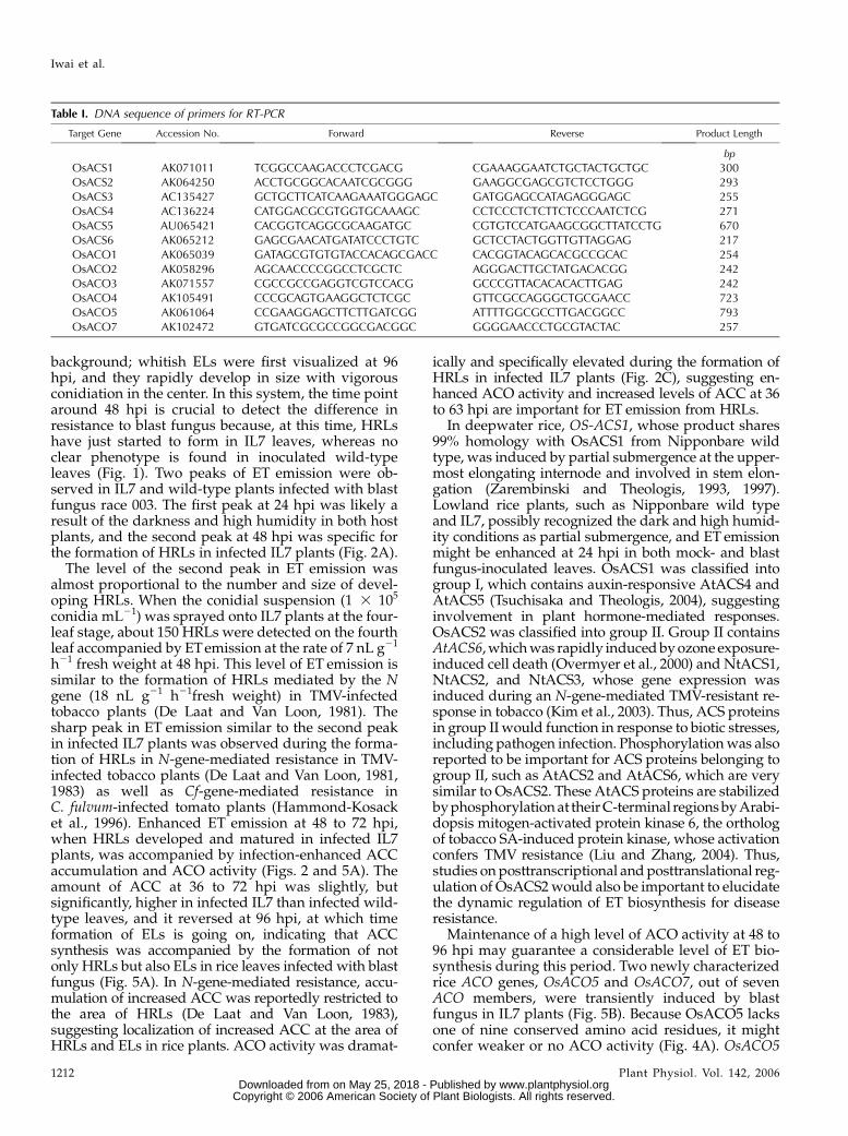
Table I. DNA sequence of primers for RT-PCR
Target Gene Accession No. Forward Reverse Product Length
bp
OsACS1 AK071011 TCGGCCAAGACCCTCGACG CGAAAGGAATCTGCTACTGCTGC 300OsACS2 AK064250 ACCTGCGGCACAATCGCGGG GAAGGCGAGCGTCTCCTGGG 293OsACS3 AC135427 GCTGCTTCATCAAGAAATGGGAGC GATGGAGCCATAGAGGGAGC 255OsACS4 AC136224 CATGGACGCGTGGTGCAAAGC CCTCCCTCTCTTCTCCCAATCTCG 271OsACS5 AU065421 CACGGTCAGGCGCAAGATGC CGTGTCCATGAAGCGGCTTATCCTG 670OsACS6 AK065212 GAGCGAACATGATATCCCTGTC GCTCCTACTGGTTGTTAGGAG 217OsACO1 AK065039 GATAGCGTGTGTACCACAGCGACC CACGGTACAGCACGCCGCAC 254OsACO2 AK058296 AGCAACCCCGGCCTCGCTC AGGGACTTGCTATGACACGG 242OsACO3 AK071557 CGCCGCCGAGGTCGTCCACG GCCCGTTACACACACTTGAG 242OsACO4 AK105491 CCCGCAGTGAAGGCTCTCGC GTTCGCCAGGGCTGCGAACC 723OsACO5 AK061064 CCGAAGGAGCTTCTTGATCGG ATTTTGGCGCCTTGACGGCC 793OsACO7 AK102472 GTGATCGCGCCGGCGACGGC GGGGAACCCTGCGTACTAC 257
Iwai et al.
1212 Plant Physiol. Vol. 142, 2006 www.plantphysiol.orgon May 25, 2018 - Published by Downloaded from
Copyright © 2006 American Society of Plant Biologists. All rights reserved.
expression was induced by blast fungus infection inboth IL7 and wild type in a similar manner, suggestingno relation to resistance. Interestingly, the maximallevel of the OsACO7 transcript was found at 48 hpi ininfected IL7, but not wild type, indicating a transcrip-tional contribution of OsACO7 to ET emission duringthe formation of HRLs in IL7. It was also increased at24 hpi in both IL7 and wild type, possibly indicating acontribution to the first peak of ET emission at 24 hpi.OsACO7 belongs to group II, as well as StACO3, whichwas induced by infection with F. eumartii in potato (Fig.4B). On the other hand, information on transcriptionalregulation of plant ACO genes is limited and we couldfind no evidence of posttranscriptional regulation ofACO in plants. Thus, the mechanism on the regulationof ACO for disease resistance remains to be solved.
Role of ET Biosynthesis in the Resistance Response
In this article, we proposed the involvement of ETbiosynthesis, but not ET itself, in resistance to blastfungus infection in rice plants. This indicates the posi-tive role of ET biosynthesis in blast fungus-dependentHRL formation and suppression of subsequent fungalgrowth. AOA treatment at 24 hpi clearly inhibitedACC synthesis and subsequent ET emission in IL7 at48 hpi, inducing EL-like lesions with vigorous hyphalgrowth instead of HRLs, which are observed in theabsence of AOA (Fig. 6, B and C). Addition of ACCcanceled the inhibitory effect of AOA, recoveringenhanced ET emission and formation of HRLs in IL7(Fig. 7A). These results indicate that ET biosynthesis isessential for Pi-i (R-gene)-mediated resistance. In thecytological studies reported by Koga (1994), hyper-sensitive cell death occurred during fungal penetra-tion and little growth of invading hyphae waspermitted after the host cell had died. At the finalstep of ET biosynthesis, endogenous ACC is convertedby ACO to equal moles of ET and cyanide, which is aninhibitor for the mitochondrial respiratory chain (byblocking cytochrome oxidase in complex IV). Thus, therole of ET itself for disease resistance should be sep-arately elucidated from the role of ET biosynthesis,which accompanies cyanide production. Using inhib-itors for ET action, such as 1-MCP and STS, weevaluated the role of ET itself for resistance to blastfungal infection. Treatment of gaseous 1-MCP (2 mLL21) for 16 h or STS (2 mM) for 1 h at 24 hpi could notsignificantly affect the formation and the phenotypesof HRLs in inoculated IL7 leaves and ELs in inoculatedwild-type leaves (Fig. 7B). These results indicate theimportance of cyanide production for resistance ratherthan ET production. This evidence is coincident withthe information that HRLs were generated in responseto avirulent fungi and bacteria such as C. fulvum, P.parasitica, and P. syringae pv glicinea in ET-insensitivemutants or transgenic plants with modified ET recep-tors (Brading, 1997; Van Loon et al., 2006). The contri-bution of cyanide for blast fungus protection was alsosuggested by the studies using an effective fungicide
metominostrobin (SSF126) in rice plants (Mizutaniet al., 1996). The agrochemical is a derivative ofstrobilurin A, which covers a wide range of antifungalspectra. SSF126 strongly inhibited mycelial growth ofblast fungus in vitro by prohibiting the mitochondrialoxidative respiration chain at the earlier period. How-ever, 20 min after the treatment, the mycelia againbegan to respire, inducing cyanide-resistant respira-tion, which is sensitive to salicyl hydroxamic acid(Mizutani et al., 1995). On the other hand, cyanide-resistant respiration of the fungus was inhibited byflavonoid compounds such as flavone, flavanone, andnaringenin, which widely exist in the plant kingdom,proposing the following mechanism that the inhibitionof cyanide-sensitive respiration by SSF-126 and inhi-bition of cyanide-resistant respiration by flavonoidscooperatively suppress fungal growth (Mizutani et al.,1996). Actually, blast fungus-induced accumulation ofa flavanone phytoalexin, sakuranetin, was detected at40 hpi, and the level was increased thereafter only inresistant rice plants (Kodama et al., 1992). The impor-tance of cyanide production via ET biosynthesis toresistance was emphasized by our results presentedhere; however, the possible cooperation of ET, which isproduced from ACC at the same time, could not beexcluded for the resistance mechanism.
The analysis of rice ACS and ACO gene familiespointed out that specific members, such as OsACS2 andOsACO7, are transcriptionally activated during theformation of HRLs in fungus-infected IL7 leaves. Theexpression profile of a gene often suggests its function,but the studies about modification of the gene productssuch as dynamic activation/inhibition or stabilization/unstabilization would also be important. Loss- or gain-of-function studies about OsACS2 and OsACO7 wouldclearly indicate the roles of these genes in the defenseagainst blast fungus in relation to ET biosynthesis. Suchexperiments have been started in our laboratory.
MATERIALS AND METHODS
Plant Materials
Rice (Oryza sativa cv Nipponbare) and the isogenic line IL7 (Ise and Horisue,
1988), which carries the R gene Pi-i against blast fungus (Magnaporthe grisea)
race 003 (isolate, Kyu89-241; Yamada et al., 1976), were grown for about 3 weeks
in a greenhouse at 25�C. The fourth leaf of 3-week-old young plants at the four-
leaf stage was used as the material in all experiments. For ETanalysis, 24 and 16
fourth leaves at the four-leaf stage were used as one sample, respectively.
Inoculation with Rice Blast Fungus
Blast fungus race 003 was grown on oatmeal medium (Difco) for 2 weeks at
26�C in the dark, and then spores were induced to form under a 20-W BLB
light (FL20S BLB; Toshiba) for 2 to 3 d at 24�C. A spore suspension (1 3 105
conidia mL21) containing 0.05% (w/v) Tween 20 was sprayed onto rice plants.
The inoculated plants were incubated at 25�C with high humidity in the dark
for 20 h and then moved to a greenhouse.
Light Microscopy
Blast fungus-inoculated leaves, cut 0.5 cm in length, were vacuum
infiltrated with water and then stained with a lactophenol-trypan blue
Ethylene Biosynthesis for Resistance to Blast Fungus
Plant Physiol. Vol. 142, 2006 1213 www.plantphysiol.orgon May 25, 2018 - Published by Downloaded from
Copyright © 2006 American Society of Plant Biologists. All rights reserved.
solution containing 10 mL of lactic acid, 10 mL of glycerol, 10 g of phenol, and
10 mg of trypan blue dissolved in 10 mL of distilled water (Koch and
Slusarenko, 1980). Leaf segments were boiled for 3 min in the stain solution
and decolorized in a chloral hydrate solution containing 2.5 g of chloral
hydrate dissolved in 1 mL of distilled water for at least 24 h. They were
mounted in the chloral hydrate solution and viewed under a microscope.
Chemical Treatments
AOA was purchased from Sigma-Aldrich. AOA was dissolved in water
and the pH adjusted to 7.0 with NaOH solution. 1-MCP was provided by
Rohm and Haas. Two millimolar STS solution was prepared by adding 20 mL
of 0.01 M silver nitrate (Sigma-Aldrich) solution to 80 mL of 0.01 M STS (Sigma-
Aldrich) solution. Inoculated fourth leaves were detached at 24 hpi and fed
with solutions of 0.1, 0.5, and 1.0 mM AOA, 1 mM ACC, both 0.5 mM AOA and
1 mM ACC, or 2 mM STS, respectively, from the cut base of freshly detached
leaf blades. Inoculated rice plants at the four-leaf stage were treated with
gaseous 1-MCP in airtight containers at 24 hpi for 16 h, following the
manufacturer’s instructions.
Measurement of ET Emission
At 0, 24, 36, 48, 63, 72, and 96 h after the inoculation, fourth leaves were
detached from the base of the leaf blade. Sixteen leaves were put into 52-mL glass
vials with 5 mL of water, sealed with a gas-proof septum, and left in a growth
cabinet at 24�C for 3 h under light. One milliliter of gas was withdrawn from the
airspace of each tube using a gas-tight syringe (Hamilton) and injected into a gas
chromatograph (Shimadzu GC-14B) equipped with an aluminum column
(Shumpak-A; Shimazu) and a flame-ionization detector for ET determination.
Determination of ACC Content
Leaf material frozen in liquid nitrogen was ground with a mortar and
pestle and stirred with a 5% (w/v) sulfosalicylic acid solution (2 mL g21 fresh
weight) for 30 min at room temperature. The concentration of ACC in the
supernatant after centrifugation at 30,000g for 30 min was determined directly
by chemical conversion to ET according to Lizada and Yang (1979), with
modifications by De Laat and Van Loon (1983).
Determination of ACO Activity
The ACO assay was performed as described by Mekhedov and Kende (1996).
Twelve mock-inoculated or blast fungus-inoculated fourth leaves from 12
individual plants (about 0.5 g fresh weight) were homogenized with 1.0 mL of
extraction buffer (100 mM Tris-HCl, pH 7.2, containing 30 mM sodium ascorbate
and 10% [v/v] glycerol) in triplicate. ACO activity was analyzed by incubation of
a total of 2.0 mL of reaction mixture (1.7 mL of extraction buffer, 50 mL of 40 mM
ACC solution, 50 mL of 2 mM FeSO4 solution, and 200 mL of leaf extract) at 30�C for
3 h in a sealed 9-mL glass vial. One milliliter of the headspace was withdrawn
and analyzed for ET in a gas chromatograph as described above.
DNA- and RNA-Blot Analyses
DNA- and RNA-blot analyses were performed using the digoxigenin
nonradioactive nucleic acid labeling and detection system (Roche), following
the manufacturer’s instructions.
Genomic DNA was isolated from rice seedlings as described by Murray
and Thompson (1980). DNA-blot analysis was performed using 5 mg of
genomic DNA from rice cv Nipponbare after digestion with EcoRI, HindIII,
and NcoI and a common probe for rice ACS or ACO genes under low
stringency conditions (two washes with 1 3 SSC and 0.1% SDS at 68�C for
20 min). Sequences of the primers for the probe for rice ACS in Figure 4C
are 5#-CAG(A/C)T(C/G)GG(C/T)CTCGCCGAGAAC-3# and 5#-GTC(A/C/G)
(A/C)A(A/C)(C/G)C(A/G/T)GGGTAGTA(A/T)GG-3#. Those for the probe
for rice ACO in Figure 5C are 5#-CTCCGCGCCCACACCGAC-3# and
5#-GGGTTGTAGAA(C/G)G(A/T)(C/G)GCG-3#.
Total RNA was extracted from rice leaves using the auxin tricarboxylic acid
method described by Nagy et al. (1988). Twenty micrograms of total RNA
were subjected to RNA-blot analysis using a specific probe under high
stringency conditions (two washes with 0.1 3 SSC and 0.1% SDS at 68�C for
20 min). Sequences of the primers for the OsPR1a (accession no. AJ278436) probe
in Figure 6C are 5#-TACGGCGAGAACATCTTCTGG-3# and 5#-GTAGTTG-
CAGGTGATGA-3#. Those for the PBZ1 (accession no. D38170) probe are
5#-AAGGTGGAGTACGAGCTCGAGG-3# and 5#-GGTGGGATATACTGGAT-
AGAGGC-3#. Those for the PAL (accession no. X16099) probe are 5#-GCA-
GAAGCTCCGCGCCGTGC-3# and 5#-TGATGGGTGTATGGCAATGG-3#.
One-Step RT-PCR
Total RNA was extracted from inoculated rice leaves using TRIzol Reagent
(Invitrogen). Also, 0.5 mg of total RNA as template was supplied for ampli-
fication of rice ACO and ACS genes using the SuperScript One-Step RT-PCR
system with Platium Taq (Invitrogen). Specific primers were designed based
on DNA sequences reported to GenBank and displayed the primer DNA
sequence listed in Table I. The specificity of the primers was checked by
excising the RT-PCR products after electrophoresis. Among 0.01 to 1 mg of
total RNA as a template for RT-PCR, amplification products for each gene
increased linearly. PCR amplification conditions were 50�C for 30 min, 96�C
for 2 min, followed by 30 cycles of 96�C for 30 s, 56�C for 30 s, and 72�C for
1 min, and then one cycle of 72�C for 5 min.
The relative transcript amounts were visualized by using a luminescent
image analyzer LAS-1000plus (Fujifilm) from the images of agarose gels after
electrophoresis.
Accession numbers of each gene are described in parentheses: OsACS1
(AK071011), OsACS2 (AK064250), OsACS3 (P0617H07.9 in AC135427),
OsACS4 (OSJNBb0006B22.3 in AC136224), OsACS5 (D46839), OsACS6
(AK065212), OsACO1 (AK058296), OsACO2 (AK071557), OsACO3 (AK065039),
OsACO4 (AK105491), OsACO5 (AK061064), OsACO6 (OJ1504_G04.8 in
AC105772), OsACO7 (AK102472), OsPR1a (AJ278436; Agrawal et al., 2000;
these cDNA clones, except OsACS4 and OsACO6, were provided from the
Rice Genome Resource Center in the National Institute of Abrobiological
Sciences [Kikuchi et al., 2003]), PBZ1 (D38170; Midoh and Iwata, 1996), and
PAL (X16099; Minami et al., 1989).
ACKNOWLEDGMENTS
We would like to thank Dr. Tokio Imbe and Dr. Ikuo Ando of the National
Agriculture and Food Research Organization for providing IL7. We also
thank Drs. Naoki Midoh and Mitiaki Iwata of Meiji Seika Kisha Ltd. and
Dr. Eiichi Minami of the National Institute of Agrobiological Sciences
for providing the PBZ1 and PAL clones, respectively. We are grateful to
Dr. Hisatoshi Kaku for advice on experiments, to Dr. Shigeo Nakamura and
Dr. Ichiro Mitsuhara for helpful suggestions, and to Hisako Ochiai for tech-
nical support.
Received June 16, 2006; accepted September 5, 2006; published September 29,
2006.
LITERATURE CITED
Agrawal GK, Jwa NS, Rakwal R (2000) A novel rice (Oryza sativa L.) acidic
PR1 gene highly responsive to cut, phytohormones, and protein phos-
phatase inhibitors. Biochem Biophys Res Commun 274: 157–165
Amrhein N, Gerhardt J (1979) Superinduction of phenylalanine ammonia-
lyase in gherkin hypocotyls caused by the inhibitor, L-alpha-aminooxy-
beta-phenylpropionic acid. Biochim Biophys Acta 583: 434–442
Brading PA (1997) Functional analysis of Cf gene-dependent defense
responses in tomato. PhD thesis. University of East Anglia, Norwich, UK
Brading PA, Hammond-Kosack KE, Parr A, Jones JD (2000) Salicylic acid
is not required for Cf-2- and Cf-9-dependent resistance of tomato to
Cladosporium fulvum. Plant J 23: 305–318
Browning JA, Frey KJ (1969) Multiline cultivars as a means of disease
control. Annu Rev Phytopathol 17: 355–382
Century KS, Lagman RA, Adkisson M, Morlan J, Tobias R, Schwartz K,
Smith A, Love J, Ronald PC, Whalen MC (1999) Short communication:
developmental control of Xa21-mediated disease resistance in rice. Plant
J 20: 231–236
Chae HS, Cho YG, Park MY, Lee MC, Eun MY, Kang BG, Kim WT (2000)
Hormonal cross-talk between auxin and ethylene differentially regu-
lates the expression of two members of the 1-aminocyclopropane-
1-carboxylate oxidase gene family in rice (Oryza sativa L.). Plant Cell
Physiol 41: 354–362
Iwai et al.
1214 Plant Physiol. Vol. 142, 2006 www.plantphysiol.orgon May 25, 2018 - Published by Downloaded from
Copyright © 2006 American Society of Plant Biologists. All rights reserved.
De Laat AMM, van Loon LC (1981) Regulation of ethylene biosynthesis in
virus-infected tobacco leaves. Plant Physiol 68: 256–260
De Laat AMM, van Loon LC (1983) The relationship between stimulated
ethylene production and symptom expression in virus-infected tobacco
leaves. Physiol Plant Pathol 22: 261–273
Delaney TP, Uknes S, Vernooij B, Friedrich L, Weymann K, Negrotto D,
Gaffney T, Gut-Rella M, Kessmann H, Ward E, et al (1994) A central
role of salicylic acid in plant disease resistance. Science 266: 1247–1250
Gaffney T, Friedrich L, Vernooij B, Negrotto D, Nye G, Uknes S, Ward E,
Kessmann H, Ryals J (1993) Requirement of salicylic acid for the
induction of systemic acquired resistance. Science 261: 754–756
Hammond-Kosack KE, Silverman P, Raskin I, Jones J (1996) Race-specific
elicitors of Cladosporium fulvum induce changes in cell morphology
and the synthesis of ethylene and salicylic acid in tomato plants car-
rying the corresponding Cf disease resistance gene. Plant Physiol 110:
1381–1394
International Rice Genome Sequencing Project (2005) The map-based
sequence of the rice genome. Nature 436: 793–800
Ise K, Horisue N (1988) Characteristics of several near-isogenic lines of rice
for blast resistance gene. Breed Sci 38: 404–405
Kikuchi S, Satoh K, Nagata T, Kawagashira N, Doi K, Kishimoto N,
Yazaki J, Ishikawa M, Yamada H, Ooka H, et al (2003) Collection,
mapping, and annotation of over 28,000 cDNA clones from japonica rice.
Science 18: 376–379
Kim CY, Liu Y, Thorne ET, Yang H, Fukushige H, Gassmann W,
Hildebrand D, Sharp RE, Zhang S (2003) Activation of a stress-responsive
mitogen-activated protein kinase cascade induces the biosynthesis of
ethylene in plants. Plant Cell 15: 2707–2718
Kim KD, Hwang BK, Koh YJ (1987) Evaluation of rice cultivars under
greenhouse conditions for adult-plant resistance to Pyricularia oryzae.
J Phytopathol 120: 310–316
Kiyosawa S (1984) Establishment of differential varieties for pathogenicity
test of rice blast fungus. Rice Genet Newsl 1: 95–97
Koch E, Slusarenko A (1990) Arabidopsis is susceptible to infection by a
downy mildew fungus. Plant Cell 2: 437–445
Kodama O, Miyakawa J, Akatsuka T, Kiyosawa S (1992) Sakuranetin,
a flavanone phytoalexin from ultraviolet-irradiated rice leaves.
Phytopathology 31: 3807–3809
Koga H (1994) Hypersensitive death, autofluorescence, and ultrastructural
change in cells of leaf sheaths of susceptible and resistant near-isogenic
lines of rice (Pi-zt) in relation to penetration and growth of Pyricularia
oryzae. Can J Bot 72: 1463–1477
Lasserre E, Bouquin T, Hernandez JA, Bull J, Pech JC, Balague C (1996)
Structure and expression of three genes encoding ACC oxidase homo-
logs from melon (Cucumis melo L.). Mol Gen Genet 251: 81–90
Liu Y, Zhang S (2004) Phosphorylation of 1-aminocyclopropane-1-carbox-
ylic acid synthase by MPK6, a stress-responsive mitogen-activated protein
kinase, induces ethylene biosynthesis in Arabidopsis. Plant Cell 16:
3386–3399
Lizada C, Yang SF (1979) A simple and sensitive assay for 1-aminocyclo-
propane-1-carboxylic acid. Anal Biochem 100: 140–145
Malamy J, Carr JP, Klessig DF, Raskin I (1990) Salicylic acid: a likely
endogenous signal in the resistance response of tobacco to viral infec-
tion. Science 250: 1002–1004
Mekhedov SI, Kende H (1996) Submergence enhances expression of a gene
encoding 1-aminocyclopropane-1-carboxylate oxidase in deepwater
rice. Plant Cell Physiol 37: 531–537
Midoh N, Iwata M (1996) Cloning and characterization of a probenazole-
inducible gene for an intracellular pathogenesis-related protein in rice.
Plant Cell Physiol 37: 9–18
Minami E, Ozeki Y, Matsuoka M, Koizuka N, Tanaka Y (1989) Structure
and some characterization of the gene for phenylalanine ammonia-lyase
from rice plants. Eur J Biochem 185: 19–25
Mizutani A, Miki N, Yukioka H, Tamura H, Masuko M (1996) A possible
mechanism of control of rice blast disease by a novel alkoxyiminoace-
tamide fungicide, SSF126. Phytopathology 86: 295–300
Mizutani A, Yukioka H, Tamura H, Miki N, Masuko M, Takeda R (1995)
Respiratory characteristics in Pyricularia oryzae exposed to a novel
alkoxyiminoacetamide fungicide. Phytopathology 85: 306–311
Murray MG, Thompson WF (1980) Rapid isolation of high molecular
weight plant DNA. Nucleic Acids Res 8: 4321–4325
Nagy F, Kay SA, Chua NN (1988) Plant Molecular Biology Manual, Vol B4.
Kluwer Academic Publishers, Dordrecht, The Netherlands
Overmyer K, Tuominen H, Kettunen R, Betz C, Langebartels C, Sandermann
H Jr, Kangasjarvi J (2000) Ozone-sensitive Arabidopsis rcd1 mutant
reveals opposite roles for ethylene and jasmonate signaling path-
ways in regulating superoxide-dependent cell death. Plant Cell 12:
1849–1862
Peiser GD, Wang T-T, Hoffman NE, Yang SF, Liu H-W, Walsh CT (1984)
Formation of cyanide from carbon 1 of 1-aminocyclopropane-1-carbox-
ylic acid during its conversion to ethylene. Proc Natl Acad Sci USA 81:
3059–3063
Rottmann WH, Peter GF, Oeller PW, Keller JA, Shen NF, Nagy BP, Taylor
LP, Campbell AD, Theologis A (1991) 1-Aminocyclopropane-1-carbox-
ylate synthase in tomato is encoded by a multigene family whose
transcription is induced during fruit and floral senescence. J Mol Biol
222: 937–961
Schweizer P, Buchala A, Dudler R, Metraux JP (1998) Induced systemic
resistance in wounded rice plants. Plant J 14: 475–481
Schweizer P, Buchala A, Silverman P, Seskar M, Raskin I, Metraux JP
(1997) Jasmonate-inducible genes are activated in rice by pathogen
attack without a concomitant increase in endogenous jasmonic acid
levels. Plant Physiol 114: 79–88
Sell S, Hehl R (2005) A fifth member of the tomato 1-aminocyclopropane-
1-carboxylic acid (ACC) oxidase gene family harbours a leucine zipper
and is anaerobically induced. DNA Seq 16: 80–82
Serek M, Sisler EC, Reid MS (1995) Effect of 1-MCP on the vase life and
ethylene response of cut flowers. Plant Growth Regul 16: 93–97
Silverman P, Seskar M, Kanter D, Schweizer P, Metraux JP, Raskin I
(1995) Salicylic acid in rice (biosynthesis, conjugation, and possible
role). Plant Physiol 108: 633–639
Sokal RR, Michener CD (1958) A statistical method for evaluating sys-
tematic relationships. University Kansas Sci Bull 28: 1409–1438
Subramaniam K, Abbo S, Ueng P (1996) Isolation of two differentially
expressed wheat ACC synthase cDNAs and the characterization of one
of their genes with root-predominant expression. Plant Mol Biol 31:
1009–1020
Tsuchisaka A, Theologis A (2004) Unique and overlapping expression
patterns among the Arabidopsis 1-amino-cyclopropane-1-carboxylate
synthase gene family members. Plant Physiol 136: 2982–3000
Van Loon LC, Geraats BP, Linthorst HJ (2006) Ethylene as a modulator of
disease resistance in plants. Trends Plant Sci 11: 184–191
Yamada M, Kiyosawa S, Yamaguchi T, Hirano T, Kobayashi T,
Kushibuchi K, Watanabe S (1976) Proposal of a new method for differ-
entiating races of Pyricularia oryzae Cavara in Japan. Ann Phytopathol
Soc Jpn 42: 216–219
Yamagami T, Tsuchisaka A, Yamada K, Haddon WF, Harden LA,
Theologis A (2003) Biochemical diversity among the 1-amino-
cyclopropane-1-carboxylate synthase isozymes encoded by the Arabi-
dopsis gene family. J Biol Chem 278: 49102–49112
Yang SF, Hoffman NE (1984) Ethylene biosynthesis and its regulation in
higher plants. Annu Rev Plant Physiol 35: 155–189
Yang Y, Qi M, Mei C (2004) Endogenous salicylic acid protects rice plants
from oxidative damage caused by aging as well as biotic and abiotic
stress. Plant J 40: 909–919
Yeh WH, Bonman JM, Lee EJ (1989) Effects of temperature, leaf wetness
duration and leaf age on partial resistance to rice blast. J Plant Prot Trop
6: 223–230
Yu Y-B, Adams DO, Yang SF (1979) 1-Aminocyclopropane-1-carboxylate
synthase, a key enzyme in ethylene biosynthesis. Arch Biochem Biophys
198: 280–286
Zanetti ME, Terrile MC, Arce D, Godoy AV, Segundo BS, Casalongue C
(2002) Isolation and characterization of a potato cDNA corresponding to
a 1-aminocyclopropane-1-carboxylate (ACC) oxidase gene differentially
activated by stress. J Exp Bot 53: 2455–2457
Zarembinski TI, Theologis A (1993) Anaerobiosis and plant growth
hormones induce two genes encoding 1-aminocyclopropane-1-carbox-
ylate synthase in rice (Oryza sativa L.). Mol Biol Cell 4: 363–373
Zarembinski TI, Theologis A (1997) Expression characteristics of OS-ACS1
and OS-ACS2, two members of the 1-aminocyclopropane-1-carboxylate
synthase gene family in rice (Oryza sativa L. cv. Habiganj Aman II)
during partial submergence. Plant Mol Biol 33: 71–77
Ethylene Biosynthesis for Resistance to Blast Fungus
Plant Physiol. Vol. 142, 2006 1215 www.plantphysiol.orgon May 25, 2018 - Published by Downloaded from
Copyright © 2006 American Society of Plant Biologists. All rights reserved.
CORRECTIONS
Vol. 142: 1202–1215, 2006
Iwai T., Miyasaka A., Seo S., and Ohashi Y. Contribution of Ethylene Biosynthesis forResistance to Blast Fungus Infection in Young Rice Plants.
The accession numbers for OsACO genes are listed incorrectly at the top right of p. 1206 andunder the ‘‘One-Step RT-PCR’’ header in the ‘‘Materials and Methods’’ section on p. 1214. Inboth instances, OsACO1 (AK058296) should be listed as OsACO1 (AK065039), OsACO2(AK071557) should be listed as OsACO2 (AK058296), and OsACO3 (AK065039) should belisted as OsACO3 (AK071557).
Vol. 126: 1196–1204, 2001
Garcı́a-Mata C. and Lamattina L. Nitric Oxide Induces Stomatal Closure and Enhances theAdaptive Plant Responses against Drought Stress.
The name of the first author, Carlos Garcı́a-Mata, was incorrectly published without ahyphen. The online version of the article has been revised.
www.plantphysiol.org/cgi/doi/10.1104/pp.109.900290
www.plantphysiol.org/cgi/doi/10.1104/pp.109.900289
Plant Physiology, May 2009, Vol. 150, p. 531, www.plantphysiol.org � 2009 American Society of Plant Biologists 531


![BMC Plant Biology BioMed Central...ethylene biosynthesis and action [8,42]. Therefore, the eto mutants are likely impaired in either the regulators or the structural enzymes of ethylene](https://static.fdocuments.in/doc/165x107/6111253c8c9a2e220c1ab5e8/bmc-plant-biology-biomed-central-ethylene-biosynthesis-and-action-842-therefore.jpg)