
MSTP Products OSN 550 _ OSN 3500 Brochure for Enterprise Business

Development/Plasticity/Repair
Continuous Neural Plasticity in the Olfactory IntrabulbarCircuitry
Diana M. Cummings and Leonardo BelluscioDevelopmental Neural Plasticity Unit, National Institute of Neurological Disorders and Stroke, National Institutes of Health, Bethesda, Maryland 20892
In the mammalian brain each olfactory bulb contains two mirror-symmetric glomerular maps linked through a set of reciprocal intrab-ulbar projections. These projections connect isofunctional odor columns through synapses in the internal plexiform layer (IPL) toproduce an intrabulbar map. Developmental studies show that initially intrabulbar projections broadly target the IPL on the oppositeside of the bulb and refine postnatally to their adult precision by 7 weeks of age in an activity-dependent manner (Marks et al., 2006). Inthis study, we sought to determine the capacity of intrabulbar map to recover its precision after disruption. Using reversible naris closurein both juvenile and adult mice, we distorted the intrabulbar map and then removed the blocks for varying survival periods. Our resultsreveal that returning normal olfactory experience can indeed drive the re-refinement of intrabulbar projections but requires 9 weeks.Since activity also affects olfactory sensory neurons (OSNs) (Suh et al., 2006), we further examined the consequence of activity deprivationon P2-expressing OSNs and their associated glomeruli. Our findings indicate that while naris closure caused a marked decrease in P2-OSNnumber and P2-glomerular volume, axonal convergence was not lost and both were quickly restored within 3 weeks. By contrast, synapticcontacts within the IPL also decreased with sensory deprivation but required at least 6 weeks to recover. Thus, we conclude that recoveryof the glomerular map precedes and likely drives the refinement of the intrabulbar map while IPL contacts recover gradually, possiblysetting the pace for intrabulbar circuit restoration.
IntroductionThe mammalian olfactory system detects chemical informationfrom the external environment and translates that informationinto neural signals that are conveyed directly to the brain. Inthe nasal cavity, each olfactory sensory neuron (OSN) ex-presses one of �1300 odorant receptors (ORs) (Buck andAxel, 1991) and responds to a distinct set of odorants (Zhuangand Matsunami, 2007). OSNs expressing the same receptor aregenerally found scattered within one of four epithelial zonesand project axons ipsilaterally, usually to a stereotypic pair ofglomeruli (Ressler et al., 1994; Vassar et al., 1994; Mombaertset al., 1996). This convergence of OSN axons produces amirror-symmetric organization of isofunctional glomerularpairs on the surface of the olfactory bulb (OB) known as the“glomerular map.” Each isofunctional pair of glomeruli is fur-ther linked through a set of intrabulbar projections mediatedby superficial tufted cells (STCs) positioned just beneath theglomerular layer. STCs extend axons that terminate on gran-ule cell dendrites in the inner plexiform layer (IPL) on theopposite side of the same OB (Liu and Shipley, 1994; Belluscioet al., 2002; Lodovichi et al., 2003). These precise reciprocalprojections form an “intrabulbar map” that may allow the two
halves of the OB to coordinate responses to odorants (forreview, see Cummings and Belluscio, 2008).
Recent studies have shown that the glomerular and intrabul-bar maps achieve specificity during development, and that bothcan be influenced by odorant-induced activity. Enhancingodorant-induced activity with odorant conditioning during de-velopment has been shown to accelerate the refinement of bothglomerular (Kerr and Belluscio, 2006) and intrabulbar circuitry(Marks et al., 2006). Long-term naris closure causes altered glo-merular refinement if it begins soon after birth (Nakatani et al.,2003; Zou et al., 2004), but not when initiated later in life. Inter-estingly, the intrabulbar map appears to be extremely responsiveto reductions in afferent activity since olfactory deprivation be-ginning either during development or adulthood results in abroadening of intrabulbar projections (Marks et al., 2006). Thus,while the axonal projections that make up the glomerular andintrabulbar maps both change with altered levels of olfactorystimulation, they do not exhibit the same degree of plasticity.
Responses to naris closure have been well characterized andinclude alterations in epithelial thickness, neuronal survival anddeath, OB size, and biochemistry (for review, see Brunjes, 1994).Since the olfactory system continuously incorporates new neu-rons into its circuitry and exhibits a tremendous capacity forreorganization, we sought to investigate its potential to recoverfrom disruption caused by a temporary reduction in odorant-induced activity. Prior work has shown that reinstating normallevels of afferent activity following naris closure leads to recoveryof OB size through neuronal replacement (Cummings et al.,1997); however, little is known about the olfactory system’s ca-pacity to reestablish the specificity of its axonal projections. Here
Received April 3, 2010; revised May 10, 2010; accepted May 18, 2010.This work was supported by the Intramural Research Program of the National Institutes of Health–National
Institute of Neurological Disorders and Stroke.Correspondence should be addressed to Dr. Leonardo Belluscio, Developmental Neural Plasticity Unit, National
Institute of Neurological Disorders and Stroke, Porter Neuroscience Research Center, Building 35, Room 3A-116, 35Convent Drive, MSC 3703, Bethesda, MD 20892-3703. E-mail: [email protected].
DOI:10.1523/JNEUROSCI.1717-10.2010Copyright © 2010 the authors 0270-6474/10/309172-09$15.00/0
9172 • The Journal of Neuroscience, July 7, 2010 • 30(27):9172–9180

we investigated whether intrabulbar projections can regain theiraccuracy following naris occlusion either before or after the in-trabulbar map had matured. Furthermore, we examined this in-trabulbar projection plasticity with respect to the coincidentchanges that occur in the olfactory epithelium (OE), the glomer-ular map, and the density of postsynaptic contacts in the internalplexiform layer of the OB.
Materials and MethodsMice. Experiments were performed on C57/B6 mice and on two trans-genic mouse lines: rI73M71 mice (Bozza et al., 2002) and P2-IRES-taulacZ mice (Mombaerts et al., 1996). All experimental subjects weremale mice. All animal procedures were in compliance with the NationalInstitutes of Health guidelines and approved by the National Institute ofNeurological Disorders and Stroke/Animal Care and Use Committee.
Reversible naris closure. rI73M71 and P2-IRES-taulacZ mice under-went reversible naris closure from 4 to 7 or 7 to 10 weeks of age usingsmall plugs as described previously (Cummings and Brunjes, 1997; Cum-mings et al., 1997). Plugs were constructed out of polyethylene (PE)tubing (PE50, Becton Dickinson), silk surgical suture thread (size 3-0),and filaments from unwaxed dental floss. Mice were briefly anesthetizedusing an inhalant anesthetic, the exterior surface of the plug was lightlycoated with a sterile lubricant (Puralube Ointment, Fougera), and plugswere gently inserted into the external naris of each mouse such that only2 mm of floss extended from the external naris. After 3 weeks of unilateralnaris closure, plugs were removed by lightly anesthetizing each mouseand gently pulling on the floss with forceps.
Neural tracer injections. Targeted injections of fluorescent dextran-tetramethylrhodamine (Invitrogen) were done as described previously(Lodovichi et al., 2003; Marks et al., 2006). Briefly, mice were anesthe-tized with ketamine (200 mg/kg)/xylazine (10 mg/kg) and maintainedwith halothane (1–3% in 100% O2). Animals were then placed in a ste-reotaxic apparatus, the scalp was resected, and a small portion of the skullover each olfactory bulb was removed. A fluorescent dye [10% dextran-tetramethylrhodamine (TMR), 3000 molecular weight, Invitrogen] wasiontophoretically injected (�10 �A, 200 ms duration, 2500 ms interval,120 pulses for 5 min) into a glomerulus through a quartz micropipette(7–10 �m tip diameter). At 12–24 h after injection, mice were killed withan overdose of 200 mg/kg ketamine and transcardially perfused with 1�PBS followed by 4% PFA. Brains were removed and postfixed, cryopro-tected in 30% sucrose, sectioned on a freezing microtome (60 �m hori-zontal sections), and mounted onto slides using Vectashield mountingmedium containing 4�,6�-diamidino-2-phenylindole (DAPI; VectorLaboratories).
Image acquisition and quantification. Images of tracer injection andprojection sites were collected using a Zeiss LSM 510 laser scanningconfocal microscope at both 10� and 20� magnifications (excitation554, emission 580). The diameter of the projection site was defined as thelongest distance between branch points of labeled axons using a linedrawn parallel to the mitral cell layer. Ratios were calculated between thediameters of the injection site and corresponding projection site, aver-aged, and reported �SEM. Comparisons of averaged projection: injec-tion site ratios were analyzed with a one-way ANOVA followed by posthoc comparisons using the Holm–Sidak method.
Epithelial tissue processing and quantification. P2-IRES-taulacZ micethat underwent reversible naris closure from 7 to 10 weeks of age werewhole-mount dissected to expose intact turbinates and X-gal stained asdescribed previously (Cummings et al., 2000). P2-LacZ-positive neuronsalong turbinate II were counted and average cell counts within the exper-imental and control sides of the nasal cavity were compared using theStudent’s t test.
OB tissue processing and glomerular volume quantification. The OBs ofP2-IRES-taulacZ mice were postfixed, cryoprotected, and sectioned (40�m) before immunohistochemical staining for �-galactosidase (�-gal;Promega; antibody dilution � 1:1000) using a standard procedure forimmunofluorescence (Cummings et al., 2000). Sections were mountedonto slides in serial order and coverslipped using Vectashield mountingmedium containing DAPI (Vector Laboratories). Confocal images of
lateral and medial �-gal-ir glomeruli were collected using a Zeiss LSM510 laser scanning confocal microscope at 40� magnification (excitation496, emission 519), and glomerular volumes measured using a stereo-logical method (Brunjes et al., 1985; Cummings et al., 1997). Briefly, eachglomerular volume was calculated by (1) measuring the area of each�-gal-ir glomerulus in every third 1.5 �m optical section using Zeiss LSMsoftware, (2) computing the average volume between adjacent measuredoptical sections, and (3) summing those volumes and adding the volumeof the glomerulus in the first and last sections in which it appeared.Comparisons of averaged lateral and medial glomerular volumes withinexperimental and control OBs were done using the Student’s t test.
Reconstruction of axonal tufts. Three-dimensional (3D) reconstruc-tions of axonal tufts (n � 3 mice per group) were performed on raw dataconfocal stacks using Imaris software (Bitplane). Reconstructions weremanually examined and edited to ensure reconstruction reliability.Quantitative parameters included the total axonal filament length andthe number of branch points. Branch point density, i.e., the number ofbranch points/filament length, was compared across groups of mice us-ing a one-way ANOVA followed by the Holm–Sidak post hoc test.
Immunofluorescence and image analysis. Mice that underwent narisclosure from 7 to 10 weeks followed by 0, 3, or 6 weeks of reopening, and10-week-old control mice were perfused transcardially with 1� PBS fol-lowed by 4% PFA. The OBs were postfixed, cryoprotected, sectioned (40�m) coronally, and processed for immunofluorescence for synaptoporin(rabbit anti-synaptoporin; Synaptic Systems; antibody dilution �1:1000) using a standard procedure for immunohistochemistry (Cum-mings et al., 2000) with an anti-rabbit secondary conjugated to Cy3(Jackson ImmunoResearch). Sections were mounted serially onto slidesand coverslipped using Vectashield mounting medium (Vector Labora-tories). Confocal images of synaptoporin-ir were collected with a ZeissLSM 510 laser scanning confocal microscope (excitation 543, emission570) using a 40�, 0.9 NA oil objective. For each group (n � 3 mice/group), a z-series was collected from each olfactory bulb at �30% and50% of the anterior and posterior extent of the OB on the medial side.Five-micrometer z-stacks encompassing both the IPL and external plex-iform layer (EPL) were collected at 1 �m intervals as 8 bit, 1024 � 1024images. For each bulb synaptoporin puncta density was quantified fromsingle images located halfway through each z-stack using Volocity ImageAnalysis Software (PerkinElmer). Images were first normalized (0 –255),then synaptoporin-positive puncta were counted based on intensitythresholding (2 SDs above the mean) and size (minimal size: 0.095 �m 2).These threshold levels were kept constant across all measured sections.Puncta counts were measured in the IPL and EPL by manually outliningeach layer of interest in every optical section using the DAPI counterstainto delineate laminar boundaries. Counts of synaptoporin-ir puncta weredivided by corresponding laminar area calculations to determine densityvalues. Data were analyzed using a one-way ANOVA, followed by theHolm–Sidak post hoc test.
ResultsIntrabulbar map recovery following naris closureTo determine the capacity of the intrabulbar map to recover fol-lowing a reduction in afferent activity, we performed reversiblenaris closure from either 4 –7 or 7–10 weeks of age in mice andthen assessed the specificity of their intrabulbar projections at 0,3, 6, or 9 weeks after removal of the naris block. At each timepoint, localized tracer injections of 10% TMR were performedand the tissue was processed for histology and analysis (n � 4 – 6in all groups) (see supplemental Table 1, available at www.jneurosci.org as supplemental material, for numbers of mice/group, average injection, and projection measurements andratios). In mice that underwent reversible naris closure from 4 to7 weeks, projection to injection measurements showed that thebroad intrabulbar projection patterns observed after 3 weeks ofolfactory deprivation (Fig. 1B,H) gradually return to controllevels by 9 weeks (Fig. 1E,K), suggesting that STC projectionspecificity does recover (Fig. 1Y). Data were analyzed using a
Cummings and Belluscio • Intrabulbar Map Recovery J. Neurosci., July 7, 2010 • 30(27):9172–9180 • 9173

one-way ANOVA that revealed significantdifferences across groups for both the 4 –7and 7–10 week blocks (F(7,28) � 52.78, p �0.001 and F(7,29) � 58.69, p � 0.001, re-spectively), yet no significant differenceswere found across control groups. Posthoc tests that compared the mean projec-tion to injection ratios of age-matched ex-perimental and control groups using theHolm–Sidak method showed significantdifferences after naris closure from 4 to 7weeks (*p � 0.001; t � 15.40), and afternaris closure followed by 3 weeks of recov-ery (*p � 0.001; t � 6.29) or 6 weeks ofrecovery (*p � 0.001; t � 4.34), but not innaris-closed animals followed by 9 weeksof recovery ( p � 0.44; t � 0.78) (Fig. 1Z).
Similarly, post hoc comparisons sho-wed that naris closure that began after theintrabulbar map had already matured,from 7 to 10 weeks, also resulted in a sig-nificant broadening of the projections ascompared to age-matched controls (*p �0.001; t � 12.04) (Fig. 1N,T). Significantdifferences in intrabulbar projectionswere observed after deprivation from 7 to10 weeks followed by 3 weeks (*p � 0.001;t � 10.23) or 6 weeks (*p � 0.001; t �3.90) of normal odorant activity com-pared to age-matched controls. However,no significant difference was found afternaris closure and 9 weeks of recovery ( p �0.16; t � 1.44) (Fig. 1Q,W,Z). Thus, olfac-tory deprivation that begins either beforeor after the intrabulbar map is maturecauses projections to broaden signifi-cantly. In both age groups, reopening thenaris leads to a gradual re-refinement ofintrabulbar projections that takes be-tween 6 and 9 weeks (Fig. 1Y,Z).
To verify that the observed changes inintrabulbar projection refinement are notunique to rI73M71 transgenic mice, wealso performed injections using C57/B6mice, the background strain for bothrI73M71 and P2-IRES-taulacZ transgenicmice. C57/B6 mice underwent naris closurefrom 7 to 10 weeks followed by targetedtracer injections at 0, 3, or 6 weeks afterblock removal (see supplemental Fig. 1,available at www.jneurosci.org as supple-mental material). The resulting projectionto injection ratios in C57/B6 mice werevirtually identical to those observed fol-lowing injections of rI73M71 transgenicmice. These data confirm that intrabulbarprojection plasticity after naris block and re-opening is a universal response and not re-stricted to a particular line of transgenic mice.
Time course of change in P2 ORNsSince studies have shown that sensory deprivation can also affectthe olfactory epithelium, we next examined the time course of
change occurring among OSNs after naris closure. Specifically,we quantified a subset of OSNs that express lacZ under the con-trol of the promoter for a single OR known as P2 using P2-IRES-taulacZ mice (Mombaerts et al., 1996). LacZ-positive cells were
Figure 1. Confocal images of discrete TMR tracer injections and axonal projections after naris closure from 4 to 7 or 7 to 10 weeks. Timelines indicate weeks of naris closure and reopening, the time points when tracer injections were done (arrows), and the side of the nasalcavity that was blocked (the right side, R). A–L, TMR tracer injections (A–F ) are shown in the dorsolateral OB of rI73M71 mice withcorrespondingintrabulbarprojectionsites(G–L).Yellowlinesrepresentthegreatest lengthsof injectionandprojectionsites. A–L, Injectionand projection series from juvenile mice following unilateral naris closure from 4 to 7 weeks of age (B, H ), along with subsequent recoveryperiods of 3 weeks (C, I ), 6 weeks (D, J ), and 9 weeks (E, K ) showing gradual refinement of the intrabulbar map. Control mice at 7 weeks(A, G) and 16 weeks (F, L) of age are included for comparison. M–X, Similar series in adult mice show tracer injections (M–R) andcorresponding projection sites (S–X ) of animals blocked from 7 to 10 weeks (N, T ) and those followed by 3 weeks (O, U ), 6 weeks (P, V ) or9 weeks (Q, W ) of recovery as well as 10-week (M, S) and 19-week (R, X ) controls, showing that the expansion and gradual re-refinementof intrabulbar projections does not have a critical period. Graphs illustrate mean projection to injection ratios (�SEM) and the trend ofprojection broadening and refinement observed in both age groups (Y ) along with the significant differences observed after the 4 –7 and7–10 week blocks (*p�0.001, t�15.40 and 12.04, respectively) as compared to age matched controls (Z). Significant differences werealso found after 4 –7 or 7–10 weeks of naris closure with 3 weeks (*p�0.001, t�6.29 and 10.23, respectively) or 6 weeks (*p�0.001,t�4.34 and *p�0.001, t�3.90, respectively) but not 9 weeks ( p�0.44, t�0.78 and p�0.16, t�1.44, respectively) of recovery(Z). R, Right side; L, left side; M, mitral cell layer. Scale bars, 50 �m.
9174 • J. Neurosci., July 7, 2010 • 30(27):9172–9180 Cummings and Belluscio • Intrabulbar Map Recovery

counted along lateral turbinate II in P2-IRES-taulacZ mice thatunderwent naris closure from 7 to 10 weeks of age followed by 3or 6 weeks of normal olfactory stimulation and compared toage-matched controls (Fig. 2A–L). Interestingly, there was a sub-stantial reduction in the number of lacZ� cells in the experimen-tal side of the nasal cavity after the 7–10 week block (*p � 0.005;t � 5.39), but not after the block and 3 weeks of recovery ( p �0.87; t � 0.17) or 6 weeks of recovery ( p � 0.97; t � �0.5) (Fig.2M). These results show that reduced levels of odorant-inducedactivity cause a striking decrease in numbers of mature P2 OSNson the deprived side of the nasal cavity. Furthermore, these dataindicate that restoring normal afferent activity leads to a relativelyrapid recovery in numbers of mature P2 OSNs.
Alterations in the glomerular mapThe sharp distinction in the recovery time line between the intra-bulbar map and P2 OSN numbers raised the question of howquickly changes occur in the glomerular layer follow alterationsin odorant-induced activity. Since OSNs send axons to appropri-ate glomeruli to form the glomerular map, we hypothesized thatany distortion in the glomerular map may also be corrected rel-atively quickly. To test this, we examined the P2–LacZ lateralglomeruli associated with the P2-IRES-taulacZ mice that under-
went (1) naris closure from 7 to 10 weeks, (2) naris closure fol-lowed by reopening for either 3 or 6 weeks, and (3) controls (Fig.3A–H). Volume measurements of immunolabeled P2-LacZ lat-eral glomeruli were collected in both the experimental and con-tralateral OBs and within animal differences were compared tothose of controls, revealing a significant, average 55.7% reductionin P2 glomerular volume after naris closure from 7 to 10 weeks(*p � 0.001; t � �6.0) (Fig. 3I). Interestingly, in as little as 3weeks after plug removal, there was no significant difference inthe volumes of P2 lateral glomeruli in experimental and con-tralateral OBs compared to controls ( p � 0.63; t � 0.52). Inaddition, no left–right differences were observed in P2 glomeru-lar volume following naris closure and 6 weeks of recovery or incontrols (Fig. 3I). Thus, the alterations in P2 lateral glomerularvolume corresponded to the changes in numbers of X-gal-containing cells observed along lateral turbinate II. Volumes ofmedial P2 glomeruli were also measured and a similar trendemerged (see supplemental Fig. 2, available at www.jneurosci.orgas supplemental material). In summary, the time course of recov-ery in the OE and within the first synaptic relay of the OB, theglomerular layer, occurs much more rapidly than the changesseen among intrabulbar map projections.
Figure 2. Photomicrographs of the lateral turbinates of P2-IRES-taulacZ mice. A–L, Mice served as controls (A, B, E, F) or underwent reversible naris closure from 7 to 10 weeks (C, D, G, H), or from7 to 10 weeks with 3 weeks (I, K ) or 6 weeks (J, L) of recovery. Lateral turbinates in the experimental side (right) of the nasal cavity are shown in C, D, I, and J, while contralateral control (left)turbinates are shown in G, H, K, and L. M, Graph shows the average number of lacZ� cells in turbinate II (�SEM), revealing a significant reduction on the experimental side in the blocked group(*p � 0.01; t � 4.5) but no significant difference in cell number after naris block and reopening for 3 or 6 weeks or in controls. These results suggest that removing the block leads to a relatively rapidupregulation of Lac-Z� cells on the previously occluded side. L, Left; R, right. Scale bar (in L): A, C, E, G, 500 �m; B, D, F, H, I–L, 350 �m.
Cummings and Belluscio • Intrabulbar Map Recovery J. Neurosci., July 7, 2010 • 30(27):9172–9180 • 9175
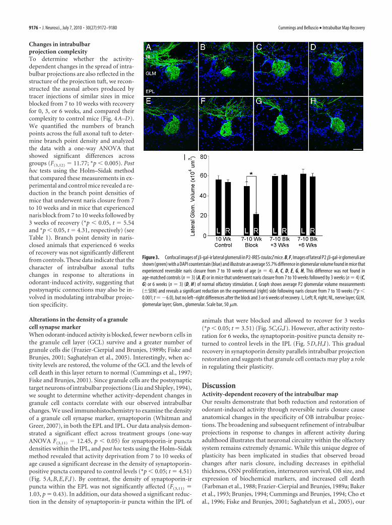
Changes in intrabulbarprojection complexityTo determine whether the activity-dependent changes in the spread of intra-bulbar projections are also reflected in thestructure of the projection tuft, we recon-structed the axonal arbors produced bytracer injections of similar sizes in miceblocked from 7 to 10 weeks with recoveryfor 0, 3, or 6 weeks, and compared theircomplexity to control mice (Fig. 4A–D).We quantified the numbers of branchpoints across the full axonal tuft to deter-mine branch point density and analyzedthe data with a one-way ANOVA thatshowed significant differences acrossgroups (F(3,12) � 11.77; *p � 0.005). Posthoc tests using the Holm–Sidak methodthat compared these measurements in ex-perimental and control mice revealed a re-duction in the branch point densities ofmice that underwent naris closure from 7to 10 weeks and in mice that experiencednaris block from 7 to 10 weeks followed by3 weeks of recovery (*p � 0.05, t � 5.54and *p � 0.05, t � 4.31, respectively) (seeTable 1). Branch point density in naris-closed animals that experienced 6 weeksof recovery was not significantly differentfrom controls. These data indicate that thecharacter of intrabulbar axonal tuftschanges in response to alterations inodorant-induced activity, suggesting thatpostsynaptic connections may also be in-volved in modulating intrabulbar projec-tion specificity.
Alterations in the density of a granulecell synapse markerWhen odorant-induced activity is blocked, fewer newborn cells inthe granule cell layer (GCL) survive and a greater number ofgranule cells die (Frazier-Cierpial and Brunjes, 1989b; Fiske andBrunjes, 2001; Saghatelyan et al., 2005). Interestingly, when ac-tivity levels are restored, the volume of the GCL and the levels ofcell death in this layer return to normal (Cummings et al., 1997;Fiske and Brunjes, 2001). Since granule cells are the postsynaptictarget neurons of intrabulbar projections (Liu and Shipley, 1994),we sought to determine whether activity-dependent changes ingranule cell contacts correlate with our observed intrabulbarchanges. We used immunohistochemistry to examine the densityof a granule cell synapse marker, synaptoporin (Whitman andGreer, 2007), in both the EPL and IPL. Our data analysis demon-strated a significant effect across treatment groups (one-wayANOVA F(3,11) � 12.45, p � 0.05) for synaptoporin-ir punctadensities within the IPL, and post hoc tests using the Holm–Sidakmethod revealed that activity deprivation from 7 to 10 weeks ofage caused a significant decrease in the density of synaptoporin-positive puncta compared to control levels (*p � 0.05; t � 4.51)(Fig. 5A,B,E,F,I). By contrast, the density of synaptoporin-irpuncta within the EPL was not significantly affected (F(3,11) �1.03, p � 0.43). In addition, our data showed a significant reduc-tion in the density of synaptoporin-ir puncta within the IPL of
animals that were blocked and allowed to recover for 3 weeks(*p � 0.05; t � 3.51) (Fig. 5C,G,I). However, after activity resto-ration for 6 weeks, the synaptoporin-positive puncta density re-turned to control levels in the IPL (Fig. 5D,H,I). This gradualrecovery in synaptoporin density parallels intrabulbar projectionrestoration and suggests that granule cell contacts may play a rolein regulating their plasticity.
DiscussionActivity-dependent recovery of the intrabulbar mapOur results demonstrate that both reduction and restoration ofodorant-induced activity through reversible naris closure causeanatomical changes in the specificity of OB intrabulbar projec-tions. The broadening and subsequent refinement of intrabulbarprojections in response to changes in afferent activity duringadulthood illustrates that neuronal circuitry within the olfactorysystem remains extremely dynamic. While this unique degree ofplasticity has been implicated in studies that observed broadchanges after naris closure, including decreases in epithelialthickness, OSN proliferation, interneuron survival, OB size, andexpression of biochemical markers, and increased cell death(Farbman et al., 1988; Frazier-Cierpial and Brunjes, 1989a; Bakeret al., 1993; Brunjes, 1994; Cummings and Brunjes, 1994; Cho etal., 1996; Fiske and Brunjes, 2001; Saghatelyan et al., 2005), our
Figure 3. Confocal images of �-gal-ir lateral glomeruli in P2-IRES-taulacZ mice. B, F, Images of lateral P2 �-gal-ir glomeruli areshown (green) with a DAPI counterstain (blue) and illustrate an average 55.7% difference in glomerular volume found in mice thatexperienced reversible naris closure from 7 to 10 weeks of age (n � 4). A, C, D, E, G, H, This difference was not found inage-matched controls (n � 3) (A, E) or in mice that underwent naris closure from 7 to 10 weeks followed by 3 weeks (n � 4) (C,G) or 6 weeks (n � 3) (D, H ) of normal olfactory stimulation. I, Graph shows average P2 glomerular volume measurements(�SEM) and reveals a significant reduction on the experimental (right) side following naris closure from 7 to 10 weeks (*p �0.001; t ��6.0), but no left–right differences after the block and 3 or 6 weeks of recovery. L, Left; R, right; NL, nerve layer; GLM,glomerular layer; Glom., glomerular. Scale bar, 50 �m.
9176 • J. Neurosci., July 7, 2010 • 30(27):9172–9180 Cummings and Belluscio • Intrabulbar Map Recovery

study presents clear evidence of this ongoing plasticity within aspecific circuit.
Previous experiments using reversible olfactory deprivationalso revealed that removal of the naris block following a period ofnaris closure resulted in increased neuronal survival and volu-metric recovery of the layers within the ipsilateral OB
(Cummings et al., 1997). Similarly, research on olfactory bulbregeneration suggests that activity can lead to substantial in-creases in the integration of new neurons into existing OB cir-cuitry (Rochefort et al., 2002; Alonso et al., 2006). These findingsalong with other work highlight the presence of long-term devel-opmental changes and/or plasticity within the OE and OB, likelyassociated with regeneration in both regions (Weiler andFarbman, 1997; Lee et al., 2009; Zou et al., 2009). Our currentfindings demonstrate that reversible naris closure, initiated for 3weeks either before or after the intrabulbar map matures, causesa spread in intrabulbar axons similar to those observed after per-manent naris closure (Marks et al., 2006). Remarkably, we showthat by reinstating normal levels of olfactory stimulation the in-trabulbar projections can gradually refine and recover their spec-ificity (Fig. 6). This suggests that the changes incurred by theolfactory bulb during sensory deprivation are not permanent butcan be reversed. Thus, in addition to the general increasesin neuronal survival and OB volume reported previously(Cummings et al., 1997), odorant-induced activity can alsorestore the specificity of the axonal connections that make upthe intrabulbar map.
Figure 4. Three-dimensional reconstructions of intrabulbar axonal tufts. Flattened images of confocal stacks of intrabulbar axonal projection sites are shown with corresponding 3D reconstruc-tions below in each case. A–D, Representative confocal images and reconstructions are shown from 10-week-old controls (A), mice that experienced naris closure from 7 to 10 weeks (B), and micethat underwent naris block followed by 3 weeks (C) or 6 weeks (D) of normal levels of afferent activity. Scale bar, 50 �m.
Table 1. Averaged process length and branch point measurements ofreconstructed intrabulbar projection sites
Group nAverage total filamentlength � SEM (�m)
Average no.of branchpts � SEM
Branch pts/totalfilament length(�m)
Controls 3 6866 � 458 560 � 57 0.0827–10 wk blk 4 9098 � 468 515 � 22 0.057*Blk � 3 wks 3 8497 � 301 517 � 7 0.061*Blk � 6 wks 3 6703 � 852 478 � 72 0.071
The table shows numbers of animals per group with total axonal filament length (�SEM), average number (no.) ofbranch points (pts) (�SEM), and the resulting average number of branch points per total filament length. Signifi-cant differences were observed in these branch point density values in 7–10 week (wk) blocked (blk) mice (*p �0.05, t � 5.54) and in mice blocked from 7–10 weeks followed by 3 weeks of recovery (*p � 0.05, t � 4.31)compared to controls. No significant difference emerged when branch point densities of mice blocked from 7–10weeks plus 6 weeks of recovery were compared to controls.
Cummings and Belluscio • Intrabulbar Map Recovery J. Neurosci., July 7, 2010 • 30(27):9172–9180 • 9177

Rapid return in OSNs and the glomerular mapGiven that intrabulbar map recovery is contingent upon olfactorysensory input, we examined activity-dependent changes in theolfactory epithelium by following P2-OSNs and their associateglomeruli. We reasoned that if olfactory input is required forintrabulbar map recovery, then it may be necessary for OSNchanges to recover before the restoration of intrabulbar projec-tion specificity, which is indeed what we found. We noted a�35% reduction in the number of P2-lacZ-expressing neuronsafter a 3 week period of naris occlusion which recovered to con-trol levels within 3 weeks of removing the naris block (Fig. 2).While the mechanism underlying these epithelial changes is un-clear, studies have shown that odorant deprivation beginningshortly after birth causes a reduction in neurogenesis and a de-crease in epithelial thickness (Farbman et al., 1988; Cummingsand Brunjes, 1994), suggesting that changes in OSN turnover arelikely involved. In addition, factors such as a variation in air flow(Scott-Johnson et al., 2000) or mucus consistency (Getchell et al.,1987; Persaud et al., 1987) that result from the naris occlusionmay also contribute to the epithelial fluctuations we observe.
At the glomerular level, our findings further demonstrate therapid recovery of OSNs in response to renewed odorant-inducedactivity. Studies have shown that the glomerular map undergoes aperiod of activity-dependent postnatal refinement during whichheterogeneous and ectopic glomeruli mature into more typicalarrangements of isofunctional glomerular pairs (Treloar et al.,2002; Zou et al., 2004). Experience also appears to play a role inthis maturation since odorant conditioning has been shown toenhance glomerular refinement (Kerr and Belluscio, 2006), andolfactory-dependent associative learning can increase the num-ber of specific OSNs and associated glomerular size (Jones et al.,2008). In this study, we observed that P2 glomeruli undergo a
reduction and subsequent restoration in volume that directlyparallels the P2-OSN changes in the epithelium. Interestingly, P2glomeruli remained intact and their position did not appear tochange as a result of odorant deprivation, suggesting that glomer-ular location and composition may be relatively stable once themap matures.
Postsynaptic changes associated with intrabulbarmap plasticityWe show that olfactory sensory input is clearly necessary to drivethe refinement and restoration of intrabulbar projections. How-ever, the remodeling process itself is very likely dependent uponpostsynaptic factors as well. For example, the elements necessaryfor axonal reorganization in the IPL depend in part upon thedestabilization, elimination, and/or addition of synaptic con-tacts. At a circuit level, intrabulbar projections form a communi-cation link between two regenerating populations of neurons:primary OSNs within the glomerulus and granule cells on theopposite side of the bulb through synaptic contacts in the IPL.Thus, synaptic fluctuations within the IPL are a separate andsomewhat complementary indicator of the plasticity that we ob-serve directly through labeling intrabulbar axons, first revealingtheir ability to expand and contract; and second demonstrating achange in their complexity as revealed by our 3D reconstructions.In addition, we demonstrate through synaptoporin staining thatactivity deprivation produced a marked decrease in synaptic den-sity within the IPL that returns to control levels following �6weeks of activity restoration (Fig. 5). These data are consistentwith studies showing that blocking olfactory sensory input causesa decrease in the number of OB granule cells (Fiske and Brunjes,2001), the postsynaptic partners of intrabulbar projections, sug-
Figure 5. Activity-dependent changes in synaptoporin-ir puncta density within the EPL and IPL. A–H, Confocal images of synaptoporin immunostaining in OB sections from 10 week control mice(A, E), mice that underwent naris closure from 7 to 10 weeks (B, F ), and mice that experienced naris closure followed by 3 weeks (C, G) or 6 weeks (D, H ) of recovery. The white dotted line in A–Ddelineates the mitral cell layer. Boxed regions of the IPL of A–D are shown in higher magnification in E–H. I, Graph shows the quantification of synaptoporin-ir puncta densities in the EPL and IPL.Naris closure led to a decrease in the density of synaptoporin-ir puncta in the IPL (*p � 0.05; t � 4.51), and this decrease persisted in mice that underwent olfactory deprivation followed by 3 weeksof recovery (*p � 0.05; t � 3.51), but not in the group that recovered for 6 weeks. In addition, no significant differences were observed in synaptoporin-ir puncta densities within the EPL. Scale bar(in H): A–D, 50 �m; E–H, 30 �m.
9178 • J. Neurosci., July 7, 2010 • 30(27):9172–9180 Cummings and Belluscio • Intrabulbar Map Recovery

gesting that intrabulbar projection plasticity may be partiallylinked to granule cell turnover.
The multiple maps of the olfactory bulb arefunctionally linkedMost sensory systems undergo periods during their developmentwhen rudimentary connections become refined and can beshaped by the stimulus environment. For example, in both thevisual and somatosensory systems, manipulating afferent activityduring certain sensitive periods alters the arrangement of corticalrepresentations (Hubel and Wiesel, 1970; Belford and Killackey,1980; Fox, 1992). Although these systems have traditionally beencharacterized as lacking plasticity beyond these early critical win-dows, recent evidence suggests that adult plasticity may exist tosome degree (Trachtenberg et al., 2002; De Paola et al., 2006; Heet al., 2007). By comparison, the olfactory system maintains acontinuous level of plasticity and regeneration both at the periph-ery in the OE, and at central levels within the bulb. While thepurpose of this plasticity is unclear, nowhere is it more obviousthan in the activity-dependent changes presented in the intrab-ulbar map. We propose that intrabulbar projections are necessaryto coordinate the communication between the two mirror-symmetric glomerular maps, which in turn work together to gen-erate specific patterns of synchronous activity that we believe arenecessary for cortical integration of signals. In a previous study,we show that intrabulbar projecting neurons can modulate thefiring of mitral cells through a timing-based gating mechanism(Zhou and Belluscio, 2008). Thus, given the anatomical specific-ity of the intrabulbar map, this mechanism could be used toselectively adjust the timing of mitral cell activity between pairs ofisofunctional glomeruli. This in turn could be used to producespecific synchronous patterns of activity that may be interpretedwithin the piriform cortex to identify specific odorants.
Our results suggest that the specificity of intrabulbar projec-tions depends upon the integrity of the sensory epithelium andglomerular map, since refinement among intrabulbar axons oc-curred only after OSN numbers and glomerular volume returnedto normal. Indeed, the recovery of intrabulbar projection speci-ficity occurs between 6 and 9 weeks following block removal (seeFig. 6), suggesting that this return to precision may depend uponmultiple factors. Olfactory bulb granule cells, the postsynaptictargets of intrabulbar axons, may also play a role in modulatingintrabulbar plasticity, possibly facilitating circuit dynamics sim-
ply through their regenerative capacity. While the precise func-tional interactions between the many cell types of the OB warrantfurther study, we propose that the intrabulbar map derives itsorganization from distinct glomerular activity patterns that aretransmitted through the odor column circuitry. Activation tim-ing could then be used to stabilize or reinforce intrabulbar con-nections between isofunctional odor columns while destabilizinginappropriate connections to allow for axonal pruning. In addi-tion, molecular factors that change with altered activity levelscould also modulate the extension or retraction of axon termi-nals. Although specific mechanisms for such a process have yet tobe determined, our results clearly indicate that sensory inputreflected in the glomerular map ultimately drives the organiza-tional maintenance and restoration of the intrabulbar map.
ReferencesAlonso M, Viollet C, Gabellec MM, Meas-Yedid V, Olivo-Marin JC, Lledo
PM (2006) Olfactory discrimination learning increases the survival ofadult-born neurons in the olfactory bulb. J Neurosci 26:10508 –10513.
Baker H, Morel K, Stone DM, Maruniak JA (1993) Adult naris closure pro-foundly reduces tyrosine hydroxylase expression in mouse olfactory bulb.Brain Res 614:109 –116.
Belford GR, Killackey HP (1980) The sensitive period in the development ofthe trigeminal system of the neonatal rat. J Comp Neurol 193:335–350.
Belluscio L, Lodovichi C, Feinstein P, Mombaerts P, Katz LC (2002) Odor-ant receptors instruct functional circuitry in the mouse olfactory bulb.Nature 419:296 –300.
Bozza T, Feinstein P, Zheng C, Mombaerts P (2002) Odorant receptor ex-pression defines functional units in the mouse olfactory system. J Neuro-sci 22:3033–3043.
Brunjes PC (1994) Unilateral naris closure and olfactory system develop-ment. Brain Res Brain Res Rev 19:146 –160.
Brunjes PC, Smith-Crafts LK, McCarty R (1985) Unilateral odor depriva-tion: effects on the development of olfactory bulb catecholamines andbehavior. Brain Res 354:1– 6.
Buck L, Axel R (1991) A novel multigene family may encode odorant recep-tors: a molecular basis for odor recognition. Cell 65:175–187.
Cho JY, Min N, Franzen L, Baker H (1996) Rapid down-regulation of ty-rosine hydroxylase expression in the olfactory bulb of naris-occludedadult rats. J Comp Neurol 369:264 –276.
Cummings DM, Belluscio L (2008) Charting plasticity in the regeneratingmaps of the mammalian olfactory bulb. Neuroscientist 14:251–263.
Cummings DM, Brunjes PC (1994) Changes in cell proliferation in the de-veloping olfactory epithelium following neonatal unilateral naris occlu-sion. Exp Neurol 128:124 –128.
Cummings DM, Brunjes PC (1997) The effects of variable periods of func-
Figure 6. Model of intrabulbar map plasticity. A, In the adult OB, isofunctional glomeruli (shown in green) are connected by STC axons (shown in red) that reciprocally project to the region of theIPL located below the “partner” glomerulus. B, If odorant-induced activity is blocked by naris closure, glomerular size becomes reduced and intrabulbar projections broaden. C, Reopening the narisfor 3 weeks results in the recovery of glomerular volume; however, the intrabulbar projections remain broad. D, Reopening the naris and recovery for �6 –9 weeks allows the intrabulbar map toreturn to control levels of precision.
Cummings and Belluscio • Intrabulbar Map Recovery J. Neurosci., July 7, 2010 • 30(27):9172–9180 • 9179

tional deprivation on olfactory bulb development in rats. Exp Neurol148:360 –366.
Cummings DM, Henning HE, Brunjes PC (1997) Olfactory bulb recoveryafter early sensory deprivation. J Neurosci 17:7433–7440.
Cummings DM, Emge DK, Small SL, Margolis FL (2000) Pattern of olfac-tory bulb innervation returns after recovery from reversible peripheraldeafferentation. J Comp Neurol 421:362–373.
De Paola V, Holtmaat A, Knott G, Song S, Wilbrecht L, Caroni P, Svoboda K(2006) Cell type-specific structural plasticity of axonal branches andboutons in the adult neocortex. Neuron 49:861– 875.
Farbman AI, Brunjes PC, Rentfro L, Michas J, Ritz S (1988) The effect ofunilateral naris occlusion on cell dynamics in the developing rat olfactoryepithelium. J Neurosci 8:3290 –3295.
Fiske BK, Brunjes PC (2001) Cell death in the developing and sensory-deprived rat olfactory bulb. J Comp Neurol 431:311–319.
Fox K (1992) A critical period for experience-dependent synaptic plasticityin rat barrel cortex. J Neurosci 12:1826 –1838.
Frazier-Cierpial L, Brunjes PC (1989a) Early postnatal cellular proliferationand survival in the olfactory bulb and rostral migratory stream of normaland unilaterally odor-deprived rats. J Comp Neurol 289:481– 492.
Frazier-Cierpial LL, Brunjes PC (1989b) Early postnatal differentiation ofgranule cell dendrites in the olfactory bulbs of normal and unilaterallyodor-deprived rats. Brain Res Dev Brain Res 47:129 –136.
Getchell ML, Zielinski B, DeSimone JA, Getchell TV (1987) Odorant stim-ulation of secretory and neural processes in the salamander olfactorymucosa. J Comp Physiol A 160:155–168.
He HY, Ray B, Dennis K, Quinlan EM (2007) Experience-dependent recov-ery of vision following chronic deprivation amblyopia. Nat Neurosci10:1134 –1136.
Hubel DH, Wiesel TN (1970) The period of susceptibility to the physiolog-ical effects of unilateral eye closure in kittens. J Physiol 206:419 – 436.
Jones SV, Choi DC, Davis M, Ressler KJ (2008) Learning-dependentstructural plasticity in the adult olfactory pathway. J Neurosci28:13106 –13111.
Kerr MA, Belluscio L (2006) Olfactory experience accelerates glomerularrefinement in the mammalian olfactory bulb. Nat Neurosci 9:484 – 486.
Lee AC, Tian H, Grosmaitre X, Ma M (2009) Expression patterns of odorantreceptors and response properties of olfactory sensory neurons in agedmice. Chem Senses 34:695–703.
Liu WL, Shipley MT (1994) Intrabulbar associational system in the rat ol-factory bulb comprises cholecystokinin-containing tufted cells that syn-apse onto the dendrites of GABAergic granule cells. J Comp Neurol346:541–558.
Lodovichi C, Belluscio L, Katz LC (2003) Functional topography of connec-tions linking mirror-symmetric maps in the mouse olfactory bulb. Neu-ron 38:265–276.
Marks CA, Cheng K, Cummings DM, Belluscio L (2006) Activity-dependent plasticity in the olfactory intrabulbar map. J Neurosci26:11257–11266.
Mombaerts P, Wang F, Dulac C, Chao SK, Nemes A, Mendelsohn M, Ed-mondson J, Axel R (1996) Visualizing an olfactory sensory map. Cell87:675– 686.
Nakatani H, Serizawa S, Nakajima M, Imai T, Sakano H (2003) Develop-mental elimination of ectopic projection sites for the transgenic OR genethat has lost zone specificity in the olfactory epithelium. Eur J Neurosci18:2425–2432.
Persaud KC, DeSimone JA, Getchell ML, Heck GL, Getchell TV (1987) Iontransport across the frog olfactory mucosa: the basal and odorant-stimulated states. Biochim Biophys Acta 902:65–79.
Ressler KJ, Sullivan SL, Buck LB (1994) Information coding in the olfactorysystem: evidence for a stereotyped and highly organized epitope map inthe olfactory bulb. Cell 79:1245–1255.
Rochefort C, Gheusi G, Vincent JD, Lledo PM (2002) Enriched odor expo-sure increases the number of newborn neurons in the adult olfactory bulband improves odor memory. J Neurosci 22:2679 –2689.
Saghatelyan A, Roux P, Migliore M, Rochefort C, Desmaisons D, Charneau P,Shepherd GM, Lledo PM (2005) Activity-dependent adjustments of theinhibitory network in the olfactory bulb following early postnatal depri-vation. Neuron 46:103–116.
Scott-Johnson PE, Blakley D, Scott JW (2000) Effects of air flow on rat elec-troolfactogram. Chem Senses 25:761–768.
Suh KS, Kim SY, Bae YC, Ronnett GV, Moon C (2006) Effects of unilateralnaris occlusion on the olfactory epithelium of adult mice. Neuroreport17:1139 –1142.
Trachtenberg JT, Chen BE, Knott GW, Feng G, Sanes JR, Welker E, SvobodaK (2002) Long-term in vivo imaging of experience-dependent synapticplasticity in adult cortex. Nature 420:788 –794.
Treloar HB, Feinstein P, Mombaerts P, Greer CA (2002) Specificity of glo-merular targeting by olfactory sensory axons. J Neurosci 22:2469 –2477.
Vassar R, Chao SK, Sitcheran R, Nunez JM, Vosshall LB, Axel R (1994)Topographic organization of sensory projections to the olfactory bulb.Cell 79:981–991.
Weiler E, Farbman AI (1997) Proliferation in the rat olfactory epithelium:age-dependent changes. J Neurosci 17:3610 –3622.
Whitman MC, Greer CA (2007) Synaptic integration of adult-generated ol-factory bulb granule cells: basal axodendritic centrifugal input precedesapical dendrodendritic local circuits. J Neurosci 27:9951–9961.
Zhou Z, Belluscio L (2008) Intrabulbar projecting external tufted cells me-diate a timing-based mechanism that dynamically gates olfactory bulboutput. J Neurosci 28:9920 –9928.
Zhuang H, Matsunami H (2007) Synergism of accessory factors in func-tional expression of mammalian odorant receptors. J Biol Chem282:15284 –15293.
Zou DJ, Feinstein P, Rivers AL, Mathews GA, Kim A, Greer CA, Mombaerts P,Firestein S (2004) Postnatal refinement of peripheral olfactory projec-tions. Science 304:1976 –1979.
Zou DJ, Chesler A, Firestein S (2009) How the olfactory bulb got its glomer-uli: a just so story? Nat Rev Neurosci 10:611– 618.
9180 • J. Neurosci., July 7, 2010 • 30(27):9172–9180 Cummings and Belluscio • Intrabulbar Map Recovery


![Glomerular Function and Structure in Living Donors ... · glomerular filtration rate (SNGFR) and glomerular capillary hydraulic pressure (P GC)[3]. Further insights into glomerular](https://static.fdocuments.in/doc/165x107/5ed58c3d3f40d10acd516aa6/glomerular-function-and-structure-in-living-donors-glomerular-filtration-rate.jpg)


