Continual renewal and replication of persistent Leishmania major ... · asites within iNOS+ cells...
10
Continual renewal and replication of persistent Leishmania major parasites in concomitantly immune hosts Michael A. Mandell a,1 and Stephen M. Beverley a,2 a Department of Molecular Microbiology, Washington University School of Medicine, St. Louis, MO 63110 Contributed by Stephen M. Beverley, December 15, 2016 (sent for review November 22, 2016; reviewed by Arturo Casadevall and Phillip Scott) In most natural infections or after recovery, small numbers of Leish- mania parasites remain indefinitely in the host. Persistent parasites play a vital role in protective immunity against disease pathology upon reinfection through the process of concomitant immunity, as well as in transmission and reactivation, yet are poorly understood. A key question is whether persistent parasites undergo replication, and we devised several approaches to probe the small numbers in persistent infections. We find two populations of persistent Leish- mania major: one rapidly replicating, similar to parasites in acute infections, and another showing little evidence of replication. Per- sistent Leishmania were not found in “safe” immunoprivileged cell types, instead residing in macrophages and DCs, ∼60% of which expressed inducible nitric oxide synthase (iNOS). Remarkably, par- asites within iNOS + cells showed normal morphology and genome integrity and labeled comparably with BrdU to parasites within iNOS − cells, suggesting that these parasites may be unexpectedly resistant to NO. Nonetheless, because persistent parasite numbers remain roughly constant over time, their replication implies that ongoing destruction likewise occurs. Similar results were obtained with the attenuated lpg2 − mutant, a convenient model that rapidly enters a persistent state without inducing pathology due to loss of the Golgi GDP mannose transporter. These data shed light on Leish- mania persistence and concomitant immunity, suggesting a model wherein a parasite reservoir repopulates itself indefinitely, whereas some progeny are terminated in antigen-presenting cells, thereby stimulating immunity. This model may be relevant to understand- ing immunity to other persistent pathogen infections. latency | quiescence | trypanosomatid protozoan parasite | vaccination | stem cell-like A s long-term infection of a host can increase a pathogen’s chances of transmission, many have evolved the ability to prolong their survival within their hosts. Leishmania, Mycobac- terium tuberculosis, Toxoplasma gondii, and latent herpesviruses can remain indefinitely within their hosts in small numbers without overt pathology. Such low-level persistent infections differ considerably from “chronic” infections, where pathogen numbers are typically much higher and often accompanied by disease symptoms (1). Unfortunately the terms persistent and/or chronic are applied inconsistently or even interchangeably across pathogens, in part reflecting the evolution of our understanding, organism-specific lexicons, and diverse mechanisms used by pathogens for long-term survival within the host (1, 2). Here we focus on persistent infections exclusively of the “low numbers” type, which are of medical importance due to their ability to reactivate to severe disease (3, 4), to serve as reservoirs for trans- mission, and to engender a protective response against subsequent infections, a process known as concomitant immunity (5). Persistence is a significant but understudied aspect of the bi- ology of parasites of the genus Leishmania, the causative agents of leishmaniasis. These parasites are responsible for an esti- mated 12 million active cases, at least 120 million asymptomatic infections, and 1.7 billion people are at risk (6–9). Leishmania sp. are transmitted as metacyclic-stage promastigotes to humans by the bite of an infected sand fly. In the skin, parasites are engulfed by phagocytic cells, where they differentiate into the amastigote stage and begin to replicate. Although most infections are asymptomatic, a significant fraction go on to produce ulcerating skin lesions; in both cases, parasites can metastasize to other sites and cause more severe disease such as visceral or mucocutane- ous leishmaniasis (9). For Leishmania major, in most human cases and experimental infections of “resistant” mouse strains, the infection is eventually controlled by an adaptive Th1 immune response, in a manner requiring inducible NOS (iNOS) (10, 11). Thereafter, the number of parasites in infected tissue declines dramatically, the lesion heals, and the host becomes im- mune to subsequent L. major infection (12, 13). However, a small and steady population of parasites remains at the site of infection and in the lymph node draining that site for the rest of the host’s life (14). These persistent parasites play vital roles in Leishmania bi- ology. Despite their limited numbers (∼1,000), persistent para- sites can be transmitted to sand fly vectors and thus could function as a transmission reservoir (6, 15, 16). Second, they pose a substantial risk to infected people in the event of immuno- suppression, as the persistent parasites can “reactivate,” leading to severe disease (17). Finally, they maintain protective immunity to subsequent Leishmania infections through concomitant immu- nity, which results in amelioration of disease pathology without sterilization of either the persistent or incoming parasite (18, 19). Indeed, healed Leishmania infections are the gold standard in anti-Leishmania immunity, and to date no other vaccination ap- proaches have proven as successful in humans (18). Importantly, treatment of persistently infected mice to achieve a sterile cure renders those mice susceptible to new infections (14, 20), sug- gesting that the persistent parasites are required for strong, long- lasting anti-Leishmania immunity. Significance Persistent parasites contribute to the maintenance of pro- tective immune responses through concomitant immunity, but our understanding of how they do so has been limited by the difficulties associated with their low numbers. Our studies in- dicate that for Leishmania substantial parasite replication oc- curs in persistent infections, with most parasites found within activated antigen-presenting cells. Parasite replication serves to maintain the infection and likely also provides a constant source of parasite antigens for immune stimulation and the maintenance of protective immunity. Collectively, these studies suggest a framework to understand concomitant immunity that may be applicable to other persistent pathogens. Author contributions: M.A.M. and S.M.B. designed research; M.A.M. performed research; M.A.M. and S.M.B. analyzed data; and M.A.M. and S.M.B. wrote the paper. Reviewers: A.C., Johns Hopkins Bloomberg School of Public Health; and P.S., University of Pennsylvania. The authors declare no conflict of interest. 1 Present address: Department of Molecular Genetics and Microbiology, University of New Mexico Health Sciences Center, Albuquerque, NM 87131. 2 To whom correspondence should be addressed. Email: [email protected]. This article contains supporting information online at www.pnas.org/lookup/suppl/doi:10. 1073/pnas.1619265114/-/DCSupplemental. www.pnas.org/cgi/doi/10.1073/pnas.1619265114 PNAS | Published online January 12, 2017 | E801–E810 MICROBIOLOGY PNAS PLUS
Transcript of Continual renewal and replication of persistent Leishmania major ... · asites within iNOS+ cells...
Continual renewal and replication of persistent Leishmaniamajor parasites in concomitantly immune hostsMichael A. Mandella,1 and Stephen M. Beverleya,2
aDepartment of Molecular Microbiology, Washington University School of Medicine, St. Louis, MO 63110
Contributed by Stephen M. Beverley, December 15, 2016 (sent for review November 22, 2016; reviewed by Arturo Casadevall and Phillip Scott)
In most natural infections or after recovery, small numbers of Leish-mania parasites remain indefinitely in the host. Persistent parasitesplay a vital role in protective immunity against disease pathologyupon reinfection through the process of concomitant immunity, aswell as in transmission and reactivation, yet are poorly understood.A key question is whether persistent parasites undergo replication,and we devised several approaches to probe the small numbers inpersistent infections. We find two populations of persistent Leish-mania major: one rapidly replicating, similar to parasites in acuteinfections, and another showing little evidence of replication. Per-sistent Leishmania were not found in “safe” immunoprivileged celltypes, instead residing in macrophages and DCs, ∼60% of whichexpressed inducible nitric oxide synthase (iNOS). Remarkably, par-asites within iNOS+ cells showed normal morphology and genomeintegrity and labeled comparably with BrdU to parasites withiniNOS− cells, suggesting that these parasites may be unexpectedlyresistant to NO. Nonetheless, because persistent parasite numbersremain roughly constant over time, their replication implies thatongoing destruction likewise occurs. Similar results were obtainedwith the attenuated lpg2− mutant, a convenient model that rapidlyenters a persistent state without inducing pathology due to loss ofthe Golgi GDP mannose transporter. These data shed light on Leish-mania persistence and concomitant immunity, suggesting a modelwherein a parasite reservoir repopulates itself indefinitely, whereassome progeny are terminated in antigen-presenting cells, therebystimulating immunity. This model may be relevant to understand-ing immunity to other persistent pathogen infections.
latency | quiescence | trypanosomatid protozoan parasite | vaccination |stem cell-like
As long-term infection of a host can increase a pathogen’schances of transmission, many have evolved the ability to
prolong their survival within their hosts. Leishmania, Mycobac-terium tuberculosis, Toxoplasma gondii, and latent herpesvirusescan remain indefinitely within their hosts in small numberswithout overt pathology. Such low-level persistent infectionsdiffer considerably from “chronic” infections, where pathogennumbers are typically much higher and often accompanied bydisease symptoms (1). Unfortunately the terms persistent and/orchronic are applied inconsistently or even interchangeably acrosspathogens, in part reflecting the evolution of our understanding,organism-specific lexicons, and diverse mechanisms used bypathogens for long-term survival within the host (1, 2). Here wefocus on persistent infections exclusively of the “low numbers”type, which are of medical importance due to their ability toreactivate to severe disease (3, 4), to serve as reservoirs for trans-mission, and to engender a protective response against subsequentinfections, a process known as concomitant immunity (5).Persistence is a significant but understudied aspect of the bi-
ology of parasites of the genus Leishmania, the causative agentsof leishmaniasis. These parasites are responsible for an esti-mated 12 million active cases, at least 120 million asymptomaticinfections, and 1.7 billion people are at risk (6–9). Leishmania sp.are transmitted as metacyclic-stage promastigotes to humans bythe bite of an infected sand fly. In the skin, parasites are engulfedby phagocytic cells, where they differentiate into the amastigote
stage and begin to replicate. Although most infections areasymptomatic, a significant fraction go on to produce ulceratingskin lesions; in both cases, parasites can metastasize to other sitesand cause more severe disease such as visceral or mucocutane-ous leishmaniasis (9). For Leishmania major, in most humancases and experimental infections of “resistant” mouse strains,the infection is eventually controlled by an adaptive Th1 immuneresponse, in a manner requiring inducible NOS (iNOS) (10, 11).Thereafter, the number of parasites in infected tissue declinesdramatically, the lesion heals, and the host becomes im-mune to subsequent L. major infection (12, 13). However, asmall and steady population of parasites remains at the site ofinfection and in the lymph node draining that site for the rest ofthe host’s life (14).These persistent parasites play vital roles in Leishmania bi-
ology. Despite their limited numbers (∼1,000), persistent para-sites can be transmitted to sand fly vectors and thus couldfunction as a transmission reservoir (6, 15, 16). Second, they posea substantial risk to infected people in the event of immuno-suppression, as the persistent parasites can “reactivate,” leadingto severe disease (17). Finally, they maintain protective immunityto subsequent Leishmania infections through concomitant immu-nity, which results in amelioration of disease pathology withoutsterilization of either the persistent or incoming parasite (18, 19).Indeed, healed Leishmania infections are the gold standard inanti-Leishmania immunity, and to date no other vaccination ap-proaches have proven as successful in humans (18). Importantly,treatment of persistently infected mice to achieve a sterile curerenders those mice susceptible to new infections (14, 20), sug-gesting that the persistent parasites are required for strong, long-lasting anti-Leishmania immunity.
Significance
Persistent parasites contribute to the maintenance of pro-tective immune responses through concomitant immunity, butour understanding of how they do so has been limited by thedifficulties associated with their low numbers. Our studies in-dicate that for Leishmania substantial parasite replication oc-curs in persistent infections, with most parasites found withinactivated antigen-presenting cells. Parasite replication servesto maintain the infection and likely also provides a constantsource of parasite antigens for immune stimulation and themaintenance of protective immunity. Collectively, these studiessuggest a framework to understand concomitant immunitythat may be applicable to other persistent pathogens.
Author contributions: M.A.M. and S.M.B. designed research; M.A.M. performed research;M.A.M. and S.M.B. analyzed data; and M.A.M. and S.M.B. wrote the paper.
Reviewers: A.C., Johns Hopkins Bloomberg School of Public Health; and P.S., Universityof Pennsylvania.
The authors declare no conflict of interest.1Present address: Department of Molecular Genetics and Microbiology, University of NewMexico Health Sciences Center, Albuquerque, NM 87131.
2To whom correspondence should be addressed. Email: [email protected].
This article contains supporting information online at www.pnas.org/lookup/suppl/doi:10.1073/pnas.1619265114/-/DCSupplemental.
www.pnas.org/cgi/doi/10.1073/pnas.1619265114 PNAS | Published online January 12, 2017 | E801–E810
MICRO
BIOLO
GY
PNASPL
US
The host immune response is important to simultaneouslyprevent reactivation and clearance of persistent parasites (21, 22).Treatment of persistently infected mice with immunosuppressivedrugs, iNOS inhibitors, or the blockade of IFN-γ signaling rapidlyresults in increased parasite numbers and the reappearance ofdisease symptoms (11). In contrast, depletion of CD4+CD25+
regulatory T cells or the blockade of IL-10 signaling results insterile cure in mice (20, 23). The mechanisms used by persistentparasites to modulate the host’s immune responses or to maintainprotective immunity are less well understood. Indeed recentstudies have drawn attention to continuing antigenic stimulationarising from the site of infection, which is better understood fromthe host’s than parasite’s perspective (24).The study of persistent parasites poses significant experimen-
tal challenges. Typically persistence is studied after resolution ofdisease in a resistant (Th1) murine model, which requires >4 moto attain (11, 25) (Fig. S1D). Furthermore, the low number ofpersistent Leishmania renders their visualization, much lesscharacterization, a daunting task (26). Here we established sev-eral methods facilitating the study of both replication and hostcellular localization of the scarce persistent parasites. Our datashow that persistent parasites constitute two populations, onereplicating and one seemingly quiescent. Similar results werefound with the lpg2− mutant L. major lacking the Golgi GDP-mannose transporter, which fails to induce disease pathologyeven in susceptible (Th2) mice but persists indefinitely for the lifeof the animal (25), where it confers strong protective immunity(14, 27). Our studies suggest a model explaining how persistentLeishmania maintain concomitant immunity in which a smallreplicating reservoir acts to maintain a perpetual infection neededfor transmission, while spinning off a pool of parasites destined forimmune destruction and stimulation. This model potentially ap-plies to other persistent pathogens.
ResultsAssay of Leishmania Replication by BrdU Incorporation in Vitro.L. major promastigotes were incubated with 0.1 mM BrdU forvarious periods, followed by fixation and staining with a rat anti-BrdU monoclonal antibody to detect incorporation (28). Todetect the parasite nuclei, we chose a pool of antibodies specificallyrecognizing Leishmania histones, as these epitopes proved durableto the harsh acid fixations needed to monitor BrdU incorporation.
BrdU was incorporated into both the kinetoplast (mitochondri-al) DNA network and the nucleus (Fig. 1A), and under condi-tions where parasites replicated with a doubling time of ∼8 h, thepercent of BrdU+ cells rose steadily to 90% over 9 h (Fig. 1B).Longer labeling times did not lead to further increase, suggestingthat this was the maximum technically achievable. Under condi-tions of slow growth (serum reduction), a lower fraction of BrdU+
parasites was observed, arising by an increase in the length of G1phase as in most eukaryotes (Fig. S1 A–C). No BrdU+ cells wereseen in stationary phase promastigotes following 24 h of labeling.L. major-infected macrophages (MΦs) grown in the presence ofBrdU for 72 h resulted in 90% BrdU+ labeling of the intracellularamastigote stage nuclei. These studies show that BrdU labelsLeishmania as expected, in both parasite stages and within MΦ.
Visualizing Persistent Leishmania and Replication in Infected Animalsin Situ. For these studies, we chose to use the classical s.c. footpadinoculation model of L. major infection, as this allowed us toput our findings in the context of a large body of literatureaddressing Leishmania pathogenesis, immunology, persistence,and vaccination (21). To detect the low numbers of “persistentinfection parasites” (PIPs), the site of footpad infection was ex-cised, the tissue fixed, and 10 μm-thick serial sections preparedand labeled with the anti-Leishmania histones for confocal mi-croscopy. In some sections, as many as 10–100 parasites wereevident, wheras others had none. Integration of parasite num-bers across serial sections from a single footpad yielded numberscomparable to those typically obtained with limiting dilutionassays (100–2,000), suggesting that this approach was capable ofdetecting most parasites.We then combined the BrdU incorporation and in situ
Leishmania detection protocols to visualize parasite replicationin infected animals. There, Leishmania replicates rapidly forthe first 4–6 wk after inoculation (13); we refer to parasitesduring this period as “acute-infection parasites” (AIPs). Bio-luminescent imaging of luciferase-expressing parasites showedthat AIPs replicate exponentially with a doubling time of ∼60 hand as such provided an ideal setting to optimize in vivo BrdUincorporation assays.Because mammals rapidly clear nucleosides from circulation
(29), we tested several different BrdU delivery protocols as de-scribed in Materials and Methods, sacrificing treated mice 24 h
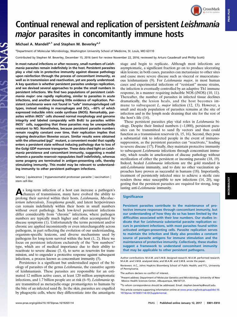
Fig. 1. BrdU incorporation assay demonstrates per-sistent parasite replication in situ. (A) Labeling of log-phase promastigotes with BrdU. (B) Parasite densityand extent of BrdU labeling of log-phase promasti-gotes cultured for 24 h in the presence of BrdU. Dark-shaded diamonds, parasite number; light-shadedsquares, percent BrdU+. (C) The effect of increasingthe number of BrdU doses (simultaneous local andsystematic injection) on the percent BrdU+ parasitesduring acute mouse footpad infections where para-sites are growing logarithmically. n = 3 mice; >1,000total parasites. (D) Confocal microscopic analysis ofBrdU incorporation of footpad PIPs. (E) Comparisonof the percent BrdU+ intracellular parasites in AIPSversus PIPs. Data points, mean percent BrdU labelingfor individual mice. Horizontal bars, mean for allmice. (F) Analysis of the BrdU-labeling intensity ofparasite nuclei. n = 20 parasites per category.Graph shows mean ± SEM; *P < 0.05; n.s., not sig-nificant by Student’s t test or ANOVA. Arrowheads,BrdU-labeled nuclei; arrows, BrdU-labeled kinetoplast(mitochondrial) DNA. (Scale bar, 5 μM.)
E802 | www.pnas.org/cgi/doi/10.1073/pnas.1619265114 Mandell and Beverley
after the start of the dosing period. Although all yielded abun-dant BrdU+ host cell nuclei, BrdU+ parasites were only detectedin mice receiving multiple i.p. and s.c. footpad injections. Thepercentage of parasites labeled increased linearly with thenumber of doses, and we adopted as the maximum practicable aregimen of 6 doses given every 3 h as a “standard” (Fig. 1C).Assuming a 24-h labeling period and a parasite doubling time of60 h, we calculated that 40% of AIPs should be BrdU+; by theprotocol above, 44 ± 6% were BrdU+, in good agreement.
PIPs Replicate in Vivo. We then tested whether PIPs incorporatedBrdU into their DNA. We defined the asymptomatic persistentinfection phase as >1 mo following the resolution of footpadswelling at the inoculation site (typically ∼4 mo postinfection;Fig. S1D). In all studies, multiple experiments, mice and sectionswere analyzed, enabling statistical tests of distribution and sig-nificance [represented as N = E (experiments)/M (mice)/P(parasites)]. We found that 19 ± 6% of PIPs showed BrdU+
nuclei (Fig. 1 D and E), about half that seen in AIPs (P <0.0001). The intensity of BrdU labeling per cell was similar forAIPs or PIPs (Fig. 1F), implying that differences reflected thenumber of replicating parasites rather than the rate of BrdUincorporation. If one assumed a homogeneously replicatingpopulation, a doubling time of about 120 h could be calculatedfor PIPs.For confirmation, we formulated an indirect assay based on
the number of parasites per cell. This approach assumes that forPIPs, host cells are initially infected by a single parasite, as thelow number renders the chance of multiple independent infec-tions unlikely. From this we inferred that host cells containingtwo or more PIPs (here termed “clusters” as opposed to sin-gletons) must have arisen exclusively through parasite replica-tion, thereby providing an indirect metric. In contrast, for in vitroMΦ infections, which are usually performed at high multiplicitiesof infection (and possibly for AIPs where parasite numbers canbe quite high), such an inference would not be valid. Thus, PIPcluster analysis provides an alternative perspective on replication.In both AIPs and PIPs, we readily found clusters containing
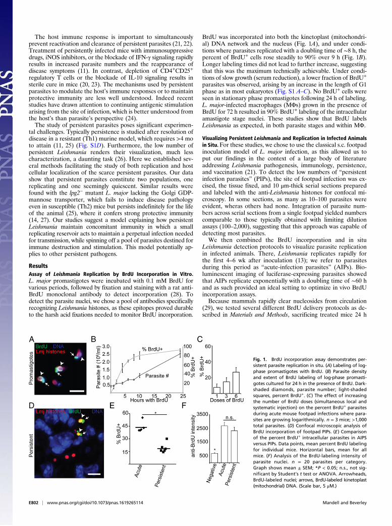
2–20 parasites per host cell in mice that were not subjected toBrdU dosing. Consistent with the results of the BrdU in-corporation assay, AIPs showed a higher proportion of parasitesfound in clusters, with a higher fraction containing >8 parasitesthan seen for PIPs (Fig. 2 A and B). Nevertheless, in persistentinfections, roughly half of all infected cells contained clusterswith two or more PIPs (Fig. 2A), and ∼80% of all PIPs werefound in clusters (Fig. 2B). This provided independent support ofPIP replication. Across all studies, singletons and clusters wereinterspersed, arguing against the possibility of segregation ofcells harboring replicating and nonreplicating PIPs.
Persistent Leishmania Show Two Populations with Different ReplicationRates: One “Fast,” One Slow/Nonreplicating. Combining the BrdUincorporation and cluster assays allowed us to test whether PIPsreplicated as a homogeneous population. We plotted the percentof parasite clusters as a function of the percent BrdU+ withinthose clusters (Fig. 2C). For a homogeneous population, a dis-tribution centered about the mean was anticipated, and indeed,this was seen with AIPs (mean BrdU+ = 44 ± 6%; Fig. 2C). Incontrast, PIPs did not show a single distribution centered around19% labeling, instead exhibiting two peaks (Fig. 2C). The firstpeak, accounting for ∼60% of the total clusters, showed noBrdU+ cells under these labeling conditions, whereas the secondpeak, accounting for the remaining 40% of clusters, showed amean BrdU+ of 57 ± 21%. The distribution and mean of thesecond population resembled that of AIP clusters (mean BrdU+
of 46 ± 29%; Fig. 2C).Although this suggested the existence of two populations, we
were concerned that the analysis excluded singleton parasites not
in clusters, about ∼20% of all PIPs. We thus reanalyzed the databy plotting the percent BrdU+ cells as a function of cluster size.Assuming homogeneous parasite replication, roughly 44% ofAIPs and 19% of PIPs should be BrdU+, regardless of thenumber of parasites per host cell. AIPs very closely matched thisprediction, with 40–50% BrdU+ uniformly across cluster size(Fig. 2D). However, for PIPs only ∼12% of the parasites in hostcells bearing 1–3 parasites were BrdU+, whereas ∼46% of theparasites within clusters containing four or more parasites wereBrdU+ (Fig. 2D), a value very close to the maximum obtainableby these labeling conditions (Fig. 1).By integrating the data shown in Fig. 2 B–D and correcting for
BrdU labeling efficiency, we conclude that there are two pop-ulations in persistent Leishmania infections: one comprisingabout 61% of the total cells, whose replication properties re-semble those of AIPs (replicating with a doubling time of ∼60 h;Fig. 1). In contrast, a second population comprises about 39% ofthe cells, which under the conditions used fail to incorporateBrdU significantly. This second population is much less abundantin AIPs (Fig. 2C; 0% BrdU+ population). Although one couldinterpret the “BrdU null” population as evidence of arrested ornonreplicating PIPs, we cannot exclude the possibility that theirreplication is just very slow, beyond the limits of our in vivo BrdUlabeling protocol.
Fig. 2. Parasite cluster analysis indicates two populations of persistentparasites: actively replicating and quiescent. (A) Frequency of infected cellsplotted as a function of the number of intracellular parasites per cell forboth AIPs and PIPs, respectively. Dark bars, persistent; light bars, acute.Clusters are defined as host cells containing two or more parasites. (B) Dis-tribution of parasites as a function of the number of parasites per infectedcell for both AIPs and PIPs. For A and B, n = 386 infected cells, 888 parasites,from 8 infected mice (PIPs) and 380 infected cells, 865 parasites, 3 mice (AIPs).Tissue sections analyzed for A and B were obtained from mice that were nottreated with BrdU. (C) Distribution of clusters as a function of the percentBrdU+ parasites within that cluster. For PIPs, n = 127 infected cells, 755 para-sites, 5 mice; for AIPs, n = 176 infected cells, 976 parasites, 5 mice. (D) Percentof BrdU+ parasites within individual clusters plotted as a function of clustersize. For PIPs, n = 167 infected cells, 237 parasites, 6 mice; for AIPs, n = 176infected cells, 976 parasites, 5 mice.
Mandell and Beverley PNAS | Published online January 12, 2017 | E803
MICRO
BIOLO
GY
PNASPL
US
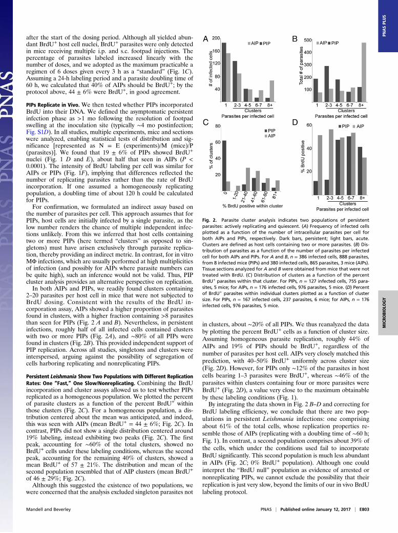
PIPs Predominantly Reside in MΦs and DCs.We next determined thelocalization of PIPs within different host cell types. Sections weretaken as before and labeled with anti-Leishmania histone anti-sera and anti-host cell type-specific markers for subsequent vi-sualization by confocal microscopy. This allowed us to confirmthat parasites were actually “within” the host cells visualizedacross all focal planes. Three cell types have been proposed ashosts for PIPs: reticular fibroblasts (RFs), MΦs, and DCs (26,30). Here, we defined RFs as ER-TR7+, DCs as CD11c+ (F4/80+
or F4/80−), and MΦ as F4/80+, CD11c−.At the footpad site of inoculation, only 2 ± 3% of PIPs resided
within ER-TR7+ RFs (Fig. 3 A and B and Table 1), despite thepresence of uninfected cells in all fields. In contrast, 79 ± 12%were found within F4/80+ cells (MΦ + DC) and 13 ± 7% werewithin CD11c+ DCs (Fig. 3 A and B and Table 1). By dual-staining footpad sections (Fig. 3 A and C), we found that 78 ±9% of PIPs were within F4/80+CD11c− MΦ, and 16 ± 6% werewithin F4/80+CD11c+ DCs (Table 1). Virtually all PIPs were res-ident within MΦ, DCs, or RFs (Fig. 3C), suggesting that all PIP-harboring host cell types had been found.A prior study reported that 43% of PIPs resided in ER-TR7+
cells in the draining lymph node (DLN) (26). When we analyzedPIPs in DLN, the number of infected ER-TR7+ RFs wassomewhat greater (10 ± 7%), but as before most cells were F4/80+
(87 ± 12%; Fig. 3 A and B and Table S1). Unlike the footpad,where most persistent parasites resided in MΦ, in the DLN mostparasites were in CDC11c+ DCs (61 ± 19%; Fig. 3 A and B andTable S1). This was confirmed by dual staining, with 30 ± 18%within F4/80+CD11c− MΦs and 61 ± 19% within F4/80+CD11c+
DCs (Fig. 3 A and C and Table S1). Again, the data showed thatvirtually all PIPs could be assigned to MΦs, DCs, or RFs. Thus,although we did not find high levels of PIPs within RFs in either
site, there was a shift from host MΦs in the footpad to DCs inthe DLN.
“Cluster” Analysis Suggests Intracellular Replication in both MΦs andDCs in Persistent Infections. Because the harsh treatments neededto visualize BrdU incorporation destroy most cell type-specificepitopes, we used cluster analysis to assess in which host cell typeparasite replication takes place. These studies showed that 73 ±11% of clusters occurred within F4/80+ cells (MΦ + DC; n = 3E/8M/168 clusters), whereas 17 ± 10% were within CD11c+ DCs (n =3E/8M/124 clusters). These values are similar to that seen with in-dividual parasites (79 ± 12% and 13 ± 7%, respectively) (Table 1).We also plotted the percent of CD11c+-infected cells as function ofthe number of parasites per cell to determine if DCs preferentiallyharbor the “static” subpopulation, which tends to be within hostcells containing <3 parasites, but did not see any obvious correla-tion. Together, these data suggest that PIP replication occurs sim-ilarly in both MΦs and DCs.
F4/80+ MΦ-Bearing PIPs Do Not Express an Alternative ActivationMarker or Gr-1 (Ly6C). Alternatively activated (M2) macrophagesMΦ might provide a more hospitable environment for PIPs, andwe tested this by reactivity with the diagnostic markers RELMαand CD206 (31). As before, PIPs were within F4/80+ cells (96 ±2%), none of which expressed RELMα (Fig. S2A). F4/80+
RELMα+ cells were seen elsewhere in the sections (Fig. S2A,Inset), and 70–80% cells were RELMα+ following IL-4 and IL-13treatment of peritoneal MΦ (PEM) (Fig. S2B). Similar resultswere obtained with CD206 (Fig. S2C). During the resolutionphase of acute infections, many Leishmania parasites are foundin inflammatory Gr-1+ cells (32, 33), which we confirmed for AIPs(71%; Fig. S2D). However, in PIPs this number drops to only 7%
Fig. 3. PIPs are predominantly found within MΦs and DCs in footpads (inoculation site) and in DLNs. (A) Representative confocal micrographs of the as-sociation between PIP nuclei (green) and indicated host cell markers in footpad (FP, Top) and DLN (Bottom) tissue. (Scale bar, 5 μM.) (B and C) Quantitation ofimages from A. Data points, mean association between parasites and markers in a mouse; horizontal bars, mean for all mice.
E804 | www.pnas.org/cgi/doi/10.1073/pnas.1619265114 Mandell and Beverley
(Fig. S2D). In combination with the prior results, the majority ofPIPs appear to reside within tissue MΦ.
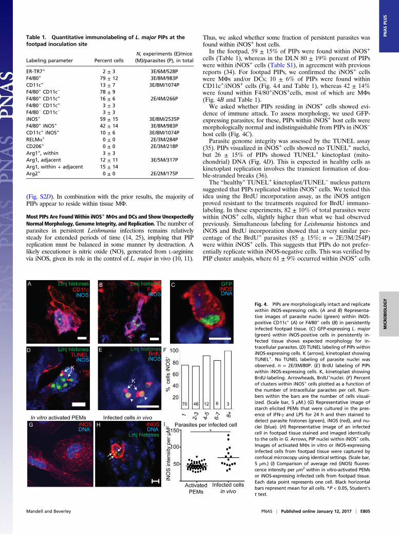
Most PIPs Are FoundWithin iNOS+ MΦs and DCs and Show UnexpectedlyNormal Morphology, Genome Integrity, and Replication.The number ofparasites in persistent Leishmania infections remains relativelysteady for extended periods of time (14, 25), implying that PIPreplication must be balanced in some manner by destruction. Alikely executioner is nitric oxide (NO), generated from L-argininevia iNOS, given its role in the control of L. major in vivo (10, 11).
Thus, we asked whether some fraction of persistent parasites wasfound within iNOS+ host cells.In the footpad, 59 ± 15% of PIPs were found within iNOS+
cells (Table 1), whereas in the DLN 80 ± 19% percent of PIPswere within iNOS+ cells (Table S1), in agreement with previousreports (34). For footpad PIPs, we confirmed the iNOS+ cellswere MΦs and/or DCs; 10 ± 6% of PIPs were found withinCD11c+/iNOS+ cells (Fig. 4A and Table 1), whereas 42 ± 14%were found within F4/80+iNOS+cells, most of which are MΦs(Fig. 4B and Table 1).We asked whether PIPs residing in iNOS+ cells showed evi-
dence of immune attack. To assess morphology, we used GFP-expressing parasites; for these, PIPs within iNOS+ host cells weremorphologically normal and indistinguishable from PIPs in iNOS−
host cells (Fig. 4C).Parasite genome integrity was assessed by the TUNEL assay
(35). PIPs visualized in iNOS+ cells showed no TUNEL+ nuclei,but 26 ± 15% of PIPs showed TUNEL+ kinetoplast (mito-chondrial) DNA (Fig. 4D). This is expected in healthy cells askinetoplast replication involves the transient formation of dou-ble-stranded breaks (36).The “healthy” TUNEL+ kinetoplast/TUNEL− nucleus pattern
suggested that PIPs replicated within iNOS+ cells. We tested thisidea using the BrdU incorporation assay, as the iNOS antigenproved resistant to the treatments required for BrdU immuno-labeling. In these experiments, 82 ± 10% of total parasites werewithin iNOS+ cells, slightly higher than what we had observedpreviously. Simultaneous labeling for Leishmania histones andiNOS and BrdU incorporation showed that a very similar per-centage of the BrdU+ parasites (85 ± 15%; n = 2E/3M/254P)were within iNOS+ cells. This suggests that PIPs do not prefer-entially replicate within iNOS-negative cells. This was verified byPIP cluster analysis, where 61 ± 9% occurred within iNOS+ cells
Table 1. Quantitative immunolabeling of L. major PIPs at thefootpad inoculation site
Labeling parameter Percent cellsN, experiments (E)/mice(M)/parasites (P), in total
ER-TR7+ 2 ± 3 3E/6M/528PF4/80+ 79 ± 12 3E/8M/983PCD11c+ 13 ± 7 3E/8M/1074PF4/80+ CD11c− 78 ± 9F4/80+ CD11c+ 16 ± 6 2E/4M/266PF4/80− CD11c+ 3 ± 3F4/80− CD11c− 3 ± 3iNOS+ 59 ± 15 3E/8M/2535PF4/80+ iNOS+ 42 ± 14 3E/8M/983PCD11c+ iNOS+ 10 ± 6 3E/8M/1074PRELMα+ 0 ± 0 2E/3M/284PCD206+ 0 ± 0 2E/3M/218PArg1+, within 3 ± 3Arg1, adjacent 12 ± 11 3E/5M/317PArg1, within + adjacent 15 ± 14Arg2+ 0 ± 0 2E/2M/175P
Fig. 4. PIPs are morphologically intact and replicatewithin iNOS-expressing cells. (A and B) Representa-tive images of parasite nuclei (green) within iNOS-positive CD11c+ (A) or F4/80+ cells (B) in persistentlyinfected footpad tissue. (C) GFP-expressing L. major(green) within iNOS-positive cells in persistently in-fected tissue shows expected morphology for in-tracellular parasites. (D) TUNEL labeling of PIPs withiniNOS-expressing cells. K (arrow), kinetoplast showingTUNEL+. No TUNEL labeling of parasite nuclei wasobserved. n = 2E/3M/80P. (E) BrdU labeling of PIPswithin iNOS-expressing cells. K, kinetoplast showingBrdU-labeling. Arrowheads, BrdU+nuclei. (F) Percentof clusters within iNOS+ cells plotted as a function ofthe number of intracellular parasites per cell. Num-bers within the bars are the number of cells visual-ized. (Scale bar, 5 μM.) (G) Representative image ofstarch elicited PEMs that were cultured in the pres-ence of IFN-γ and LPS for 24 h and then stained todetect parasite histones (green), iNOS (red), and nu-clei (blue). (H) Representative image of an infectedcell in footpad tissue stained and imaged identicallyto the cells in G. Arrows, PIP nuclei within iNOS+ cells.Images of activated MΦs in vitro or iNOS-expressinginfected cells from footpad tissue were captured byconfocal microscopy using identical settings. (Scale bar,5 μm.) (I) Comparison of average red (iNOS) fluores-cence intensity per μm2 within in vitro-activated PEMsor iNOS-expressing infected cells from footpad tissue.Each data point represents one cell. Black horizontalbars represent mean for all cells. *P < 0.05, Student’st test.
Mandell and Beverley PNAS | Published online January 12, 2017 | E805
MICRO
BIOLO
GY
PNASPL
US
(n = 3E/8M/329 clusters). A similar percentage was seen acrossall cluster sizes, suggesting that neither the replicating nor non-replicating PIPs were preferentially associated with iNOS (Fig.4F). This established that PIPs replicate comparably in bothiNOS+ and iNOS− host cells.Interestingly, about half of the uninfected F4/80+ cells adja-
cent to PIP-infected host cells also were iNOS+ (50 ± 15%; n = 3mice/11 fields/343 cells). In contrast, more distant cells wererarely iNOS+. These data show that “bystander” cell activationoccurs during persistent infections, as has also been observed inacute infections (37), and raise the possibility that once released,PIPs may enter into either iNOS+ or iNOS− cells.
Evidence That Multiple Inoculations of BrdU Do Not Perturb PIPReplication or iNOS Expression. Multiple injections were requiredin the vicinity of the parasite infection site to obtain sufficientBrdU labeling of both AIPs and PIPs (Fig. 1C). This promptedus to ask whether this necessary experimental perturbation couldcause induction and/or inhibition of PIP replication in somemanner. We compared PIP replication in BrdU-treated micewith that of untreated mice by the “cluster analysis” method. PIPclustering from BrdU-treated mice was determined by imagingtissue sections stained to detect parasite histones and iNOS,whereas the data from control mice were determined as part ofthe analysis of the host cell types containing persistent Leish-mania (Fig. 3). This comparison showed that the distribution ofparasite clusters was indistinguishable in experiments in whichBrdU labeling was performed from those where it was not (Fig.S3; P > 0.6 by two-way ANOVA). We also tested whether theBrdU administration protocol affected the level of iNOS ex-pression in infected host cells. In tissue from BrdU-treated mice,65 ± 21% of the parasites were within iNOS+ cells, not signifi-cantly different than that seen in mice that had not been sub-jected to BrdU treatment (59 ± 15%; P = 0.38). Thus, neitherthe multiple inoculations nor the presence of BrdU appear toresult in detectable perturbation of either iNOS expression orPIP replication in vivo.
How Do PIPs Survive Within iNOS Host Cells?We considered severalmodels to account for the unexpected abundance, survival, andmorphological normality of PIPs within iNOS+ host cells. Methodsfor direct in situ detection of NO are not well developed norreadily applied to the low levels of tissue PIPs seen; thus, ourattempts to establish NO production by PIP-infected iNOS+ cellswere unsuccessful (on both control and test samples). Neverthe-less, we found that iNOS expression in PIP-infected cells wassomewhat higher than in IFN-γ/LPS-activated PEMs (1.6-fold;Fig. 4 G–I; P < 0.001) that kill L. major promastigotes in aniNOS-dependent manner (38), indicating that PIP survival is notexplained by reduced iNOS expression. Next, we asked whetherhost or parasite arginase was up-regulated, which could deprivecells of the essential substrate for NO synthesis. PIP-bearing hostcells were scored for expression of iNOS and arginase 1 (cytosolic)or 2 (mitochondrial) (39), as were adjacent cells in physical con-tact with the parasite-infected cell, as these could deplete argininein their immediate vicinity (40). Although 83 ± 14% of PIPs werewithin iNOS+ cells in these experiments, only 3 ± 3% were withiniNOS+Arg1+ cells, and only 12 ± 11% of PIPs in iNOS+ wereadjacent to Arg1+ cells (Fig. S4A and Table 1). This result is inkeeping with our finding that PIPs were not found within MΦsshowing hallmarks of alternative activation. We did not detect anyspatial association between PIPs and Arg2+ host cells (Table 1),which could be seen distantly from parasites (>75 μm; Fig. S4B).Lastly, although L. major also express arginase, promastigotes donot express enough to affect NO production by activated MΦs invitro (41). Our studies revealed that arginase levels were reducedtwofold in PIPs relative to cultured promastigotes (Fig. S4 C–E;
P = 10−6). Thus, up-regulation of arginase does not contribute toPIP survival in iNOS+ cells.PIPs could be intrinsically resistant to NO, as there is increasing
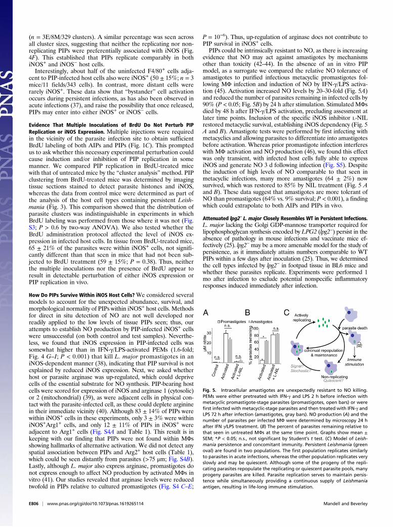
evidence that NO may act against amastigotes by mechanismsother than toxicity (42–44). In the absence of an in vitro PIPmodel, as a surrogate we compared the relative NO tolerance ofamastigotes to purified infectious metacyclic promastigotes fol-lowing MΦ infection and induction of NO by IFN-γ/LPS activa-tion (45). Activation increased NO levels by 20–30-fold (Fig. 5A)and reduced the number of parasites remaining in infected cells by90% (P < 0.05; Fig. 5B) by 24 h after stimulation. Stimulated MΦsdied by 48 h after IFN-γ/LPS activation, precluding assessment atlater time points. Inclusion of the specific iNOS inhibitor L-NILrestored metacyclic survival, establishing iNOS dependency (Fig. 5A and B). Amastigote tests were performed by first infecting withmetacyclics and allowing parasites to differentiate into amastigotesbefore activation. Whereas prior promastigote infection interfereswith MΦ activation and NO production (46), we found this effectwas only transient, with infected host cells fully able to expressiNOS and generate NO 3 d following infection (Fig. S5). Despitethe induction of high levels of NO comparable to that seen inmetacyclic infections, many more amastigotes (64 ± 2%) nowsurvived, which was restored to 85% by NIL treatment (Fig. 5 Aand B). These data suggest that amastigotes are more tolerant ofNO than promastigotes (64% vs. 9% survival; P < 0.001), a findingwhich could extrapolate to both AIPs and PIPs in vivo.
Attenuated lpg2– L. major Closely Resembles WT in Persistent Infections.L. major lacking the Golgi GDP-mannose transporter required forlipophosphoglycan synthesis encoded by LPG2 (lpg2−) persist in theabsence of pathology in mouse infections and vaccinate mice ef-fectively (25). lpg2−may be a more amenable model for the study ofpersistence, as it immediately attains numbers comparable to WTPIPs within a few days after inoculation (25). Thus, we determinedthe cell types infected by lpg2− in footpad tissue in BL6 mice andwhether these parasites replicate. Experiments were performed 1mo after infection to exclude potential nonspecific inflammatoryresponses induced immediately after infection.
Fig. 5. Intracellular amastigotes are unexpectedly resistant to NO killing.PEMs were either pretreated with IFN-γ and LPS 2 h before infection withmetacyclic promastigote-stage parasites (promastigotes, open bars) or werefirst infected with metacyclic-stage parasites and then treated with IFN-γ andLPS 72 h after infection (amastigotes, gray bars). NO production (A) and thenumber of parasites per infected MФ were determined by microscopy 24 hafter IFN γ/LPS treatment. (B) The percent of parasites remaining relative tothat seen in untreated MФs at the same time point. Graphs show mean ±SEM; *P < 0.05; n.s., not significant by Student’s t test. (C) Model of Leish-mania persistence and concomitant immunity. Persistent Leishmania (greenoval) are found in two populations. The first population replicates similarlyto parasites in acute infections, whereas the other population replicates veryslowly and may be quiescent. Although some of the progeny of the repli-cating parasites repopulate the replicating or quiescent parasite pools, manyprogeny parasites are killed. Parasite replication serves to maintain persis-tence while simultaneously providing a continuous supply of Leishmaniaantigen, resulting in life-long immune stimulation.
E806 | www.pnas.org/cgi/doi/10.1073/pnas.1619265114 Mandell and Beverley
For most properties, lpg2− PIPs were similar to WT, includingthe extent of BrdU labeling and residence primarily within tissueMΦ and DCs, as assessed by labeling for F4/80, CDC11c, iNOS,RELMα, ER-TR7, and Arg2 (Table S2). These data suggest thatlpg2− parasites provide an excellent model for long-term persis-tent L. major.For three markers, modest differences were seen. First, tran-
siently elevated levels of lpg2− PIPs were found in CD206+ cellsafter 1 mo (50 ± 26%; P < 0.05), which declined to the WT levels(4 ± 5%) after 5 mo (Fig. S6A and Table S1). Second, the fre-quency of lpg2− PIPs within CD11c+ cells was elevated threefoldrelative to WT (46 ± 29%; Fig. S6B; P < 0.006; Table S1), andtheir association with Arg1 elevated fivefold (Fig. S6C and TableS1; P = 0.002; 37% within and 35% adjacent). Unlike CD206localization, these differences did not return to WT after 5 mo(Fig. S6C). Whether these differences contribute to the ability oflpg2− to vaccinate (14, 27) will be addressed in future studies.
DiscussionPersistent Infection Leishmania Comprise Two Populations, One FastReplicating and a Second Showing Little if any Replication. Becausethe number of persistent parasites remains steady over longperiods of time in L. major following resolution of infectionpathology, an unanswered question was whether these parasiteslay in a quiescent, nonreplicating state. As in other pathogens,this has profound consequences to strategies used for MΦ sur-vival and immune evasion. Here, we demonstrate that a majorfraction of PIPs replicate comparably to AIPs, using three in-dependent approaches. First, we formulated a parasite clusterassay, reasoning that host cells bearing more than one parasitearose via parasite replication rather than multiple infections. Suchclusters were identified both in footpads and DLNs (Figs. 2 and 3 Aand B), indirectly suggesting PIP replication at both sites. Second,by the TUNEL assay, we detected significant levels of kinetoplast(mitochondrial) DNA replication (Fig. 4). The definitive evidencefor PIP replication came from BrdU incorporation, which revealedthat a substantial percentage of PIPs were BrdU+ (Fig. 1). Thus,many PIPs undergo active replication.As the fraction of BrdU+ PIPs was about half of that seen with
AIPs (19 vs. 44%; Fig. 1E), we asked whether this arose fromdifferences in overall replication rate or population heteroge-neity. This was tested by quantitative analysis of the distributionof BrdU+ parasites versus cluster size (Fig. 2), postulating thatclusters comprising multiple parasites/host cells were a sign ofparasite replication. As expected, AIPs showed the distributionexpected for a homogeneously replicating population, with sim-ilar BrdU labeling across all cluster sizes. In contrast, PIPsshowed two populations—one of whose properties closely re-sembled that of AIPs and a second of small clusters—showing noBrdU incorporation by our labeling protocol; indeed, BrdU−
“singletons” were most numerous (Fig. 2C). Although we cannotrule out that these parasites are very slowly replicating by themethods available, we estimate that about 39% of PIPs fall intothis nonreplicating class.
Do Slowly/Nonreplicating PIPs Enter a Quiescent Metabolic State?Potentially, slowly/nonreplicating BrdU− PIPs have entered adifferent metabolic state than the actively replicating BrdU+
PIPs. Indeed, recently it was shown that progressive chronic in-fections of Leishmania mexicana were comprised of slowly rep-licating parasite populations that were metabolically relativelyquiescent (47). However, the methods used there for the study oflarge numbers of parasites in chronic infections are not exten-sible to the low levels of PIPs seen in L. major nor for mixedpopulations showing widely varying replication rates. Thus, otherapproaches will be required to explore the metabolic status ofthe slowly/nonreplicating L. major PIPs. NO synthesis may act todampen parasite metabolism in AIPs (42, 43), and this likely
occurs with PIPs as well. The quiescent forms of many pathogensshow differences in gene expression from actively replicatingforms (48, 49). Similar knowledge would aid our understandingof Leishmania persistence considerably.
Is There a “Safe” Cell for Leishmania in Persistent Infections? Res-olution of L. major infections following infection of resistantmouse strains is known to be dependent on the production ofNO via iNOS (34). To account for the emergence of PIPs, in-vestigators have posited the existence “safe cells” free from im-mune attack (26, 50). Although RFs or alternatively activatedMΦs were attractive candidates to harbor persistent organisms(26, 31), few PIPs could be found within them (Table 1).Instead, virtually all PIPs were found in cells often considered
“unsafe”—namely, MΦs or DCs—especially given that most ofthese expressed iNOS at levels comparable to that seen in clas-sically activated MΦs (Table 1 and Fig. 4). PIPs in iNOS+ hostcells appeared strikingly normal: their replication profiles re-sembled those parasites within iNOS− host cells, as did theirmorphology (amastigote-like) and genome integrity.This raised the question as to how PIPs could withstand the
presumptive action of iNOS through NO production. Using MΦinfections in vitro as a surrogate for AIPs and PIPs, we found thesurvival of amastigotes to greatly exceed that of infectiousmetacyclics (Fig. 5). Correspondingly, several studies suggest that theaction of NO against amastigotes may be based more on meta-bolic inhibition rather than killing (42, 43), arising from a tissue-wide “collective” effect (44). This would likely be less effective atlow PIP densities seen in persistent infections. We saw no evi-dence of attenuation of NO production by substrate competitionwith host or parasite arginase nor of other previously describedmechanisms of modulating or rendering iNOS activity ineffective(51, 52). An interesting question concerns the contribution ofreactive oxidant species, as infections of phox– mice lacking themajor phagocyte oxidase (gp91/phox) often yield uncontrolledinfections, albeit less reproducibly and progressing much lessrapidly than seen in infections of iNOS knockout mice (53, 54).However, the lack of gp91 oxidase-specific inhibitors has pre-cluded tests of its role specifically in PIP control, as shown ele-gantly using specific inhibitors of iNOS (11).The most plausible scenario accounting for current data is that
PIP-bearing host cells synthesize NO via iNOS expression, towhich PIPs are largely but perhaps not entirely resistant. Wehave as yet not seen evidence of PIPs destruction microscopi-cally, although the rapidity by which killed parasites disappear(Fig. 5B) (35) suggests the anticipated “corpses” might be diffi-cult to visualize in vivo.
lpg2– as a Model for Persistence. Previous work has shown thatalthough attenuated in acute infections and unable to inducepathology, the L. major lpg2− mutant can persist indefinitely innumbers comparable to WT persistent parasites (25) and canvaccinate susceptible (BALB/c) mice against virulent challenge(27). Because lpg2− effectively enters the “persistent” phase veryquickly, we proposed that it could provide a more convenientmodel than the study of healed persistent WT infections, whichrequire >5 mo to establish. This prediction is now supported byour finding that after only 1 mo lpg2− was indistinguishable fromWT PIPs in most respects, including the extent of replication andresidence primarily with MΦs and DCs, the majority of whichwere iNOS+ (Table S1). Preliminary data also suggest that lpg2−
recruits Foxp3+ cells to the site of infection in BALB/c mice, aphenomenon reported for WT PIPs (20). Modest differenceswere seen with a few markers (CD206 transiently, Arg1, andCD11c; Fig. S5), which may reflect the absence of phosphogly-cans or other metabolites in the lpg2− and could contribute to theimmune response and ability to vaccinate (25, 27, 55). Overall,
Mandell and Beverley PNAS | Published online January 12, 2017 | E807
MICRO
BIOLO
GY
PNASPL
US
these data support the continued use of lpg2– parasites as amodel of Leishmania persistence and vaccination.
A Model of Leishmania Persistence and Concomitant Immunity. Ourdata show that PIPs comprise two populations of cells: onereplicating similarly to logarithmically growing acute phase par-asites (∼60 h doubling time), and a second population replicatingmuch less rapidly, and possibly not at all. However, despitereplication of the fast population, estimated to comprise ∼61%of the total cells, PIP numbers do not increase over time, im-plying that parasite replication is matched by killing. Thus, theoffspring of replicating parasites have three potential fates: Theymay continue active replication, enter a nonreplicating state, orbe destroyed (Fig. 5C). Similarly, nonreplicating parasites mayremain dormant or at some frequency resume replication. Howthese eventual fates are decided is not yet known. This couldbe under cellular regulation and control, transitioning betweenan active, replicating state and a potentially quiescent non-replicating state, as seen in other pathogens exhibiting dormancyor latency. Alternatively, the fact that both replicating and quies-cent PIPs reside in similarly unsafe host cells suggests a stochasticmodel, where forces favoring parasite persistence (such as IL-10/Treg-dependent suppression) are balanced against those favoringdestruction (56), leading to maintenance of roughly constantnumbers. In reality, knowledge is limited concerning how “steady”persistent Leishmania numbers actually are, with current data be-ing mostly of low temporal resolution and high granularity. Thus, amodel wherein parasite numbers oscillate within some range overtime in response to these competing forces could be invoked.Strong perburbations of the host’s response could further pushpersistent parasites along either path, resulting in reactivation orsterile cure (11, 20).The ongoing replication and destruction of persistent Leish-
mania provides an attractive model for concomitant immunityand a compelling rationale for the superiority of “live” vaccina-tion strategies in Leishmania. Killed parasites arising as abyproduct of parasite replication would be a good source of an-tigen for maintaining a robust anti-Leishmania response at thevery site of initial infection (24). Provision of “dead antigen” insitu would also overcome the ability of live parasites to inhibitantigen presentation (57–59). Thus, from the host’s perspective,persistent parasites serve as a continually self-renewing stimula-tory vaccine. In some respects, the model proposed for PIP rep-lication and “termination” is reminiscent of a stem cell cycle (Fig.S7) with some of the progeny of replicating PIPs serving as acontinually self-renewing “stem” maintaining parasite persis-tence, whereas the remainder meet their terminal fate in antigen-presenting cells.The continually replicating immunogen model may provide a
new perspective with which to view the immune response toother persistent pathogens. For most of these, the experimentalfocus has been on the role of the immune system in pathogensuppression and containment, leaving the question of whetherthis contributes to resistance to subsequent challenges as seenwith L. major. Following resolution of primary infections andthe generation of persistent (Toxoplasma bradyzoites) or latentstates (herpesviruses), sporadic subclinical reactivations oftenoccur, leading to the synthesis of antigen and immune stimulation(60, 61). There is evidence that both of these persistent/latentinfections may confer protection to rechallenge (62–64). Simi-larly, there is evidence that latent M. tuberculosis may undergolimited replication (65) and that latent, asymptomatic infectionsare associated with significant resistance to active disease (66).Future work will be needed to establish whether a continuallyrenewing “stem-like” immunogen model is relevant to these orother persistent pathogens.
Materials and MethodsParasite Strains and Culture. L. major strain LV39c5 (Rho/SU/59/P) and itsderivative lpg2− (Δlpg2::HYG/Δlpg2::HYG) (25) were used in all experiments,except for the promastigote experiments show in Fig. 1 and Fig. S1, whichused L. major strain Friedlin V1 (MHOM/IL/80/Friedlin). Parasites were grownat 26 °C in M199 medium (US Biologicals) supplemented with 40 mM4-(2-hydroxyethyl)-1-piperazine-ethanesulfonic acid (Hepes) pH 7.4, 50 μMadenosine, 1 μg·mL−1 biotin, 5 μg·mL−1 hemin, 2 μg·mL−1 biopterin, and10% (vol/vol) heat-inactivated FCS (67). “Slow-growing” promastigote cul-tures were grown in RPMI 1640 + L-glutamine (Invitrogen) supplementedwith 37 mM Hepes pH 7.4, 47 μM adenosine, 0.93 μg·mL−1 biotin, 4.7 μg·mL−1
hemin, 1.9 μg·mL−1 biopterin, and 0.9% (vol/vol) heat-inactivated FCS (68).The WT LV39c5 parasites used here expressed GFP from the ribosomal locus(SSU::IR1SAT-GFP) and were generated by transfecting SwaI-cut plasmidB3538 into WT LV39c5 as described (67) and selecting for resistance to100 μM nourseothricin and bright green fluorescence. The clone used in thisstudy exhibited virulence similar to WT in BALB/c mice. Null mutants of theparasite arginase gene (Δarg::HYG/Δarg::PAC) (69) were cultured in theabove media supplemented with 50 mM putrescine. Infective metacyclic-stage parasites were recovered using the density gradient centrifugationmethod (70). Propidium iodide staining of promastigotes was performed asdescribed (71).
Antibodies Used. L. major nuclei were detected with a pool of rabbit anti-bodies raised against L. major histones H2A, H2Avariant, H2B, H3, and H4(pooled at a ratio of 3:2:3:3:1 by titer, kindly provided by I. L. K. Wong,Washington University School of Medicine, St. Louis) (72). For some experi-ments, this pool was used at a dilution of 1:750. For others, this pool ofantibodies was directly conjugated to Alexafluor488 according to themanufacturer’s protocol (Invitrogen) and used at a final concentration of0.15 mg·mL−1. GFP was detected with a chicken anti-GFP antibody (AbCam)at a final concentration of 0.02 mg·mL−1. F4/80 was detected with a ratmonoclonal antibody (clone A3-1, AbD Serotec) diluted to 1:250. CD11c wasdetected with a hamster monoclonal antibody (clone N418, eBioscience)diluted to 1:250. ER-TR7 was detected with a rat monoclonal antibody (BMABiomedicals) used at a final concentration of 0.01 mg·mL−1. iNOS was de-tected with a rabbit anti-iNOS (BD Transduction Labs) used at 1 μg·mL−1.Relmα was detected with a rabbit polyclonal antibody (Abcam) used at0.8 μg·mL−1. BrdU was detected with a rat monoclonal antibody (Abcam) usedat 10 μg·mL−1. Goat anti-Arg1 (Santa Cruz) and goat anti-Arg2 (Santa Cruz)were used at 2 μg·mL−1. Rat anti-Gr1 mAb (clone RB6-8C5; kindly providedby L. D. Sibley, Washington University School of Medicine, St. Louis) was usedat a 1:250 dilution. Rabbit anti-Leishmania arginase (kindly provided byB. Ullman, Oregon Health Sciences University, Portland, OR) was used at a1:1,000 dilution. The following antisera were screened for reactivity to nitro-tyrosine in IFN-γ/LPS-stimulated MΦs: rabbit anti-nitrotyrosine (Santa CruzBiotechnology, sc-55256; Millipore, #06–284; Abcam, ab50185), mouse anti-nitrotyrosine (Santa Cruz Biotechnology, sc-32757), and rat anti-nitrotyrosine(Abcam, ab6479).
The following fluorescent secondary antibodies were used: Alexafluor555goat anti-rabbit, Alexafluor633 goat anti-rabbit, Alexafluor488 goat anti-rat,Alexafluor555 goat anti-rat, Alexafluor633 goat anti-rat, Alexafluor568 goatanti-hamster, Alexafluor488 goat anti-chicken, Alexaflour555 donkey anti-goat, and Alexafluor647 donkey anti-rabbit (Invitrogen, all used at 2 μg·mL−1
concentrations).
Statement Identifying Institutional Committee Approving Animal Experiments.Animal handling and experiments were carried out in strict accordance withthe recommendations in the Guide for the Care and Use of LaboratoryAnimals of the US National Institutes of Health (73). Animal studies wereapproved by the Animal Studies Committee at Washington University(protocol 20090086) in accordance with the Office of Laboratory AnimalWelfare’s guidelines and the Association for Assessment and Accreditationof Laboratory Animal Care International.
Mouse Infections. Female C57BL/6J mice (6–10 wk old; Jackson Labs) wereinjected s.c. in the left hind footpad with either 105 metacyclic WT or 106
metacyclic lpg2– parasites. Following infection with WT parasites, the micedeveloped lesions that resolved, as determined by the absence of footpadswelling relative to the uninfected foot, ∼4 mo after infection. For thepurposes of this study, persistent infections were defined as any time >1 mofollowing the resolution of footpad swelling. Unless otherwise indicated,studies with lpg2− were performed between 1 and 2 mo following infection.Possible lpg2− revertants showing amastigote virulence (74), defined here as
E808 | www.pnas.org/cgi/doi/10.1073/pnas.1619265114 Mandell and Beverley
having >150 parasites in a single section, were excluded from analysis. PEMswere harvested and infected as described (75).
MΦ Infections. Starch-elicited PEMs were harvested and infected as described(75). Cells were activated with 100 U·mL−1 recombinant IFN-γ (Chemicon)and 100 ng·mL−1 LPS (Sigma) with or without the iNOS-inhibitor L-NIL(Cayman Chemical) at a concentration of 10 μM either immediately beforeor 72 h after infection. Nitrite production (determined by the Greiss assay;Sigma) and parasite survival were determined 24 h after MΦ stimulation. Forthese experiments, “percent survival” is defined as the ratio of the numberof parasites per 100 MΦs compared with the same statistic in MΦs that werenot treated with LPS/IFN-γ. For these experiments, percent survival is definedas the ratio of the number of parasites per 100 PEMs at either 24 or 96 hafter infection versus the number of parasites per 100 PEMs 24 h earlier. Togenerate alternatively activated (M2) MΦs, PEMs were treated with mediacontaining 100 U·mL−1 each of recombinant IL-4 (BD Pharmingen) and IL-13(BD Pharmingen) for 48 h.
BrdU Staining and in Vitro Labeling Experiments. For all BrdU immunode-tection experiments, paraformaldehyde-fixed samples were permeabilizedwith 0.1% Triton-X-100 in PBS for 15 min, washed in distilled water, and thenimmersed in 2MHCl for 40min to denature theDNA. After extensivewashingwith PBS, the samples were incubated in a blocking buffer containing PBS and5% (vol/vol) normal goat sera and 0.1% (vol/vol) Triton-X-100 and were thenstained as described in the text in the methods above. All BrdU stains used a2-h incubation with the anti-BrdU antibody.
To test BrdU labeling in promastigotes in culture, either log- or stationary-phase L. major promastigotes were cultured in M199 media containing0.1 mM BrdU (Sigma) for the indicated time, after which they were fixed in4% (wt/vol) paraformaldehyde in PBS for 10 min and stained as describedabove. To test BrdU labeling in infected MΦs in vitro, PEMS were harvestedand infected as described with stationary-phase parasites (75). Two hoursafter parasites were added, the MΦs were washed to remove extracellularparasites and placed in media containing 0.1 mM BrdU, where they weremaintained. At 2 h, 1 d, 2 d, and 5 d postinfection, samples were fixed andstained as described above.
BrdU Incorporation Assay in Vivo. Several different methods were attemptedto administer BrdU to the infected mice. In the preferred method, infectedmice were injected every 3 h for 18 h with 200 μL of PBS containing 4mg·mL−1
BrdU into the peritoneal cavity and 50 μL of this solution directly into theinfected footpad, yielding a total dose of 6 mg BrdU. Mice were killed andfrozen tissue sections prepared 24 h after the first dose of BrdU. In additionto our standard approach, we also tried administering BrdU in the drinkingwater (1 mg·mL−1) via infusion using osmotic pumps (Alzet #2001D, 7.2 mgtotal dose) and single i.p. injections of 200 μL of PBS containing 4 mg·mL−1
BrdU. At 24 h after the first dose, the mice were euthanized and footpadtissue prepared and stained as described above. Volocity image analysissoftware (Improvision) was used to assist quantitation.
Tissue Preparation and Histological Staining. After euthanasia, infected drainingpopliteal lymph nodes or feet were harvested. The infected tissue was thenfixed for 1 h at room temperature in 4% (wt/vol) paraformaldehyde in PBS.After fixation, tissues were incubated at 4 °C for 1 h in 10% and 20% (wt/vol)sucrose in PBS, followed by an overnight incubation in 30% sucrose. The tis-sues were then embedded in O.C.T. compound (Ted Pella, Inc.), cut into 10 μm-thick sections using a cryostat, and mounted onto microscope slides.
Unless otherwise indicated, tissue sections were stained as follows. Slideswere washed in PBS, and tissues were then blocked and permeabilized in
PBS containing 5% (vol/vol) normal goat sera (Vector laboratories) and 0.1%Triton-X-100 for 30 min. The sections were then stained with primary anti-bodies for 1 h. Unbound antibody was then washed off in PBS, and primaryantibodies were detected with fluorescent secondary antibodies for 40 min,followed by a second wash in PBS. For some experiments, we needed tosimultaneously stain tissue sections with different antibodies that were bothgenerated in rabbits. To do this, the tissue was stained with an unlabeledrabbit primary antibody and a fluorescently labeled secondary antibody asdescribed above. Next, the tissue was blocked for 30 min with a buffercontaining 5% (vol/vol) normal rabbit sera (Sigma Aldrich), and then thesecond fluorescently conjugated primary antibody was used.
For TUNEL staining of the tissue sections, after all primary and secondaryantibody staining was finished, the sections were stained with the In Situ CellDeath Detection Kit, TMR Red (Roche), according to the manufacturer’sprotocol. All sections were mounted in ProLong Gold reagent (Invitrogen).
Confocal Microscopy and Image Analysis.Microscopy was performed on a Zeiss510 META confocal laser scanning microscope. Image analysis was performedusing ImageJ or Volocity software.
Comparison of BrdU Staining Intensity. Samples were stained to detect par-asite histones and BrdU, and three-dimensional confocal image stacks wereacquired. Parasite nuclei were manually circumscribed in ImageJ software,and the average intensity in the anti-BrdU channelwas determined from two-dimensional image stacks. As a control for comparison of iNOS staining in-tensity, PEMs were activated with media containing 100 U·mL−1 recombinantIFN-γ (Chemicon) and 100 ng·mL−1 LPS (Sigma). Twenty-four hours later, thecells were fixed in 4% (vol/vol) paraformaldehyde and stained in parallelwith footpad tissue sections from PIP-infected mice with antibodies againstL. major histones and iNOS, and nuclei were stained with TOPRO-3 (Invi-trogen). To determine the fluorescence intensity of iNOS per cross-sectionalarea, the outline of each cell from a confocal stack was traced in Volocitysoftware (Improvision), and the sum intensity of all “red” pixels (iNOS) in theselected area was divided by the total number of pixels in that area, yieldingthe mean pixel intensity for the cross-section.
Comparison of Arginase Staining Intensity Between Persistent Parasites andPromastigotes. Footpad tissue sections infected with persistent WT (PIPs) orlog-phase WT promastigotes were labeled to detect parasite arginase withanti-Leishmania arginase antibody and parasite histones (with the fluo-rescently conjugated anti-histone antibody). Confocal images were acquiredusing identical settings, and then Volocity image analysis software was usedto determine the total arginase fluorescence intensity on a per-cell basis.Confocal stacks were compressed into a single plane, and then the totalarginase fluorescence intensity was determined within a 2.28-μm radiuscircle centered on parasite nuclei.
Statistics. Data are presented as the geometric mean ± SE. P values arecalculated by the Student’s t test method or ANOVA.
ACKNOWLEDGMENTS. We thank I.L.K. Wong for providing the antibodiesagainst L. major histones, L. D. Sibley for providing the antibodies againstF4/80 and CD11c, B. Ullman for providing the anti-Leishmania arginase an-tibody, and W. Beatty, C. Bogdan, D. E. Dobson, S. Khader, T. Margolis,D. Sacks, L. D. Sibley, T. Vickers, H. W. Virgin, and J. Uzonna, as well as thereferees, for discussions and/or comments on the manuscript. This work wasfunded by NIH Grants R01 AI31078 and AI29646 (to S.M.B.) and a Berg/MorseGraduate Fellowship (to M.A.M.).
1. Monack DM, Mueller A, Falkow S (2004) Persistent bacterial infections: The interfaceof the pathogen and the host immune system. Nat Rev Microbiol 2(9):747–765.
2. Virgin HW, Wherry EJ, Ahmed R (2009) Redefining chronic viral infection. Cell 138(1):30–50.
3. Pawlowski A, Jansson M, Sköld M, Rottenberg ME, Källenius G (2012) Tuberculosisand HIV co-infection. PLoS Pathog 8(2):e1002464.
4. Roizman B, Whitley RJ (2013) An inquiry into the molecular basis of HSV latency andreactivation. Annu Rev Microbiol 67:355–374.
5. Brown SP, Grenfell BT (2001) An unlikely partnership: Parasites, concomitant immu-nity and host defence. Proc Biol Sci 268(1485):2543–2549.
6. Singh OP, Hasker E, Sacks D, Boelaert M, Sundar S (2014) Asymptomatic Leish-mania infection: A new challenge for Leishmania control. Clin Infect Dis 58(10):1424–1429.
7. Pigott DM, et al. (2014) Global distribution maps of the leishmaniases. eLife 3:3.8. WHO (2010) Control of the Leishmaniases: WHO Expert Committee on the Control of
Leishmaniases. World Health Organization Technical Reports 949:1–186.
9. Bañuls AL, et al. (2011) Clinical pleiomorphism in human leishmaniases, with specialmention of asymptomatic infection. Clin Microbiol Infect 17(10):1451–1461.
10. Wei XQ, et al. (1995) Altered immune responses in mice lacking inducible nitric oxidesynthase. Nature 375(6530):408–411.
11. Stenger S, Donhauser N, Thüring H, Röllinghoff M, Bogdan C (1996) Reactivation oflatent leishmaniasis by inhibition of inducible nitric oxide synthase. J Exp Med 183(4):1501–1514.
12. Launois P, Louis JA, Milon G (1997) The fate and persistence of Leishmania major inmice of different genetic backgrounds: An example of exploitation of the immunesystem by intracellular parasites. Parasitology 115(Suppl):S25–S32.
13. Belkaid Y, et al. (2000) A natural model of Leishmania major infection reveals aprolonged “silent” phase of parasite amplification in the skin before the onset oflesion formation and immunity. J Immunol 165(2):969–977.
14. Okwor I, Uzonna J (2008) Persistent parasites and immunologic memory in cutaneousleishmaniasis: Implications for vaccine designs and vaccination strategies. ImmunolRes 41(2):123–136.
Mandell and Beverley PNAS | Published online January 12, 2017 | E809
MICRO
BIOLO
GY
PNASPL
US
15. Svobodová M, Votýpka J, Nicolas L, Volf P (2003) Leishmania tropica in the black rat(Rattus rattus): Persistence and transmission from asymptomatic host to sand flyvector Phlebotomus sergenti. Microbes Infect 5(5):361–364.
16. Kimblin N, et al. (2008) Quantification of the infectious dose of Leishmania majortransmitted to the skin by single sand flies. Proc Natl Acad Sci USA 105(29):10125–10130.
17. Aebischer T (1994) Recurrent cutaneous leishmaniasis: A role for persistent parasites?Parasitol Today 10(1):25–28.
18. Sacks DL (2014) Vaccines against tropical parasitic diseases: A persisting answer to apersisting problem. Nat Immunol 15(5):403–405.
19. Mandell MA, Beverley SM (2016) Concomitant immunity induced by persistentLeishmania major does not preclude secondary re-infection: Implications for geneticexchange, diversity and vaccination. PLoS Negl Trop Dis 10(6):e0004811.
20. Belkaid Y, Piccirillo CA, Mendez S, Shevach EM, Sacks DL (2002) CD4+CD25+ regula-tory T cells control Leishmania major persistence and immunity. Nature 420(6915):502–507.
21. Sacks D, Noben-Trauth N (2002) The immunology of susceptibility and resistance toLeishmania major in mice. Nat Rev Immunol 2(11):845–858.
22. Kaye P, Scott P (2011) Leishmaniasis: Complexity at the host-pathogen interface. NatRev Microbiol 9(8):604–615.
23. Belkaid Y, et al. (2001) The role of interleukin (IL)-10 in the persistence of Leishmaniamajor in the skin after healing and the therapeutic potential of anti-IL-10 receptorantibody for sterile cure. J Exp Med 194(10):1497–1506.
24. Pagán AJ, et al. (2013) Tracking antigen-specific CD4+ T cells throughout the courseof chronic Leishmania major infection in resistant mice. Eur J Immunol 43(2):427–438.
25. Späth GF, et al. (2003) Persistence without pathology in phosphoglycan-deficientLeishmania major. Science 301(5637):1241–1243.
26. Bogdan C, et al. (2000) Fibroblasts as host cells in latent leishmaniosis. J Exp Med191(12):2121–2130.
27. Uzonna JE, Späth GF, Beverley SM, Scott P (2004) Vaccination with phosphoglycan-deficient Leishmania major protects highly susceptible mice from virulent challengewithout inducing a strong Th1 response. J Immunol 172(6):3793–3797.
28. Gratzner HG (1982) Monoclonal antibody to 5-bromo- and 5-iododeoxyuridine: Anew reagent for detection of DNA replication. Science 218(4571):474–475.
29. Tattersall MHN, Brown B, Frei E, 3rd (1975) The reversal of methotrexate toxicityby thymidine with maintenance of antitumour effects. Nature 253(5488):198–200.
30. Moll H, Flohé S, Röllinghoff M (1995) Dendritic cells in Leishmania major-immunemice harbor persistent parasites and mediate an antigen-specific T cell immune re-sponse. Eur J Immunol 25(3):693–699.
31. Gordon S (2003) Alternative activation of macrophages. Nat Rev Immunol 3(1):23–35.32. De Trez C, et al. (2009) iNOS-producing inflammatory dendritic cells constitute the
major infected cell type during the chronic Leishmania major infection phase ofC57BL/6 resistant mice. PLoS Pathog 5(6):e1000494.
33. Goncalves R, Zhang X, Cohen H, Debrabant A, Mosser DM (2011) Platelet activationattracts a subpopulation of effector monocytes to sites of Leishmania major infection.J Exp Med 208(6):1253–1265.
34. Stenger S, Thüring H, Röllinghoff M, Bogdan C (1994) Tissue expression of induciblenitric oxide synthase is closely associated with resistance to Leishmania major. J ExpMed 180(3):783–793.
35. Prina E, Roux E, Mattei D, Milon G (2007) Leishmania DNA is rapidly degraded fol-lowing parasite death: An analysis by microscopy and real-time PCR. Microbes Infect9(11):1307–1315.
36. Liu B, Liu Y, Motyka SA, Agbo EE, Englund PT (2005) Fellowship of the rings: Thereplication of kinetoplast DNA. Trends Parasitol 21(8):363–369.
37. Carvalho LP, Pearce EJ, Scott P (2008) Functional dichotomy of dendritic cells fol-lowing interaction with Leishmania braziliensis: Infected cells produce high levels ofTNF-alpha, whereas bystander dendritic cells are activated to promote T cell re-sponses. J Immunol 181(9):6473–6480.
38. Green SJ, Meltzer MS, Hibbs JB, Jr, Nacy CA (1990) Activated macrophages destroyintracellular Leishmania major amastigotes by an L-arginine-dependent killingmechanism. J Immunol 144(1):278–283.
39. Popovic PJ, Zeh HJ, 3rd, Ochoa JB (2007) Arginine and immunity. J Nutr 137(6, Suppl2):1681S–1686S.
40. Bronte V, Serafini P, Mazzoni A, Segal DM, Zanovello P (2003) L-arginine metab-olism in myeloid cells controls T-lymphocyte functions. Trends Immunol 24(6):302–306.
41. Muleme HM, et al. (2009) Infection with arginase-deficient Leishmania major revealsa parasite number-dependent and cytokine-independent regulation of host cellulararginase activity and disease pathogenesis. J Immunol 183(12):8068–8076.
42. Müller AJ, et al. (2013) Photoconvertible pathogen labeling reveals nitric oxide con-trol of Leishmania major infection in vivo via dampening of parasite metabolism. CellHost Microbe 14(4):460–467.
43. Olekhnovitch R, Bousso P (2015) Induction, propagation, and activity of host nitricoxide: Lessons from Leishmania infection. Trends Parasitol 31(12):653–664.
44. Olekhnovitch R, Ryffel B, Müller AJ, Bousso P (2014) Collective nitric oxide productionprovides tissue-wide immunity during Leishmania infection. J Clin Invest 124(4):1711–1722.
45. Liew FY, Millott S, Parkinson C, Palmer RM, Moncada S (1990) Macrophage killing ofLeishmania parasite in vivo is mediated by nitric oxide from L-arginine. J Immunol144(12):4794–4797.
46. Liu D, Uzonna JE (2012) The early interaction of Leishmania with macrophages anddendritic cells and its influence on the host immune response. Front Cell InfectMicrobiol 2:83.
47. Kloehn J, Saunders EC, O’Callaghan S, Dagley MJ, McConville MJ (2015) Character-ization of metabolically quiescent Leishmania parasites in murine lesions using heavywater labeling. PLoS Pathog 11(2):e1004683.
48. Cleary MD, Singh U, Blader IJ, Brewer JL, Boothroyd JC (2002) Toxoplasma gondiiasexual development: Identification of developmentally regulated genes and distinctpatterns of gene expression. Eukaryot Cell 1(3):329–340.
49. Betts JC, Lukey PT, Robb LC, McAdam RA, Duncan K (2002) Evaluation of a nutrientstarvation model of Mycobacterium tuberculosis persistence by gene and proteinexpression profiling. Mol Microbiol 43(3):717–731.
50. Mirkovich AM, Galelli A, Allison AC, Modabber FZ (1986) Increased myelopoiesisduring Leishmania major infection in mice: Generation of ‘safe targets’, a possibleway to evade the effector immune mechanism. Clin Exp Immunol 64(1):1–7.
51. Pandit L, Kolodziejska KE, Zeng S, Eissa NT (2009) The physiologic aggresome medi-ates cellular inactivation of iNOS. Proc Natl Acad Sci USA 106(4):1211–1215.
52. Miller BH, et al. (2004) Mycobacteria inhibit nitric oxide synthase recruitment tophagosomes during macrophage infection. Infect Immun 72(5):2872–2878.
53. Blos M, et al. (2003) Organ-specific and stage-dependent control of Leishmania majorinfection by inducible nitric oxide synthase and phagocyte NADPH oxidase. Eur JImmunol 33(5):1224–1234.
54. Diefenbach A, et al. (1998) Type 1 interferon (IFNalpha/β) and type 2 nitric oxidesynthase regulate the innate immune response to a protozoan parasite. Immunity8(1):77–87.
55. Liu D, et al. (2009) Leishmania major phosphoglycans influence the host early immuneresponse by modulating dendritic cell functions. Infect Immun 77(8):3272–3283.
56. Peters N, Sacks D (2006) Immune privilege in sites of chronic infection: Leishmania andregulatory T cells. Immunol Rev 213:159–179.
57. Courret N, et al. (1999) Presentation of the Leishmania antigen LACK by infectedmacrophages is dependent upon the virulence of the phagocytosed parasites. Eur JImmunol 29(3):762–773.
58. Reiner NE, Ng W, McMaster WR (1987) Parasite-accessory cell interactions in murineleishmaniasis. II. Leishmania donovani suppresses macrophage expression of class Iand class II major histocompatibility complex gene products. J Immunol 138(6):1926–1932.
59. Fruth U, Solioz N, Louis JA (1993) Leishmania major interferes with antigen pre-sentation by infected macrophages. J Immunol 150(5):1857–1864.
60. van Lint AL, et al. (2005) Latent infection with herpes simplex virus is associated withongoing CD8+ T-cell stimulation by parenchymal cells within sensory ganglia. J Virol79(23):14843–14851.
61. Theil D, et al. (2003) Latent herpesvirus infection in human trigeminal ganglia causeschronic immune response. Am J Pathol 163(6):2179–2184.
62. Brandão GP, et al. (2011) Susceptibility to re-infection in C57BL/6 mice with re-combinant strains of Toxoplasma gondii. Exp Parasitol 128(4):433–437.
63. Hiszczy�nska-Sawicka E, Gatkowska JM, Grzybowski MM, Długo�nska H (2014) Veteri-nary vaccines against toxoplasmosis. Parasitology 141(11):1365–1378.
64. Centifanto-Fitzgerald YM, Varnell ED, Kaufman HE (1982) Initial herpes simplex virustype 1 infection prevents ganglionic superinfection by other strains. Infect Immun35(3):1125–1132.
65. Gideon HP, Flynn JL (2011) Latent tuberculosis: What the host “sees”? Immunol Res50(2-3):202–212.
66. Esmail H, Barry CE, 3rd, Young DB, Wilkinson RJ (2014) The ongoing challenge oflatent tuberculosis. Philos Trans R Soc Lond B Biol Sci 369(1645):20130437.
67. Kapler GM, Coburn CM, Beverley SM (1990) Stable transfection of the human parasiteLeishmania major delineates a 30-kilobase region sufficient for extrachromosomalreplication and expression. Mol Cell Biol 10(3):1084–1094.
68. Vickers TJ, et al. (2006) Biochemical and genetic analysis of methylenetetrahy-drofolate reductase in Leishmania metabolism and virulence. J Biol Chem 281(50):38150–38158.
69. Reguera RM, Balaña-Fouce R, Showalter M, Hickerson S, Beverley SM (2009) Leish-mania major lacking arginase (ARG) are auxotrophic for polyamines but retain in-fectivity to susceptible BALB/c mice. Mol Biochem Parasitol 165(1):48–56.
70. Späth GF, Beverley SM (2001) A lipophosphoglycan-independent method for isolationof infective Leishmania metacyclic promastigotes by density gradient centrifugation.Exp Parasitol 99(2):97–103.
71. Akopyants NS, et al. (2009) Demonstration of genetic exchange during cyclical de-velopment of Leishmania in the sand fly vector. Science 324(5924):265–268.
72. Anderson BA, et al. (2013) Kinetoplastid-specific histone variant functions are con-served in Leishmania major. Mol Biochem Parasitol 191(2):53–57.
73. Committee on Care and Use of Laboratory Animals (2011) Guide for the Care and Useof Laboratory Animals (National Academies Press, Washington, DC), 8th Ed.
74. Späth GF, Lye LF, Segawa H, Turco SJ, Beverley SM (2004) Identification of a com-pensatory mutant (lpg2-REV) of Leishmania major able to survive as amastigoteswithin macrophages without LPG2-dependent glycoconjugates and its significance tovirulence and immunization strategies. Infect Immun 72(6):3622–3627.
75. Capul AA, Hickerson S, Barron T, Turco SJ, Beverley SM (2007) Comparisons of mu-tants lacking the Golgi UDP-galactose or GDP-mannose transporters establish thatphosphoglycans are important for promastigote but not amastigote virulence inLeishmania major. Infect Immun 75(9):4629–4637.
E810 | www.pnas.org/cgi/doi/10.1073/pnas.1619265114 Mandell and Beverley