Photoelectric Effect Photoelectric Effect (How Einstein really became famous!)
17ARRT 2017 Limited Scope Examination Handbook
Copyright © 2014 by The American Registry of Radiologic Technologists. All rights reserved. Reproduction in whole or part is not permitted without the written consent of the ARRT.
CONTENT SPECIFICATIONS FOR THE LIMITED SCOPE OF PRACTICE IN RADIOGRAPHY EXAMINATION
ARRT® Board Approved: January 2014 Implementation Date: January 2015
The purpose of the Limited Scope of Practice in Radiography Examination, which is developed and administered by The American Registry of Radiologic Technologists® (ARRT®) on behalf of state licensing agencies, is to assess the knowledge and cognitive skills underlying the intelligent performance of the tasks typically required of operators of radiographic equipment used to radiograph selected anatomic regions (chest, extremities, etc.). ARRT administers the examination to state approved candidates under contractual arrangement with the state and provides the results directly to the state. This examination is not associated with any type of certification and registration by the ARRT.
The knowledge and skills covered by the examination were determined by administering a comprehensive practice analysis survey to a nationwide sample of radiographers and adopting a subset of the tasks developed for the radiography task inventory as the limited scope task inventory. The task inventory appears in Attachment D of this document. The content specifications for the limited scope examination identify the knowledge areas underlying performance of the tasks on the limited scope task inventory. Every content category can be linked to one or more activities on the task inventory.
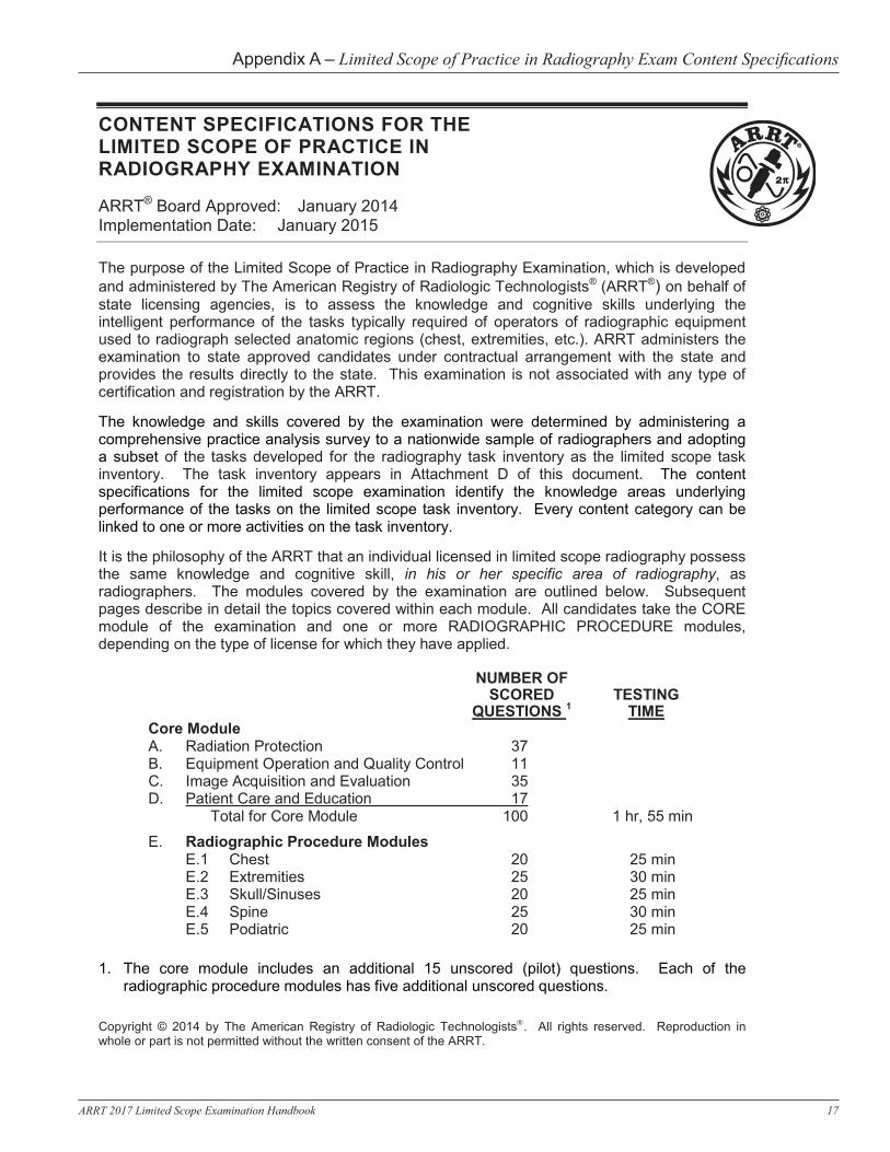
It is the philosophy of the ARRT that an individual licensed in limited scope radiography possess the same knowledge and cognitive skill, in his or her specific area of radiography, as radiographers. The modules covered by the examination are outlined below. Subsequent pages describe in detail the topics covered within each module. All candidates take the CORE module of the examination and one or more RADIOGRAPHIC PROCEDURE modules, depending on the type of license for which they have applied.
NUMBER OF SCORED TESTING
QUESTIONS 1 TIME Core Module A. Radiation Protection 37 B. Equipment Operation and Quality Control 11 C. Image Acquisition and Evaluation 35 D. Patient Care and Education 17
Total for Core Module 100 1 hr, 55 min
E. Radiographic Procedure ModulesE.1 Chest 20 25 min E.2 Extremities 25 30 min E.3 Skull/Sinuses 20 25 min E.4 Spine 25 30 min E.5 Podiatric 20 25 min
1. The core module includes an additional 15 unscored (pilot) questions. Each of theradiographic procedure modules has five additional unscored questions.
Appendix A – Limited Scope of Practice in Radiography Exam Content Specifications

18 ARRT 2017 Limited Scope Examination Handbook
ARRT, page 2
A. RADIATION PROTECTION (37)
1. Biological Aspects of Radiation (7)
A. Radiosensitivity
1. dose-response relationships 2. relative tissue radiosensitivities
(e.g., LET, RBE) 3. cell survival and recovery (LD50) 4. oxygen effect
B. Somatic Effects
1. short-term versus long-term effects 2. acute versus chronic effects 3. carcinogenesis 4. organ and tissue response (e.g., eye,
thyroid, breast, bone marrow, skin, gonadal)
C. Acute Radiation Syndromes
1. CNS 2. hemopoietic 3. GI
D. Embryonic and Fetal Risks
E. Genetic Impact
1. genetic significant dose 2. goals of gonadal shielding
F. Photon Interactions with Matter
1. Compton effect 2. photoelectric absorption 3. coherent (classical) scatter 4. attenuation by various tissues
a. thickness of body part (density) b. type of tissue (atomic number)
2. Minimizing Patient Exposure (13)
A. Exposure Factors
1. kVp 2. mAs
B. Shielding
1. rationale for use 2. types 3. placement
C. Beam Restriction
1. purpose of primary beam restriction 2. types (e.g., collimators)
D. Filtration
1. effect on skin and organ exposure 2. effect on average beam energy 3. NCRP recommendations (NCRP Report
No. 102, minimum filtration in useful beam)
E. Exposure Reduction
1. patient positioning 2. patient communication 3. digital imaging 4. pediatric dose reduction 5. ALARA
F. Image Receptors (e.g., types, relative speed, digital versus film)
(Section A continues on the following page)
Appendix A – Limited Scope of Practice in Radiography Exam Content Specifications

19ARRT 2017 Limited Scope Examination Handbook
ARRT, page 3
A. RADIATION PROTECTION (continued) 3. Personnel Protection (9)
A. Sources of Radiation Exposure
1. primary x-ray beam 2. secondary radiation
a. scatter b. leakage
3. patient as source
B. Basic Methods of Protection
1. time 2. distance 3. shielding
C. Protective Devices
1. types 2. attenuation properties 3. minimum lead equivalent (NCRP
Report No. 102)
4. Radiation Exposure and Monitoring (8)
A. Units of Measurement*
1. absorbed dose 2. dose equivalent 3. exposure
B. Dosimeters
1. types 2. proper use
C. NCRP Recommendations for Personnel Monitoring (NCRP Report No. 116)
1. occupational exposure 2. public exposure 3. embryo/fetus exposure 4. ALARA and dose equivalent limits 5. evaluation and maintenance of personnel
dosimetry records
* Conventional units are generally used. However,
questions referenced to specific reports (e.g., NCRP) will use SI units to be consistent with such reports.
Appendix A – Limited Scope of Practice in Radiography Exam Content Specifications

20 ARRT 2017 Limited Scope Examination Handbook
ARRT, page 4
B. EQUIPMENT OPERATION AND QUALITY CONTROL (11)
1. Principles of Radiation Physics (3)
A. X-Ray Production
1. source of free electrons (e.g., thermionic emission)
2. acceleration of electrons 3. focusing of electrons 4. deceleration of electrons
B. Target Interactions
1. bremsstrahlung 2. characteristic
C. X-Ray Beam
1. frequency and wavelength 2. beam characteristics
a. quality b. quantity c. primary versus remnant (exit)
3. inverse square law 4. fundamental properties (e.g., travel in
straight lines, ionize matter)
2. Imaging Equipment (4)
A. Components of Radiographic Unit (fixed or mobile)
1. operating console 2. x-ray tube construction
a. electron sources b. target materials c. induction motor
3. manual exposure controls 4. beam restriction devices
B. X-Ray Generator, Transformers, and Rectification System (basic principles)
C. Components of Digital Imaging (CR and DR)
1. PSP - photo-stimulable phosphor 2. flat panel detectors - direct and indirect 3. CR reader components 4. CR plate erasure 5. equipment cleanliness (imaging plates,
CR plates)
3. Quality Control of Imaging Equipment and Accessories (4)
A. Beam Restriction
1. light field to radiation field alignment 2. central ray alignment
B. Recognition and Reporting of Malfunctions
C. Digital Imaging Receptor Systems
1. artifacts (e.g., non-uniformity, erasure) 2. maintenance (e.g., detector fog) 3. display monitor quality assurance
D. Shielding Accessories (e.g., lead apron and glove testing)
Appendix A – Limited Scope of Practice in Radiography Exam Content Specifications

21ARRT 2017 Limited Scope Examination Handbook
ARRT, page 5
C. IMAGE ACQUISITION AND EVALUATION (35) 1. Selection of Technical Factors (17)
A. Factors Affecting Radiographic Quality. Refer to Attachment C to clarify terms that may occur on the exam. (X indicates topics covered on the examination)
1. Receptor Exposure
2. Contrast
3. Spatial
Resolution 4.
Distortion
a. mAs X
b. kVp X X
c. OID X (air gap) X X
d. SID X X X
e. focal spot size X
f. filtration X X
g. beam restriction X X
h. motion X
i. anode heel effect X j. patient factors (size,
pathology) X X X X
k. angle (tube, part, or receptor) X X
B. Technique Charts
1. pre-programmed techniques – anatomically programmed radiography (APR)
2. caliper measurement 3. fixed versus variable kVp 4. special considerations
a. anatomic and pathologic factors b. pediatrics
C. Digital Imaging Characteristics
1. spatial resolution a. pixel b. sampling frequency c. DEL (detector element) size d. matrix size
2. image signal (exposure related)
a. dynamic range b. quantum mottle (noise) c. SNR (signal to noise ratio) d. CNR (contrast to noise ratio)
D. Film Screen Characteristics
1. density 2. contrast 3. recorded detail
(Section C continues on the following page)
Appendix A – Limited Scope of Practice in Radiography Exam Content Specifications

22 ARRT 2017 Limited Scope Examination Handbook
ARRT, page 6
C. IMAGE ACQUISITION AND EVALUATION (continued)
2. Image Processing and Quality Assurance (6) A. Image Identification
1. methods (e.g., photographic, radiographic, electronic)
2. legal considerations (e.g., patient data, examination data)
B. Film Screen Processing
1. film storage 2. components*
a. developer b. fixer
3. maintenance/malfunction a. start up and shut down procedure b. possible causes of malfunction
(e.g., improper temperature, contamination, replenishment, water flow)
C. Digital Imaging Processing
1. histogram a. value of interest (VOI) b. rescaling
2. grayscale a. bit depth b. LUT
3. edge enhancement 4. equalization 5. smoothing 6. electronic masking
D. Image Display
1. viewing conditions (e.g., luminance, ambient lighting)
2. spatial resolution (e.g., pixel size, pixel pitch)
3. window level and width function
E. Digital Image Display Informatics
1. DICOM 2. PACS 3. RIS (modality worklist) 4. HIS
3. Criteria for Image Evaluation (12) A. Receptor Exposure (e.g., mAs, distance)
B. Exposure Indicator Determination
C. Contrast/Gray Scale (e.g., kVp, filtration)
D. Recorded Detail/Spatial Resolution (e.g., motion, poor film-screen contact)
E. Distortion (e.g., magnification, OID, SID)
F. Demonstration of Anatomical Structures (e.g., positioning, tube-part-image receptor alignment)
G. Identification Markers (e.g., anatomical, patient, date)
H. Patient Considerations (e.g., pathologic conditions)
I. Image Artifacts, Digital and Film Screen
J. Fog (e.g., age, chemical, radiation, temperature, safelight)
K. Noise
L. Acceptable Range of Exposure
M. Gross Exposure Error (e.g., mottle, light or dark, low contrast)
* Specific chemicals in the processing solutions will not be covered (e.g., glutaraldehyde).
Appendix A – Limited Scope of Practice in Radiography Exam Content Specifications
23ARRT 2017 Limited Scope Examination Handbook
ARRT, page 7
D. PATIENT CARE AND EDUCATION (17)
1. Ethical and Legal Aspects (3) A. Patient’s Rights
1. informed consent (e.g., written, oral, implied)
2. confidentiality (e.g., HIPAA) 3. additional rights (e.g., Patient’s Bill
of Rights) a. privacy b. extent of care (e.g., DNR) c. access to information d. living will; health care proxy e. research participation
B. Legal Issues 1. examination documentation (e.g., patient
history, clinical diagnosis) 2. common terminology (e.g., battery,
negligence, malpractice) 3. legal doctrines (e.g., respondeat superior,
res ipsa loquitur) 4. restraints versus immobilization
C. Professional Ethics 2. Interpersonal Communication (3)
A. Modes of Communication 1. verbal/written 2. nonverbal (e.g., eye contact, touching)
B. Challenges in Communication 1. patient characteristics 2. explanation of medical terms 3. strategies to improve understanding 4. cultural diversity (e.g., language barriers)
C. Patient Education (e.g., explanation of current procedure)
3. Infection Control (5)
A. Terminology and Basic Concepts 1. asepsis
a. medical b. surgical c. sterile technique
2. pathogens a. fomites, vehicles, vectors b. nosocomial infections
B. Cycle of Infection 1. pathogen 2. source or reservoir of infection 3. susceptible host 4. method of transmission
a. contact (direct, indirect) b. droplet c. airborne/suspended d. common vehicle e. vector borne
C. Standard Precautions
1. handwashing 2. gloves, gowns 3. masks 4. medical asepsis (e.g., equipment
disinfection)
D. Additional or Transmission-Based Precautions
1. airborne (e.g., respiratory protection, negative ventilation)
2. droplet (e.g., mask, restricted patient placement)
3. contact (e.g., gloves, gown, restricted patient placement)
E. Disposal of Contaminated Materials
1. linens 2. needles 3. patient supplies (e.g., tubes, emesis basin)
(Section D continues on the following page)
Appendix A – Limited Scope of Practice in Radiography Exam Content Specifications
24 ARRT 2017 Limited Scope Examination Handbook
ARRT, page 8
D. PATIENT CARE AND EDUCATION (continued)
4. Physical Assistance and Transfer (3)
A. Patient Transfer and Movement
1. body mechanics (e.g., balance, alignment, movement)
2. patient transfer
B. Assisting Patients with Medical Equipment (e.g., oxygen delivery systems)
C. Routine Monitoring
1. equipment (e.g., stethoscope, sphygmomanometer)
2. vital signs (e.g., blood pressure, pulse, respiration)
3. physical signs and symptoms (e.g., motor control, severity of injury)
4. documentation
5. Medical Emergencies (3)
A. Allergic Reactions (e.g., latex)
B. Cardiac or Respiratory Arrest (e.g., CPR)
C. Physical Injury or Trauma
D. Other Medical Disorders (e.g., seizures, diabetic reactions)
Appendix A – Limited Scope of Practice in Radiography Exam Content Specifications

25ARRT 2017 Limited Scope Examination Handbook
ARRT, page 9
E. RADIOGRAPHIC PROCEDURES
The specific positions and projections within each anatomic region that may be covered on the examination are listed in Attachment A. A guide to positioning terminology appears in Attachment B.
RADIOGRAPHIC PROCEDURE MODULE 1 1. Chest A. Routine B. Other
# QUESTIONS PER MODULE 2
16 4 TOTAL 20
FOCUS OF QUESTIONS 3 1. Positioning (topographic
landmarks, body positions, path of central ray, etc.)
emphasis: high
2. Anatomy (including
physiology, basic pathology, and related medical terminology)
emphasis: medium
3. Technical Factors
(including adjustments for circumstances such as body habitus, trauma, pathology, breathing techniques, casts, splints, soft tissue for foreign body, etc.)
emphasis: low
4. Equipment and
Accessories (grids or Bucky, compensating filter, automatic exposure control [AEC], automatic collimation, dedicated chest unit)
emphasis: low
2. Extremities A. Lower (toes, foot,
calcaneus, ankle, tibia, fibula, knee, patella, and distal femur)
B. Upper (fingers, hand, wrist, forearm, elbow, and humerus)
C. Pectoral Girdle (shoulder, scapula, clavicle, and acromioclavicular joints)
11 11 3 TOTAL 25
3. Skull/Sinuses A. Skull B. Paranasal Sinuses C. Facial Bones (nasal
bones, orbits)
8 8 4 TOTAL 20
4. Spine A. Cervical Spine B. Thoracic Spine C. Lumbar Spine D. Sacrum, Coccyx, and
Sacroiliac Joints E. Scoliosis Series
8 6 8 2 1 TOTAL 25
5. Podiatric A. Foot and Toes B. Ankle C. Calcaneus (os calcis)
14 5 1 TOTAL 20
Notes: 1. Examinees take one or more radiographic procedure modules, depending on the type of license
they have applied for. Each radiographic procedure module has 20 or 25 scored test questions, depending on the module (see chart above). The number of questions within a module should be regarded as approximate values.
2. Each of the radiographic procedure modules has five additional unscored questions. 3. The radiographic procedure modules may include questions about the four areas listed under
FOCUS OF QUESTIONS on the right side of the chart. The podiatric module does not include questions from the equipment and accessories section.
Appendix A – Limited Scope of Practice in Radiography Exam Content Specifications

26 ARRT 2017 Limited Scope Examination Handbook
ARRT, page 10
Attachment A Radiographic Positions and Projections
I. Chest
A. Chest 1. PA or AP upright 2. lateral upright 3. AP Lordotic 4. AP supine 5. lateral decubitus 6. anterior and posterior
obliques II. Extremities
A. Toes 1. AP, entire foot 2. oblique toe 3. lateral toe
B. Foot 1. AP angle toward heel 2. medial oblique 3. lateral oblique 4. mediolateral 5. lateromedial 6. sesamoids, tangential 7. AP weight-bearing 8. lateral weight-bearing
C. Calcaneus (os calcis) 1. lateral 2. plantodorsal, axial 3. dorsoplantar, axial
D. Ankle 1. AP 2. AP mortise 3. mediolateral 4. oblique, 45º internal 5. lateromedial 6. AP stress views
E. Tibia, Fibula 1. AP 2. lateral 3. oblique
F. Knee 1. AP 2. lateral 3. AP weight-bearing 4. lateral oblique 45º 5. medial oblique 45º 6. PA 7. PA axial – intercondylar
fossa (tunnel) G. Patella
1. lateral 2. supine flexion 45º (Merchant) 3. PA 4. prone flexion 90º (Settegast) 5. prone flexion 55º (Hughston)
H. Femur (Distal) 1. AP 2. mediolateral
I. Fingers 1. PA entire hand 2. PA finger only 3. lateral 4. oblique 5. AP thumb 6. oblique thumb 7. lateral thumb
J. Hand
1. PA 2. lateral 3. oblique
K. Wrist 1. PA 2. oblique 45º 3. lateral 4. PA for scaphoid 5. scaphoid (Stecher) 6. carpal canal
L. Forearm 1. AP 2. lateral
M. Elbow 1. AP 2. lateral 3. external oblique 4. internal oblique 5. AP partial flexion 6. axial trauma (Coyle)
N. Humerus 1. AP 2. lateral 3. AP neutral 4. scapular Y 5. transthoracic lateral
O. Shoulder 1. AP internal and external
rotation 2. inferosuperior axial 3. posterior oblique (Grashey) 4. tangential 5. AP neutral 6. transthoracic lateral 7. scapular Y
P. Scapula 1. AP 2. lateral, anterior oblique 3. lateral, posterior oblique
Q. Clavicle 1. AP 2. AP angle 15-30º cephalad 3. PA angle 15-30º caudad
R. Acromioclavicular joints - AP bilateral with and without weights
III. Skull/Sinuses
A. Skull 1. AP axial (Towne) 2. lateral 3. PA (Caldwell) 4. PA 5. submentovertex (full basal)
B. Facial Bones 1. lateral 2. parietoacanthial (Waters) 3. PA (Caldwell) 4. PA (modified Waters)
C. Nasal Bones 1. parietoacanthial (Waters) 2. lateral 3. PA (Caldwell)
D. Orbits
1. parietoacanthial (Waters) 2. lateral 3. PA (Caldwell)
E. Paranasal Sinuses 1. lateral 2. PA (Caldwell) 3. parietoacanthial (Waters) 4. submentovertex (full basal) 5. open mouth parietoacanthial
(Waters) IV. Spine
A. Cervical spine 1. AP angle cephalad 2. AP open mouth 3. lateral 4. anterior oblique 5. posterior oblique 6. lateral swimmers 7. lateral flexion and extension
B. Thoracic Spine 1. AP 2. lateral, breathing 3. lateral, expiration
C. Lumbar Spine 1. AP 2. PA 3. lateral 4. L5-S1 lateral spot 5. posterior oblique 45º 6. anterior oblique 45º 7. AP L5-S1, 30-35º cephalad 8. AP right and left bending 9. lateral flexion and extension
D. Sacrum and Coccyx 1. AP sacrum, 15-25º cephalad 2. AP coccyx, 10-20º caudad 3. lateral sacrum and coccyx,
combined 4. lateral sacrum or coccyx,
separate E. Sacroiliac Joints
1. AP 2. posterior oblique 3. anterior oblique
F. Scoliosis Series 1. AP/PA scoliosis series
(Ferguson) V. Podiatric
A. Foot and Toes 1. dorsal plantar (DP)* 2. medial oblique 3. lateral oblique 4. lateral* 5. sesamoidal axial*
B. Ankle* 1. AP* 2. mortise* 3. AP medial oblique* 4. AP lateral oblique* 5. lateral*
C. Calcaneus (os calcis) 1. axial calcaneal* 2. Harris and Beath (ski-jump)*
*weight-bearing
Appendix A – Limited Scope of Practice in Radiography Exam Content Specifications

27ARRT 2017 Limited Scope Examination Handbook
ARRT, page 11
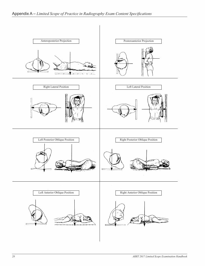
Attachment B Standard Terminology
for Positioning and Projection Radiographic View: Describes the body part as seen by the image receptor or other recording
medium, such as a fluoroscopic screen. Restricted to the discussion of a radiograph or image. Radiographic Position: Refers to a specific body position, such as supine, prone, recumbent,
erect, or Trendelenburg. Restricted to the discussion of the patient’s physical position. Radiographic Projection: Restricted to the discussion of the path of the central ray. POSITIONING TERMINOLOGY A. Lying Down 1. supine − lying on the back 2. prone − lying face downward 3. decubitus − lying down with a horizontal x-ray beam 4. recumbent − lying down in any position B. Erect or Upright 1. anterior position − facing the image receptor 2. posterior position − facing the radiographic tube 3. oblique position − erect or lying down a. anterior (facing the image receptor)
i. left anterior oblique body rotated with the left anterior portion closest to the image receptor
ii. right anterior oblique body rotated with the right anterior portion closest to the image receptor
b. posterior (facing the radiographic tube) i. left posterior oblique body rotated with the left posterior portion
closest to the image receptor ii. right posterior oblique body rotated with the right posterior portion
closest to the image receptor
Appendix A – Limited Scope of Practice in Radiography Exam Content Specifications
28 ARRT 2017 Limited Scope Examination Handbook
ARRT, page 12
Anteroposterior Projection Posteroanterior Projection
Right Lateral Position Left Lateral Position
Left Posterior Oblique Position Right Posterior Oblique Position
Left Anterior Oblique Position Right Anterior Oblique Position
Appendix A – Limited Scope of Practice in Radiography Exam Content Specifications

29ARRT 2017 Limited Scope Examination Handbook
ARRT, page 13
Attachment C ARRT Standard Definitions
Term Film-Screen Radiography Term Digital Radiography includes both computed radiography and direct radiography.
Computed Radiography (CR) systems use storage phosphors to temporarily store energy representing the image signal. The phosphor then undergoes a process to extract the latent image.
Direct Radiography (DR) systems have detectors that directly capture and readout an electronic image signal.
Recorded Detail
The sharpness of the structural lines as recorded in the radiographic image.
Spatial Resolution
The sharpness of the structural edges recorded in the image.
Receptor Exposure
The amount of radiation striking the image receptor.
Receptor Exposure
The amount of radiation striking the image receptor.
Density Radiographic density is the degree of blackening or opacity of an area in a radiograph due to the accumulation of black metallic silver following exposure and processing of a film. Density = Log incident light intensity
transmitted light intensity
Brightness Brightness is the measurement of the luminance of a monitor calibrated in units of candela (cd) per square meter on a monitor or soft copy.
Density on a hard copy is the same as film.
Contrast Radiographic contrast is defined as the visible differences between any two selected areas of density levels within the radiographic image.
Scale of Contrast refers to the number of densities visible (or the number of shades of gray).
Long Scale is the term used when slight differences between densities are present (low contrast) but the total number of densities is increased.
Short Scale is the term used when considerable or major differences between densities are present (high contrast) but the total number of densities is reduced.
Contrast Image contrast or display contrast is determined primarily by the processing algorithm (mathematical codes used by the software to provide the desired image appearance). The default algorithm determines the initial processing codes applied to the image data.
Scale of Contrast is synonymous to “gray scale” and is linked to the bit depth of the system. “Gray scale” is used instead of “scale of contrast” when referring to digital images.
Film Latitude
The inherent ability of the film to record a long range of density levels on the radiograph.
Film latitude and film contrast depend upon the sensitometric properties of the film and the processing conditions, and are determined directly from the characteristic H and D curve.
Dynamic Range
The range of exposures that may be captured by a detector.
The dynamic range for digital imaging is much larger than film.
Film Contrast
The inherent ability of the film emulsion to react to radiation and record a range of densities.
Receptor Contrast
The fixed characteristic of the receptor. Most digital receptors have an essentially linear response to exposure. This is impacted by contrast resolution (the smallest exposure change or signal difference that can be detected). Ultimately, contrast resolution is limited by the dynamic range and the quantization (number of bits per pixel) of the detector.
Exposure Latitude
The range of exposure factors which will produce a diagnostic radiograph.
Exposure Latitude
The range of exposures which produces quality images at appropriate patient dose.
Subject Contrast
The difference in the quantity of radiation transmitted by a particular part as a result of the different absorption characteristics of the tissues and structures making up that part.
Subject Contrast
The magnitude of the signal difference in the remnant beam.
Appendix A – Limited Scope of Practice in Radiography Exam Content Specifications

30 ARRT 2017 Limited Scope Examination Handbook
ARRT, page 14
Attachment D Task Inventory for Limited Scope of Practice in Radiography
Activity Content Categories
1. Confirm patient’s identity. D.2.
2. Evaluate patient’s ability to understand and comply with requirements for the requested examination.
D.2.
3. Obtain pertinent medical history. D.1.B.1., D.2.
4. Examine imaging examination requisition to verify accuracy and completeness of information (e.g., patient history, clinical diagnosis, physicians orders).
D.1.B.
5. Respond as appropriate to imaging study inquiries from patients. D.2.
6. Assume responsibility for medical equipment attached to patients (e.g., IVs, oxygen) during the imaging procedures.
D.4.B.
7. Follow environmental protection standards for handling and disposing of biohazardous materials (e.g., sharps, blood and body fluids).
D.3.E.
8. Provide for patient safety, comfort, and modesty. D.1.C., D.4., D.1.A.
9. Notify appropriate personnel of adverse events or incidents (e.g., patient fall, wrong patient imaged).
C.2.A., D.1.B.1., D.4.
10. Communicate scheduling delays to waiting patients. D.2.
11. Verify or obtain patient consent as necessary. D.1.A.1.
12. Communicate relevant information to others (e.g., MDs, RNs, other radiology personnel.
D.
13. Explain procedure instructions to patient or patient’s family. D.2.
14. Practice standard precautions. D.3.B., D.3.C.
15. Follow appropriate procedures when in contact with patient in isolation. D.3.C., D.3.D.
16. Select immobilization devices, when indicated, to prevent patient’s movement and/or ensure patients safety.
D.1.B.4.
17. Use proper body mechanics and/or mechanical transfer devices when assisting patient.
D.4.A.1.
18. Use sterile or aseptic technique when indicated. D.3.A.
19. Obtain vital signs. D.4.C.
20. Recognize and communicate the need for prompt medical attention. D.5., D.4.C.3.
21. Administer emergency care. D.5., D.4.C.3.
22. Explain post-procedural instructions to patient or patient’s family. D.2.C.
23. Maintain confidentiality of patient’s information. D.1.A.2., D.1.C.
24. Clean, disinfect or sterilize facilities and equipment, and dispose of contaminated items in preparation for next examination.
D.3.A., D.3.E.
Appendix A – Limited Scope of Practice in Radiography Exam Content Specifications
31ARRT 2017 Limited Scope Examination Handbook
*Applies to specific modules
ARRT, page 15
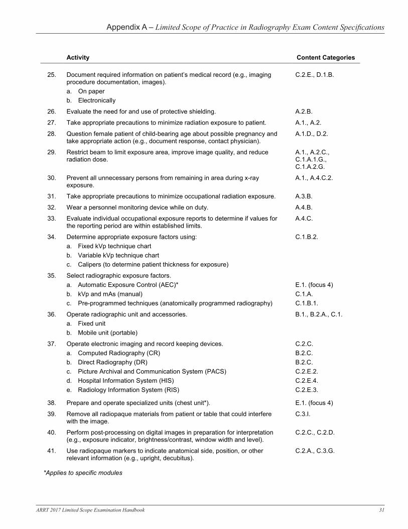
Activity Content Categories
25. Document required information on patient’s medical record (e.g., imaging procedure documentation, images). a. On paper b. Electronically
C.2.E., D.1.B.
26. Evaluate the need for and use of protective shielding. A.2.B.
27. Take appropriate precautions to minimize radiation exposure to patient. A.1., A.2.
28. Question female patient of child-bearing age about possible pregnancy and take appropriate action (e.g., document response, contact physician).
A.1.D., D.2.
29. Restrict beam to limit exposure area, improve image quality, and reduce radiation dose.
A.1., A.2.C., C.1.A.1.G., C.1.A.2.G.
30. Prevent all unnecessary persons from remaining in area during x-ray exposure.
A.1., A.4.C.2.
31. Take appropriate precautions to minimize occupational radiation exposure. A.3.B.
32. Wear a personnel monitoring device while on duty. A.4.B.
33. Evaluate individual occupational exposure reports to determine if values for the reporting period are within established limits.
A.4.C.
34. Determine appropriate exposure factors using: a. Fixed kVp technique chart b. Variable kVp technique chart c. Calipers (to determine patient thickness for exposure)
C.1.B.2.
35. Select radiographic exposure factors. a. Automatic Exposure Control (AEC)* b. kVp and mAs (manual) c. Pre-programmed techniques (anatomically programmed radiography)
E.1. (focus 4) C.1.A. C.1.B.1.
36. Operate radiographic unit and accessories. a. Fixed unit b. Mobile unit (portable)
B.1., B.2.A., C.1.
37. Operate electronic imaging and record keeping devices. a. Computed Radiography (CR) b. Direct Radiography (DR) c. Picture Archival and Communication System (PACS) d. Hospital Information System (HIS) e. Radiology Information System (RIS)
C.2.C. B.2.C. B.2.C. C.2.E.2. C.2.E.4. C.2.E.3.
38. Prepare and operate specialized units (chest unit*). E.1. (focus 4)
39. Remove all radiopaque materials from patient or table that could interfere with the image.
C.3.I.
40. Perform post-processing on digital images in preparation for interpretation (e.g., exposure indicator, brightness/contrast, window width and level).
C.2.C., C.2.D.
41. Use radiopaque markers to indicate anatomical side, position, or other relevant information (e.g., upright, decubitus).
C.2.A., C.3.G.
Appendix A – Limited Scope of Practice in Radiography Exam Content Specifications

32 ARRT 2017 Limited Scope Examination Handbook
*Applies to specific modules
ARRT, page 16
Activity Content Categories
42. Add electronic annotations on digital images to indicate position, or other relevant information (e.g., upright, decubitus, standing, weight-bearing).
C.2.A., C.3.G.
43. Use film-screen cassettes and automatic film processing. C.1.D., C.2.B.
44. Select equipment and accessories (e.g., grid, compensating filter, shielding) for the examination requested.
A.2.B., E.1. (focus 4)
45. Explain breathing instructions prior to making the exposure.* C.1.A.3.H., D.2.C., E.1.E.2.C., E.4. (focus 3)
46. Position patient to demonstrate the desired anatomy using body landmarks. E., C.3.F.
47. Modify exposure factors for circumstances such as involuntary motion, casts and splints, pathological conditions, or patient’s inability to cooperate.
C.1.B.3., C.1.A.
48. Verify accuracy of patient identification on image. C.2.A., C.3.G.
49. Evaluate images for diagnostic quality. C.3.
50. Determine corrective measures if image is not of diagnostic quality and take appropriate action.
C.3.
51. Store and handle image receptor in a manner which will reduce the possibility of artifact production.
B.3.C., B.2.C.5., B.2.C.3., C.2., C.3.J., C.3.I.
52. Visually inspect, recognize, and report malfunctions in the imaging unit and accessories.
B.3.B.
53. Recognize the need for basic evaluations of radiographic equipment and accessories. a. Light field to radiation field alignment b. Central-ray alignment c. Shielding accessories (e.g., lead aprons and gloves)
B.3.A.1. B.3.A.2. B.3.D.
54. Perform routine maintenance on digital equipment. a. Perform start-up or shut-down b. Erase CR plate c. Equipment cleanliness (e.g., imaging plates, CR cassettes) d. Recognize and report malfunctions
B.2.C.3. B.2.C.4. B.2.C.5. B.3.B.
Position patient, x-ray tube, and image receptor to produce the following diagnostic images:
55. Chest E.1.A.
56. Cervical spine E.4.A.
57. Thoracic spine E.4.B.
58. Scoliosis series E.4.C.
59. Lumbar spine E.4.D.
60. Sacrum and coccyx E.4.E.
Appendix A – Limited Scope of Practice in Radiography Exam Content Specifications

33ARRT 2017 Limited Scope Examination Handbook
ARRT, page 17 V 2015.09.17
Activity Content Categories
61. Sacroiliac joints E.4.D.
62. Skull E.3.A.
63. Facial bones E.3.C.
64. Nasal bones E.3.C.
65. Orbits E.3.C.
66. Paranasal sinuses E.3.B.
67. Toes E.2.A., E.5.A.
68. Foot E.2.A., E.5.A.
69. Calcaneus (os calcis) E.2.A., E.5.C.
70. Ankle E.2.A., E.5.B.
71. Tibia, fibula E.2.A.
72. Knee E.2.A.
73. Patella E.2.A. 74. Distal femur E.2.A. 75. Fingers E.2.A. 76. Hand E.2.A. 77. Wrist E.2.A. 78. Forearm E.2.A. 79. Elbow E.2.A. 80. Humerus E.2.A. 81. Shoulder E.2.A. 82. Scapula E.2.A. 83. Clavicle E.2.A. 84. Acromioclavicular joints E.2.A. 85. Soft tissue/foreign body E.2. (focus 3)
Appendix A – Limited Scope of Practice in Radiography Exam Content Specifications


![Higher Photoelectric Effect Questions - Larbert High School25375]11._H_Photoelectric_Effect... · Higher Photoelectric Effect Questions 1. a) ... "One of the important factors affecting](https://static.fdocuments.in/doc/165x107/5af772cf7f8b9aac248bdbf3/higher-photoelectric-effect-questions-larbert-high-2537511hphotoelectriceffecthigher.jpg)