Gastric Emptying Measurements: Delayed and Complex Emptying Patterns Without AppropriateCorrection
4/23/2007
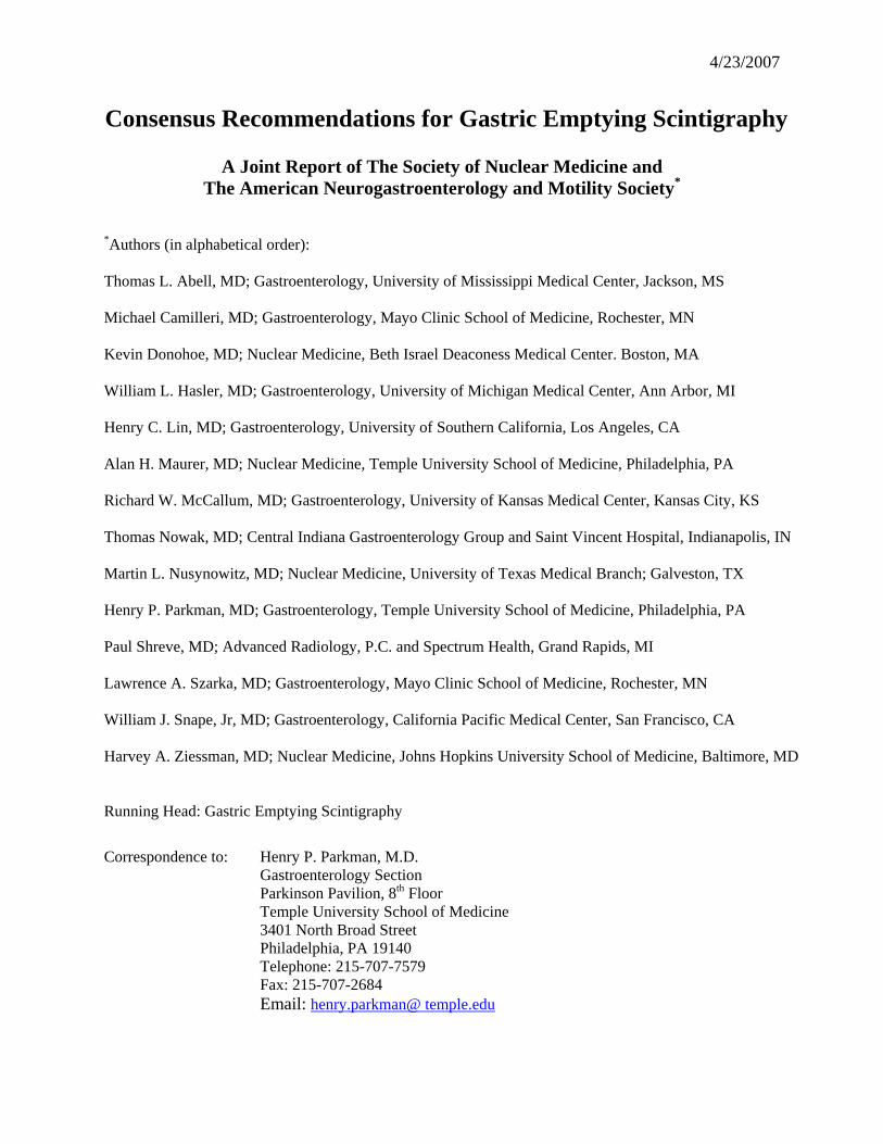
Consensus Recommendations for Gastric Emptying Scintigraphy
A Joint Report of The Society of Nuclear Medicine and The American Neurogastroenterology and Motility Society*
*Authors (in alphabetical order): Thomas L. Abell, MD; Gastroenterology, University of Mississippi Medical Center, Jackson, MS Michael Camilleri, MD; Gastroenterology, Mayo Clinic School of Medicine, Rochester, MN Kevin Donohoe, MD; Nuclear Medicine, Beth Israel Deaconess Medical Center. Boston, MA William L. Hasler, MD; Gastroenterology, University of Michigan Medical Center, Ann Arbor, MI Henry C. Lin, MD; Gastroenterology, University of Southern California, Los Angeles, CA Alan H. Maurer, MD; Nuclear Medicine, Temple University School of Medicine, Philadelphia, PA Richard W. McCallum, MD; Gastroenterology, University of Kansas Medical Center, Kansas City, KS Thomas Nowak, MD; Central Indiana Gastroenterology Group and Saint Vincent Hospital, Indianapolis, IN Martin L. Nusynowitz, MD; Nuclear Medicine, University of Texas Medical Branch; Galveston, TX Henry P. Parkman, MD; Gastroenterology, Temple University School of Medicine, Philadelphia, PA Paul Shreve, MD; Advanced Radiology, P.C. and Spectrum Health, Grand Rapids, MI Lawrence A. Szarka, MD; Gastroenterology, Mayo Clinic School of Medicine, Rochester, MN William J. Snape, Jr, MD; Gastroenterology, California Pacific Medical Center, San Francisco, CA Harvey A. Ziessman, MD; Nuclear Medicine, Johns Hopkins University School of Medicine, Baltimore, MD Running Head: Gastric Emptying Scintigraphy
Correspondence to: Henry P. Parkman, M.D.
Gastroenterology Section Parkinson Pavilion, 8th Floor Temple University School of Medicine 3401 North Broad Street Philadelphia, PA 19140 Telephone: 215-707-7579 Fax: 215-707-2684 Email: henry.parkman@ temple.edu
4/23/2007
ABSTRACT
This consensus statement from members of the American Neurogastroenterology and
Motility Society and the Society of Nuclear Medicine recommends a standardized method for
measuring gastric emptying by scintigraphy (GES). A low-fat, egg white meal with imaging at
0, 1, 2, 4 hours after meal ingestion, as described by a published multicenter protocol, provides
standardized information about normal and delayed gastric emptying. Adoption of this
standardized protocol will resolve the lack of uniformity of testing, add reliability and credibility
to the results, and improve the clinical utility of the gastric emptying test.
INTRODUCTION
Gastric emptying scintigraphy (GES) is commonly performed to evaluate patients with
symptoms that suggest an alteration of gastric emptying and/or motility (1Maurer 2006). The
first use of radionuclides to measure gastric emptying was published in 1966 (2Griffith 1966).
Since then, it has become the standard for the measurement of gastric motility in clinical practice
because it provides a physiologic, noninvasive, quantitative measurement of solid or liquid
gastric emptying (3Camilleri 1998).
While GES has been considered the standard for measuring gastric emptying, there is a
lack of standardization of the test, including differences in meals used, patient positioning,
frequency, and duration of imaging. There are differences in the quantitative data reported, e.g.,
half-time of emptying, rate of emptying (per cent per minute), or the percent retention or
emptying at different time points during the study. Normal values often have not been
established for some of the protocols used, and the performance characteristics of the test with
the specified meal may not have been established or published.
Lack of standardization limits the clinical utility of the test, and presents problems for
patients and their physicians as the latter try to interpret study results from other institutions. This
often leads to repeat testing using a different protocol with which the gastroenterologist has
greater familiarity and confidence. This practice presents problems for the patient as insurance
companies may not reimburse a second test, or may require the patient undergo the test at a
center where the protocol is suboptimal. Discrepant results between two studies can further
complicate patient management.
4/23/2007
Several professional societies (Society of Nuclear Medicine [SNM], American
Gastroenterological Association [AGA], and the American Neurogastroenterology and Motility
Society [ANMS]) have developed guidelines for performing GES (4Parkman 2004,
5Donohoe1999, 6Donohoe2004, 7Lin 2005). However, there has not been a consensus on a
protocol that meets both the clinicians’ needs and the services available at most nuclear medicine
facilities. For GES to attain its full clinical potential there is a need for closer collaboration
between nuclear medicine physicians and referring gastroenterologists to develop such a
consensus protocol
This manuscript provides consensus guidelines from members of the ANMS and the
SNM. Our goal was to propose a single, standardized protocol for performing GES that meets
the needs of clinicians, imaging specialists, and patients.
The process leading to this joint report involved three meetings. At the first meeting
(Philadelphia, April 2006), the participants focused on the needs of the patient and the referring
physician, reviewed current methods considered best practice for performing GES, and
addressed unresolved areas requiring further research or clarification. At the second meeting at
the Society of Nuclear Medicine annual meeting (San Diego, June 2006), there were
presentations and discussion of the same topics which led to the drafting of a manuscript
summarizing a consensus protocol. At a third meeting at the ANMS annual meeting (Boston,
September 2006), a consensus protocol was discussed and critiqued. This was followed by final
discussions and revisions to this manuscript.
This manuscript represents this group’s current recommendation on how to perform a
solid-meal GES test for clinical practice, using readily available technology and normative data
which can provide clinicians with standardized results. Throughout this process, we have
recognized that the current consensus may not imply unanimity of opinion. In addition, this
document does not answer all the questions raised during these discussions.
However, this manuscript addresses those aspects that the multidisciplinary group
consider were in the greatest need of immediate standardization – the meal, the frequency of
imaging, the duration of the test, and normative data. Technical issues concerning the acquisition
and processing parameters are less controversial and do not contribute to wide variations in
methodology. These have been addressed elsewhere (5Donohoe 1999, 6Donohoe 2004) and are
4/23/2007
summarized in Appendix 1. The manuscript also identifies areas for future study, but there was
overwhelming consensus on the pressing need to recommend a standardized GES test.
DISORDERS OF GASTRIC EMPTYING
Both rapid and delayed gastric emptying can cause similar symptoms. Hence, it is
important to evaluate patients for both rapid and delayed gastric emptying in the same test. It is
therefore appropriate to review some of the relevant clinical information on symptoms related to
disorders of gastric emptying.
Delayed Gastric Emptying
Gastroparesis is diagnosed when symptoms such as nausea, vomiting, early satiety,
postprandial fullness, abdominal discomfort and pain are associated with objective evidence of
delayed gastric emptying in the absence of obstruction and typically with impairment in
maintenance of normal nutrition using standard food (4Parkman 2004, 8Soykan 1998). These
symptoms are also reported by some individuals with the postprandial distress subtype of
functional dyspepsia (9Tack 2006). Symptoms that suggest delayed gastric emptying in patients
with dyspepsia are primarily postprandial fullness, nausea and vomiting (10Stanghellini 1996,
11Sarnelli 2003). In patients with diabetes, symptoms that have been associated with delayed
gastric emptying are abdominal bloating/fullness and upper abdominal pain (12Jones 2001,
13Samson 2003). Some studies, however, have shown a poor correlation between the rate of
solid gastric emptying and the severity of gastric symptoms (12Jones 2001, 13Samsom 2003,
14Horowitz 1991). Furthermore, some clinical trials of drug therapy for gastroparesis show
variable symptomatic benefit from pharmacologic stimulation of gastric emptying (15Snape
1982, 16Patterson 1999). In addition, some patients symptoms improve with prokinetic agents
but gastric emptying remains unchanged (16Patterson 1999). Investigations are attempting to
determine if factors other than a global delay in gastric emptying such as impaired fundic
accommodation, antral distension, antral hypomotility, gastric dysrhythmias, visceral
hypersensitivity, or psychological disturbances explain, in part, the symptoms experienced by
patients with suspected gastroparesis (1Maurer 2006).
Symptoms of gastroparesis may be quantified by a validated symptom questionnaire, the
Gastroparesis Cardinal Symptom Index (GCSI) (17Revicki 2003, 18Revicki 2004). The GCSI is
4/23/2007
based on three subscales (postprandial fullness/early satiety, nausea/vomiting, and bloating) and
represents a subset of the longer Patient Assessment of Upper Gastrointestinal Disorders-
Symptoms (PAGI-SYM) that quantifies symptoms of gastroparesis, dyspepsia, and
gastroesophageal reflux disease (19Revicki 2004).
Rapid Gastric Emptying
Rapid gastric emptying is the major factor in dumping syndrome, characteristically seen
after surgery for peptic ulcer disease with and without vagotomy (20Hasler, 21Eagon). The early
symptoms of “dumping” occurring in the initial hour after meal ingestion include diarrhea,
abdominal discomfort, nausea, bloating, and vasomotor symptoms. Some of these symptoms
may be difficult to distinguish from gastroparesis symptoms. The late dumping syndrome
presents with diaphoresis, palpitations, weakness, and fainting which are secondary to reactive
hypoglycemia from exaggerated insulin release.
Rapid gastric emptying of solids has been demonstrated in some patients with
unexplained nausea, vomiting, bloating, and fullness (22Lin 1999). Rapid GE has recently been
reported in some patients with functional dyspepsia (23Delgado 2004). Rapid gastric emptying
also occurs in some diabetic patients, particularly in the early stages of Type II diabetics
(24Schwartz 1996). Many of these patients have symptoms indistinguishable from those of
gastroparesis. Rapid movement of food from the stomach into the small bowel with small bowel
distension may explain pain and nausea, symptoms similar to those described in patients with
delayed gastric emptying. Rapid emptying has been recently observed as an accompanying
factor in adult patients with cyclic vomiting syndrome (25Namin 2006, 26Fajardo 2005). In one
study, rapid gastric emptying was more common than delayed GE in symptomatic patients with
autonomic dysfunction (27Lawal 2006).
FACTORS TO CONSIDER IN THE MEASUREMENT OF GASTRIC EMPTYING
Measurements of gastric emptying are influenced by a variety of factors which must be
considered when performing GES (28Christian 1983). Factors that may influence the
performance and negatively influence the clinical validity of a GES are: 1) short duration of
testing; 2) extrapolated t-1/2 measurements; 3) unknown meal composition and amount
4/23/2007
consumed; 4) medications at the time of the test; 5) poor or unknown glycemic control at the
time of the test; and 6) vomiting a portion of the meal.
Patient–Related Factors to be considered for Gastric Emptying Tests
Patient-related factors that need to be considered in gastric emptying tests include
medications, tobacco smoking, hyperglycemia, and gender (1Maurer 2006, 3Camilleri 1998).
Patients may be taking medications to intentionally affect gastric emptying (e.g.,
prokinetic agents) or mediations that alter emptying as a side effect (e.g., narcotic analgesics).
Depending on the reason for the test, patients may need to discontinue these medications. The
period of time off medication before the test should be based on the drug half-life; for most
medications, this will be 48-72 hours (3Camilleri 1998). Opiate analgesic medications and
anticholinergic agents delay gastric emptying and, if not discontinued prior to the study, could
result in a false diagnosis of delayed gastric emptying. Prokinetics may lead to a normal gastric
emptying result in a patient with gastroparesis. Serotonin receptor (5-HT-3) antagonists, such as
ondansetron, which have little effect on gastric emptying, may be given for severe symptoms of
nausea and vomiting before performance of GES (29Nielson 1990).
Tobacco smoking has been shown in early studies to slow gastric emptying (30Nowak
1987, 31Miller 1989). This effect may not be related to nicotine (31Miller 1989, 32Wong 1999).
Recent studies with 13C-octanoic breath testing, however, have shown conflicting results
(33Sanaka 2005). It is recommended that patients abstain from smoking the morning of the test
and throughout time gastric emptying is being imaged.
Hyperglycemia can delay gastric emptying. Although some data suggest that even
modest degrees of hyperglycemia (>144 mg/dl) retards gastric emptying (34Schvarcz 1997), it is
not clear what level produces clinically significant delay in gastric emptying (35Horowitz 2002).
Marked hyperglycemia with serum glucose levels > 288 mg/dL significantly delays gastric
emptying in diabetic patients when compared to euglycemia (36Fraser 1990). The general
consensus is that blood glucose should be under reasonable control on the day of a GES to obtain
a reliable measurement of gastric emptying. For diabetics, it is recommended that serum glucose
be measured prior to the study, noted in the report, and if the blood glucose is greater than 275
mg/dl on the morning of the test, the glucose should be lowered with insulin to <275 mg/dl, or
the study should be rescheduled for another day when the blood glucose is under better control.
4/23/2007
Some centers administer insulin if the blood glucose level immediately before GES is >180
mg/dl and do not start the test until the glucose is less than 180 mg/dl (Horowitz personal
communication).
Pre-menopausal women are reported in some studies to have slower gastric emptying
than men (37Datz 1987, 38Gill 1987). A few centers use separate reference values for pre-
menopausal women (10Stanghellini 1996) or limit testing to the first week of the menstrual cycle
before estrogen and progesterone peak. The latter approach is suggested since separate values
are not generally available for men and women. The four hour test of Tougas et al recommended
in these guidelines did not show variability attributable to menstrual phase and gender (39Tougas
2001). A study of postmenopausal women administered estrogen or progesterone or
combination to mimic normal hormone levels in pre-menopausal women did not demonstrate
any effect of these sex hormones on gastric emptying (40Gonene 2006).
Technical Factors in Gastric Emptying Tests
There are many reasons for the current diversity of imaging and analysis of GES
protocols: an individual center’s meal and analysis preferences, different camera and computer
systems, scheduling constraints, and processing software.
Meals currently used for measuring gastric emptying consist of a variety of foods,
including chicken liver, eggs, egg whites, oatmeal, or pancakes. The content of the meal used is
one of the most important variables needing standardization since gastric emptying depends on
meal composition. Solids empty more slowly than liquids and digestible solids emptying more
rapidly than indigestible residue (1Maurer 2006). Additionally, emptying of fats is slow
compared to emptying of proteins or carbohydrates. The caloric content and volume of the test
meal will also alter gastric emptying. Incomplete meal ingestion can lead to values suggesting
more rapid emptying. Vomiting a portion of the ingested meal after the initial baseline image
may lead to lower subsequent estimated gastric retention values, so that gastric emptying appears
faster than it was.
Reported values of gastric emptying are influenced by the duration of testing and the
method of analysis. Half times (T-1/2 values) of emptying may be potentially less accurate than
percentages of retention measured at fixed time points, particularly in individuals with very
prolonged emptying in which extrapolation is needed to calculate the half time if it was not
4/23/2007
actually reached during the test. Surveys of nuclear medicine centers show wide variations in
reporting rates of emptying. In a study of Canadian health care facilities, 40% of centers did not
validate a normal range for their gastric emptying test meal and the range of normal values
varied considerably: 20% of centers used 2 standard deviations about the mean (SD), 26% used 1
SD, 6% used 1.5 SD for normal ranges (41House 1997).
Individual preferences of both referring physicians and the imaging specialists have
influenced local decisions on how to conduct GES. Currently, studies vary in length from as
short as 60 minutes up to 4 hours. Studies have shown that extending measurements out to 4
hours increases the detection rate of delayed emptying (42Guo 2001, 43Ziesman 2007). As more
has been learned about optimal methodology, some imaging centers have changed their
protocols, but many centers have been reluctant to perform a 4 hour procedure because
reimbursement is not commensurate with the time and effort needed. Reimbursement for gastric
emptying scintigraphy is substantially lower than for other imaging procedures that can be done
in less time on the same equipment. This economic reality however should not be an impediment
to providing optimum patient care. Under current Center for Medicare and Medicaid Services
(CMS) guidelines, any future increase in RVU assignment (reimbursement) will not occur until
the medical community demonstrates a consensus on the requirement for more comprehensive
imaging.
TOWARDS STANDARDIZATION: Validation of the proposed regimen
One multi-institutional protocol has been published which investigated a large number of
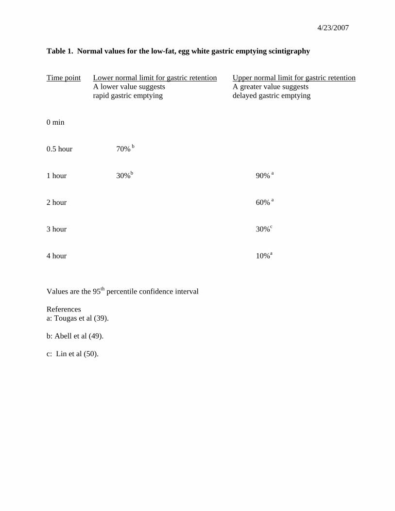
normal subjects and established normal values (39Tougas 2000). One hundred and twenty-three
normal subjects from 11 medical institutions in the United States, Canada, and Europe were
studied in a multi-institutional and multinational study. Normal values published from other
protocols were often based on smaller numbers of subjects (typically 10-35) at single institutions
(44Cremonini et al). The percent gastric emptying was quantified at several time intervals after
consumption of a low-fat, egg-white meal. The Tc-99m sulfur colloid radiolabeled meal
consisted of the equivalent of 2 large eggs (Eggbeaters®), 2 slices bread and jam with water.
Imaging was performed in the anterior and posterior projections at only four time points (0, 1, 2,
and 4 hours). The geometric mean activity of decay-corrected counts (square root of the product
of the anterior and posterior counts) was determined at each imaging time. Normal values were

4/23/2007
established using the median and 95th percentile because the data were skewed, particularly at 4
hours. These authors defined the upper limits of gastric retention (95th percentile) of the 123
men and women at each of three time points: 1, 2, and 4 hours (Table 1). Delayed gastric
emptying (gastric retention) was determined to be > 90% at 1 hour, >60% at 2 hours, and >10%
gastric retention at 4 hours. These results currently provide the largest published data base for a
standardized GES protocol (39Tougas 2000).
Imaging at only four time points can actually simplify scheduling and permit more
efficient use of imaging equipment. This helps alleviate some of the concerns at some imaging
centers that GES is too time consuming, and the efficiencies in camera use are associated with
little loss of test accuracy (45Vijayamkumar 2006, 46Camilleri 1991). Multiple gastric emptying
tests can be performed on one camera in a single day if the starting times are staggered.
Another important factor supporting use of the Tougas’ protocol is the data suggesting
the superiority of a longer, 4 hour study rather than the 2 hour study (39Tougas 2000, 42Guo
2001, 43Ziessman 2007). The first reported studies promoting 4 hour imaging were from the
Mayo Clinic in 1991 and 1995 (46Camilleri 1991, 47Thomforde 1995). In a study of 35 patients,
the 4 hour time interval was more sensitive for detection of delayed gastric emptying than the
two hour time-point. The Temple University group reported in 2001 on 127 consecutive patients
referred for clinical gastric emptying studies (42Guo 2001). GES was performed with a
conventional egg labeled sandwich with water. Imaging was performed at 0, 0.5, 1, 2, 3, 4
hours. The data suggested that the 3 and 4 hour imaging times detected more abnormal gastric
emptying than 2 hour images, e.g., 50% of patients were abnormal at 3-4 hours versus 33% at 2
hours. Data from Johns Hopkins University has found similar results using the Tougas meal
(48Goetze 2005, 42Ziessman 2007). In an investigation of 175 patients, 34 studies were
abnormal at 2 hours. The 4 hour retention identified 11 additional abnormal patients who had
normal emptying at two hours, a 29% increase in the number of abnormal studies.
Any GES protocol should be able to identify both rapid and slow gastric emptying.
Although the symptoms of rapid emptying may be similar to those of delayed emptying
(23Delgado-Aros 2004), the treatment is quite different. Use of a standardized protocol across
all medical centers which includes standard time points (e.g. 0, 1, 2, and 4 hours) offers the
potential of consistently defining both delayed and rapid GE. The early 1 hour time point can be
used for determining rapid gastric emptying; whereas the later 2 and 4 hour time points are used
4/23/2007
for determining delayed gastric emptying. The study of Tougas et al did not establish values for
rapid emptying. Reanalysis of data from the original Tougas manuscript suggests 32% is the
lower 95% confidence interval for normal emptying suggesting <30% retention at 1 hour
(49Abell 2006) is indicative of rapid gastric emptying (Table 1). Other recent studies also
suggest that 30% is an optimal threshold for 3 hour empyting data (43Ziessman 2007).
ITEMS FOR FURTHER INVESTIGATION
It is anticipated that GES will need to be further optimized as more studies using this
protocol become available. The consensus participants therefore identified areas where more
information is needed (Table 2).
Optimal Timing of Imaging
Some patients with delayed emptying at 2 hours normalize their emptying at the 4 hour
time point and some individuals with normal emptying at 2 hours have delayed emptying at 4
hours (42Guo 2001, 43Zeissman). The clinical importance of delayed emptying at only certain
time points is unknown. There needs to be better understanding of the use of multiple time
points in combination (e.g., 2 and 4 hours). Data from Guo et al. suggest that the 3 hour time
period might be as sensitive as a four hour study in detecting delayed gastric emptying (42Guo et
al 1991). Although the original report for the Tougas et al data had no 3 hour measurement
point, more recent studies suggest the upper limit of normal is 28% gastric retention at 3 hours
after meal ingestion (50Lin 2006). A recent study using the Tougas meal have shown that the 3
hour time point is nearly comparable to the 4 hour value in detecting patients with delayed
gastric emptying (50Lin 2006).
For rapid gastric emptying, currently the one hour gastric emptying value is
recommended. However, the normal values were generated with a smaller subgroup of normal
subjects and currently published only in abstract form for the Tougas meal (49Abell 2006).
More data are needed for delineation of the cutoffs to determine rapid gastric emptying. Recent
investigations also suggest that rapid emptying is detected at 15-60 minutes after meal ingestion
(22Lin 1999, 23Delgado-Aros 2004). It is conceivable that earlier time points, such as a 30
minute postprandial value, may be better than one hour value for detection of rapid gastric
emptying.
4/23/2007
The time points for measuring gastric emptying may be further optimized in the future.
Presently, however, there are insufficient data to recommend the 30 minute and 3 hour time
points for routine use (Table 2). The current consensus recommendation is to obtain images at a
minimum at 0, 1, 2, and 4 hours after meal ingestion as described originally (39Tougas 2000)
Composition of Meal
This consensus document proposes a low fat meal used as the initial screening test for
gastric emptying. In some patients, a low fat, egg white meal may not prove to be an adequate
functional challenge, especially for patients who report symptom exacerbations after eating lipid-
rich foods (22Lin 1999). Meal composition may need to be altered depending on the patient’s
specific symptoms. Some European investigators have suggested adding butter (10 grams) to the
low fat meal to increase the fat content. This brings the meal to a caloric content of 345 kcal
with nutritional composition of 69% carbohydrate, 22% protein, 7% fat, 2% fiber. Other
alternative meals may also be useful for patients with egg allergies or intolerance to eggs, and
patients with gluten-sensitive enteropathy. Liquid Ensure nutrient supplement or an oatmeal
meal are used by some centers. However, specific normal databases will be needed for these
alternative meals before they are used clinically.
Glycemic Control
More uniform management of diabetic patients undergoing GES is needed. The
consensus opinion is to measure and record the blood glucose level of diabetic patients just prior
to the study. The study should be performed if the patient is under reasonable glucose control,
that is, fasting glucose of <275 mg/dl. There is still no consensus, however, on what should be
done with insulin and oral hypoglycemic agents on the day of the study. Generally, half of the
usual morning dose of insulin is given with the radiolabeled test meal. Postprandial glucose is
not usually measured to determine if hyperglycemia develops postprandially during the gastric
emptying test; postprandial hyperglycemia could prolong gastric emptying. Hypoglycemia may
also influence gastric emptying or increase the patient’s symptoms. Postprandial glucose
monitoring at 2 and 4 hours may be considered in the future.
Monitoring of Symptoms
4/23/2007
Monitoring symptoms during the gastric emptying test is currently being performed in
clinical research studies (13Samsom 2003) to determine whether delay in gastric emptying
correlates with symptoms. If the gastric emptying test is normal, this simultaneous recording of
symptoms will inform whether the meal was an appropriate provocative test to induce the typical
postprandial symptoms.
Assessment of Severity
Since there is not a close correlation between a delay in gastric emptying and symptoms,
the gastric emptying test alone should probably not be used for grading the severity of the
clinical disorder of gastroparesis. Grading the severity of the delay in gastric emptying has been
performed in clinical research studies and might be used clinically (51Arts 2005). Grading for
severity of delayed gastric emptying based on 4 hour value in groups related to the standard
deviation of the normal results:
Grade 1 (mild): 11-20% retention at 4 hours
Grade 2 (moderate): 21-35% retention at 4 hours
Grade 3 (severe): 36-50% retention at 4 hours.
Grade 4 (very severe): >50% retention at 4 hours
On the other hand, a combination of the degree of gastric emptying delay and the nutritional
needs or approaches necessary to support the patients’ hydration and nutrition provide a better
assessment of severity and facilitate the approach to management. In a recent review, mild delay
was designated as 11-15%, moderate 16-35% and severe >35% retention at 4 hours (52Camilleri
2007).
Establishing normal values for the post-gastric surgery group
There is no appropriate “normal” data base to use for patients that are symptomatic after
gastric surgical procedures. In postsurgical patients, the altered anatomy is likely to alter gastric
emptying compared to normal subjects without gastric surgery (53Fich 1990). In general, there
is delay in the emptying of solids and accelerated emptying of liquids (53Fich 1900). After Roux
gastrectomy, there may be retention of solids in both gastric remnant and the Roux limb
(54Miedema 1992). The normal gastric emptying of the Tougas meal in patients with different
degrees of partial gastric resections (e.g., antrectomy) and different drainage procedures is not

4/23/2007
known. This is also relevant with surgical reconstruction of the stomach, especially post-
bariatric surgery of the stomach.
Assessment of proximal and distal gastric function
A unique advantage of GES is that it can characterize more completely the complex
physiology of GE including proximal (fundal) and distal (antral) function. Scintigraphy permits
analysis of the intragastric distribution of the test meal between the fundus and antrum. The
routine GE imaging can be used to measure both regional and total GE. Visual inspection of
fundal and antral gastric emptying and quantification of regional emptying with fundic and antral
regions of interest can be helpful for defining abnormal physiology and explaining dyspeptic
symptoms especially when global GE values are normal (55Troncon 1999, 56Gonlachanvit
2006, 57Karamanolis 2007). Studies have shown an association between symptoms of nausea,
early satiety, abdominal distention and acid reflux with proximal gastric retention; whereas
vomiting is associated more with delayed distal GE (56Gonlachanvit 2006). However more
validation of intragastric distribution and regional emptying are necessary before this is
incorporated into clinical practice.
Utility of measuring the lag time
The lag phase is the time required for commencement of gastric emptying of solid
particles. It represents the time for solid food to be triturated into small particles that are then
passed through the pylorus. The lag phase can be measured as the time from meal ingestion to
first appearance of the radiolabeled solids in the proximal small bowel. This approach often
requires frequent imaging for at least the first 60 minutes of the gastric emptying study, as the
normal values have been reported to be 20±10 minutes (SD) (43Ziessman 2007). It is not certain
whether this physiological information is important for clinical management. Gastroparesis is
often associated with a prolonged lag phase and slow post-lag emptying rate (58Camilleri 1986).
However, a prolonged solid gastric emptying lag phase has been implicated in some
investigations as the cause of delayed emptying in certain diseases and shortening of the lag
phase by prokinetic drugs as the reason for improved emptying (59Ziessman 1992). One report
has suggested the lag phase to be a sensitive indicator of the efficacy of drugs used to treat
diabetic gastroparesis (60Mackie 1991). Recent reports with the Tougas meal suggest that the
4/23/2007
length of the lag phase does not correlate with gastric emptying and does not add additional
information to the percent retention at specified times (43Ziessman 2007).
Use of mathematical models for parameter estimation
Curve fitting to the data points for gastric retention over time may provide useful
information on gastric emptying (61Elashoff 1983, 62Siegel 1988, 63Nusynowitz 1994,
64Camilleri 1985). The gastric emptying curve can be analyzed in several mathematical ways to
determine both the emptying rate and lag phase. A curve fitting procedure such as a dual
exponential equation (modified power exponential) has been used, namely:
y = 100[1 – (1- e –kt) b]
where y is the percent remaining at time t, k is an exponential emptying rate constant (fraction of
amount present at time t) and b is the y intercept of the terminal exponential with slope = -k. The
lag time can be then calculated as ln(b)/k. Slope and lag time may be helpful to interpret
borderline results. Using curve fitting techniques and a similar meal to the low fat Tougas meal,
the upper limit of normal for the lag phase is approximately 45 minutes (63Nusynowitz 1994). If
considering the curve fitting approach for gastric interpretation, several additional data points
should be obtained such that imaging occurs at 0, 0.5, 1, 2, 3, 4 hours.
SUMMARY CONSENSUS RECOMMENDATIONS FOR A STANDARDIZED GASTRIC
EMPTYING PROCEDURE
This consensus document concludes that, at the current time, the most universally
acceptable meal is the low-fat, egg white meal as described by Tougas et al. Eggbeaters® or
other equivalent generic commercial brands of liquid egg white are available and acceptable for
use. Currently this meal and protocol has the largest normative database (39Tougas 2000). GES
should be performed with imaging, at a minimum, at 0, 1, 2, and 4 hours after radiolabeled meal
ingestion.
Ultimately, to have value, any standardized protocol must be followed closely. Our
current consensus recommendation including the technical details on how to perform GES is
presented in Appendix 1. In order to facilitate better standardization, a number of sample
documents are presented. A patient information form for those undergoing the test is contained
in Appendix 2 (Reference: ANMS web site: www.motilitysociety.org). A form for all patients
4/23/2007
to complete to provide important information to assist in study interpretation is contained in
Appendix 3.
This consensus statement recommends use of a single, standardized GES protocol.
Adoption of this standard is important to improve how gastric emptying studies are used to direct
patient care. The authors recognize that any consensus protocol has limitations. However, the
one currently recommended has the largest database of normal values. Adopting this protocol
will solve the problem of non-uniformity of protocols across institutions and will be a vast
improvement over the diverse methods currently in use. Other questions and issues will need to
be addressed in the months and years ahead before GES attains its full clinical potential (Table
2). With continued close collaboration between imaging specialists and gastroenterologists, this
important physiologic test will continue to serve as a valuable tool in the diagnosis and
management of gastric motility disorders.
4/23/2007
References
1. Maurer AH, Parkman HP. Update on Gastrointestinal Scintigraphy. Seminars in Nuclear Medicine 2006;36:110-118 2. Griffith GA, Owen Gm, Kirkman S, et al. Measurement of the rate of gastric emptying using chromium-51. Lancet 1966; 1:1244-1245. 3. Camilleri M, Hasler W. Parkman HP, Quigley EMM, Soffer E. Measurement of gastroduodenal motility in the GI laboratory. Gastroenterology 1998;115:747-762. 4. Parkman HP, Hasler WL, Fisher RS. American Gastroenterological Association technical review on the diagnosis and treatment of gastroparesis. Gastroenterology 2004;127:1592-1622. 5. Donohoe KJ, Maurer AH, Ziessman HA, Urbain JL, Royal HD. Procedure guideline for gastric emptying and motility - Society of Nuclear Medicine. J Nucl Med. 1999;40(7):1236-9. 6. Donohoe KJ, Maurer AH, Ziessman HA, et al. Society of Nuclear Medicine Procedure Guideline for Gastric Emptying and Motility. June 4, 2004. 7. Lin HC, Prather C, Fisher RS, et al – The AMS Task Force Committee on Gastrointestinal Transit. Measurement of Gastrointestinal Transit. Dig Dis Sci 2005;50:989-1004. 8. Soykan I, Sivri B, Sarosiek I, Kierran B, McCallum RW. Demography, clinical characteristics, psychological profiles, treatment and long-term follow-up of patients with gastroparesis. Dig Dis Sci 1998;43:2398-2404. 9. Tack J, Talley NJ, Camilleri M, Holtmann G, Hu P, Malagelada JR, Stanghellini V. Functional gastroduodenal disorders. Gastroenterology 2006;130(5):1466-79. 10. Stanghellini V, Tosetti C, Paternico A, Barbara G, Morselli-Labate AM, Monetti N, Marengo M, Corinaldesi R. Risk indicators of delayed gastric emptying of solids in patients with functional dyspepsia. Gastroenterology 1996;110,1036-1042. 11. Sarnelli G, Caenepeel P, Geypens B, Janssens J, Tack J. Symptoms associated with impaired gastric emptying of solids and liquids in functional dyspepsia. Am J Gastroenterol. 2003;98(4):783-8. 12. Jones KL, Wishart JM, Russo A, Berry MK, Stevens JE, Horowitz M. Predictors of delayed gastric emptying in diabetes. Diabetes Care 2001;24:1264-1269. 13. Samsom M, Vermeijden JR, Smout AJ, et al. Prevalence of delayed gastric emptying in diabetic patients and relationship to dyspeptic symptoms: a prospective study in unselected diabetic patients. Diab Care 2003; 26: 3116-3122. 14. Horowitz M, Maddox AF, Wishart JM, et al. Relationships between oesophageal transit and solid and liquid gastric emptying in diabetes mellitus. Eur J Nuc Med 1991; 18: 229-234. 15. Snape WJ, Battle WM, Schwartz SS, Braunstein SN, Goldstein HA, Alavi A. Metoclopramide to treat gastroparesis due to diabetes mellitus. Ann Intern Med 1982;96:444-446. 16. Patterson D, Abell T, Rothstein R, Koch K, Barnett J. A double-blind multicenter comparison of domperidone and metoclopramide in the treatment of diabetic patients with symptoms of gastroparesis. Am J Gastroenterol 1999;94:1230-1234. 17. Revicki DA, Rentz AM, Dubois D, Kahrilas P, Stanghellini V, Talley NJ, Tack J. Development and validation of a patient-assessed gastroparesis symptoms severity measure: the Gastroparesis Cardinal Symptom Index. Aliment Pharmacol Ther 2003;18:141-150. 18. Revicki DA, Rentz AM, Dubois D, Kahrilas P, Stanghellini V, Talley NJ, Tack J. Gastroparesis Cardinal Symptom Index (GCSI): development and validation of a patient reported assessment of severity of gastroparesis symptoms. Qual Life Res 2004;13(4):833-44. 19. Revicki DA, Rentz AM, Tack J, Stanghellini V, Talley NJ, Kahrilas P, de la Loge C, Trudeau E, Dubois D. Responsiveness and Interpretation of a Symptom Severity Index specific to upper gastrointestinal disorders. Clinical Gastroenterol Hepatol 2004;2:769-777. 20. Hasler WH. Dumping Syndrome. Curr Treat Options Gastroenterol. 2002;5(2):139-145. 21. Eagon JC, Miedema BW, Kelly KA. Postgastrectomy syndromes. Surgical Clinics NA 1992;72:445-465.
4/23/2007
22. Lin HC, Van Citters GW, Zhao XT, Waxman A. Fat intolerance depends on rapid gastric emptying. Dig Dis Sci 1999; 44: 330-335. 23. Delgado-Aros S, Camilleri M, Cremonini F, et al. Contributions of gastric volumes and gastric emptying to meal size and postmeal symptoms in functional dyspepsia. Gastroenterology 2004; 127: 1685-1694. 24. Schwartz JG, Green GM, Guan D, et al. Rapid gastric emptying of a solid pancake meal in type II diabetic patients. Diabetes Care 1996;19:468-471. 25. Namin F, Jitan P, Joker I, Lin Z, Dusing R, McCallum RW. Clinical hallmarks of cyclic vomiting syndrome (CVS) in adults and role of long-term tricyclic therapy. Gastroenterology 2006;130 (Suppl 2):A601. (abstract) 26. Fajardo NR, Locke GR, Talley NJ. Cyclic vomiting syndrome is associated with rapid early gastric emptying (abstract). Am J Gastroenterol 2005; 100:143. (abstract) 27. Lawal A, Barboi A, Krasnow A, Hellman R, Jaradeh S, Massey BT. Rapid gastric emtpying is more common than gastroparesis in patients with autonomic dysfunction. Am J Gastroenterol 2007 (in press). 28. Christian PE, Datz FL, Sorenson JA, et al. Technical factors in gastric emptying studies. J Nucl Med 1983; 24: 264–268. 29. Nielsen OH, Hvid-Jacobsen K, Lund P, Langoholz E. Gastric emptying and subjective symptoms of nausea: lack of effects of a 5-hydroxytryptamine-3 antagonist ondansetron on gastric emptying in patients with gastric stasis syndrome. Digestion 1990;46(2):89-96. 30. Nowak A, Jondeerko K, Kaezor R, Nowak S, Skrzypek D. Cigarette smoking delays gastric emptying of a radiolabelled solid food in healthy smokers. Scand J Gastroenterol. 1987; 22(1):54-8. 31. Miller G, Palmer KR, Smith B, Ferrington C, Merrick MV. Smoking delays gastric emptying of solids. Gut. 1989;30(1): 50-3. 32. Wong, PWK, Kadakia SC, McBiles M. Acute effect of nicotine patch on gastric emptying of liquid and solid contents in healthy subjects. Dig Dis Sci. 1999; 44(11): 2165-71. 33. Sanaka, M, Anjiki H, Tsutsumi H, Abe K, Kaakami T, Saitoh, M, Yamamoto T, Ishii T, Kuyama Y. Effect of cigarette smoking on gastric emptying of solids in Japanese smokers: a crossover study using the 13C-octanoic acid breath test. 2005; 40(6): 578-82. 34. Schvarcz E, Palmer M, Aman J, Horowitz M, Stridsberg M, Berne C. Physiological hyperglycemia slows gastric emptying in normal subjects and patients with insulin-dependent diabetes mellitus. Gastroenterology 1997;113:60-66. 35. Horowitz M, O’Donovan D, Jones KL, Feinle C, Rayner CK, Samsom M. Gastric emptying in diabetes: clinical significance and treatment. Diabetes Medicine 2002;19:177-194. 36. Fraser R, Horowitz M, maddox A, Chatterton B, Harding P, Dent J. Hyperglcaemia slows gastric emptying in type 1 diabetes mellitus. Diabetologia 1990;30:675-680. 37. Datz FL. Christian PE, Moore J. Gender-related differences in gastric emptying. J Nucl Med 1987;28:1204-1207. 38. Gill RC, Murphy PD, Hooper HR, Bowes KL, Kingma YJ. Effect of the menstrual cycle on gastric emptying. Digestion 1987;36:168-174. 39. Tougas G, Eaker EY, Abell TL, et al. Assessment of gastric emptying using a low fat meal: establishment of international control values. Am J Gastroenterol 2000; 95: 1456-1462. 40. Gonenne J, Esfandyari T, Camilleri M, Burton DD, Stephens DA, Baxter KL, Zinsmeister AR, Bharucha AE: Effect of female sex hormone supplementation and withdrawal on gastrointestinal and colonic transit in postmenopausal women. Neurogastroenterol Motil 2006; 18:911-918. 41. House A, Champion MC, Chamberlain M. National survey of radionuclide gastric emptying studies. Can J Gastroenterol 1997; 11: 317-321. 42. Guo J-P, Maurer AH, Urbain J-L, Fisher RS, Parkman HP. Extending gastric emptying scintigraphy from two to four hours detects more patients with gastroparesis. Dig Dis Sci 2001;46:24-29. 43. Ziessman HA, Bonta DV, Goetze S, Ravich WJ. Experience with a new simplified and standardized four-hour gastric emptying protocol. J Nucl Med 2007;48:568-572.
4/23/2007
44. Cremonini F, Mullan BP, Camilleri M, Burton DD, Rank MR. Performance characteristics of scintigraphic transit measurements for studies of experimental therapies. Aliment Pharmacol Ther 2002;16(10):1781-90. 45. Vijayamkumar V, Briscoe EG, Boysen TL, Jimenez YM. Assessment of the practical role of a low-fat-meal solid gastric emptying study. J Nucl Med Technol 2006;34:82-85. 46. Camilleri M, Zinsmeister AR, Greydanus MP, Brown ML, Proano M. Towards a less costly but accurate test of gastric emptying and small bowel transit. Dig Dis Sci. 1991;36(5):609-15. 47. Thomforde GM, Camilleri M, Phillips SF, et al. Evaluation of an inexpensive screening scintigraphic test of gastric emptying. J Nucl Med 1995; 36:93-96. 48. Goetze S, Ravich WJ, Ziessman HA. Increased detection of abnormal gastric emptying at 4 hours compared to two hours using a standardized protocol. J Nucl Med 2005; 46:320p. 49. Abell TL, Starkebaum W, Abidi N, Liu A. How common is rapid gastric emptying in gastroparesis. Gastroenterology 2006 (abstract) 50. Lin Z, Sarosiak I, McCallum RW. Optimal detection of gastroparesis. Am J Gastro 2006 abstract. 51. Arts J, Caenepeel P, Verbeke K, Tack J. Influence of erythromycin on gastric emptying and meal related symptoms in functional dyspepsia with delayed gastric emptying. Gut. 2005;54(4):455-60. 52. Camilleri M. Clinical practice. Diabetic gastroparesis. N Engl J Med. 2007; 356: 820-951. 53. Fich A, Neri M, Camilleri M, Kelly KA, Phillips SF. Stasis syndromes following gastric surgery: clinical and motility features of 60 symptomatic patients. J Clin Gastroenterol 1990;12(5):505-12. 54. Miedema BW, Kelly KA, Camilleri M, Hanson RB, Zinsmeister AR, O'Connor MK, Brown ML: Human gastric and jejunal transit and motility after Roux gastrojejunostomy. Gastroenterology 103:1133-1143, 1992. 55. Troncon LE, Bennett RJ, Ahluwalia NK, Thompson DG. Abnormal intragastric distribution of food during gastric emptying in functional dyspepsia patients. Gut 1994;35:327-332. 56. Gonlachanvit S, Maurer AH, Fisher RS, Parkman HP. Regional Gastric Emptying Abnormalities in Functional Dyspepsia and Gastroesophageal Reflux Disease. Neurogastroenterology and Motility 2006;18(10):894-904. 57. Karamanolis G, Caenepeel P, Arts J, Tack J. Determinants of symptom pattern in idiopathic severely delayed gastric emptying: gastric emptying rate or proximal stomach dysfunction? Gut 2007;56:29-36. 58. Camilleri M, Brown ML, Malagelada JR. Relationship between impaired gastric emptying and abnormal gastrointestinal motility. Gastroenterology. 1986;91(1):94-9. 59. Ziessman HA, Fahey FH, Collen MJ. Biphasic solid and liquid gastric emptying in normal controls and diabetics using continuous acquisition in LAO view. Dig Dis Sci 1992;37(5):744-50. 60. Mackie AD, Ferrington C, Cowan S, Merrick MV, Baird JD, Palmer KR. The effects of renzapride, a novel prokinetic agent, in diabetic gastroparesis. Aliment Pharmacol Ther 1991;5(2):135-42. 61. Elashoff JD, Reedy TJ, Meyer JH. Methods for analyzing gastric emptying. Am J Physiol. 1983; 244:G701-250. 62. Siegel JA, Urbain JL, Adler LP, Charkes ND, Maurer AH, Krevsky B, Knight LC, Fisher RS, Malmud LS. Biphasic nature of gastric emptying. Gut 1988;29(1):85-9. 63. Nusynowitz ML, Benedetto AR. The lag phase of gastric emptying: clinical, mathematical and in vitro studies. J Nucl Med. 1994;35(6):1023-7. 64. Camilleri M, Malagelada JR, Brown ML, Becker G, Zinsmeister AR. Relation between antral motility and gastric emptying of solids and liquids in humans. Am J Physiol. 1985;249(5 Pt 1):G580-5.
4/23/2007
Table 1. Normal values for the low-fat, egg white gastric emptying scintigraphy Time point Lower normal limit for gastric retention Upper normal limit for gastric retention A lower value suggests A greater value suggests rapid gastric emptying delayed gastric emptying 0 min 0.5 hour 70% b 1 hour 30%b 90% a 2 hour 60% a 3 hour 30%c 4 hour 10%a Values are the 95th percentile confidence interval References a: Tougas et al (39). b: Abell et al (49). c: Lin et al (50).
4/23/2007
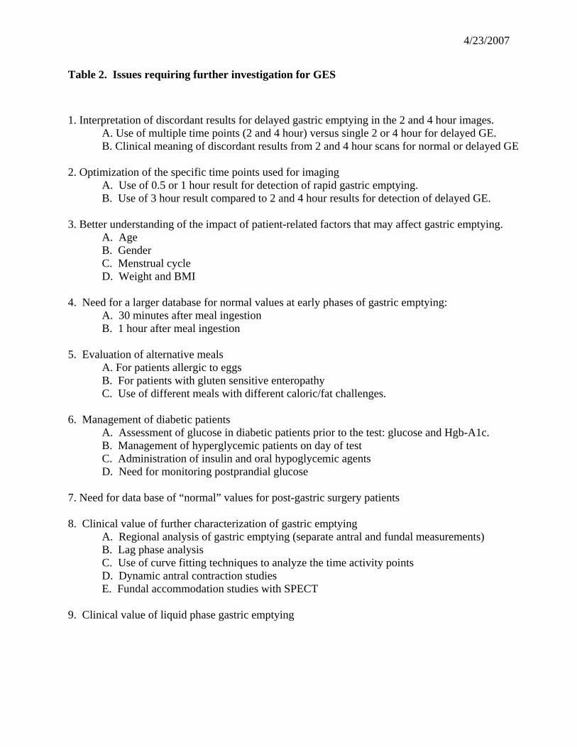
Table 2. Issues requiring further investigation for GES
1. Interpretation of discordant results for delayed gastric emptying in the 2 and 4 hour images. A. Use of multiple time points (2 and 4 hour) versus single 2 or 4 hour for delayed GE. B. Clinical meaning of discordant results from 2 and 4 hour scans for normal or delayed GE 2. Optimization of the specific time points used for imaging A. Use of 0.5 or 1 hour result for detection of rapid gastric emptying. B. Use of 3 hour result compared to 2 and 4 hour results for detection of delayed GE. 3. Better understanding of the impact of patient-related factors that may affect gastric emptying. A. Age B. Gender C. Menstrual cycle D. Weight and BMI 4. Need for a larger database for normal values at early phases of gastric emptying: A. 30 minutes after meal ingestion B. 1 hour after meal ingestion 5. Evaluation of alternative meals A. For patients allergic to eggs B. For patients with gluten sensitive enteropathy C. Use of different meals with different caloric/fat challenges. 6. Management of diabetic patients A. Assessment of glucose in diabetic patients prior to the test: glucose and Hgb-A1c. B. Management of hyperglycemic patients on day of test C. Administration of insulin and oral hypoglycemic agents D. Need for monitoring postprandial glucose 7. Need for data base of “normal” values for post-gastric surgery patients 8. Clinical value of further characterization of gastric emptying A. Regional analysis of gastric emptying (separate antral and fundal measurements) B. Lag phase analysis C. Use of curve fitting techniques to analyze the time activity points D. Dynamic antral contraction studies E. Fundal accommodation studies with SPECT 9. Clinical value of liquid phase gastric emptying

4/23/2007
Appendix 1. Consensus Method for Performing Gastric Emptying Scintigraphy I. Patient Preparation The referring physician should determine what medications are to be continued prior to GES. Prokinetic agents that enhance gastric emptying such as metoclopramide (Reglan), tegaserod (Zelnorm), erythromycin, domperidone (Motilium) are generally stopped at least 2 days prior to the test. Opiate analgesic medications delay gastric emptying and should also be stopped 2 days before the test. These include: Demerol, codeine, morphine, oxycontin, percodan, percocet. Anticholinergic antispasmodic agents such as Bentyl, Donnatal, Levsin, Robinul are usually stopped for 2 days prior to the test. It is preferable to study menstruating females during the first 10 days of their menstrual cycle to improve the interpretation of the study and to prevent administration of radionuclide to a potentially pregnant woman. The study should be performed in the morning after an overnight fast. If this is not possible, patients should be fasting for at least 6 hours prior to the test. The patient may take medications with some water on arising before coming for the test. Patients should refrain from smoking the morning of the test and throughout the time of imaging. Diabetic patients should test and record their blood glucose prior to the study (fasting). Generally the fasting glucose should be < 275 mg/dl. At the time the meal is ingested, diabetic patients should self administer their insulin at a dose that is generally ½ what they normally take. Subjects should be instructed that they will be in the imaging facility for at least 4 hours after meal ingestion and advised to bring reading material, a personal music player or other material to occupy them for the time of the study. For the time between images, the subjects can be sitting, standing, or walking but should remain in close proximity to the nuclear medicine section. II. Meal Preparation and ingestion The standard scintigraphic meal for gastric emptying should consist of an egg-white meal (Egg Beaters® or generic equivalent) radiolabeled with 0.5 -1 mCi 99mTc. The meal has a caloric value of 255 kcal (nutritional composition: 72% carbohydrate, 24% protein, 2% fat, and 2% fiber). An allergy to eggs is a contraindication to this meal. Items needed for Gastric Emptying Meal
4 oz.(120 gm, equal to approximately two large eggs) Liquid egg white; 99% real eggs, cholesterol free, fat free, low calorie) 2 slices of white bread (120 kcal), Strawberry jam (30 g, 74 kcal) Water (120 ml). Technetium-99m sulfur colloid, 0.5 -1 mCi
The eggs can be cooked either scrambled on a hot skillet (nonstick frying pan) or microwaved in an appropriately shielded container. The eggs are served with toast, jam, and water. To prepare the meal, the liquid egg is poured into a bowl, mixed with 0.5 - 1 mCi99Tc sulfur-colloid marker,
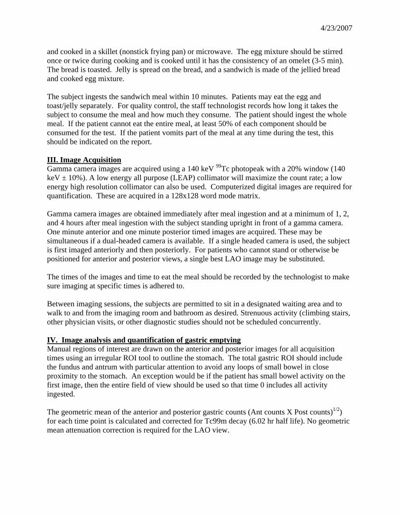
4/23/2007
and cooked in a skillet (nonstick frying pan) or microwave. The egg mixture should be stirred once or twice during cooking and is cooked until it has the consistency of an omelet (3-5 min). The bread is toasted. Jelly is spread on the bread, and a sandwich is made of the jellied bread and cooked egg mixture. The subject ingests the sandwich meal within 10 minutes. Patients may eat the egg and toast/jelly separately. For quality control, the staff technologist records how long it takes the subject to consume the meal and how much they consume. The patient should ingest the whole meal. If the patient cannot eat the entire meal, at least 50% of each component should be consumed for the test. If the patient vomits part of the meal at any time during the test, this should be indicated on the report. III. Image Acquisition Gamma camera images are acquired using a 140 keV 99Tc photopeak with a 20% window (140 keV ± 10%). A low energy all purpose (LEAP) collimator will maximize the count rate; a low energy high resolution collimator can also be used. Computerized digital images are required for quantification. These are acquired in a 128x128 word mode matrix. Gamma camera images are obtained immediately after meal ingestion and at a minimum of 1, 2, and 4 hours after meal ingestion with the subject standing upright in front of a gamma camera. One minute anterior and one minute posterior timed images are acquired. These may be simultaneous if a dual-headed camera is available. If a single headed camera is used, the subject is first imaged anteriorly and then posteriorly. For patients who cannot stand or otherwise be positioned for anterior and posterior views, a single best LAO image may be substituted. The times of the images and time to eat the meal should be recorded by the technologist to make sure imaging at specific times is adhered to. Between imaging sessions, the subjects are permitted to sit in a designated waiting area and to walk to and from the imaging room and bathroom as desired. Strenuous activity (climbing stairs, other physician visits, or other diagnostic studies should not be scheduled concurrently. IV. Image analysis and quantification of gastric emptying Manual regions of interest are drawn on the anterior and posterior images for all acquisition times using an irregular ROI tool to outline the stomach. The total gastric ROI should include the fundus and antrum with particular attention to avoid any loops of small bowel in close proximity to the stomach. An exception would be if the patient has small bowel activity on the first image, then the entire field of view should be used so that time 0 includes all activity ingested. The geometric mean of the anterior and posterior gastric counts (Ant counts X Post counts)1/2) for each time point is calculated and corrected for Tc99m decay (6.02 hr half life). No geometric mean attenuation correction is required for the LAO view.

4/23/2007
The final results are expressed as percent remaining in the stomach at each time point with the total gastric counts normalized to 100% for the time t=0 (first image set immediately after meal ingestion). V. Reporting The percent remaining in the stomach at each time point is reported. The report should contain the normal values at the key time points: 1 hour (37-90%), 2 hr (30-60%) and 4 hrs (0-10%). A graph of the values plotted as a function of time may also be included in the report as a visual summary of the study results. The report should mention the fasting blood glucose if the patient is diabetic. The report should mention medications that the patient was taking within the last 24 hours of the test that may affect gastric emptying. The report should document the amount of the meal ingested, the total time taken to ingest the meal, and if any vomiting of the meal occurred postprandially. The report should also describe any other unusual findings which are observed in the images such as: abnormal esophageal retention of the meal, hiatal hernia, fundal wrap, lack of fundic accommodation, evidence for retained old food particles.
4/23/2007
Appendix 2. Sample Patient Instruction Sheet for Gastric Emptying Scintigraphy Your doctor has ordered a test that will permit evaluation of how food moves through your stomach. These studies will be performed in the Nuclear Medicine Department. Preparation for the Gastric Emptying Test: • You should not eat any food after midnight the night before the test. If you smoke, do not smoke
beginning on the morning of your test and thoughout the time you are having the pictures of gastric emptying recorded. You may smoke after you are instructed that the test is completed.
• Some medications are generally stopped for this test. This should be discussed with your doctor or
health care provider. Drugs that affect gastric emptying such as Reglan (metoclopramide), Zelnorm (tegaserod), erythromycin, Motilium (domperidone) and antispasmodics such as Bentyl, Donnatal, Levsin, Robinul are usually stopped for three days prior to this test. Do not take any laxatives on the day before or any time during your study.
• Unless otherwise directed by your doctor, the following pain medications should not be taken for two days
prior to your test: Pain medications such as Demerol, codeine, morphine, oxycontin, percodan, percocet. Sedatives or tranquilizers, such as Valium, Librax, Ativan or Thorazine.
• Unless otherwise directed by your physician, you may continue your normal medications that could
be taken with a small amount of water or juice up to two hours prior to your study. You should not drink coffee or tea.
• If you have diabetes, skipping breakfast may affect your need for diabetic medication. If you are a
diabetic and on insulin, we request that you bring your regular morning dose of insulin with you. You can take this with the meal that will be given to you. We may reduce your insulin dose to adjust for the small size of the breakfast. Often half of your insulin is taken with the test meal. If you take oral hypoglycemic medications, generally these are taken with the meal in Nuclear Medicine. If there are any questions concerning your dose of insulin this should be discussed with your physician, radiologist, or the Nuclear Medicine physician performing the test.
• If you have diabetes, we also ask you to bring your glucose monitoring equipment to the test. We
will ask you to check your glucose before the test and possibly during or after the test. • Women, please note: This test should not be performed if you are pregnant. Inform your physician
or nurse if you are pregnant or think you may be pregnant. You will be asked if there is the possibility of pregnancy. Often the test is best scheduled for females during the first ten days after the menstrual cycle.
Description of the Gastric Emptying Test: • For this test, you will be asked to eat a meal that consists of liquid egg meal (equivalent to 2 whole
eggs) on toast together with water. The meal has been labeled with an isotope that will permit pictures to be taken as the meal passes through the stomach and the GI tract.
• Pictures of short duration are acquired with you standing in front of the nuclear medicine gamma camera. Between the images you will be permitted to walk about and continue normal activities. It is suggested that you bring some reading material and/or a "Walkman" or “iPod” if you have personal music preferences. These studies try to simulate normal daily activities. The nuclear medicine rooms may be cooler than the rest of the hospital, and you may want to bring a sweater with you.
• The gastric emptying test generally takes four hours once it is started.
4/23/2007
Appendix 3. Sample Nuclear Medicine Information Sheet for Patients Undergoing Gastric Emptying Scintigraphy to Fill Out Your name: Today’s Date: Your doctor:
Your age: Your weight: Your height:
1. What is your main symptom for undergoing the gastric emptying test today?
(Please circle the best answer or write in):
Heartburn Chest pain Nausea Vomiting
Abdominal pain Bloating/Distension Constipation Diarrhea
Other symptom:____________________
2. Do you have diabetes? No Yes
If yes, How long have you had diabetes? _______ years
What medications do you take: insulin pills
Did you measure your glucose this morning before the test? No Yes
If yes, what was the value?:_________
3. Do you take any pain medications? These include Percocet, Percodan, Demerol, Tylox, Tylenol #3,
Oxycodone, Duragesic (Fentynal) patch, Methadone, and others. No Yes
If yes, Which one(s):
How often do you take this:
When did you last take this type of medicine?
4. Do you take any medications to speed up your GI tract - stomach or colon? These include medications
such as Reglan, Zelnorm, Domperidone, and Erythromycin. No Yes
If yes, Which one(s):
When did you last take this type of medicine?
5. List any other medications you currently take:
6. Have you had surgery on your GI tract – the esophagus, stomach, or colon? No Yes
If yes, please describe