


Connective tissue the review
50
PRESENTED BY K.GOPALASATHEESKUMAR, M.PHARM., PHARMACOLOGY. [email protected] 1 CONNECTIVE TISSUE [email protected] 02/21/2022
-
Upload
gopalasatheeskumar-k -
Category
Health & Medicine
-
view
30 -
download
0
Transcript of Connective tissue the review

05/01/[email protected] 2
INTRODUCTION
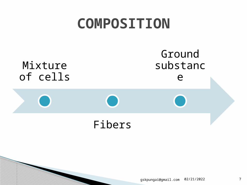
COMPOSITION OF CONNECTIVE TISSUE
TYPES OF CONNECTIVE TISSUE
FUNCTIONS OF CONNECTIVE TISSUE
HISTOPATHOLOGY
STAINING OF CONNECTIVE TISSUE
REFERENCES
CONTENTS

05/01/[email protected] 4

05/01/[email protected] 6
Tissues
Types of tissues Epithelial tissue: protection, secretion, and absorption Connective tissue: support, bind, and protect organs Muscle tissue: contraction Nervous tissue: information to other cells
CONNECTIVE TISSUE
Connects, supports, binds, or separates different types of tissues and organs in the body
Develops from the mesoderm
Found in b/w other tissues everywhere in the body, including nervous system
INTRODUCTION

05/01/[email protected] 8
FIBROBLASTS
MACROPHAGES
MAST CELLS
PLASMA CELLS
LEUKOCYTES
ADIPOCYTES (FAT CELLS)
MIXTURE OF CELLS
05/01/[email protected] 9
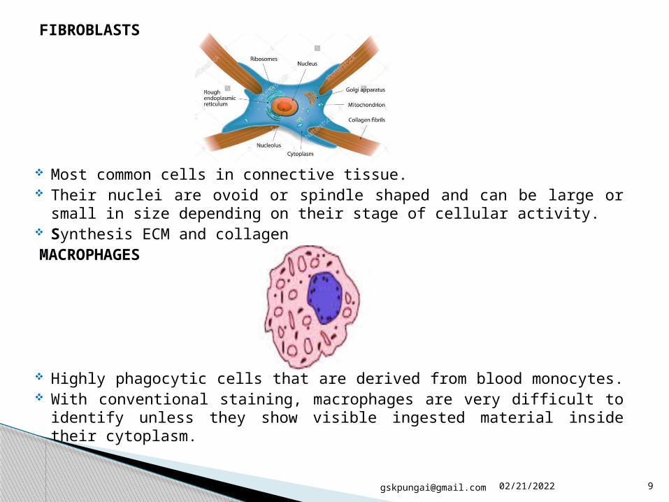
FIBROBLASTS
Most common cells in connective tissue. Their nuclei are ovoid or spindle shaped and can be large or small in
size depending on their stage of cellular activity. Synthesis ECM and collagenMACROPHAGES
Highly phagocytic cells that are derived from blood monocytes. With conventional staining, macrophages are very difficult to identify
unless they show visible ingested material inside their cytoplasm.
05/01/[email protected] 10
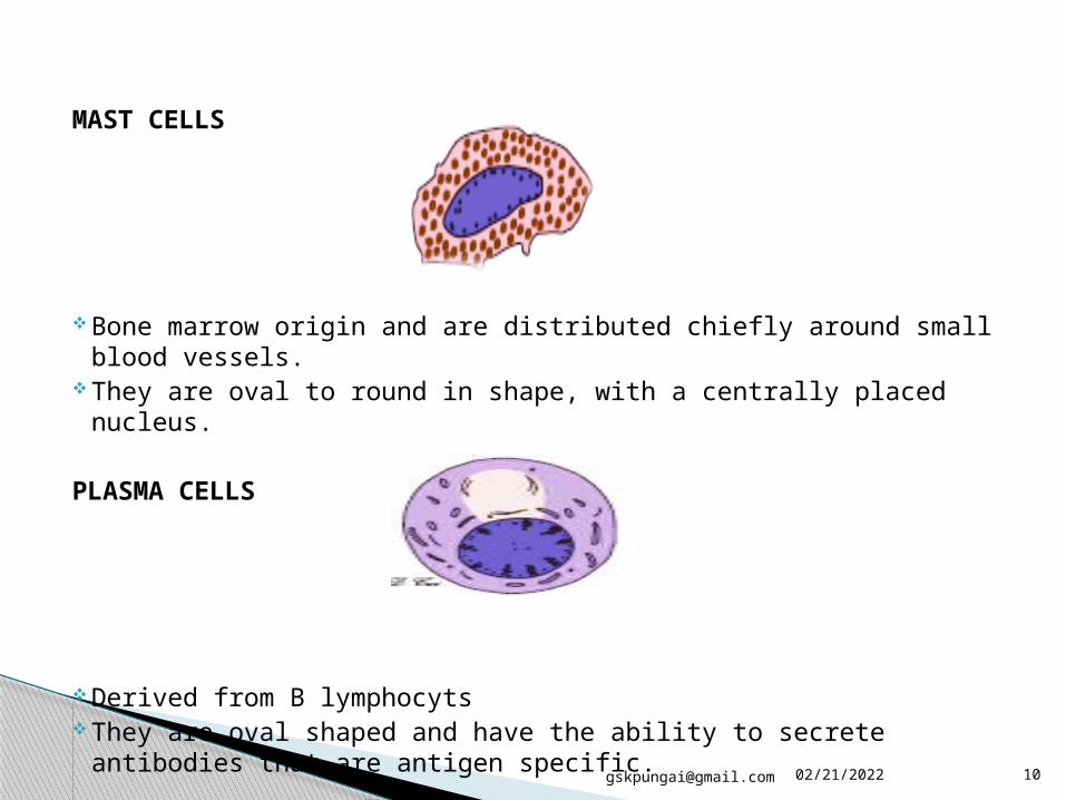
MAST CELLS
Bone marrow origin and are distributed chiefly around small blood vessels.
They are oval to round in shape, with a centrally placed nucleus.
PLASMA CELLS
Derived from B lymphocyts They are oval shaped and have the ability to secrete antibodies
that are antigen specific.

05/01/[email protected] 11
LEUKOCYTES
White blood cells, are considered the transient cells of connective tissue. They migrate from the blood vessels into connective tissue by the process of
diapedesis.
ADIPOCYTES
Arise from undifferentiated mesenchymal cells of connective tissue. They gradually accumulate
cytoplasmic fat, which results in a significant flattening of the nucleus in the periphery of the cell.

05/01/[email protected] 12
Ground substance is a clear, viscous substance with a high water content, but with very little morphologic structure.
When stained with basic dyes (periodic acid-Schiff [PAS]), it appears amorphous, it appears as a clear space.
Its major component is Glycosaminoglycans (GAGs), which are long, un-branched chains of polysaccharides with repeating disaccharide units.
Most GAGs are covalently bonded to a large central protein to form larger molecules called proteoglycans.
GROUND SUBSTANCE

05/01/[email protected] 14
Collagen COLLAGEN FIBERS are the most common and widespread
fibers in connective tissue. 25-35% of body The collagen molecule (tropo-collagen) is a product of the
fibroblast. Each collagen molecule is 300 nm in length and consists of
three polypeptide amino acid chains. Elastic Elastic fibers have a very resilient nature (stretch and recoil),
which is important in areas like the lungs, aorta, and skin. They are composed of two proteins, elastin and fibrillin, and
do not have a banding pattern.Reticular RETICULAR FIBERS are small-diameter fibers. Supporting mesh in soft tissues(liver,bone marrow and
lymphatic system)

05/01/[email protected] 15
Connective Tissue Proper
Specialized Connective
Tissues
Embryonic Connective
tissue
Supportive connective
Tissues
Fluid connective
tissue
TYPES OF CONNECTIVE TISSUE

05/01/[email protected] 19
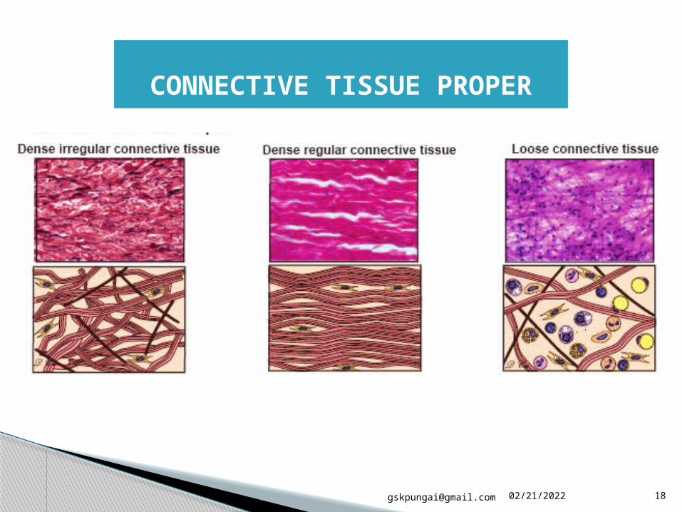
Dense irregular connective tissue is a cushion like tissue, which provides great strength against pressure-induced stresses on structures or organs.
“Dense” refers to the high abundance of collagen fibers (but fewer cells) compared to loose connective tissue.
“Irregular” indicates that the orientation of the fiber bundles is in many different directions (or randomly oriented bundles).
Contains mostly collagen fibers with a lesser number of other fibers such as elastic fibers.
The skin has a thick layer of dense irregular connective tissue, with fibers arranged in various directions to resist stretching forces in any direction.
It is prominent in the dermis of the skin, mammary glands, and capsules of many organs.
Function - provide strength.
DENSE IRREGULAR CT

05/01/[email protected] 20
This type of tissue is composed of coarse collagen bundles that is densely packed and oriented into parallel cylinders.
Long, thin fibroblasts are found among the fiber bundles and are oriented in
the same direction as the fibers.
The nuclei of the fibroblasts are visible, but the cytoplasm is not easily seen.
The thick bundles of collagen fibers fill the intercellular spaces.
Dense regular connective tissue provides resistance to traction forces in tendons and ligaments.
Function = provide strong attachment between various structures
DENSE REGULAR CT

05/01/[email protected] 21
Loose connective tissue is also called areolar connective tissue. This type of connective tissue has abundant ground substance,
with many connective tissue cells and relatively few fibers. It is richly vascularized, flexible, and not highly resistant to
stress. The lamina propria of the digestive tract is an extreme example
of loose connective tissue. This tissue lies immediately beneath the thin epithelium of the
gut, which is one place where the body’s defense mechanisms initially attack bacteria and pathogens.
Therefore, plasma cells, mast cells, leukocytes, and fibroblasts are common in this area.
Loose connective tissue is characterized by loosely arranged, woven connective fibers, abundant ground substance, and tissue fluid, which contains the rich array of connective tissue cells.
LOOSE CONNECTIVE TISSUE

05/01/[email protected] 22
Adipose connective tissue
Reticular connective tissue
Elastic connective tissue
SPECIALIZED CONNECTIVE TISSUES

05/01/[email protected] 24
Adipose tissue is a special form of connective tissue and has a rich neurovascular supply.
Adipocytes (fat cells) are scattered within a loose collagenous supporting tissue in this unilocular adipose tissue.
Each adipose cell contains a single large drop of lipid; it has a thin rim of cytoplasm around the lipid, and its flattened nucleus is located in the periphery of the cell.
Adipocytes are the primary site for storage of energy, and lipid deposition and mobilization are regulated by hormonal factors (steroids, insulin, thyroid hormone, etc.).
Adipocytes also play a role in the synthesis of some hormones such as leptin.
During childhood, the adipocyte numbers may increase depending on nutrition and other factors, but in adulthood, adipocyte numbers normally remain constant.
ADIPOSE CONNECTIVE TISSUE

05/01/[email protected] 25
Reticular tissue is a specialized loose connective tissue that provides a delicate supporting framework for many highly cellular organs, such as endocrine glands, lymphoid organs, the spleen, and the liver.
They are arranged in a net like framework to support parenchymal cells, in this example, pancreatic cells.
The inset drawing represents the organization of reticular fibers and pancreatic cells.
These fibers consist of collagen type III, which forms a mesh like network that supports the liver cells and holds these cells together.
There is a sinusoid running between the reticular fibers, which appears as empty space here.
RETICULAR CONNECTIVE TISSUE

05/01/[email protected] 26
Elastic connective tissue consists predominately of elastic material, and this allows distension and recoil of the structure.
This tissue can be found in some vertebral ligaments, arterial walls, and in the bronchial tree.
Thick bundles of elastic lamellae are arranged in parallel wavy sheets, with the smooth muscle cells and collagen fibers insinuated between alternating lamellae.
The elastic fibers are formed by elastin and fibrillin micro-fibrils. Elastic connective tissue is able to recoil after stretching. This property in large arteries helps to moderate the extremes of
pressure associated with the cardiac cycle. Abnormal expression of the fibrillin (FBN1) gene is associated
with abnormal elastic tissue disease.
ELASTIC CONNECTIVE TISSUE

05/01/[email protected] 27
Mesenchymal connective
tissue
Mucous connective
tissue
EMBRYONIC CONNECTIVE TISSUES

05/01/[email protected] 29
Mesenchyme (mesenchymal connective tissue) is found in the developing structures in the embryo.
It contains scattered reticular fibers and mesenchymal cells, which have irregular, star or spindle shapes and pale-stained cytoplasm.
These cells exhibit cytoplasmic processes, which often give the cells a stellate appearance.
Mesenchymal cells are relatively unspecialized and are capable of differentiating into different cell types in mature tissue cells, such as cartilages, bones, and muscles.
These blood cells contain a nucleus in each cell. This is the characteristic of their immature state (a nucleated red blood cells are characteristic of the mature state and are found in adult tissues).
Interestingly enough, some vertebrates, such as frogs and chickens, have nucleated red blood cells in the adult state.
MESENCHYMAL CONNECTIVE TISSUE

05/01/[email protected] 30
An example of mucous connective tissue that has an abundance of a jellylike matrix with some fine aggregates of collagen fibers and stellate-shaped fibroblasts is shown.
It is found in the umbilical cord and sub dermal connective tissue of the embryo.
Mucous tissue is a major constituent of the umbilical cord, where it is referred to as Wharton jelly.
In this example, the viscous ground substance has been stained with a special stain to reveal jelly like mucin, which contains hyaluronic acid and glycoproteins.
Collagen fibers and large stellate-shaped fibroblasts (not mesenchymal cells) predominate in the mucous tissue.
MUCOUS CONNECTIVE TISSUE

05/01/[email protected] 32
Bone cells (osteocytes) are surrounded by a matrix of collagen fibres strengthened by inorganic salts, especially calcium and phosphate.
This provides bones with their characteristic strength and rigidity.
BONE

05/01/[email protected] 33

05/01/[email protected] 34
BONE
Compact bone – solid or dense appearance
Spongy or cancellous bone –’spongy’ or fine
honeycomb appearance.
TYPES OF BONE

05/01/[email protected] 35
Found on ends of long bone
Contains bony bars and plates called trabeculae separated by irregular spaces
Blood cells are made within the red marrow found in the spongy bone
SPONGY BONE

05/01/[email protected] 36
COMPACT BONE
Found in the outer portion of long bones
Consists of many cylindrical-shaped units called osteon

05/01/[email protected] 37
Cartilage has a flexible rubbery matrix.
It is found in organs like,
External ear
Tip of the nose or
The “Adam’s apple” (thyroid cartilage of the larynx).
CARTILAGE
05/01/[email protected] 39
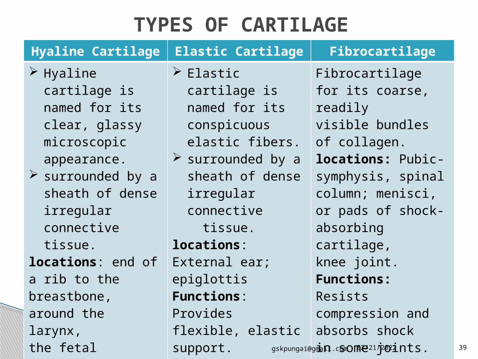
Hyaline Cartilage Elastic Cartilage Fibrocartilage Hyaline cartilage is
named for its clear, glassy microscopic appearance.
surrounded by a sheath of dense irregular connective tissue.
locations: end of a rib to the breastbone, around the larynx, the fetal skeleton.Functions: Eases joint movements; holds airway open during respiration; moves vocal cords during speech , growth zones of long bones of children.
Elastic cartilage is named for its conspicuous elastic fibers.
surrounded by a sheath of dense irregular connective
tissue.locations: External ear; epiglottisFunctions: Provides flexible, elastic support.
Fibrocartilage for its coarse, readilyvisible bundles of collagen.locations: Pubic-symphysis, spinal column; menisci, or pads of shock-absorbing cartilage,knee joint.Functions: Resists compression and absorbs shockin some joints.
TYPES OF CARTILAGE

05/01/[email protected] 41
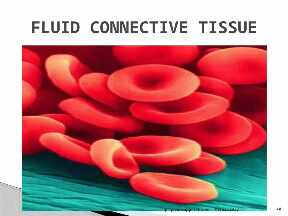
BLOOD Connective tissue composed
of cells suspended in liquid matrix called plasma
Red blood cells (erythrocytes) – carries oxygen
White blood cells (leukocytes) – fights infection
Platelets – fragment of giant cells found in bone marrow

05/01/[email protected] 42
Type Connective TissueCells
Main Locations Main Functions
Dense irregularconnective tissue
Predominantlyfibroblasts; otherconnective tissuecells occasionallypresent
Dermis of the skin,capsules of manyorgans
Resists stressfrom alldirections;protects organs
Dense regularconnective tissue
Predominantlyfibroblasts; otherconnective tissuecells occasionallypresent
Tendons, ligaments Providesresistance totraction forces
Looseconnective tissue
Fibroblasts,macrophages,adipocytes, mastcells, plasma cells,leukocytes
Lamina propriaof gastrointestinaltract; around thenerves and vessels(in adventitia layer)
Provides protection,suspension, andsupport; conduitfor vessels andnerves; environmentfor immunedefense function
Adiposeconnective tissue
Predominantlyadipocytes (fat cells);fibroblasts and otherconnective tissuecells occasionallypresent
Hypodermis of theskin, mammaryglands, and aroundmany organs
Providesenergy storage,insulation;cushioning oforgans; hormonesecretion
Types of connective tissue

05/01/[email protected] 43
Reticularconnective tissue
Fibroblasts,reticular cells,hepatocytes,smooth musclecells, Schwann cellsdepending on thelocation
Liver, pancreas,lymph nodes,spleen, and bonemarrow
Provides supportiveframeworkfor hematopoieticand parenchymalorgans
Elasticconnective tissue
Predominantlyfibroblasts orsmooth muscle cells;other connectivetissue cells occasionallypresent
Vertebral ligaments,walls of the largearteries
Provides flexiblesupport for thetissue; reducespressure on thewalls of theArteries
Mesenchymalconnective tissue
Mesenchymal cells Embryonicmesoderm
Gives rise to allconnective tissuetypes
Mucousconnective tissue
Spindle-shapedfibroblasts
Umbilical cord,subdermal layerof the fetus, dentalpulp of the developingteeth, nucleuspulposus of the disk
Provides cushionto protect theblood vessels inthe umbilical cord.

05/01/[email protected] 44
Providing a medium for oxygen and nutrients to diffuse from capillaries to cells.
Wraps around and cushions and protects organs
Stores nutrients
Internal support for organs
As tendon and ligaments, it protects joints and attaches muscles to bone and each other
Runs through organ capsules and in deep layers of skin giving strength
FUNCTIONS OF CONNECTIVE TISSUE

05/01/[email protected] 45
Fibroblasts are responsible for synthesis of various fibers and extracellular matrix components, such as collagen, elastic, and reticular fibers.
Macrophages contain many lysosomes and are involved in the removal of cell debris and the ingestion of foreign substances ; they also aid in antigen presentation to the immune system.
Adipocytes function to store neutral fats for energy or production of heat and are involved in hormone secretion.
Mast cells contain many granules, indirectly participate in allergic reactions, and act against microbial invasion.
Plasma cells are derived from B lymphocytes and are responsible for the production of antibodies in the immune response.
Lymphocytes participate in the immune response and protect against foreign invasion.
Neutrophils are the first line of defense against bacterial invasion. Eosinophils have anti-parasitic activity and moderate allergic reactions. Basophils have a (primary) function similar to mast cells; they mediate
hypersensitivity reactions.
FUNCTIONS OF THE CELLS IN CONNECTIVE TISSUE

05/01/[email protected] 47
1 in 10 people have a connective tissue disorder
Congenital disease – Marfan syndrome Ehlers-Danlos syndrome Myxomatous degeneration – Pathological weakness Mixed connective tissue disease – Autoimmune disease Systemic lupus erythematosus(SLE) Scurvy – Vit-C – collagen synthesis
PATHOLOGICAL TERMS Urticaria: An itchy skin eruption Pruritis: Itching of the skin Cirrhosis: An abnormal liver condition Jaundice: Yellow staining of the skin Coagulopathy: A disorder that prevents the normal clotting process Necrosis: Irreversible cell changes that occur as a result of cell death
CONNECTIVE TISSUE DISORDER
05/01/[email protected] 48
Van Gieson’s stain- staining of collagen and other connective tissue
Masson’s trichrome stain- distinguishing cells from surrounding connective tissue
Mallory’s trichrome stain- examining the collagen of connective tissue
Aniline blue stain
Eosin- stain of blood cells
Reticulin stain- visualize reticular fiber
STAINING OF CONNECTIVE TISSUE

05/01/[email protected] 49
Human Anatomy second edition by Kenneth S. Saladin, “Histology: the study of tissues”. Anatomy& Physiology in Health and Illness 12th Edition by Ross and Wilson, “The cells,
tissues and organization of the body”. Fundamentals of histology by GP Verma, “Connective tissue”. Anatomy& Physiology for nurses by Inderbir Singh, “General connective tissue” Human anatomy & Physiology by Biology-about.com, “Connective tissue Types and
examples”. Biology Encyclopedia by Biologyreference.com, “Connective tissue”.
REFERENCES