Connective Tissue Growth Factor Neutralization Aggravates ... · Background: Connective tissue...
7
CTGF Contributes to Inhibit Psoriasis Vol. 30, No. 1, 2018 47 Received December 27, 2016, Revised July 24, 2017, Accepted for publication July 31, 2017 Corresponding author: Keigo Ikeda, Department of Internal Medicine and Rheumatology, Juntendo University Urayasu Hospital, 2-1-1 Tomioka Urayasu-shi, Chiba 279-0021, Japan. Tel: 81-47-353-3111, Fax: 81-47- 381-5054, E-mail: [email protected] This is an Open Access article distributed under the terms of the Creative Commons Attribution Non-Commercial License (http://creativecommons. org/licenses/by-nc/4.0) which permits unrestricted non-commercial use, distribution, and reproduction in any medium, provided the original work is properly cited. Copyright © The Korean Dermatological Association and The Korean Society for Investigative Dermatology pISSN 1013-9087ㆍeISSN 2005-3894 Ann Dermatol Vol. 30, No. 1, 2018 https://doi.org/10.5021/ad.2018.30.1.47 ORIGINAL ARTICLE Connective Tissue Growth Factor Neutralization Aggravates the Psoriasis Skin Lesion: The Analysis of Psoriasis Model Mice and Patients Kunihiro Hayakawa 1 , Keigo Ikeda 1,2 , Maki Fujishiro 1 , Yuko Yoshida 1 , Takuya Hirai 1 , Hiroshi Tsushima 1 , Tomoko Miyashita 2 , Shinji Morimoto 1,2 , Yasushi Suga 3 , Kenji Takamori 1 , Hideoki Ogawa 1 , Iwao Sekigawa 1,2 1 Institutes for Environmental and Gender Specific Medicine, Juntendo University Graduate School of Medicine, Departments of 2 Internal Medicine and Rheumatology and 3 Dermatology, Juntendo University Urayasu Hospital, Chiba, Japan Background: Connective tissue growth factor (CTGF) is a multifunctional cellular protein and playing a role as a cen- tral mediator in tissue remodeling and fibrosis. The physio- logical function of CTGF in psoriasis is unknown. Objective: The purpose of this study was to investigate the function of CTGF in psoriasis using the established imiquimod (IMQ)- induced psoriasis murine model and psoriasis patients. Methods: Anti-CTGF monoclonal antibody was applied to IMQ induced psoriasis mice and those skin were clinically, pathologically and immunologically analyzed. Additionally, CTGF expression was analyzes using skin samples and plas- ma from psoriasis patients. Results: CTGF expression was ob- served in the dermis from both IMQ-induced psoriatic mice and psoriasis patients. CTGF inhibition using an anti-CTGF antibody slightly worsened IMQ-induced dermatitis. In addi- tion, the increase of CTGF showed tendency to suppress the psoriatic dermatitis through inhibition of suprabasal cells proliferation and macrophage infiltration in the skin. CTGF was also detected significantly higher in plasma from psor- iasis patients comparing with healthy control. Conclusion: Our findings suggest that CTGF could contribute to the heal- ing rather than the worsening of psoriasis skin lesions. (Ann Dermatol 30(1) 47∼53, 2018) -Keywords- Connective tissue growth factor, Dermatitis, Imiquimod, Monoclonal antibodies, Psoriasis INTRODUCTION Connective tissue growth factor (CTGF, also known as CCN2) is a member of the CCN family of proteins that contribute to a variety of cellular processes, including an- giogenesis, chondrogenesis, wound healing, and organ fibrosis 1 . CTGF is known to be a full-length molecule con- sisting of four conserved domains, i.e., insulin-like growth factor-binding protein domain (module 1: M1), von Wille- brand factor type C domain (module 2: M2), thrombo- spondin type I repeat domain (module 3: M3) and C-termi- nal cysteine-knot I repeat domain (module 4: M4). CTGF overexpression is induced by transforming growth factor-β in many pro-fibrotic conditions such as scleroderma 1,2 . In articular tissue, CTGF is produced by chondrocytes and maintains cartilage tissue homeostasis via an autocrine process 3 . We previously demonstrated that tumor necrosis factor (TNF)-α inhibited CTGF production in human chondrocytes, whereas TNF-α induced CTGF in human synovial cells 4 . Based on those results, we found that a neutralizing anti-CTGF monoclonal antibody significantly ameliorated arthritis in collagen-induced mice 5 . Psoriasis is a chronic inflammatory skin disease charac-
Transcript of Connective Tissue Growth Factor Neutralization Aggravates ... · Background: Connective tissue...

CTGF Contributes to Inhibit Psoriasis
Vol. 30, No. 1, 2018 47
Received December 27, 2016, Revised July 24, 2017, Accepted for publication July 31, 2017
Corresponding author: Keigo Ikeda, Department of Internal Medicine and Rheumatology, Juntendo University Urayasu Hospital, 2-1-1 Tomioka Urayasu-shi, Chiba 279-0021, Japan. Tel: 81-47-353-3111, Fax: 81-47- 381-5054, E-mail: [email protected]
This is an Open Access article distributed under the terms of the Creative Commons Attribution Non-Commercial License (http://creativecommons.org/licenses/by-nc/4.0) which permits unrestricted non-commercial use, distribution, and reproduction in any medium, provided the original work is properly cited.
Copyright © The Korean Dermatological Association and The Korean Society for Investigative Dermatology
pISSN 1013-9087ㆍeISSN 2005-3894Ann Dermatol Vol. 30, No. 1, 2018 https://doi.org/10.5021/ad.2018.30.1.47
ORIGINAL ARTICLE
Connective Tissue Growth Factor Neutralization Aggravates the Psoriasis Skin Lesion: The Analysis of Psoriasis Model Mice and Patients
Kunihiro Hayakawa1, Keigo Ikeda1,2, Maki Fujishiro1, Yuko Yoshida1, Takuya Hirai1, Hiroshi Tsushima1, Tomoko Miyashita2, Shinji Morimoto1,2, Yasushi Suga3, Kenji Takamori1, Hideoki Ogawa1, Iwao Sekigawa1,2
1Institutes for Environmental and Gender Specific Medicine, Juntendo University Graduate School of Medicine, Departments of 2Internal Medicine and Rheumatology and 3Dermatology, Juntendo University Urayasu Hospital, Chiba, Japan
Background: Connective tissue growth factor (CTGF) is a multifunctional cellular protein and playing a role as a cen-tral mediator in tissue remodeling and fibrosis. The physio-logical function of CTGF in psoriasis is unknown. Objective: The purpose of this study was to investigate the function of CTGF in psoriasis using the established imiquimod (IMQ)- induced psoriasis murine model and psoriasis patients. Methods: Anti-CTGF monoclonal antibody was applied to IMQ induced psoriasis mice and those skin were clinically, pathologically and immunologically analyzed. Additionally, CTGF expression was analyzes using skin samples and plas-ma from psoriasis patients. Results: CTGF expression was ob-served in the dermis from both IMQ-induced psoriatic mice and psoriasis patients. CTGF inhibition using an anti-CTGF antibody slightly worsened IMQ-induced dermatitis. In addi-tion, the increase of CTGF showed tendency to suppress the psoriatic dermatitis through inhibition of suprabasal cells proliferation and macrophage infiltration in the skin. CTGF was also detected significantly higher in plasma from psor-iasis patients comparing with healthy control. Conclusion:
Our findings suggest that CTGF could contribute to the heal-ing rather than the worsening of psoriasis skin lesions. (Ann Dermatol 30(1) 47∼53, 2018)
-Keywords-Connective tissue growth factor, Dermatitis, Imiquimod, Monoclonal antibodies, Psoriasis
INTRODUCTION
Connective tissue growth factor (CTGF, also known as CCN2) is a member of the CCN family of proteins that contribute to a variety of cellular processes, including an-giogenesis, chondrogenesis, wound healing, and organ fibrosis1. CTGF is known to be a full-length molecule con-sisting of four conserved domains, i.e., insulin-like growth factor-binding protein domain (module 1: M1), von Wille-brand factor type C domain (module 2: M2), thrombo-spondin type I repeat domain (module 3: M3) and C-termi-nal cysteine-knot I repeat domain (module 4: M4). CTGF overexpression is induced by transforming growth factor-β
in many pro-fibrotic conditions such as scleroderma1,2. In articular tissue, CTGF is produced by chondrocytes and maintains cartilage tissue homeostasis via an autocrine process3. We previously demonstrated that tumor necrosis factor (TNF)-α inhibited CTGF production in human chondrocytes, whereas TNF-α induced CTGF in human synovial cells4. Based on those results, we found that a neutralizing anti-CTGF monoclonal antibody significantly ameliorated arthritis in collagen-induced mice5.Psoriasis is a chronic inflammatory skin disease charac-
K Hayakawa, et al
48 Ann Dermatol
terized by epidermal hyperplasia and dermal infiltration of immune cells. Cytokines, such as interleukin-17A and TNF-α, produced by T helper cells are implicated in dis-ease pathogenesis6. Since the development of biological agents, anti-TNF-α antibodies have been used for the treatment of psoriasis7. Anti-TNF-α antibody therapy also improved the treatment of rheumatoid arthritis (RA). Psoriasis and RA have a commonality in terms of pathogenesis and treatment8. However, the function and role of CTGF are unknown in psoriasis.In this study, we investigated the function of CTGF in psoriasis using the established imiquimod (IMQ)-induced psoriasis murine model9 and samples from psoriasis patients.
MATERIALS AND METHODSExperimental conditions
BALB/c mice were obtained from Charles River Japan (Yokohama, Japan). Experiments were performed in com-pliance with institutional guidelines and were approved by the Institutional Animal Care and Use Committee (IACUC) of Juntendo University (Approval no. 2900053). At eight weeks of age, mice were shaved on the back skin and left ear, and 62.5 μl and 3.125 μl of 5% IMQ cream (Beselna Cream; Mochida Pharmaceutical, Tokyo, Japan) were respectively applied to these areas for six days. One day before IMQ application, 200 μg of anti-CTGF mono-clonal antibody (Nosan Corporation, Yokohama, Japan) was intraperitoneally injected. An objective scoring system called the psoriasis severity index, which considers eryth-ema, scaling, and thickness, was used, and skin lesions were scored from 0 to 4. The cumulative score (erythema plus scaling plus thickening) was also employed for meas-uring severity (scored from 0 to 12)9.
Patients and samples
The human subject research component was approved by the Juntendo University Urayasu Hospital Institutional Review Board (IRB no. 2009-009). After informed consent, 4-mm punch biopsies were obtained from both psoriasis patients (n=6) and other disease patients with normal con-dition of skin (n=4). The diagnosis was confirmed by both pathologists and dermatologists. Plasma was also col-lected from patients with psoriasis (n=28) and healthy control (n=8).
Pathological analysis
Mouse and human skin samples were fixed in a 20% for-malin solution and embedded in paraffin, sectioned, and subjected to hematoxylin-eosin (H&E) staining, immuno-fluorescence, and immunohistochemistry. Epidermal and
dermal thickness were determined at five positions per section by measuring the average distance from the basal lamina to the bottom of the stratum corneum using a BZ-II Analyzer ver. 1.1 (Keyence, Osaka, Japan). For immuno-fluorescence staining, back skin samples were blocked and stained using goat anti-CTGF antibody (Santa Cruz Biotechnology, Santa Cruz, CA, USA), rat anti-Ki-67 anti-body (eBioscience, Sun Diego, CA, USA), rat anti-CD3 an-tibody (Abcam plc, Cambridge, UK), and biotin conjugated Armenian hamster anti-Vγ3 antibody (Leinco Technologies Inc., St. Louis, MO, USA). Sections were further incubated with Alexa Fluor 488 conjugated anti-goat IgG (Invitrogen, Carlsbad, CA, USA) or anti-rat IgG (Invitrogen), and Alexa Fluor 594 conjugated streptavidin (BioLegend, Sun Diego, CA, USA). Nuclei were stained with 4',6-diamidino-2-phe-nylindole. For immunohistochemical staining, sections were incubated with primary rat anti-F4/80 antibody (Abcam plc, Cambridge, UK), rat anti-Ly-6G antibody (BD Biosciences, San Diego, CA, USA), Armenian hamster an-ti-CD11c antibody (BioLegend), and rat anti-PDCA1 anti-body (DENDRITICS, Lyon, France). Sections were further incubated with horseradish peroxidase-conjugated anti-rat IgG (Dako, Glostrup, Denmark) and anti-Armenian hamster IgG secondary antibody (Jackson Immunoresearch Laboratories, West Grove, PA, USA). Immunoreactivity was detected by incubating with a DAB+ Liquid kit (Dako). Sections were then counterstained with hematoxylin. For evaluation of leukocyte infiltration in the skin, three re-searchers quantified the staining of three sections from all mice. The numbers of cells positive for F4/80, Ly-6G, PDCA-1, CD3, and Vγ3 were quantified in each section. Photomicrographs were taken using a BZ-9000 microscope (Keyence).
Immunoblotting analysis
Skin tissues were collected and homogenized. Ten micro-grams of total protein was loaded in each well. The pri-mary antibodies used were anti-CTGF antibody (Santa Cruz) and anti-GAPDH antibody (Millipore, Billerica, MA, USA). Densitometric analysis was performed using ImageJ soft-ware (National Institute of Health, Bethesda, MD, USA).
Sandwich enzyme-linked immunosorbent assay analysis
Human CTGF enzyme-linked immunosorbent assay (ELISA) analysis was performed as described before10,11. Briefly, plasma samples were added to 96 well-plates (ThemoFisher Scientific, Waltham, MA, USA) that were coated with a monoclonal antibody against human CTGF modules. A bi-otin–labeled anti-human CTGF monoclonal antibody and a peroxidase-conjugated streptavidin (Jackson Immuno Research, West Grove, PA, USA) were added, Substrate

CTGF Contributes to Inhibit Psoriasis
Vol. 30, No. 1, 2018 49
Fig. 1. Connective tissue growth factor (CTGF) expression was increased in back skin lesions from imiquimod (IMQ)-induced psoriatic-like mice compared with control IMQ-untreated mice. BALB/c mice (n=4∼6 per groups) were treated with IMQ cream on shaved back skin. On day 6 after IMQ treatment, samples were collected from back skin. (A) Representative immunofluorescent staining of CTGF (green) in back skins of control and IMQ-treated mice. Nuclei were stained with 4',6-diamidino-2-phenylindole (blue). Arrowheads showed CTGF-expressing cells; bar: 50 μm. White line represents the border between the epidermis and dermis. (B) CTGF expression in back skins of control and IMQ-treated mice was evaluated by western blot analysis; scatterplot showing the results of densitometric analysis. IMQ(−): IMQ-untreated control mice, IMQ(+): IMQ-treated mice, GAPDH: glyceraldehyde-3-phosphate dehydrogenase.
and stop solution were sequentially added. The optical density at 450 nm was read to microplate reader (ARVO SX; Perkin Elmer, MA, USA), and measure the CTGF concentration.
Statistical analysis
Data were expressed as the mean±standard deviation and analyzed using GraphPad Prism 6 software (La Jolla, CA, USA). Statistical significance was determined by the Mann-Whitney U test, 2-way ANOVA, and unpaired t-test with Welch’s correction. A p-value <0.05 was considered statistically significant.
RESULTS
To identify CTGF expression in IMQ-induced psoriatic mice, we used immunohistological analyses and Western blotting (Fig. 1). Immunofluorescence tissue staining re-vealed that CTGF expression was increased in IMQ-in-duced psoriatic back dermal lesions compared with con-trol IMQ untreated mice (Fig. 1A). To confirm CTGF pro-tein expression levels, we performed a Western blot anal-ysis using back skin samples. CTGF protein expression showed a tendency to increase in IMQ-induced psoriatic mice relative to untreated mice (Fig. 1B, left). However, the increased expression of CTGF was not statistically sig-nificant using this method (Fig. 1B, right).

K Hayakawa, et al
50 Ann Dermatol
Fig. 2. Psoriatic scores were worsened because of blocking connective tissue growth factor (CTGF) in imiquimod (IMQ)-treated mice. BALB/c mice were treated daily with IMQ cream on shaved back skin and the left ear. One day before IMQ application, 200 μg of anti-CTGF monoclonal antibody was intraperitoneally injected (n=4∼6 per groups). (A) Representative skin condition of anti-CTGF treated and untreated mice. These photos were taken on day 5 after IMQ application. (B) Erythema, scaling, and thickness of back skin was scored daily using a scale from 0 to 4; the cumulative score (0∼12) is depicted. Symbols indicate mean score+standard deviation (n=4∼6). **p<0.01, ***p<0.001, ****p<0.0001, by 2-way ANOVA (IMQ[+] vs. IMQ[+], anti-CTGF). (C) Epidermal and dermal thicknesses were measured in back skin samples. (D) Epidermal thickness in ear samples. (E) Immunohistochemically staining of Ki-67 (green) in back skin. Nuclei were stained with 4',6-diamidino-2-phenylindole (blue); bar: 50 μm. Yellow line represents the border between the epidermis and dermis. (F) Scatterplot of epidermal Ki-67+ cell number. *p<0.05 by Mann-Whitney U test (IMQ[+] vs. IMQ[+], anti-CTGF).
CTGF Contributes to Inhibit Psoriasis
Vol. 30, No. 1, 2018 51
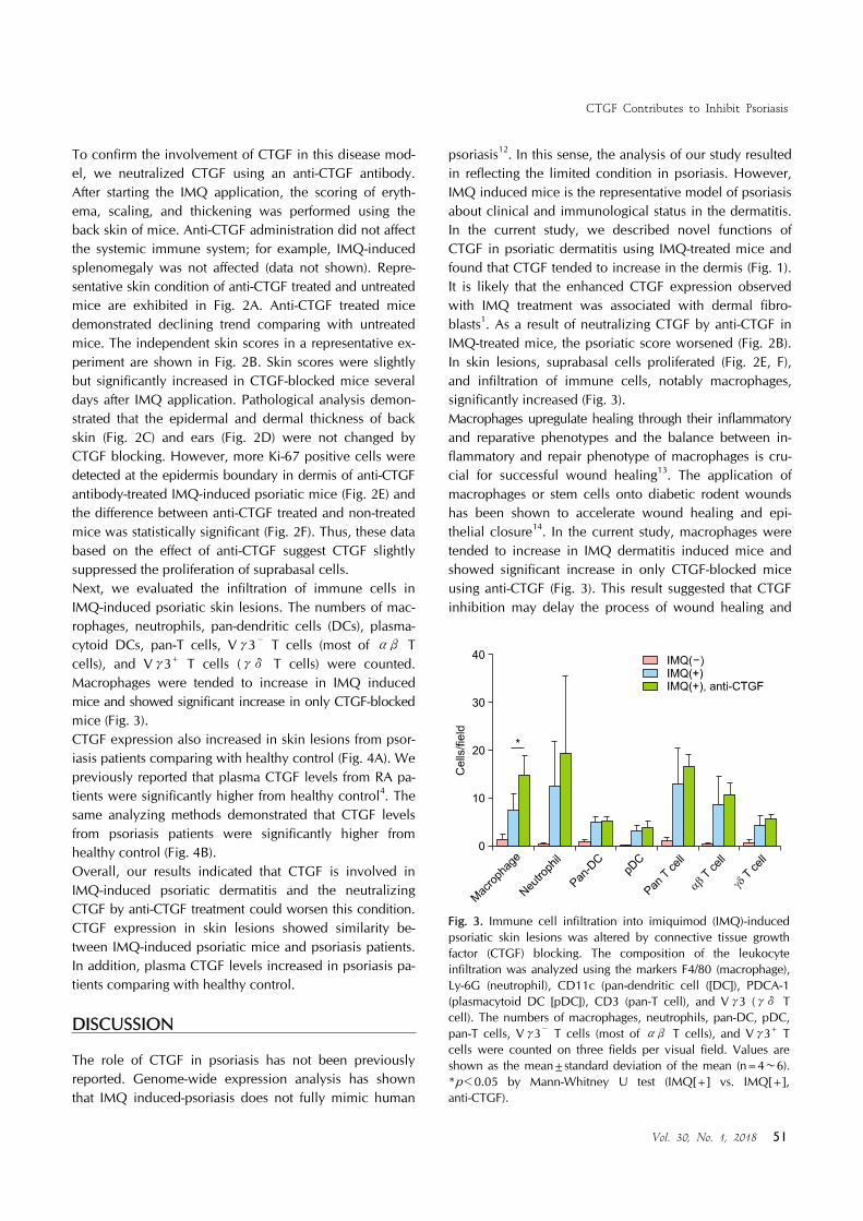
Fig. 3. Immune cell infiltration into imiquimod (IMQ)-induced psoriatic skin lesions was altered by connective tissue growth factor (CTGF) blocking. The composition of the leukocyte infiltration was analyzed using the markers F4/80 (macrophage), Ly-6G (neutrophil), CD11c (pan-dendritic cell ([DC]), PDCA-1 (plasmacytoid DC [pDC]), CD3 (pan-T cell), and Vγ3 (γδ T cell). The numbers of macrophages, neutrophils, pan-DC, pDC, pan-T cells, Vγ3− T cells (most of αβ T cells), and Vγ3+ T cells were counted on three fields per visual field. Values are shown as the mean±standard deviation of the mean (n=4∼6). *p<0.05 by Mann-Whitney U test (IMQ[+] vs. IMQ[+], anti-CTGF).
To confirm the involvement of CTGF in this disease mod-el, we neutralized CTGF using an anti-CTGF antibody. After starting the IMQ application, the scoring of eryth-ema, scaling, and thickening was performed using the back skin of mice. Anti-CTGF administration did not affect the systemic immune system; for example, IMQ-induced splenomegaly was not affected (data not shown). Repre-sentative skin condition of anti-CTGF treated and untreated mice are exhibited in Fig. 2A. Anti-CTGF treated mice demonstrated declining trend comparing with untreated mice. The independent skin scores in a representative ex-periment are shown in Fig. 2B. Skin scores were slightly but significantly increased in CTGF-blocked mice several days after IMQ application. Pathological analysis demon-strated that the epidermal and dermal thickness of back skin (Fig. 2C) and ears (Fig. 2D) were not changed by CTGF blocking. However, more Ki-67 positive cells were detected at the epidermis boundary in dermis of anti-CTGF antibody-treated IMQ-induced psoriatic mice (Fig. 2E) and the difference between anti-CTGF treated and non-treated mice was statistically significant (Fig. 2F). Thus, these data based on the effect of anti-CTGF suggest CTGF slightly suppressed the proliferation of suprabasal cells.Next, we evaluated the infiltration of immune cells in IMQ-induced psoriatic skin lesions. The numbers of mac-rophages, neutrophils, pan-dendritic cells (DCs), plasma-cytoid DCs, pan-T cells, Vγ3− T cells (most of αβ T cells), and Vγ3+ T cells (γδ T cells) were counted. Macrophages were tended to increase in IMQ induced mice and showed significant increase in only CTGF-blocked mice (Fig. 3). CTGF expression also increased in skin lesions from psor-iasis patients comparing with healthy control (Fig. 4A). We previously reported that plasma CTGF levels from RA pa-tients were significantly higher from healthy control4. The same analyzing methods demonstrated that CTGF levels from psoriasis patients were significantly higher from healthy control (Fig. 4B). Overall, our results indicated that CTGF is involved in IMQ-induced psoriatic dermatitis and the neutralizing CTGF by anti-CTGF treatment could worsen this condition. CTGF expression in skin lesions showed similarity be-tween IMQ-induced psoriatic mice and psoriasis patients. In addition, plasma CTGF levels increased in psoriasis pa-tients comparing with healthy control.
DISCUSSION
The role of CTGF in psoriasis has not been previously reported. Genome-wide expression analysis has shown that IMQ induced-psoriasis does not fully mimic human
psoriasis12. In this sense, the analysis of our study resulted in reflecting the limited condition in psoriasis. However, IMQ induced mice is the representative model of psoriasis about clinical and immunological status in the dermatitis. In the current study, we described novel functions of CTGF in psoriatic dermatitis using IMQ-treated mice and found that CTGF tended to increase in the dermis (Fig. 1). It is likely that the enhanced CTGF expression observed with IMQ treatment was associated with dermal fibro-blasts1. As a result of neutralizing CTGF by anti-CTGF in IMQ-treated mice, the psoriatic score worsened (Fig. 2B). In skin lesions, suprabasal cells proliferated (Fig. 2E, F), and infiltration of immune cells, notably macrophages, significantly increased (Fig. 3).Macrophages upregulate healing through their inflammatory and reparative phenotypes and the balance between in-flammatory and repair phenotype of macrophages is cru-cial for successful wound healing13. The application of macrophages or stem cells onto diabetic rodent wounds has been shown to accelerate wound healing and epi-thelial closure14. In the current study, macrophages were tended to increase in IMQ dermatitis induced mice and showed significant increase in only CTGF-blocked mice using anti-CTGF (Fig. 3). This result suggested that CTGF inhibition may delay the process of wound healing and

K Hayakawa, et al
52 Ann Dermatol
Fig. 4. Connective tissue growth factor (CTGF) expression was increased in skin lesions and plasma from psoriasis patients. (A) Representative immunofluorescent staining of CTGF (green) in skins of healthy control (n=4) and psoriasis patients (n=6). Nuclei were stained with 4',6-diamidino-2-phenylindole (blue). Arrowheads showed CTGF-expressing cells; bar: 50 μm. White line represents the border between the epidermis and dermis. (B) CTGF in plasma from healthy control (HC) and psoriasis patients was detected by a sandwich enzyme-linked immunosorbent assay (ELISA) analysis. Blue circle: HC (n=10), red rectangle: psoriasis patients (psoriasis, n=28). Values are shown as the mean±standard deviation of the mean. ****p<0.0001 by unpaired t-test with Welch’s correction.
CTGF could induce repair phenotype of macrophages to improve skin lesions in psoriasis. Recent reports showed CTGF contribute wound repair,15,16 and both CTGF and macrophages are involved in tissue remodeling17. However, another reports showed CTGF function as chemotactic factor for some lymphocytes18. In vivo CTGF function is still unclear. At least, our results suggested that reduction of CTGF by anti-CTGF could have an effect on worsening skin condition in psoriasis.We demonstrated CTGF expression increased in skin le-sions and plasma from psoriasis patients comparing with healthy control (Fig. 4). Taken together, CTGF could con-tribute to improve psoriatic skin lesions through inducing repair phenotype of macrophage in both IMQ-induced psoriatic mice and psoriasis patient. In summary, the present data based on the effect of an-ti-CTGF treatment demonstrated that CTGF suppressed the progression of IMQ-induced psoriatic dermatitis. Additio-nally, CTGF expression was increased in skin lesions and plasma from psoriasis patients. Our findings suggest that CTGF could contribute to the healing rather than the wor-
sening of psoriasis skin lesions including the induction of reparative macrophage.
ACKNOWLEDGMENT
This study was supported in part by a Grant-in-Aid (S1311011) from MEXT (Ministry of Education, Culture, Sports, Science and Technology) Supported Program for the Strategic Research Foundation at Private University and by a JSPS (Japan Society for the Promotion of Science) KAKENHI grant numbers JP26860265 (to KH) and JP15K08107 (to KI). The authors would like to thank Ms. Megumi Morioka (Nosan Corporation) for the provision of anti-CTGF monoclonal antibody, Dr. Mitsutoshi Tominaga, Dr. Utako Kimura and the members of our laboratory for technical support and helpful discussion.
CONFLICTS OF INTEREST
The authors have nothing to disclose.

CTGF Contributes to Inhibit Psoriasis
Vol. 30, No. 1, 2018 53
REFERENCES
1. Leask A, Denton CP, Abraham DJ. Insights into the molecular mechanism of chronic fibrosis: the role of connective tissue growth factor in scleroderma. J Invest Dermatol 2004;122:1-6.
2. Igarashi A, Nashiro K, Kikuchi K, Sato S, Ihn H, Grotendorst GR, et al. Significant correlation between connective tissue growth factor gene expression and skin sclerosis in tissue sections from patients with systemic sclerosis. J Invest Dermatol 1995;105:280-284.
3. Takigawa M, Nakanishi T, Kubota S, Nishida T. Role of CTGF/HCS24/ecogenin in skeletal growth control. J Cell Physiol 2003;194:256-266.
4. Nozawa K, Fujishiro M, Kawasaki M, Kaneko H, Iwabuchi K, Yanagida M, et al. Connective tissue growth factor promotes articular damage by increased osteoclastogenesis in patients with rheumatoid arthritis. Arthritis Res Ther 2009;11:R174.
5. Nozawa K, Fujishiro M, Kawasaki M, Yamaguchi A, Ikeda K, Morimoto S, et al. Inhibition of connective tissue growth factor ameliorates disease in a murine model of rheumatoid arthritis. Arthritis Rheum 2013;65:1477-1486.
6. Lowes MA, Suárez-Fariñas M, Krueger JG. Immunology of psoriasis. Annu Rev Immunol 2014;32:227-255.
7. Reich K, Nestle FO, Papp K, Ortonne JP, Evans R, Guzzo C, et al. Infliximab induction and maintenance therapy for moderate-to-severe psoriasis: a phase III, multicentre, double-blind trial. Lancet 2005;366:1367-1374.
8. Coates LC, FitzGerald O, Helliwell PS, Paul C. Psoriasis, psoriatic arthritis, and rheumatoid arthritis: is all inflammation the same? Semin Arthritis Rheum 2016;46:291-304.
9. van der Fits L, Mourits S, Voerman JS, Kant M, Boon L, Laman JD, et al. Imiquimod-induced psoriasis-like skin inflammation in mice is mediated via the IL-23/IL-17 axis. J
Immunol 2009;182:5836-5845. 10. Miyazaki O, Kurashita S, Fukamachi I, Endo K, Ng PS,
Takehara K. Subtraction method for determination of N-terminal connective tissue growth factor. Ann Clin Biochem 2010;47:205-211.
11. Fujishiro M, Yamaguchi A, Kawasaki M, Nozawa K, Takasaki Y, Takamori K, et al. The detection of plasma levels of connective tissue growth factor in rheumatoid arthritis patients. Clin Exp Rheumatol 2012;30:145-146.
12. Swindell WR, Johnston A, Carbajal S, Han G, Wohn C, Lu J, et al. Genome-wide expression profiling of five mouse models identifies similarities and differences with human psoriasis. PLoS One 2011;6:e18266.
13. DiPietro LA. Wound healing: the role of the macrophage and other immune cells. Shock 1995;4:233-240.
14. Waugh HV, Sherratt JA. Macrophage dynamics in diabetic wound dealing. Bull Math Biol 2006;68:197-207.
15. Alfaro MP, Deskins DL, Wallus M, DasGupta J, Davidson JM, Nanney LB, et al. A physiological role for connective tissue growth factor in early wound healing. Lab Invest 2013;93:81-95.
16. Henshaw FR, Boughton P, Lo L, McLennan SV, Twigg SM. Topically applied connective tissue growth factor/CCN2 improves diabetic preclinical cutaneous wound healing: potential role for CTGF in human diabetic foot ulcer healing. J Diabetes Res 2015;2015:236238.
17. Riley KG, Pasek RC, Maulis MF, Dunn JC, Bolus WR, Kendall PL, et al. Macrophages are essential for CTGF- mediated adult β-cell proliferation after injury. Mol Metab 2015;4:584-591.
18. Cicha I, Yilmaz A, Klein M, Raithel D, Brigstock DR, Daniel WG, et al. Connective tissue growth factor is overexpressed in complicated atherosclerotic plaques and induces mo-nonuclear cell chemotaxis in vitro. Arterioscler Thromb Vasc Biol 2005;25:1008-1013.