Conj Pneu 6A With PspA
9
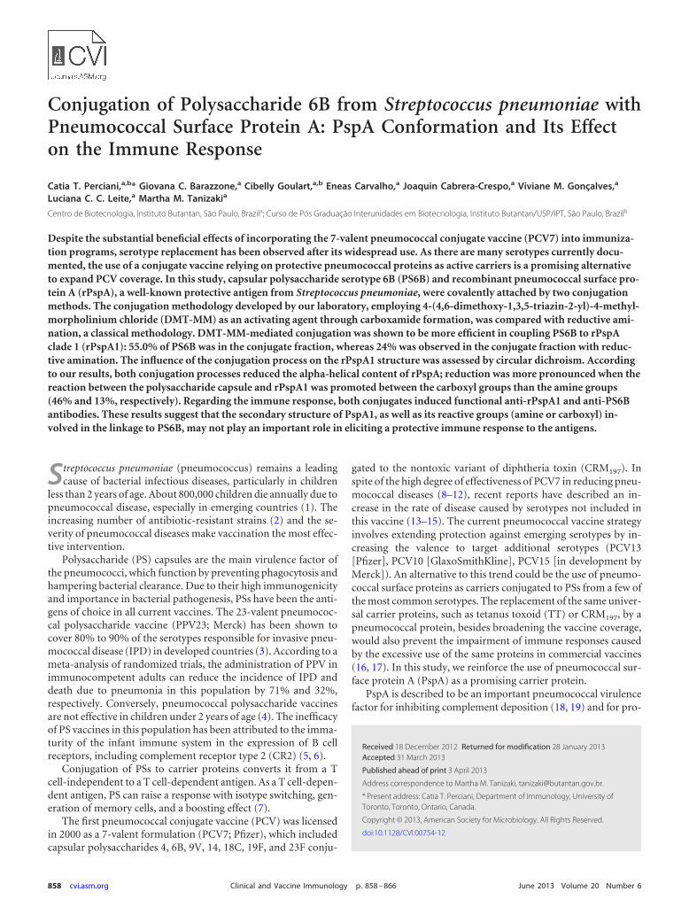
Conjugation of Polysaccharide 6B from Streptococcus pneumoniae with Pneumococcal Surface Protein A: PspA Conformation and Its Effect on the Immune Response Catia T. Perciani, a,b * Giovana C. Barazzone, a Cibelly Goulart, a,b Eneas Carvalho, a Joaquin Cabrera-Crespo, a Viviane M. Gonçalves, a Luciana C. C. Leite, a Martha M. Tanizaki a Centro de Biotecnologia, Instituto Butantan, São Paulo, Brazil a ; Curso de Pós Graduação Interunidades em Biotecnologia, Instituto Butantan/USP/IPT, São Paulo, Brazil b Despite the substantial beneficial effects of incorporating the 7-valent pneumococcal conjugate vaccine (PCV7) into immuniza- tion programs, serotype replacement has been observed after its widespread use. As there are many serotypes currently docu- mented, the use of a conjugate vaccine relying on protective pneumococcal proteins as active carriers is a promising alternative to expand PCV coverage. In this study, capsular polysaccharide serotype 6B (PS6B) and recombinant pneumococcal surface pro- tein A (rPspA), a well-known protective antigen from Streptococcus pneumoniae, were covalently attached by two conjugation methods. The conjugation methodology developed by our laboratory, employing 4-(4,6-dimethoxy-1,3,5-triazin-2-yl)-4-methyl- morpholinium chloride (DMT-MM) as an activating agent through carboxamide formation, was compared with reductive ami- nation, a classical methodology. DMT-MM-mediated conjugation was shown to be more efficient in coupling PS6B to rPspA clade 1 (rPspA1): 55.0% of PS6B was in the conjugate fraction, whereas 24% was observed in the conjugate fraction with reduc- tive amination. The influence of the conjugation process on the rPspA1 structure was assessed by circular dichroism. According to our results, both conjugation processes reduced the alpha-helical content of rPspA; reduction was more pronounced when the reaction between the polysaccharide capsule and rPspA1 was promoted between the carboxyl groups than the amine groups (46% and 13%, respectively). Regarding the immune response, both conjugates induced functional anti-rPspA1 and anti-PS6B antibodies. These results suggest that the secondary structure of PspA1, as well as its reactive groups (amine or carboxyl) in- volved in the linkage to PS6B, may not play an important role in eliciting a protective immune response to the antigens. S treptococcus pneumoniae (pneumococcus) remains a leading cause of bacterial infectious diseases, particularly in children less than 2 years of age. About 800,000 children die annually due to pneumococcal disease, especially in emerging countries (1). The increasing number of antibiotic-resistant strains (2) and the se- verity of pneumococcal diseases make vaccination the most effec- tive intervention. Polysaccharide (PS) capsules are the main virulence factor of the pneumococci, which function by preventing phagocytosis and hampering bacterial clearance. Due to their high immunogenicity and importance in bacterial pathogenesis, PSs have been the anti- gens of choice in all current vaccines. The 23-valent pneumococ- cal polysaccharide vaccine (PPV23; Merck) has been shown to cover 80% to 90% of the serotypes responsible for invasive pneu- mococcal disease (IPD) in developed countries (3). According to a meta-analysis of randomized trials, the administration of PPV in immunocompetent adults can reduce the incidence of IPD and death due to pneumonia in this population by 71% and 32%, respectively. Conversely, pneumococcal polysaccharide vaccines are not effective in children under 2 years of age (4). The inefficacy of PS vaccines in this population has been attributed to the imma- turity of the infant immune system in the expression of B cell receptors, including complement receptor type 2 (CR2) (5, 6). Conjugation of PSs to carrier proteins converts it from a T cell-independent to a T cell-dependent antigen. As a T cell-depen- dent antigen, PS can raise a response with isotype switching, gen- eration of memory cells, and a boosting effect (7). The first pneumococcal conjugate vaccine (PCV) was licensed in 2000 as a 7-valent formulation (PCV7; Pfizer), which included capsular polysaccharides 4, 6B, 9V, 14, 18C, 19F, and 23F conju- gated to the nontoxic variant of diphtheria toxin (CRM 197 ). In spite of the high degree of effectiveness of PCV7 in reducing pneu- mococcal diseases (8–12), recent reports have described an in- crease in the rate of disease caused by serotypes not included in this vaccine (13–15). The current pneumococcal vaccine strategy involves extending protection against emerging serotypes by in- creasing the valence to target additional serotypes (PCV13 [Pfizer], PCV10 [GlaxoSmithKline], PCV15 [in development by Merck]). An alternative to this trend could be the use of pneumo- coccal surface proteins as carriers conjugated to PSs from a few of the most common serotypes. The replacement of the same univer- sal carrier proteins, such as tetanus toxoid (TT) or CRM 197 , by a pneumococcal protein, besides broadening the vaccine coverage, would also prevent the impairment of immune responses caused by the excessive use of the same proteins in commercial vaccines (16, 17). In this study, we reinforce the use of pneumococcal sur- face protein A (PspA) as a promising carrier protein. PspA is described to be an important pneumococcal virulence factor for inhibiting complement deposition (18, 19) and for pro- Received 18 December 2012 Returned for modification 28 January 2013 Accepted 31 March 2013 Published ahead of print 3 April 2013 Address correspondence to Martha M. Tanizaki, [email protected]. * Present address: Catia T. Perciani, Department of Immunology, University of Toronto, Toronto, Ontario, Canada. Copyright © 2013, American Society for Microbiology. All Rights Reserved. doi:10.1128/CVI.00754-12 858 cvi.asm.org Clinical and Vaccine Immunology p. 858 – 866 June 2013 Volume 20 Number 6
-
Upload
vikramreddy041302 -
Category
Documents
-
view
241 -
download
0
Transcript of Conj Pneu 6A With PspA
Conjugation of Polysaccharide 6B from Streptococcus pneumoniae withPneumococcal Surface Protein A: PspA Conformation and Its Effecton the Immune Response
Catia T. Perciani,a,b* Giovana C. Barazzone,a Cibelly Goulart,a,b Eneas Carvalho,a Joaquin Cabrera-Crespo,a Viviane M. Gonçalves,a
Luciana C. C. Leite,a Martha M. Tanizakia
Centro de Biotecnologia, Instituto Butantan, São Paulo, Brazila; Curso de Pós Graduação Interunidades em Biotecnologia, Instituto Butantan/USP/IPT, São Paulo, Brazilb
Despite the substantial beneficial effects of incorporating the 7-valent pneumococcal conjugate vaccine (PCV7) into immuniza-tion programs, serotype replacement has been observed after its widespread use. As there are many serotypes currently docu-mented, the use of a conjugate vaccine relying on protective pneumococcal proteins as active carriers is a promising alternativeto expand PCV coverage. In this study, capsular polysaccharide serotype 6B (PS6B) and recombinant pneumococcal surface pro-tein A (rPspA), a well-known protective antigen from Streptococcus pneumoniae, were covalently attached by two conjugationmethods. The conjugation methodology developed by our laboratory, employing 4-(4,6-dimethoxy-1,3,5-triazin-2-yl)-4-methyl-morpholinium chloride (DMT-MM) as an activating agent through carboxamide formation, was compared with reductive ami-nation, a classical methodology. DMT-MM-mediated conjugation was shown to be more efficient in coupling PS6B to rPspAclade 1 (rPspA1): 55.0% of PS6B was in the conjugate fraction, whereas 24% was observed in the conjugate fraction with reduc-tive amination. The influence of the conjugation process on the rPspA1 structure was assessed by circular dichroism. Accordingto our results, both conjugation processes reduced the alpha-helical content of rPspA; reduction was more pronounced when thereaction between the polysaccharide capsule and rPspA1 was promoted between the carboxyl groups than the amine groups(46% and 13%, respectively). Regarding the immune response, both conjugates induced functional anti-rPspA1 and anti-PS6Bantibodies. These results suggest that the secondary structure of PspA1, as well as its reactive groups (amine or carboxyl) in-volved in the linkage to PS6B, may not play an important role in eliciting a protective immune response to the antigens.
Streptococcus pneumoniae (pneumococcus) remains a leadingcause of bacterial infectious diseases, particularly in children
less than 2 years of age. About 800,000 children die annually due topneumococcal disease, especially in emerging countries (1). Theincreasing number of antibiotic-resistant strains (2) and the se-verity of pneumococcal diseases make vaccination the most effec-tive intervention.
Polysaccharide (PS) capsules are the main virulence factor ofthe pneumococci, which function by preventing phagocytosis andhampering bacterial clearance. Due to their high immunogenicityand importance in bacterial pathogenesis, PSs have been the anti-gens of choice in all current vaccines. The 23-valent pneumococ-cal polysaccharide vaccine (PPV23; Merck) has been shown tocover 80% to 90% of the serotypes responsible for invasive pneu-mococcal disease (IPD) in developed countries (3). According to ameta-analysis of randomized trials, the administration of PPV inimmunocompetent adults can reduce the incidence of IPD anddeath due to pneumonia in this population by 71% and 32%,respectively. Conversely, pneumococcal polysaccharide vaccinesare not effective in children under 2 years of age (4). The inefficacyof PS vaccines in this population has been attributed to the imma-turity of the infant immune system in the expression of B cellreceptors, including complement receptor type 2 (CR2) (5, 6).
Conjugation of PSs to carrier proteins converts it from a Tcell-independent to a T cell-dependent antigen. As a T cell-depen-dent antigen, PS can raise a response with isotype switching, gen-eration of memory cells, and a boosting effect (7).
The first pneumococcal conjugate vaccine (PCV) was licensedin 2000 as a 7-valent formulation (PCV7; Pfizer), which includedcapsular polysaccharides 4, 6B, 9V, 14, 18C, 19F, and 23F conju-
gated to the nontoxic variant of diphtheria toxin (CRM197). Inspite of the high degree of effectiveness of PCV7 in reducing pneu-mococcal diseases (8–12), recent reports have described an in-crease in the rate of disease caused by serotypes not included inthis vaccine (13–15). The current pneumococcal vaccine strategyinvolves extending protection against emerging serotypes by in-creasing the valence to target additional serotypes (PCV13[Pfizer], PCV10 [GlaxoSmithKline], PCV15 [in development byMerck]). An alternative to this trend could be the use of pneumo-coccal surface proteins as carriers conjugated to PSs from a few ofthe most common serotypes. The replacement of the same univer-sal carrier proteins, such as tetanus toxoid (TT) or CRM197, by apneumococcal protein, besides broadening the vaccine coverage,would also prevent the impairment of immune responses causedby the excessive use of the same proteins in commercial vaccines(16, 17). In this study, we reinforce the use of pneumococcal sur-face protein A (PspA) as a promising carrier protein.
PspA is described to be an important pneumococcal virulencefactor for inhibiting complement deposition (18, 19) and for pro-
Received 18 December 2012 Returned for modification 28 January 2013Accepted 31 March 2013
Published ahead of print 3 April 2013
Address correspondence to Martha M. Tanizaki, [email protected].
* Present address: Catia T. Perciani, Department of Immunology, University ofToronto, Toronto, Ontario, Canada.
Copyright © 2013, American Society for Microbiology. All Rights Reserved.
doi:10.1128/CVI.00754-12
858 cvi.asm.org Clinical and Vaccine Immunology p. 858–866 June 2013 Volume 20 Number 6
tecting pneumococci from killing by apolactoferrin (20). Thisprotein is widely known to be immunogenic and protective (21,22) and is present in all pneumococcal strains (23). According tosequence identities, PspA molecules have been classified into fam-ilies and clades: family 1 (clades 1 and 2), family 2 (clades 3, 4, and5), and family 3 (clade 6) (24). More than 90% of clinical isolatesare distributed in family 1 or family 2 (25, 26).
Our group has previously demonstrated that conjugation ofrecombinant PspA (rPspA) to different PSs either maintains orincreases its immunogenicity: (i) rPspA family 1, clade 1, conju-gated to PS23F induced higher protection against lethal challengethan the nonconjugated rPspA (52), and (ii) rPspA family 2, clade3, conjugated to polysaccharide serotype 14 (PS14) induced anti-bodies with a higher efficiency in complement deposition andhigher opsonophagocytic activity than the nonconjugated protein(27). To extend these studies, PS6B was conjugated to rPspA fam-ily 1, clade 1, using two different methods of conjugation: thechemical linkage of PS6B either to the carboxyl groups or to theamine groups of rPspA. The focus of this study was to elucidatethe influence of the method of conjugation on the efficiency ofcoupling PS6B to rPspA, on the secondary structure of the pro-tein, and on the protective immune response induced against eachantigen (PS6B and rPspA).
The improvement of conjugation yields also represents a cur-rent effort in the development of new conjugates. Improved con-jugation yields increase the possibility of achieving an affordablemanufacturing process. In the studies described herein, themethod of conjugation through the carboxyl groups previouslydescribed by us (27, 52) was extended to the conjugation of PS6Bwith to rPspA clade 1 (rPspA1), and its efficiency was comparedwith that of reductive amination, a classical method used to obtainPCV7 and PCV13.
MATERIALS AND METHODSMaterials. Recombinant PspA clade 1 (rPspA1) and S. pneumoniae poly-saccharide serotype 6B (PS6B) were produced in the Fermentation Labo-ratory of the Instituto Butantan (29–32). 1,8-Diaminooctane (OCT) and4-(4,6-dimethoxy-1,3,5-triazin-2-yl)-4-methylmorpholinium chloride(DMT-MM) were from Sigma-Aldrich (St. Louis, MO). S. pneumoniaestrains (245/00 and 679/99) were generously supplied by Instituto AdolfoLutz (São Paulo, Brazil). The strains were maintained as frozen stocks(�80°C) in Todd-Hewitt broth supplemented with 0.5% yeast extract(THY) with 10% glycerol.
Polysaccharide activation. Before activation, PS6B (10 mg/ml) washydrolyzed with HCl (0.5 M) under agitation at 80°C for 1 h in a refluxsystem, followed by neutralization with NaOH to pH 7.5. HydrolyzedPS6B was oxidized with NaIO4 at a final concentration of 10 mM in 10mM phosphate buffer (pH 7.5) for 30 min in the dark (3:2 molar ratio ofthe PS6B repeating unit to NaIO4). The reaction was quenched by addingglycerol (10 eq). Oxidized PS6B was purified from the remaining glyceroland from low-molecular-weight oxidation products through chromatog-raphy using Sephadex G-25 gel filtration medium packed in an XK 26/40column (GE Healthcare) and elution with 10 mM phosphate buffer (pH7.5). The purified oxidized PS6B was lyophilized and resuspended to afinal concentration of 10 mg/ml. The extent of oxidation was estimated bythe bicinchoninic acid (BCA) colorimetric method (33) with glucose asthe standard.
Polysaccharide derivatization. Oxidized PS6B (10 mg/ml) was incu-bated with OCT in a ratio of 100 mol of OCT per mol of aldehyde in PS.The reaction proceeded for 24 h in 10 mM phosphate buffer (pH 7.5).Sodium cyanoborohydride (NaBH3CN) was then added at the same pro-portion used for OCT in order to reduce the Schiff’s base generated and to
favor the formation of the PS6B-OCT product. Sodium borohydride in aratio of 100 mol per mol of aldehyde in PS was dissolved in 2% NaOH(final volume, 100 �l) and added to the solution to stop the reaction. Theproduct, PS6B-OCT, was purified by gel filtration chromatography usingSephadex G-25 packed in an XK 26/40 column (GE Healthcare) and elu-tion with 10 mM phosphate buffer (pH 7.5). The extent of the reactionwith OCT was estimated by the trinitrobenzenesulfonic acid (TNBS)method (34), using TNBS (Sigma-Aldrich) with OCT as the standard.After purification, the PS6B-OCT was lyophilized and stored at �20°C.
rPspA1 modification. rPspA1 (15 mg/ml) was treated with formalde-hyde (5%) in the presence of a 5 M solution of sodium cyanoborohydridein 2% NaOH (10 �l per ml of reaction mixture) for 5 days at roomtemperature. Modified PspA1 (mPspA1) was purified by gel filtrationchromatography using Sephadex G-25 packed in an XK 26/40 column(GE Healthcare) and eluted with 10 mM phosphate buffer (pH 7.5). TherPspA1 lysine content after the modification reaction was compared tothat of rPspA1 by estimation using the TNBS method (34). After purifi-cation, mPspA1 was lyophilized and stored at �20°C.
PS6B-rPspA1 conjugation. (i) Conjugation using DMT-MM.mPspA1 (10 mg/ml) was activated with 0.1 M DMT-MM, followed by theaddition of PS6B-OCT (15 mg/ml) (mass ratio, 1:1) in 10 mM phosphatebuffer (pH 7.5) for 48 h. The PS6B-OCT-mPspA1 conjugate was dialyzedagainst 10 mM phosphate buffer (pH 7.5) and purified by hydrophobicchromatography in phenyl-Sepharose 6 Fast Flow High Sub packed in anXK 16/20 column (GE Healthcare), using descending gradient elutionfrom 1 M to 0 M (NH4)2SO4, starting in 50 ml and ending in 189 ml of theelution volume. The chromatography was performed using an ÄKTAPrime system (GE Healthcare) with a flow rate of 3 ml/min. The conjugatefraction was dialyzed against 1 mM sodium phosphate buffer (pH 7.5) andstored lyophilized at �20°C.
(ii) Reductive amination method. Oxidized PS6B was incubated withrPspA1 (recombinant PspA1 in its native form) for 15 days in a ratio of 1:1(wt/wt) and final concentration of 5.5 mg/ml each in the presence ofsodium cyanoborohydride (twice the PS mass) and 0.1% phenol. After 15days, sodium borohydride was added to reduce the remaining aldehydegroups. The PS6B-rPspA1 conjugate was dialyzed against 10 mM sodiumphosphate buffer (pH 7.5). The product was purified by hydrophobicchromatography in phenyl-Sepharose 6 Fast Flow High Sub packed in anXK 16/20 column (GE Healthcare), using descending gradient elutionfrom 1 M to 0 M (NH4)2SO4, starting in 50 ml and ending in 180 ml of theelution volume. The chromatography was performed using an ÄKTAPrime system (GE Healthcare) with a flow rate of 3 ml/min. The conjugatefraction was dialyzed against 1 mM sodium phosphate buffer (pH 7.5) andstored lyophilized at �20°C.
Analytical procedures. (i) Measurement of PS. The quantities ofPS6B were measured by the phenol-sulfuric acid method (35) with a smallmodification: the reaction volumes were reduced 5 times, but the propor-tions of the reagents were maintained. Rhamnose was used as the stan-dard.
(ii) Measurement of protein. The concentration of rPspA1 was as-sayed by the method of Lowry (36), using a Bio-Rad DC protein assay kit(Bio-Rad, Hercules, CA) and bovine serum albumin as the standard.
(iii) Determination of molecular size. The molecular size of hydro-lyzed PS6B was determined in Sephacryl S-400 packed in an XK16/100column (GE Healthcare), using 0.2 M NaCl as the mobile phase at 1ml/min. The column was calibrated with dextrans (Sigma-Aldrich) ofknown sizes (2,000 kDa, 229 kDa, 70 kDa, 40 kDa, and 10 kDa).
CD analysis. The circular dichroism (CD) spectra were obtained on aJasco J-810 spectropolarimeter (Japan Spectroscopic, Tokyo, Japan) at20°C. The measurements were performed at wavelengths from 185 to 260nm and intervals of 0.1 nm in a 0.1-cm-path cell. All samples were previ-ously dialyzed against 10 mM sodium phosphate buffer, pH 7.5. The spec-tra presented are the averages of five scans, and the data obtained werereported as molar ellipticity (degrees·cm2·dmol�1). A baseline measure-ment with sodium phosphate buffer was subtracted from each spectrum;
Conjugation of Pneumococcal PS6B with PspA
June 2013 Volume 20 Number 6 cvi.asm.org 859
for each PS-protein conjugate, a measurement with the same amount ofPS was also subtracted from the spectrum (the measurements for oxidizedPS6B and PS6B-OCT were subtracted from the PS6B-rPspA1 and PS6B-OCT-mPspA1 spectra, respectively). The secondary structure deconvolu-tion analyses were performed with Dichroweb software (37), using theCDSSTR algorithm (38).
Immunization procedures. BALB/c mice (female, 8 weeks old, and 6per group) were obtained from the local breeding facility of the Universi-dade Federal de São Paulo (UNIFESP) and were immunized intraperito-neally (i.p.) on days 1, 14, and 28. The same dose of PS6B (15 �g) wasestablished for both conjugates, and as the mass ratio of PS6B/rPspA1varied in the conjugates produced, the rPspA1 dose also varied (describedin detail in Table 1). The controls (coadministered compounds) wereprepared to contain the same mass of protein and PS contained in theirrespective conjugates. All samples (500 �l per mouse) were prepared in0.9% saline solution with 50 �g of Al(OH)3 as the adjuvant. Sera werecollected from mice on the 41st day by retro-orbital bleeding and kept at�20°C before use.
ELISA. Antibodies to rPspA1 were determined by conventional directenzyme-linked immunosorbent assay (ELISA). PolySorp 96-well plates(Nunc) were coated with 0.1 �g per well of rPspA1 in 0.05 M sodiumbicarbonate buffer (pH 9.6) overnight at 4°C. The plates were then washedwith phosphate-buffered saline (PBS) containing 0.05% Tween 20(PBS-T) and blocked for 1 h at 37°C with PBS containing nonfat driedmilk (10%). After this incubation time, the plates were washed withPBS-T and then incubated with serial dilutions of serum from individualmice in PBS for 1 h at 37°C. The plates were then washed with PBS-T andloaded with peroxidase-conjugated goat anti-mouse IgG (Sigma-Aldrich,St. Louis, MO) in PBS. After a new incubation for 1 h at 37°C, the plateswere washed and incubated for 15 min at room temperature in the darkwith 40 �g of o-phenylenediamine (Sigma) and 0.5 �l of 3% hydrogenperoxide in 0.1 M citrate buffer (pH 5.0). The reaction was stopped byaddition of 50 �l of 4 M sulfuric acid. The optical density was measured at492 nm using an ELISA reader (Multiskan EX; Labsystems Uniscience)(39). The titer was defined as the dilution of serum that measured 0.1 at anoptical density at 492 nm (OD492).
Complement deposition assay. S. pneumoniae strain 245/00, bearinghomologous PspA (serotype 14, PspA clade 1), was plated on blood agar,followed by growth in THY to an OD600 of 0.4 to 0.5 (concentration,approximately 108 CFU/ml). The samples were centrifuged at 4,000 � gfor 3 min, and the pellets were washed once with PBS and resuspended inthe same buffer. Sera from mice immunized with conjugates or controlsamples had their complement previously inactivated by heating at 56°Cfor 30 min and were added to the pneumococcus suspension at a finalconcentration of 10%; the mixture was incubated for 30 min at 37°C. Afterthis incubation period, the bacteria were washed once with PBS and thenincubated with 100 �l per well of 10% fresh-frozen normal mouse serum(NMS) from naive BALB/c mice in gelatin Veronal buffer (Sigma) for 30min at 37°C. The bacteria were washed again with PBS, followed by an-other incubation with 100 �l of fluorescein isothiocyanate (FITC)-conju-gated goat antiserum to mouse complement C3 (MP Biomedicals) at adilution of 1:500 on ice for 30 min in the dark. In the last step, the bacteriawere washed twice with PBS and then resuspended in 1% formaldehydefor analysis in a FACSCanto flow cytometer (BD Biosciences).
Opsonophagocytic assay (OPA). S. pneumoniae strains 245/00 (sero-type 14, PspA clade 1, used to evaluate the opsonic activity of anti-PspAantibodies) and 679/99 (serotype 6B, PspA clade 3, used to evaluate theopsonic activity of anti-PS6B antibodies) were grown in THY to an OD600
of 0.4 to 0.5 (concentration, approximately 108 CFU/ml) and harvested bycentrifugation at 4,000 � g for 3 min. The pellets were washed once withPBS and resuspended in Hank’s buffer (Invitrogen) containing 0.1% gel-atin. Aliquots of bacteria containing 2.5 � 106 CFU were incubated withheat-inactivated pooled test sera at a final dilution of 1:16 for 30 min at37°C. Sera from mice injected with saline plus Al(OH)3 were used as acontrol for all the assays. The samples were then incubated with 10% NMSdiluted in opsono-buffer (Hank’s buffer containing 0.1% gelatin) at 37°Cfor 30 min. Following incubation, the samples were washed once with PBSand then incubated with 4 � 105 peritoneal cells diluted in opsono-bufferat 37°C for 30 min with shaking (200 rpm). Peritoneal cells were obtainedas previously described (40). The reaction was stopped by cooling on icefor 5 min. Tenfold dilutions of the samples were plated on blood agarplates in triplicate. The plates were incubated at 37°C in a 5% CO2 incu-bator, and the numbers of pneumococcal CFU recovered were countedafter 20 h (40).
Statistical analysis. The significance of differences in the final pneu-mococcal counts in the protection studies was assessed using a one-wayanalysis of variance (ANOVA), followed by Tukey’s multiple-comparisontest. For all comparisons, a P value of �0.05 was considered to representstatistical significance.
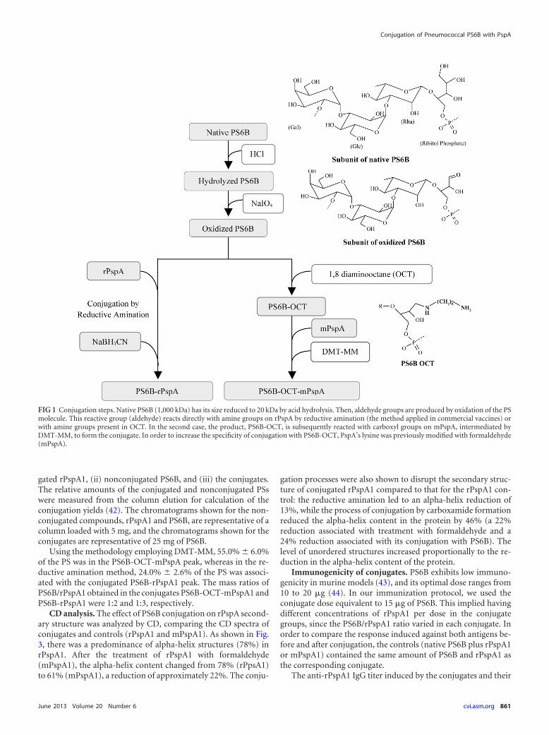
RESULTSConjugation and purification of PS6B-rPspA1. Two differentconjugates were synthesized: PS6B-OCT-mPspA1 (with an eight-carbon spacer molecule) and PS6B-rPspA1 (with no spacer mol-ecule). PS6B-OCT-mPspA1 was prepared by the method devel-oped in our laboratory (28). The steps of the conjugation processare represented in Fig. 1 and described above in detail in the ex-perimental protocols. The acid hydrolysis of native PS6B reducedits size from 1,000 kDa to approximately 20 kDa. The aldehydegroups were obtained by a mild oxidation condition with NaIO4
that resulted in 5 aldehydes per PS6B molecule (approximately0.16 aldehyde per PS6B repeating unit). Eighty percent of the al-dehydes inserted in the PS6B molecule were linked to the spacermolecule OCT, resulting in 4 OCT molecules per PS6B (approx-imately 0.128 OCT molecule per PS6B repeating unit). PS6B-rPspA1 was obtained by the currently used reductive aminationmethod (41), using the same hydrolyzed and oxidized PS.
The rPspA1 employed in the DMT-MM-mediated conjuga-tion was previously treated with formaldehyde in order to avoidintermolecular reactions and precipitation during conjugate syn-thesis. This modification process incorporates methyl groups inabout 70% of the ε-amine groups of PspA lysine residues; methylincorporation has been proven not to interfere with rPspA immu-nogenicity (27).
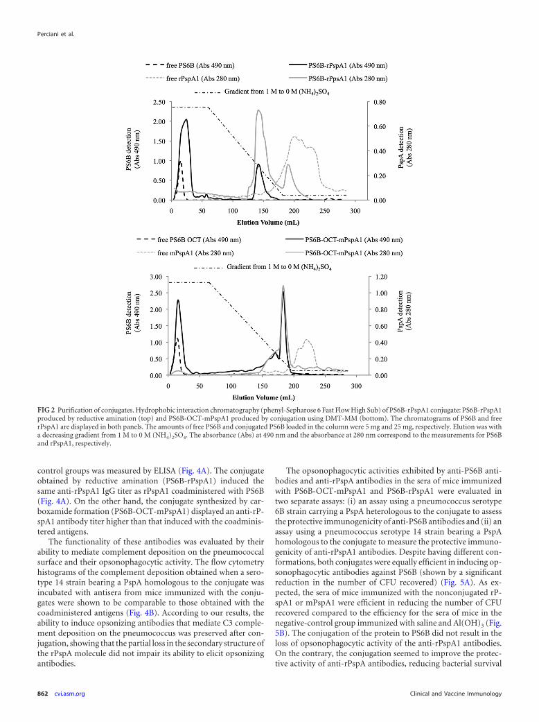
The conjugates were purified using hydrophobic interactionchromatography (HIC) (Fig. 2). PS6B, a hydrophilic molecule,did not bind to phenyl-Sepharose and eluted in the flowthroughfraction. rPspA1, which contains hydrophobic domains, inter-acted strongly with the resin and eluted after the end of the gradi-ent. The conjugates combine the characteristics of both PS6B andrPspA and eluted in the last third of the decreasing ammoniumsulfate gradient, allowing separation of the reagents and products.The conjugate elution was characterized by the coincidence of PSand protein detection in the same elution volume. Figure 2 showsthe chromatograms of PS6B-OCT-mPspA (top) and PS6B-PspA(bottom) with 3 overlapping chromatograms each: (i) nonconju-
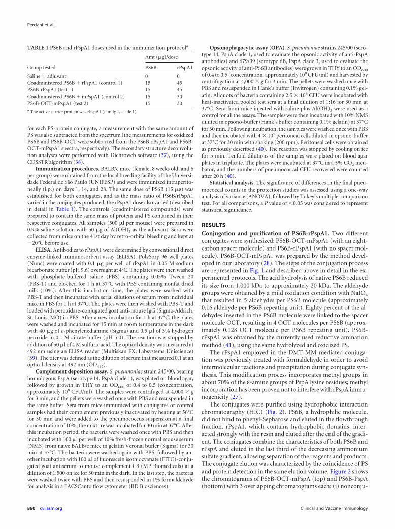
TABLE 1 PS6B and rPspA1 doses used in the immunization protocola
Group tested
Amt (�g)/dose
PS6B rPspA1
Saline � adjuvant 0 0Coadministered PS6B � rPspA1 (control 1) 15 45PS6B-rPspA1 (test 1) 15 45Coadministered PS6B � mPspA1 (control 2) 15 30PS6B-OCT-mPspA1 (test 2) 15 30a The active carrier protein was rPspA1 (family 1, clade 1).
Perciani et al.
860 cvi.asm.org Clinical and Vaccine Immunology
gated rPspA1, (ii) nonconjugated PS6B, and (iii) the conjugates.The relative amounts of the conjugated and nonconjugated PSswere measured from the column elution for calculation of theconjugation yields (42). The chromatograms shown for the non-conjugated compounds, rPspA1 and PS6B, are representative of acolumn loaded with 5 mg, and the chromatograms shown for theconjugates are representative of 25 mg of PS6B.
Using the methodology employing DMT-MM, 55.0% � 6.0%of the PS was in the PS6B-OCT-mPspA peak, whereas in the re-ductive amination method, 24.0% � 2.6% of the PS was associ-ated with the conjugated PS6B-rPspA1 peak. The mass ratios ofPS6B/rPspA1 obtained in the conjugates PS6B-OCT-mPspA1 andPS6B-rPspA1 were 1:2 and 1:3, respectively.
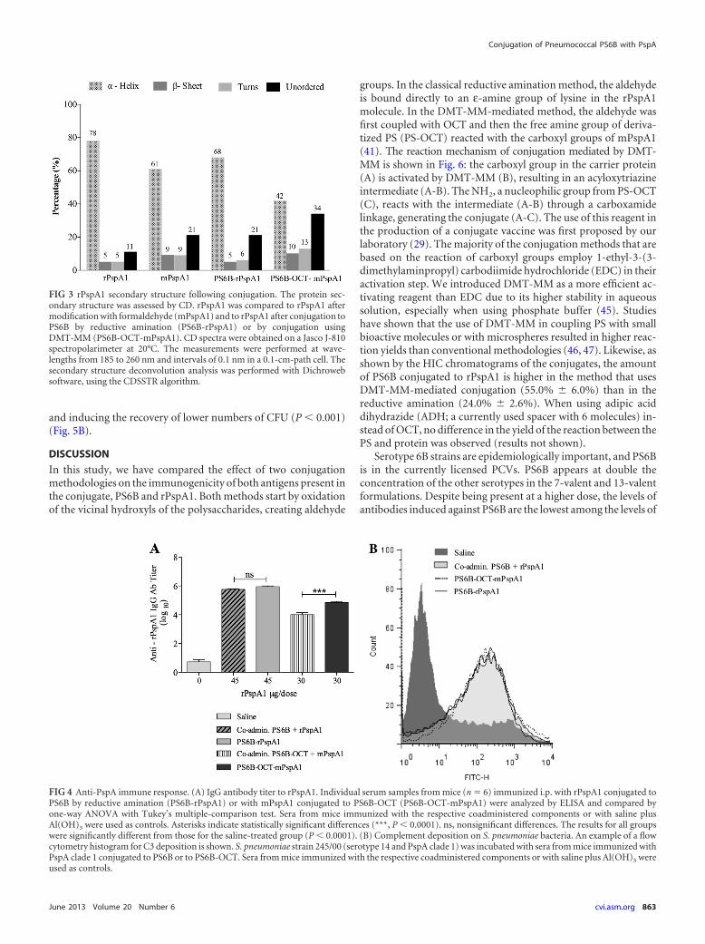
CD analysis. The effect of PS6B conjugation on rPspA second-ary structure was analyzed by CD, comparing the CD spectra ofconjugates and controls (rPspA1 and mPspA1). As shown in Fig.3, there was a predominance of alpha-helix structures (78%) inrPspA1. After the treatment of rPspA1 with formaldehyde(mPspA1), the alpha-helix content changed from 78% (rPpsA1)to 61% (mPspA1), a reduction of approximately 22%. The conju-
gation processes were also shown to disrupt the secondary struc-ture of conjugated rPspA1 compared to that for the rPspA1 con-trol: the reductive amination led to an alpha-helix reduction of13%, while the process of conjugation by carboxamide formationreduced the alpha-helix content in the protein by 46% (a 22%reduction associated with treatment with formaldehyde and a24% reduction associated with its conjugation with PS6B). Thelevel of unordered structures increased proportionally to the re-duction in the alpha-helix content of the protein.
Immunogenicity of conjugates. PS6B exhibits low immuno-genicity in murine models (43), and its optimal dose ranges from10 to 20 �g (44). In our immunization protocol, we used theconjugate dose equivalent to 15 �g of PS6B. This implied havingdifferent concentrations of rPspA1 per dose in the conjugategroups, since the PS6B/rPspA1 ratio varied in each conjugate. Inorder to compare the response induced against both antigens be-fore and after conjugation, the controls (native PS6B plus rPspA1or mPspA1) contained the same amount of PS6B and rPspA1 asthe corresponding conjugate.
The anti-rPspA1 IgG titer induced by the conjugates and their
FIG 1 Conjugation steps. Native PS6B (1,000 kDa) has its size reduced to 20 kDa by acid hydrolysis. Then, aldehyde groups are produced by oxidation of the PSmolecule. This reactive group (aldehyde) reacts directly with amine groups on rPspA by reductive amination (the method applied in commercial vaccines) orwith amine groups present in OCT. In the second case, the product, PS6B-OCT, is subsequently reacted with carboxyl groups on mPspA, intermediated byDMT-MM, to form the conjugate. In order to increase the specificity of conjugation with PS6B-OCT, PspA’s lysine was previously modified with formaldehyde(mPspA).
Conjugation of Pneumococcal PS6B with PspA
June 2013 Volume 20 Number 6 cvi.asm.org 861
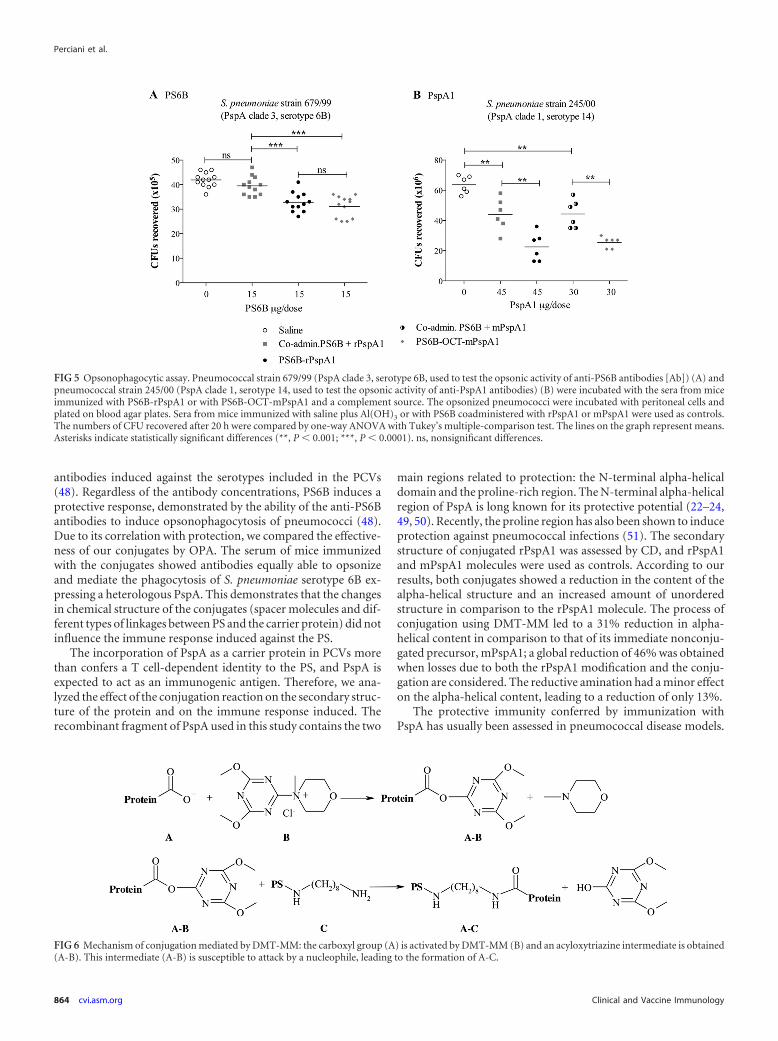
control groups was measured by ELISA (Fig. 4A). The conjugateobtained by reductive amination (PS6B-rPspA1) induced thesame anti-rPspA1 IgG titer as rPspA1 coadministered with PS6B(Fig. 4A). On the other hand, the conjugate synthesized by car-boxamide formation (PS6B-OCT-mPspA1) displayed an anti-rP-spA1 antibody titer higher than that induced with the coadminis-tered antigens.
The functionality of these antibodies was evaluated by theirability to mediate complement deposition on the pneumococcalsurface and their opsonophagocytic activity. The flow cytometryhistograms of the complement deposition obtained when a sero-type 14 strain bearing a PspA homologous to the conjugate wasincubated with antisera from mice immunized with the conju-gates were shown to be comparable to those obtained with thecoadministered antigens (Fig. 4B). According to our results, theability to induce opsonizing antibodies that mediate C3 comple-ment deposition on the pneumococcus was preserved after con-jugation, showing that the partial loss in the secondary structure ofthe rPspA molecule did not impair its ability to elicit opsonizingantibodies.
The opsonophagocytic activities exhibited by anti-PS6B anti-bodies and anti-rPspA antibodies in the sera of mice immunizedwith PS6B-OCT-mPspA1 and PS6B-rPspA1 were evaluated intwo separate assays: (i) an assay using a pneumococcus serotype6B strain carrying a PspA heterologous to the conjugate to assessthe protective immunogenicity of anti-PS6B antibodies and (ii) anassay using a pneumococcus serotype 14 strain bearing a PspAhomologous to the conjugate to measure the protective immuno-genicity of anti-rPspA1 antibodies. Despite having different con-formations, both conjugates were equally efficient in inducing op-sonophagocytic antibodies against PS6B (shown by a significantreduction in the number of CFU recovered) (Fig. 5A). As ex-pected, the sera of mice immunized with the nonconjugated rP-spA1 or mPspA1 were efficient in reducing the number of CFUrecovered compared to the efficiency for the sera of mice in thenegative-control group immunized with saline and Al(OH)3 (Fig.5B). The conjugation of the protein to PS6B did not result in theloss of opsonophagocytic activity of the anti-rPspA1 antibodies.On the contrary, the conjugation seemed to improve the protec-tive activity of anti-rPspA antibodies, reducing bacterial survival
FIG 2 Purification of conjugates. Hydrophobic interaction chromatography (phenyl-Sepharose 6 Fast Flow High Sub) of PS6B-rPspA1 conjugate: PS6B-rPspA1produced by reductive amination (top) and PS6B-OCT-mPspA1 produced by conjugation using DMT-MM (bottom). The chromatograms of PS6B and freerPspA1 are displayed in both panels. The amounts of free PS6B and conjugated PS6B loaded in the column were 5 mg and 25 mg, respectively. Elution was witha decreasing gradient from 1 M to 0 M (NH4)2SO4. The absorbance (Abs) at 490 nm and the absorbance at 280 nm correspond to the measurements for PS6Band rPspA1, respectively.
Perciani et al.
862 cvi.asm.org Clinical and Vaccine Immunology
and inducing the recovery of lower numbers of CFU (P � 0.001)(Fig. 5B).
DISCUSSION
In this study, we have compared the effect of two conjugationmethodologies on the immunogenicity of both antigens present inthe conjugate, PS6B and rPspA1. Both methods start by oxidationof the vicinal hydroxyls of the polysaccharides, creating aldehyde
groups. In the classical reductive amination method, the aldehydeis bound directly to an ε-amine group of lysine in the rPspA1molecule. In the DMT-MM-mediated method, the aldehyde wasfirst coupled with OCT and then the free amine group of deriva-tized PS (PS-OCT) reacted with the carboxyl groups of mPspA1(41). The reaction mechanism of conjugation mediated by DMT-MM is shown in Fig. 6: the carboxyl group in the carrier protein(A) is activated by DMT-MM (B), resulting in an acyloxytriazineintermediate (A-B). The NH2, a nucleophilic group from PS-OCT(C), reacts with the intermediate (A-B) through a carboxamidelinkage, generating the conjugate (A-C). The use of this reagent inthe production of a conjugate vaccine was first proposed by ourlaboratory (29). The majority of the conjugation methods that arebased on the reaction of carboxyl groups employ 1-ethyl-3-(3-dimethylaminpropyl) carbodiimide hydrochloride (EDC) in theiractivation step. We introduced DMT-MM as a more efficient ac-tivating reagent than EDC due to its higher stability in aqueoussolution, especially when using phosphate buffer (45). Studieshave shown that the use of DMT-MM in coupling PS with smallbioactive molecules or with microspheres resulted in higher reac-tion yields than conventional methodologies (46, 47). Likewise, asshown by the HIC chromatograms of the conjugates, the amountof PS6B conjugated to rPspA1 is higher in the method that usesDMT-MM-mediated conjugation (55.0% � 6.0%) than in thereductive amination (24.0% � 2.6%). When using adipic aciddihydrazide (ADH; a currently used spacer with 6 molecules) in-stead of OCT, no difference in the yield of the reaction between thePS and protein was observed (results not shown).
Serotype 6B strains are epidemiologically important, and PS6Bis in the currently licensed PCVs. PS6B appears at double theconcentration of the other serotypes in the 7-valent and 13-valentformulations. Despite being present at a higher dose, the levels ofantibodies induced against PS6B are the lowest among the levels of
FIG 3 rPspA1 secondary structure following conjugation. The protein sec-ondary structure was assessed by CD. rPspA1 was compared to rPspA1 aftermodification with formaldehyde (mPspA1) and to rPspA1 after conjugation toPS6B by reductive amination (PS6B-rPspA1) or by conjugation usingDMT-MM (PS6B-OCT-mPspA1). CD spectra were obtained on a Jasco J-810spectropolarimeter at 20°C. The measurements were performed at wave-lengths from 185 to 260 nm and intervals of 0.1 nm in a 0.1-cm-path cell. Thesecondary structure deconvolution analysis was performed with Dichrowebsoftware, using the CDSSTR algorithm.
FIG 4 Anti-PspA immune response. (A) IgG antibody titer to rPspA1. Individual serum samples from mice (n � 6) immunized i.p. with rPspA1 conjugated toPS6B by reductive amination (PS6B-rPspA1) or with mPspA1 conjugated to PS6B-OCT (PS6B-OCT-mPspA1) were analyzed by ELISA and compared byone-way ANOVA with Tukey’s multiple-comparison test. Sera from mice immunized with the respective coadministered components or with saline plusAl(OH)3 were used as controls. Asterisks indicate statistically significant differences (***, P � 0.0001). ns, nonsignificant differences. The results for all groupswere significantly different from those for the saline-treated group (P � 0.0001). (B) Complement deposition on S. pneumoniae bacteria. An example of a flowcytometry histogram for C3 deposition is shown. S. pneumoniae strain 245/00 (serotype 14 and PspA clade 1) was incubated with sera from mice immunized withPspA clade 1 conjugated to PS6B or to PS6B-OCT. Sera from mice immunized with the respective coadministered components or with saline plus Al(OH)3 wereused as controls.
Conjugation of Pneumococcal PS6B with PspA
June 2013 Volume 20 Number 6 cvi.asm.org 863
antibodies induced against the serotypes included in the PCVs(48). Regardless of the antibody concentrations, PS6B induces aprotective response, demonstrated by the ability of the anti-PS6Bantibodies to induce opsonophagocytosis of pneumococci (48).Due to its correlation with protection, we compared the effective-ness of our conjugates by OPA. The serum of mice immunizedwith the conjugates showed antibodies equally able to opsonizeand mediate the phagocytosis of S. pneumoniae serotype 6B ex-pressing a heterologous PspA. This demonstrates that the changesin chemical structure of the conjugates (spacer molecules and dif-ferent types of linkages between PS and the carrier protein) did notinfluence the immune response induced against the PS.
The incorporation of PspA as a carrier protein in PCVs morethan confers a T cell-dependent identity to the PS, and PspA isexpected to act as an immunogenic antigen. Therefore, we ana-lyzed the effect of the conjugation reaction on the secondary struc-ture of the protein and on the immune response induced. Therecombinant fragment of PspA used in this study contains the two
main regions related to protection: the N-terminal alpha-helicaldomain and the proline-rich region. The N-terminal alpha-helicalregion of PspA is long known for its protective potential (22–24,49, 50). Recently, the proline region has also been shown to induceprotection against pneumococcal infections (51). The secondarystructure of conjugated rPspA1 was assessed by CD, and rPspA1and mPspA1 molecules were used as controls. According to ourresults, both conjugates showed a reduction in the content of thealpha-helical structure and an increased amount of unorderedstructure in comparison to the rPspA1 molecule. The process ofconjugation using DMT-MM led to a 31% reduction in alpha-helical content in comparison to that of its immediate nonconju-gated precursor, mPspA1; a global reduction of 46% was obtainedwhen losses due to both the rPspA1 modification and the conju-gation are considered. The reductive amination had a minor effecton the alpha-helical content, leading to a reduction of only 13%.
The protective immunity conferred by immunization withPspA has usually been assessed in pneumococcal disease models.
FIG 5 Opsonophagocytic assay. Pneumococcal strain 679/99 (PspA clade 3, serotype 6B, used to test the opsonic activity of anti-PS6B antibodies [Ab]) (A) andpneumococcal strain 245/00 (PspA clade 1, serotype 14, used to test the opsonic activity of anti-PspA1 antibodies) (B) were incubated with the sera from miceimmunized with PS6B-rPspA1 or with PS6B-OCT-mPspA1 and a complement source. The opsonized pneumococci were incubated with peritoneal cells andplated on blood agar plates. Sera from mice immunized with saline plus Al(OH)3 or with PS6B coadministered with rPspA1 or mPspA1 were used as controls.The numbers of CFU recovered after 20 h were compared by one-way ANOVA with Tukey’s multiple-comparison test. The lines on the graph represent means.Asterisks indicate statistically significant differences (**, P � 0.001; ***, P � 0.0001). ns, nonsignificant differences.
FIG 6 Mechanism of conjugation mediated by DMT-MM: the carboxyl group (A) is activated by DMT-MM (B) and an acyloxytriazine intermediate is obtained(A-B). This intermediate (A-B) is susceptible to attack by a nucleophile, leading to the formation of A-C.
Perciani et al.
864 cvi.asm.org Clinical and Vaccine Immunology
A restricted repertoire of pneumococcal strains is virulent in mu-rine models, and these strains usually bear capsular polysaccha-rides 3, 6A, and 6B (21). In the present case, the selection of astrain virulent for mice mainly relies on strains bearing capsulartype 3. The selection of a strain bearing serogroup 6 would impairthe analysis of the immune response induced by PspA. The strainscarrying PspA clade 1 and serotype 3 were shown to be highlypathogenic, causing rapid sepsis and death in the mice, while testsusing serotypes different from serotypes 3, 6A, and 6B did notcause disease in the animals (data not shown). In the absence of asuitable pneumococcal disease model, the OPA was the assay ofchoice for evaluating the functional activity of anti-PspA antibod-ies (27, 40).
Notably, both conjugation processes preserved PspA’s anti-genic properties, including the ability to induce antibodies capa-ble of mediating complement deposition and phagocytosis. Theseresults would indicate that the primary sequence of amino acidresidues in rPspA1, rather than its secondary structure, is probablyassociated with the induction of protective antibodies.
Our main goals with this study were to investigate whetherrPspA1 could act as a carrier protein for PS6B and whether theconjugation would disrupt rPspA1’s structure, affecting its immu-nogenicity. We observed that, when conjugated, rPspA1 inducedlower levels of antibodies, although it had higher opsonophago-cytic activity than when it was nonconjugated (Fig. 5B). The ratioof the opsonophagocytic activity per unit of antibody titer torPspA1 was 15% higher for the conjugated rPspA1 than for thecoadministered rPspA1 (data from Fig. 4A and 5A). A hypothesisfor this observation is that surface PspA1, contrary to rPspA1,interacts with other pneumococcal surface components, includ-ing the capsular polysaccharide, and the charge distribution ofconjugated rPspA1 (positively charged protein and negativelycharged PS) more closely resembles that of the conformationalstructure of PspA expressed on the bacterial surface.
In conclusion, despite the fact that the circular dichroism anal-ysis has shown that the conjugation alters the secondary structureof rPspA1, the immunological assays have demonstrated thatthese alterations do not affect its ability to induce a protectiveimmune response. Furthermore, the conjugation strategy usingdifferent chemical linkages does not seem to impair the immuno-genicity of rPspA1 or PS6B and, consequently, does not impose anobstacle to implementation of the more economical methodol-ogy. Therefore, our results support the use of rPspA1 as an anti-genic carrier protein and reinforce the use of DMT-MM-mediatedconjugation as a valuable strategy to be considered in conjugationprocesses.
ACKNOWLEDGMENT
This work was supported by grants from the Fundação de Amparo àPesquisa do Estado de São Paulo (FAPESP).
REFERENCES1. O’Brien KL, Wolfson LJ, Watt JP, Henkle E, Deloria-Knoll M, McCall
N, Lee E, Mulholland K, Levine OS, Cherian T. 2009 Burden of diseasecaused by Streptococcus pneumoniae in children younger than 5 years:global estimates. Lancet 374:893–902.
2. Linares J, Ardanuy C, Pallares R, Fenoll A. 2010. Changes in antimicro-bial resistance, serotypes and genotypes in Streptococcus pneumoniaeover a 30-year period. Clin. Microbiol. Infect. 16:402– 410.
3. Fedson DS, Nicolas-Spony L, Klemets P, van der Linden M, Marques A,Salleras L, Samson SI. 2011. Pneumococcal polysaccharide vaccination
for adults: new perspectives for Europe. Expert Rev. Vaccines 10:1143–1167.
4. Cornu C, Yzebe D, Leophonte P, Gaillat J, Boissel JP, Cucherat M.2001. Efficacy of pneumococcal polysaccharide vaccine in immunocom-petent adults: a meta-analysis of randomized trials. Vaccine 19:4780 –4790.
5. Griffioen AW, Rijkers GT, Janssens-Korpela P, Zegers BJ. 1991. Pneu-mococcal polysaccharides complexed with C3d bind to human B lympho-cytes via complement receptor type 2. Infect. Immun. 59:1839 –1845.
6. Timens W, Boes A, Rozeboom-Uiterwijk T, Poppema S. 1989. Imma-turity of the human splenic marginal zone in infancy. Possible contribu-tion to the deficient infant immune response. J. Immunol. 143:3200 –3206.
7. Guttormsen HK, Sharpe AH, Chandraker AK, Brigtsen AK, SayeghMH, Kasper DL. 1999. Cognate stimulatory B-cell–T-cell interactions arecritical for T-cell help recruited by glycoconjugate vaccines. Infect. Im-mun. 67:6375– 6384.
8. Redelings MD, Sorvillo F, Simon P, Mascola L. 2005. Declining earlychildhood mortality from invasive pneumococcal disease: the impact ofvaccination. Arch. Pediatr. Adolesc. Med. 159:195–196.
9. Roush SW, Murphy TV. 2007. Historical comparisons of morbidity andmortality for vaccine-preventable diseases in the United States. JAMA298:2155–2163.
10. Kellner JD, Vanderkooi OG, MacDonald J, Church DL, Tyrrell GJ,Scheifele DW. 2009. Changing epidemiology of invasive pneumococcaldisease in Canada, 1998-2007: update from the Calgary-area Streptococ-cus pneumoniae research (CASPER) study. Clin. Infect. Dis. 49:205–212.
11. Ruckinger S, van der Linden M, Reinert RR, von Kries R, BurckhardtF, Siedler A. 2009. Reduction in the incidence of invasive pneumococcaldisease after general vaccination with 7-valent pneumococcal conjugatevaccine in Germany. Vaccine 27:4136 – 4141.
12. Grijalva CG, Nuorti JP, Arbogast PG, Martin SW, Edwards KM, GriffinMR. 2007. Decline in pneumonia admissions after routine childhood im-munisation with pneumococcal conjugate vaccine in the USA: a time-series analysis. Lancet 369:1179 –1186.
13. Chibuk TK, Robinson JL, Hartfield DS. 2010. Pediatric complicatedpneumonia and pneumococcal serotype replacement: trends in hospital-ized children pre and post introduction of routine vaccination with pneu-mococcal conjugate vaccine (PCV7). Eur. J. Pediatr. 169:1123–1128.
14. Leach AJ, Morris PS, McCallum GB, Wilson CA, Stubbs L, BeissbarthJ, Jacups S, Hare K, Smith-Vaughan HC. 2009. Emerging pneumococcalcarriage serotypes in a high-risk population receiving universal 7-valentpneumococcal conjugate vaccine and 23-valent polysaccharide vaccinesince 2001. BMC Infect. Dis. 9:121. doi:10.1186/1471-2334-9-121.
15. Munoz-Almagro C, Jordan I, Gene A, Latorre C, Garcia-Garcia JJ,Pallares R. 2008. Emergence of invasive pneumococcal disease caused bynonvaccine serotypes in the era of 7-valent conjugate vaccine. Clin. Infect.Dis. 46:174 –182.
16. Borrow R, Dagan R, Zepp F, Hallander H, Poolman J. 2011. Glycocon-jugate vaccines and immunointeractions and implication for vaccinationschedules. Expert Rev. Vaccines 10:1621–1631.
17. Dagan R, Poolman J, Siegrist CA. 2010. Glycoconjugate vaccines andimmune interference: a review. Vaccine 28:5513–5523.
18. Ren B, Szalai AJ, Thomas O, Hollingshead SK, Briles DE. 2003. Bothfamily 1 and family 2 PspA proteins can inhibit complement depositionand confer virulence to a capsular serotype 3 strain of Streptococcus pneu-moniae. Infect. Immun. 71:75– 85.
19. Tu AH, Fulgham RL, McCrory MA, Briles DE, Szalai AJ. 1999. Pneu-mococcal surface protein A inhibits complement activation by Strepto-coccus pneumoniae. Infect. Immun. 67:4720 – 4724.
20. Shaper M, Hollingshead SK, Benjamin WH, Jr, Briles DE. 2004. PspAprotects Streptococcus pneumoniae from killing by apolactoferrin, andantibody to PspA enhances killing of pneumococci by apolactoferrin [cor-rected]. Infect. Immun. 72:5031–5040.
21. Briles DE, Hollingshead SK, King J, Swift A, Braun PA, Park MK,Ferguson LM, Nahm MH, Nabors GS. 2000. Immunization of humanswith recombinant pneumococcal surface protein A (rPspA) elicits anti-bodies that passively protect mice from fatal infection with Streptococcuspneumoniae bearing heterologous PspA. J. Infect. Dis. 182:1694 –1701.
22. Wu HY, Nahm MH, Guo Y, Russell MW, Briles DE. 1997. Intranasalimmunization of mice with PspA (pneumococcal surface protein A) canprevent intranasal carriage, pulmonary infection, and sepsis with Strepto-coccus pneumoniae. J. Infect. Dis. 175:839 – 846.
Conjugation of Pneumococcal PS6B with PspA
June 2013 Volume 20 Number 6 cvi.asm.org 865
23. Crain MJ, Waltman WD, II, Turner JS, Yother J, Talkington DF,McDaniel LS, Gray BM, Briles DE. 1990. Pneumococcal surface proteinA (PspA) is serologically highly variable and is expressed by all clinicallyimportant capsular serotypes of Streptococcus pneumoniae. Infect. Im-mun. 58:3293–3299.
24. Hollingshead SK, Becker R, Briles DE. 2000. Diversity of PspA: mosaicgenes and evidence for past recombination in Streptococcus pneumoniae.Infect. Immun. 68:5889 –5900.
25. Hollingshead SK, Baril L, Ferro S, King J, Coan P, Briles DE. 2006.Pneumococcal surface protein A (PspA) family distribution among clini-cal isolates from adults over 50 years of age collected in seven countries. J.Med. Microbiol. 55:215–221.
26. Pimenta FC, Ribeiro-Dias F, Brandileone MC, Miyaji EN, Leite LC,Sgambatti de Andrade AL. 2006. Genetic diversity of PspA types amongnasopharyngeal isolates collected during an ongoing surveillance study ofchildren in Brazil. J. Clin. Microbiol. 44:2838 –2843.
27. Santamaria R, Goulart C, Perciani CT, Barazzone GC, Carvalho R,Goncalves VM, Leite LC, Tanizaki MM. 2011. Humoral immune re-sponse of a pneumococcal conjugate vaccine: capsular polysaccharide se-rotype 14-lysine modified PspA. Vaccine 29:8689 – 8695.
28. Barazzone GC, Perciani CT, Raw I, Tanizaki MM. July 2011. Método deconjugação de polissacarídeo capsular a uma proteína carregadora, parauso como antígeno vacinal contra bactérias encapsuladas, utilizando oreagente cloreto de 4-(4,6-dimetoxi-1,3,5-triazin-2-il)-4-metilmorfolino(DMT-MM). Brazilian patent PI0904528-7.
29. Barazzone GC, Carvalho RJ, Kraschowetz S, Horta ACL, Sargo C, SilvaAJ, Tanizaki MM, Cabrera-Crespo J, Gonçalves VM. 2011. Productionand purification of recombinant fragment of pneumococcal surface pro-tein A (PspA) in Escherichia coli. Proc. Vaccinol. 4:27–35.
30. Carvalho RJ, Cabrera-Crespo J, Tanizaki MM, Gonçalves VM. 2012.Development of production and purification processes of recombinantfragment of pneumococcal surface protein A in Escherichia coli usingdifferent carbon sources and chromatography sequences. Appl. Micro-biol. Biotechnol. 94:683– 694.
31. Silva M, Cabrera-Crespo J, Sbrogio-Almeida ME, Miyaji EN, Ho PL,Leite LC, Lopes AP. 2007. Optimizing expression of Streptococcus pneu-moniae surface protein a, PspA: serocross-reactivity within families ofantisera induced against clades 1 and 3. Mol. Biotechnol. 37:146 –154.
32. Gonçalves VM, Takagi M, Carmo TS, Albani SMF, Pinto JV, Zangi-rolami TC, Giordano RC, Tanizaki MM, Cabrera-Crespo J. 2007. Sim-ple and efficient method of bacterial polysaccharides purification for vac-cines production using hydrolytic enzymes and tangential flowultrafiltration, p 250 –257. In Mendez-Vilas A (ed), Communicating cur-rent research and educational topics and trends in applied microbiology.Formatex, Badajoz, Spain.
33. Tyllianakis PE, Kakabakos SE, Evangelatos GP, Ithakissios DS. 1994.Direct colorimetric determination of solid-supported functional groupsand ligands using bicinchoninic acid. Anal. Biochem. 219:335–340.
34. Qi XY, Keyhani NO, Lee YC. 1988. Spectrophotometric determinationof hydrazine, hydrazides, and their mixtures with trinitrobenzenesulfonicacid. Anal. Biochem. 175:139 –144.
35. Dubois M, Gilles KA, Hamilton JK, Rebers PA, Smith F. 1956. Color-imetric method for determination of sugars and related substances. Anal.Chem. 28:350 –356.
36. Fryer HJ, Davis GE, Manthorpe M, Varon S. 1986. Lowry protein assayusing an automatic microtiter plate spectrophotometer. Anal. Biochem.153:262–266.
37. Whitmore L, Wallace BA. 2008. Protein secondary structure analyses
from circular dichroism spectroscopy: methods and reference databases.Biopolymers 89:392– 400.
38. Compton LA, Johnson WC, Jr. 1986. Analysis of protein circular dichr-oism spectra for secondary structure using a simple matrix multiplication.Anal. Biochem. 155:155–167.
39. Darrieux M, Moreno AT, Ferreira DM, Pimenta FC, de Andrade AL,Lopes AP, Leite LC, Miyaji EN. 2008. Recognition of pneumococcalisolates by antisera raised against PspA fragments from different clades. J.Med. Microbiol. 57:273–278.
40. Goulart C, Darrieux M, Rodriguez D, Pimenta FC, Brandileone MC, deAndrade AL, Leite LC. 2011. Selection of family 1 PspA molecules capableof inducing broad-ranging cross-reactivity by complement depositionand opsonophagocytosis by murine peritoneal cells. Vaccine 29:1634 –1642.
41. Anderson P, Pichichero ME, Insel RA. 1985. Immunogens consisting ofoligosaccharides from the capsule of Haemophilus influenzae type b cou-pled to diphtheria toxoid or the toxin protein CRM197. J. Clin. Invest.76:52–59.
42. Lee CH, Kuo WC, Beri S, Kapre S, Joshi JS, Bouveret N, LaForce FM,Frasch CE. 2009. Preparation and characterization of an immunogenicmeningococcal group A conjugate vaccine for use in Africa. Vaccine 27:726 –732.
43. Fairchild RL, Braley-Mullen H. 1983. Characterization of the murineimmune response to type 6 pneumococcal polysaccharide. Infect. Immun.39:615– 622.
44. Chu RS, McCool T, Greenspan NS, Schreiber JR, Harding CV. 2000.CpG oligodeoxynucleotides act as adjuvants for pneumococcal polysac-charide-protein conjugate vaccines and enhance antipolysaccharide im-munoglobulin G2a (IgG2a) and IgG3 antibodies. Infect. Immun. 68:1450 –1456.
45. Gilles MA, Hudson AQ, Borders CL, Jr. 1990. Stability of water-solublecarbodiimides in aqueous solution. Anal. Biochem. 184:244 –248.
46. Farkas P, Bystricky S. 2007. Efficient activation of carboxyl polysaccha-rides for the preparation of conjugates. Carbohydr. Polymers 68:187–190.
47. Schlottmann SA, Jain N, Chirmule N, Esser MT. 2006. A novel chem-istry for conjugating pneumococcal polysaccharides to Luminex micro-spheres. J. Immunol. Methods 309:75– 85.
48. Vesikari T, Wysocki J, Chevallier B, Karvonen A, Czajka H, Arsène JP,Lommel P, Dieussaert I, Schuerman L. 2009. Immunogenicity of the10-valent pneumococcal non-typeable Haemophilus influenzae protein Dconjugate vaccine (PHiD-CV) compared to the licensed 7vCRM vaccine.Pediatr. Infect. Dis. J. 28(4 Suppl):S66 –S76.
49. McDaniel LS, Ralph BA, McDaniel DO, Briles DE. 1994. Localization ofprotection-eliciting epitopes on PspA of Streptococcus pneumoniae be-tween amino acid residues 192 and 260. Microb. Pathog. 17:323–337.
50. McDaniel LS, Sheffield JS, Swiatlo E, Yother J, Crain MJ, Briles DE.1992. Molecular localization of variable and conserved regions of pspAand identification of additional pspA homologous sequences in Strepto-coccus pneumoniae. Microb. Pathog. 13:261–269.
51. Daniels CC, Coan P, King J, Hale J, Benton KA, Briles DE, Hollings-head SK. 2010. The proline-rich region of pneumococcal surface proteinsA and C contains surface-accessible epitopes common to all pneumococciand elicits antibody-mediated protection against sepsis. Infect. Immun.78:2163–2172.
52. Csordas FC, Perciani CT, Darrieux M, Goncalves VM, Cabrera-CrespoJ, Takagi M, Takagi M, Sbrogio-Almeida ME, Leite LC, Tanizaki MM.2008. Protection induced by pneumococcal surface protein A (PspA) isenhanced by conjugation to a Streptococcus pneumoniae capsular poly-saccharide. Vaccine 26:2925–2929.
Perciani et al.
866 cvi.asm.org Clinical and Vaccine Immunology