Congenital Nasal Masses - University of Texas Medical Branch€¦ · Outline •Common Causes of...
53
Congenital Nasal Masses Naren Venkatesan, MD Mentor: Harold Pine, MD The University of Texas Medical Branch Department of Otolaryngology Grand Rounds Presentation December 2013 Series Editor: Francis B. Quinn, Jr., MD, FACS Archivist: Melinda Stoner Quinn, MSICS
Transcript of Congenital Nasal Masses - University of Texas Medical Branch€¦ · Outline •Common Causes of...
Congenital Nasal Masses
Naren Venkatesan, MD
Mentor: Harold Pine, MD
The University of Texas Medical Branch
Department of Otolaryngology
Grand Rounds Presentation
December 2013
Series Editor: Francis B. Quinn, Jr., MD, FACS Archivist: Melinda Stoner Quinn, MSICS
Outline
• Common Causes of Pediatric Nasal Obstruction
• Embryology
• Nasal Dermoid
• Nasal Glioma
• Encephalocele
• Nasolacrimal Duct Cysts
• Thornwaldt Cyst
• Brief Word on Pediatric Nasal Tumors
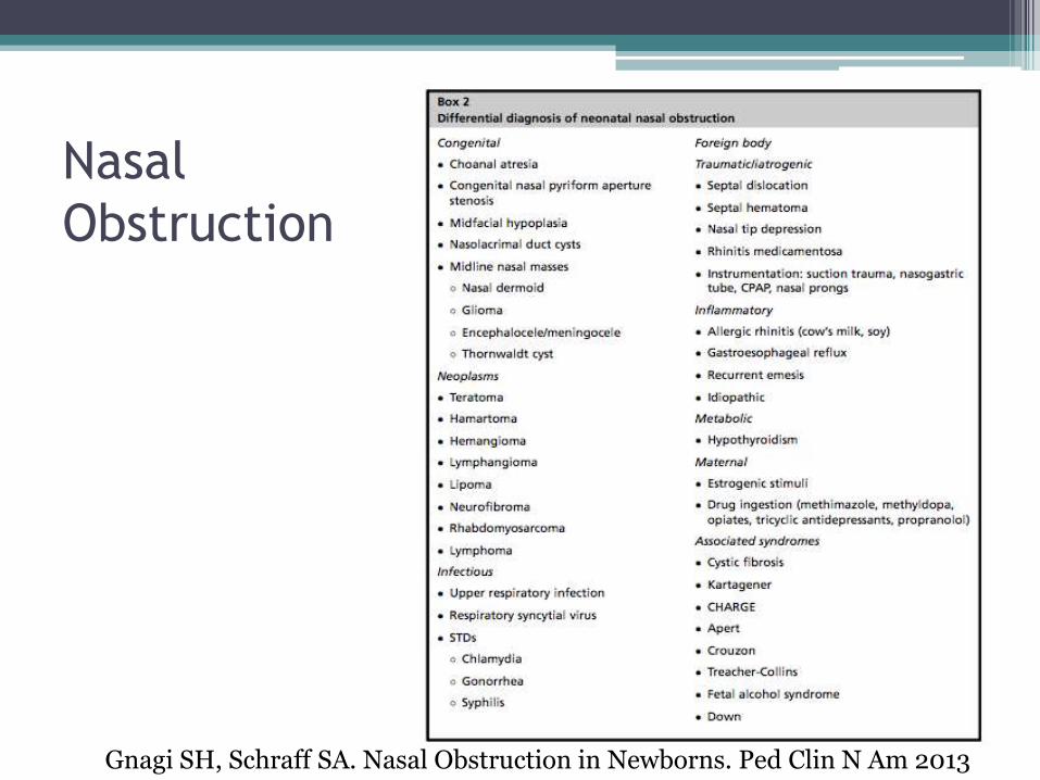
Nasal
Obstruction
Gnagi SH, Schraff SA. Nasal Obstruction in Newborns. Ped Clin N Am 2013
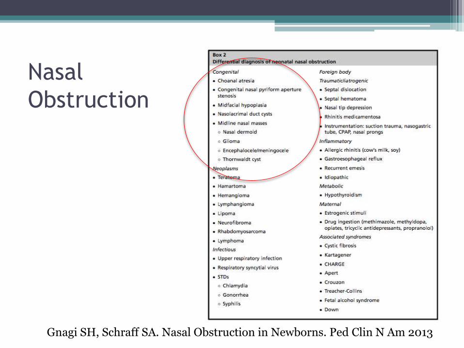
Nasal
Obstruction
Gnagi SH, Schraff SA. Nasal Obstruction in Newborns. Ped Clin N Am 2013
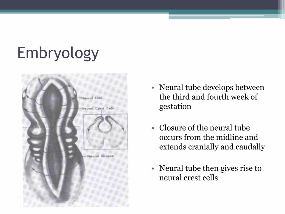
Embryology
• Neural tube develops between the third and fourth week of gestation
• Closure of the neural tube occurs from the midline and extends cranially and caudally
• Neural tube then gives rise to neural crest cells
Embryology
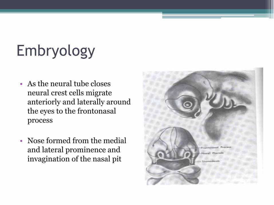
• As the neural tube closes neural crest cells migrate anteriorly and laterally around the eyes to the frontonasalprocess
• Nose formed from the medial and lateral prominence and invagination of the nasal pit
Embryology
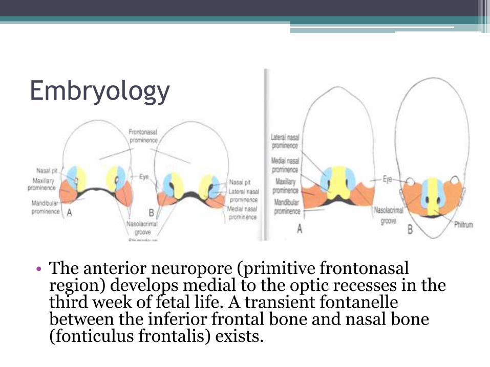
• The anterior neuropore (primitive frontonasalregion) develops medial to the optic recesses in the third week of fetal life. A transient fontanellebetween the inferior frontal bone and nasal bone (fonticulus frontalis) exists.
Embryology
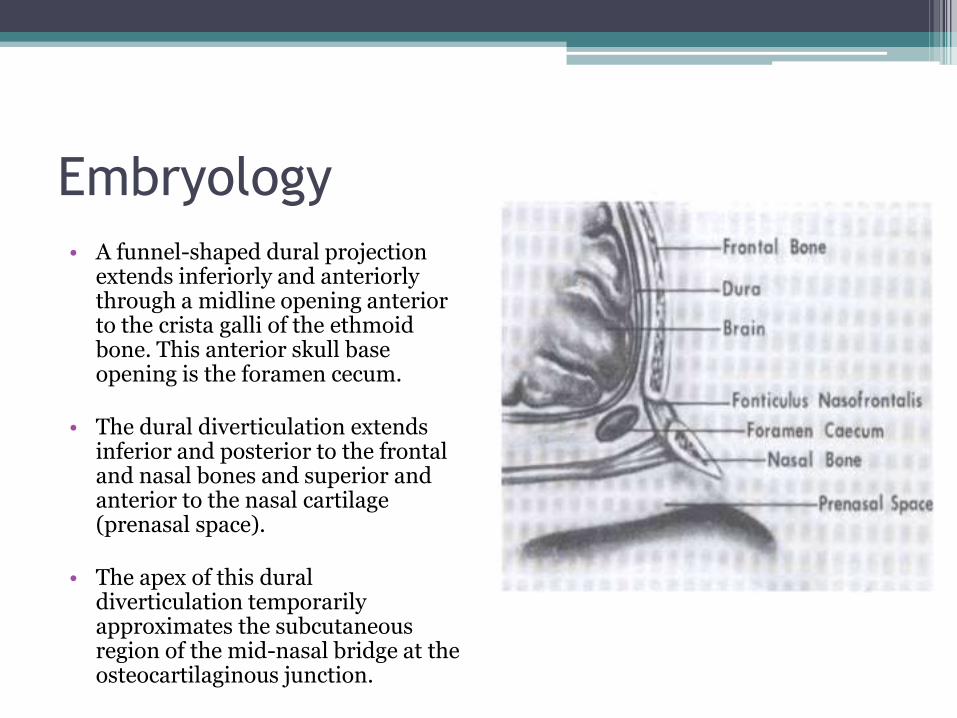
• A funnel-shaped dural projection extends inferiorly and anteriorlythrough a midline opening anterior to the crista galli of the ethmoidbone. This anterior skull base opening is the foramen cecum.
• The dural diverticulation extends inferior and posterior to the frontal and nasal bones and superior and anterior to the nasal cartilage (prenasal space).
• The apex of this duraldiverticulation temporarily approximates the subcutaneous region of the mid-nasal bridge at the osteocartilaginous junction.
Embryology – Normal Development
(Speaker Notes for next slide)
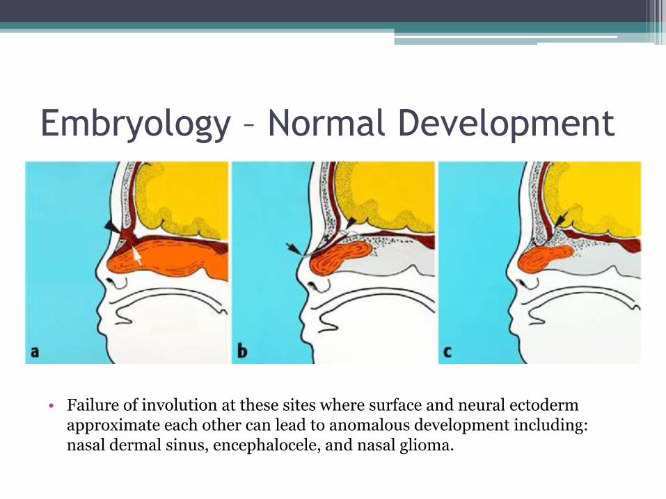
Fig. 1 Normal anterior neuropore development.
• A : Sagittal color graphic of the fetal anterior neuropore shows the transient osteo-cartilaginous gap at the fonticulus frontalis(arrowhead) and the prenasal space (arrow).
• B: In the fourth week to the seventh week of gestation, a transient dural diverticulum extends through the plane of the foramen cecum (Arrowhead ), coursing through the prenasal space and terminating at the skin of the nasal bridge (arrow).
• C: With normal regression of the dural diverticulum, a blind-ending foramen cecum is left (arrow ). This lies just anterior to the crista galli
Embryology – Normal Development
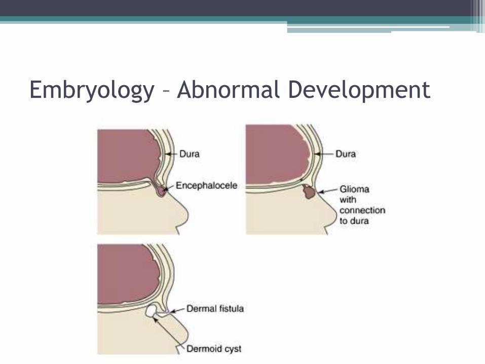
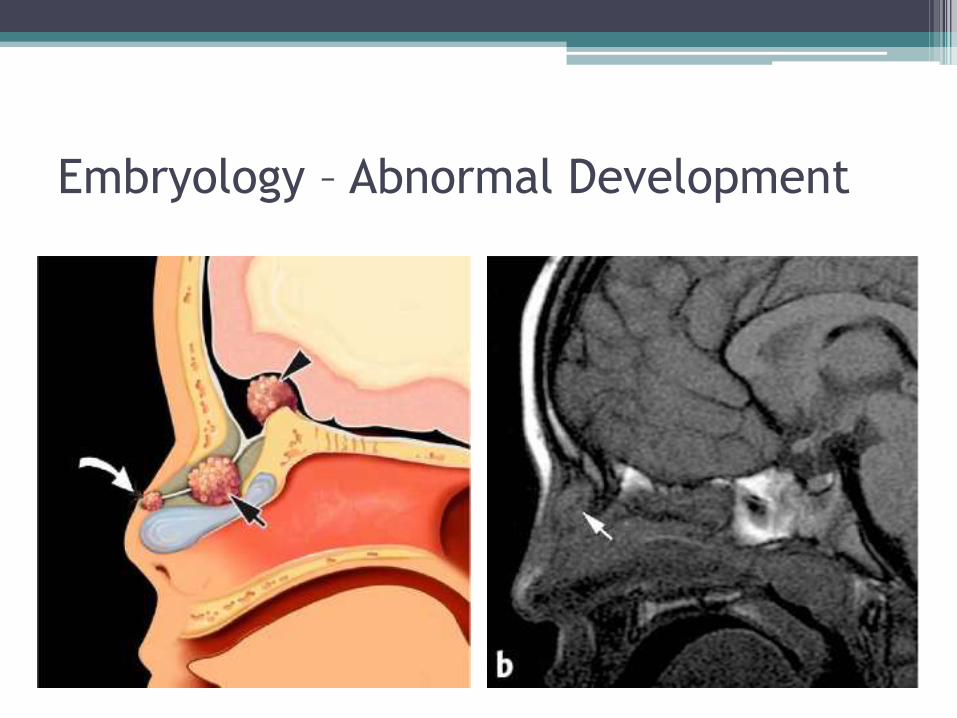
• Failure of involution at these sites where surface and neural ectoderm approximate each other can lead to anomalous development including: nasal dermal sinus, encephalocele, and nasal glioma.
Embryology – Normal Development
(speaker notes for next slide)
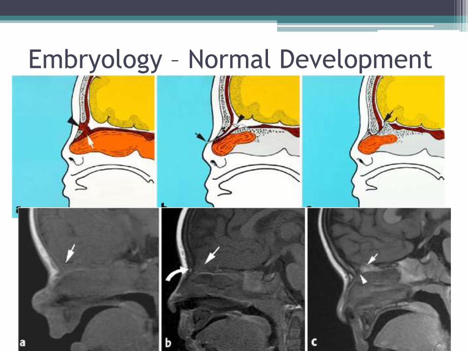
Normal development of the frontonasal region, MRI assessment.
• A: Sagittal T1-W image of a newborn shows isointense signal of the basal frontal lobes, nasal septal cartilage, and crista galli (arrow).
• B: At 6 months, there is normal T1 hyperintensity within the nasal bone (curved arrow) and slight T1 hyperintensity of the crista galli (arrow).
• C: Sagittal T1-W image of a normal 14-month-old boy shows a cock’ s comb shape and hyperintense signal of the crista galli(arrow). Note the foramen cecum located posterior to the nasal process of the frontal bone (arrowhead)
Embryology – Normal Development
Embryology – Abnormal Development
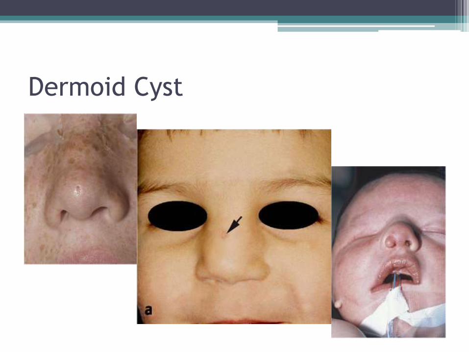
Dermoid Cyst
Dermoid Cyst
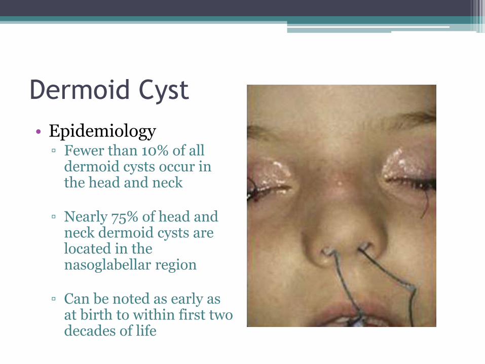
• Epidemiology▫ Fewer than 10% of all
dermoid cysts occur in the head and neck
▫ Nearly 75% of head and neck dermoid cysts are located in the nasoglabellar region
▫ Can be noted as early as at birth to within first two decades of life
Dermoid Cyst
• Embryology:
▫ Cysts consist of squamous cell epithelium containing epidermal appendages
▫ Epidermal elements are displaced during intramembranous growth phase of nasal bones
▫ Location can be several:
Fronto-temporal region
Orbital region
Nasoglabellar region
Dermoid Cyst
• Embryology:
▫ Cysts consist of squamouscell epithelium containing epidermal appendages
▫ Epidermal elements are displaced during intramembranous growth phase of nasal bones
▫ Location can be several:
Fronto-temporal region
Orbital region
Nasoglabellar region
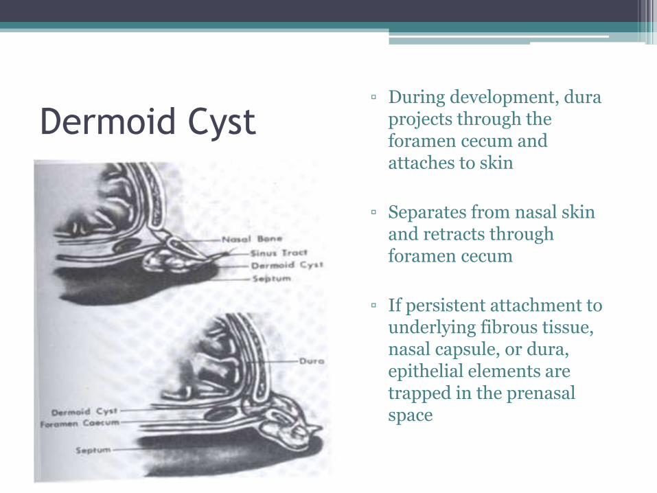
▫ During development, duraprojects through the foramen cecum and attaches to skin
▫ Separates from nasal skin and retracts through foramen cecum
▫ If persistent attachment to underlying fibrous tissue, nasal capsule, or dura, epithelial elements are trapped in the prenasalspace
Embryology – Abnormal Development
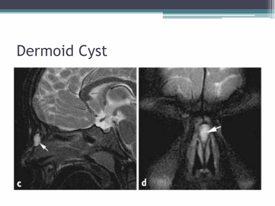
Dermoid Cyst
Dermoid Cyst
Nasoglabellar Cysts
▫ Up to 50% may have a fistula or sinus tract
▫ Tract traverses via the cribiform plate or foramen cecum
▫ Tract attaches to dura, falx cerebri, or other intracranial structures
▫ Diff Dx: Nasolabial cyst, premaxillary cyst, nasopalatine cyst, Jacobson organ cyst
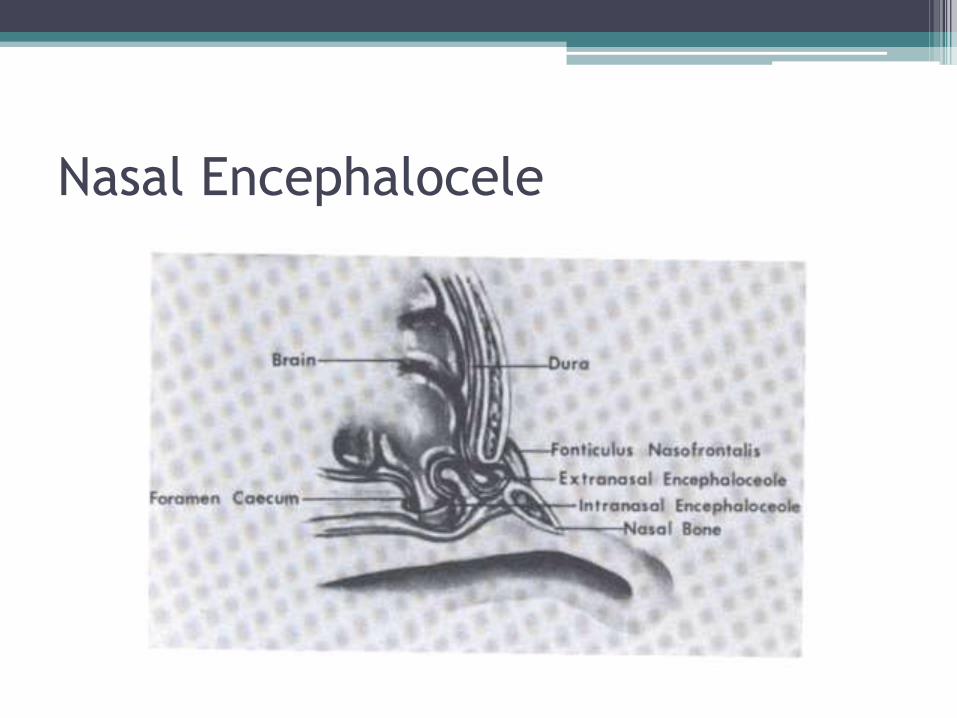
Nasal Encephalocele
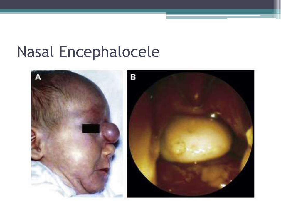
• Extracranial protrusions of meninges, CSF fluid, and neural tissue
• Meningoceles present similarly without herniation of brain tissue
• May present as external or internal nasal masses
• Described by location of dehiscence in the skull base
Nasal Encephalocele
• Presentation:
▫ Pale
▫ Compressible
▫ Pulsatile
▫ Transilluminate with light
▫ Positive Furstenberg’s Test (expansion with compression of Jugular Vein)
▫ Expansion may also be triggered by crying or straining
Nasal Encephalocele
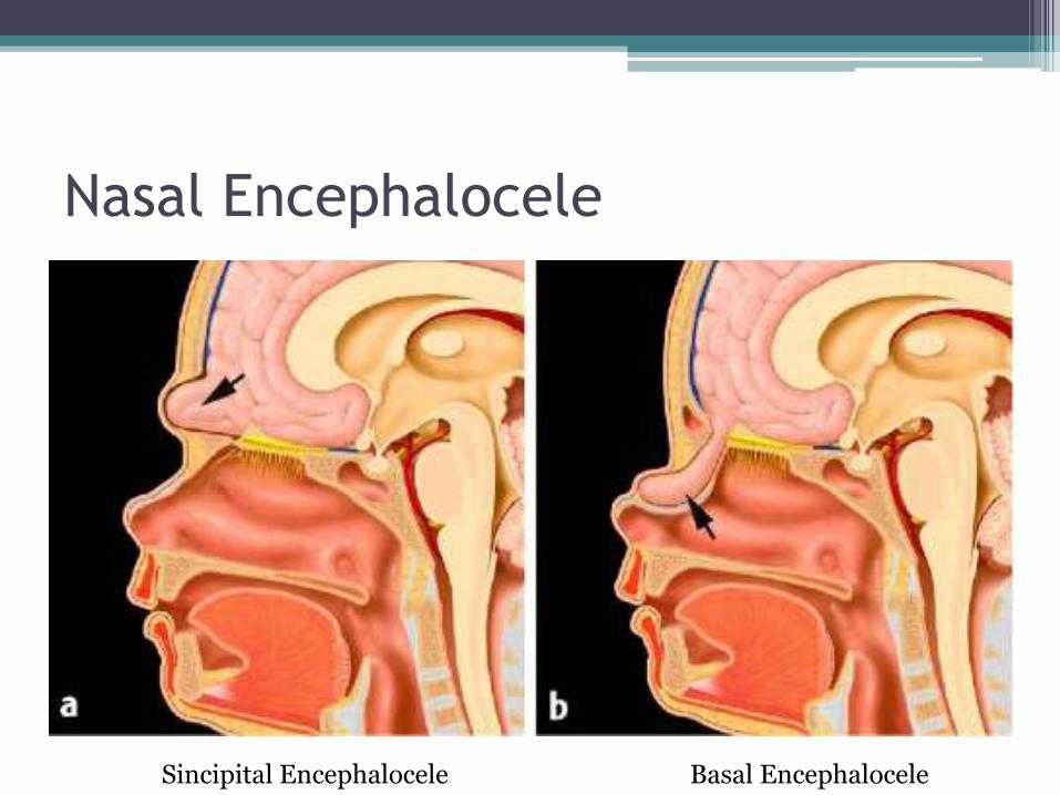
Nasal Encephalocele
Sincipital Encephalocele Basal Encephalocele
Nasal Encephalocele
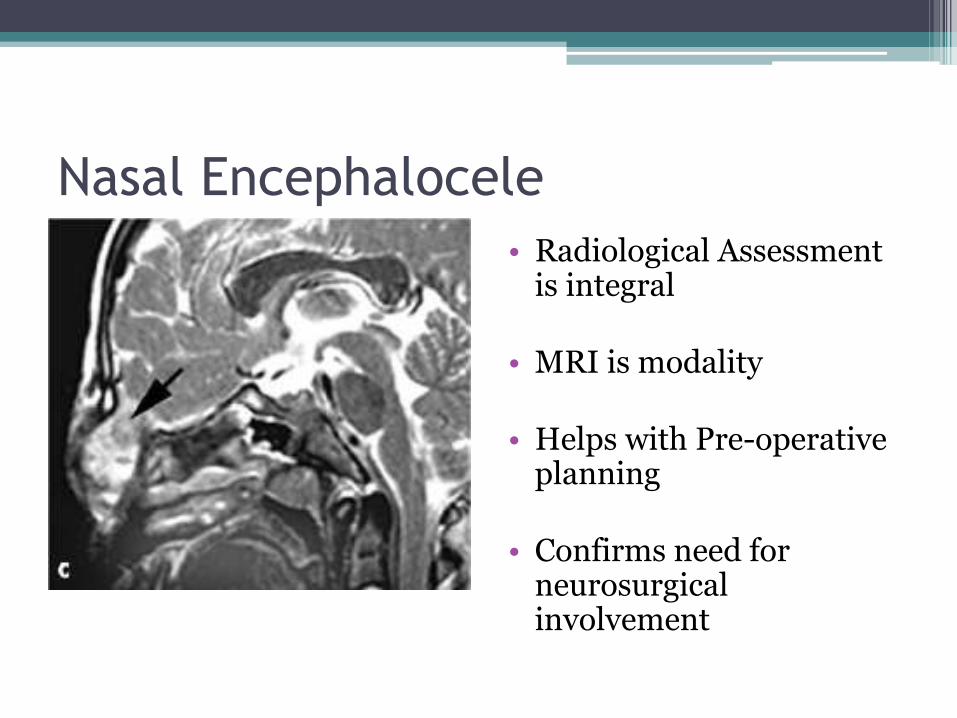
Nasal Encephalocele
• Radiological Assessment is integral
• MRI is modality
• Helps with Pre-operative planning
• Confirms need for neurosurgical involvement
Nasal Glioma
• Encephalocele without intracranial connection
• Also known as:
▫ Benign congenital nasal neuroectodermal tumor
▫ Nasal cerebral heterotopia
▫ Glial heterotopia
Nasal Glioma
• Hypotheses on formation
▫ Develop from extracranial rests of glial tissue
▫ Abnormal closure of fonticulus nasofrontalis
▫ Another theory is that they are possibly encephaloceles which have lost CSF connection
Nasal Glioma
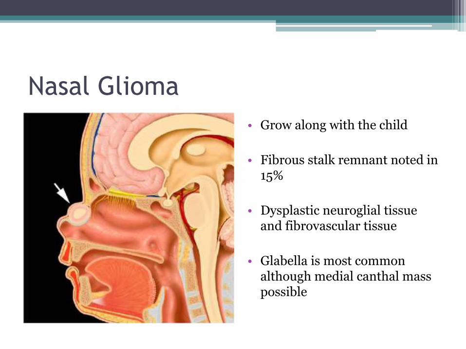
• Grow along with the child
• Fibrous stalk remnant noted in 15%
• Dysplastic neuroglial tissue and fibrovascular tissue
• Glabella is most common although medial canthal mass possible
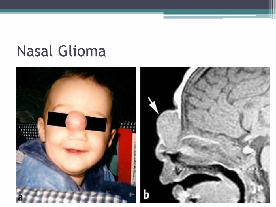
Nasal Glioma
• Most identified at birth or in infancy
• Appear well-circumscribed, bluish or reddish in color, and telangiectatic surface
• Sincipital vs Basal
• 30% have intranasal presentations
▫ Lateral Nasal Wall
▫ Middle Turbinate
▫ Nasal Septum
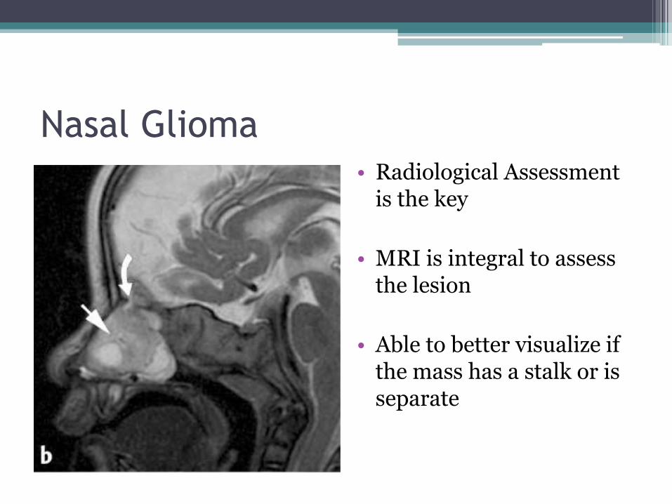
Nasal Glioma• Radiological Assessment
is the key
• MRI is integral to assess the lesion
• Able to better visualize if the mass has a stalk or is separate
Nasal Glioma
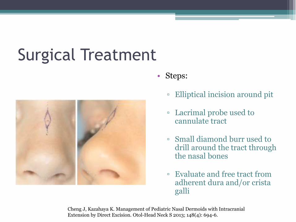
Surgical Treatment• Steps:
▫ Elliptical incision around pit
▫ Lacrimal probe used to cannulate tract
▫ Small diamond burr used to drill around the tract through the nasal bones
▫ Evaluate and free tract from adherent dura and/or cristagalli
Cheng J, Kazahaya K. Management of Pediatric Nasal Dermoids with Intracranial Extension by Direct Excision. Otol-Head Neck S 2013; 148(4): 694-6.
Surgical Treatment
• For Gliomas and Encephaloceles:
▫ Nasal Evaluation and Pre-operative imaging
▫ Entities involving the dura or intracranial contents mandate neurosurgical evaluation
▫ Lesions restricted to the nasal cavity may be removed endoscopically or via a lateral rhinotomyapproach
Duncan NO. Combined Approach for Complete Excision of Congenital Nasal Masses in Children. Oper Tech Head Neck S 1994; 5(1): 18-21.
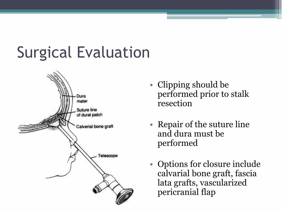
Surgical Evaluation
• Clipping should be performed prior to stalk resection
• Repair of the suture line and dura must be performed
• Options for closure include calvarial bone graft, fascia lata grafts, vascularizedpericranial flap

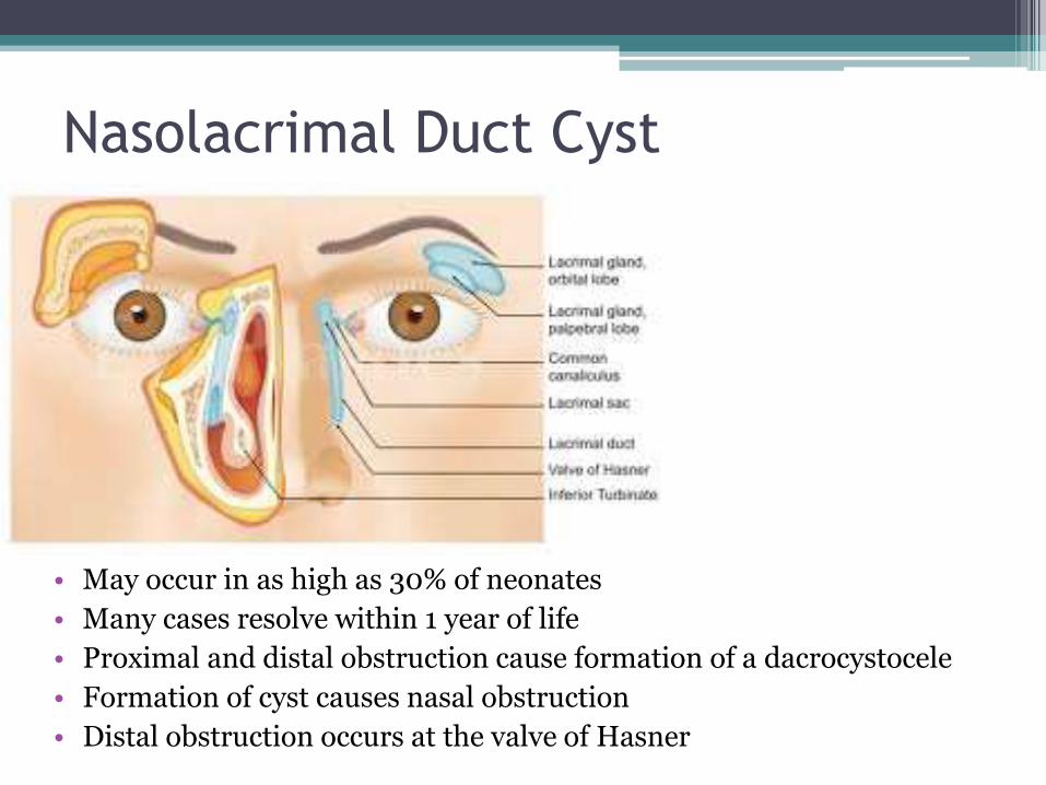
Nasolacrimal Duct Cyst
• May occur in as high as 30% of neonates
• Many cases resolve within 1 year of life
• Proximal and distal obstruction cause formation of a dacrocystocele
• Formation of cyst causes nasal obstruction
• Distal obstruction occurs at the valve of Hasner
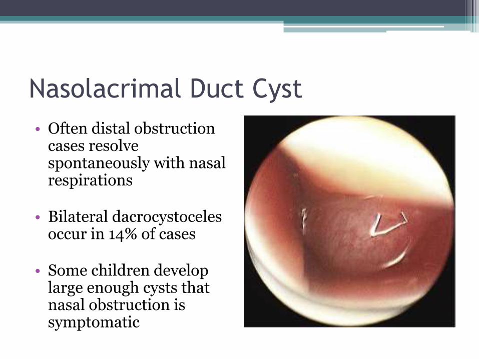
Nasolacrimal Duct Cyst
• Often distal obstruction cases resolve spontaneously with nasal respirations
• Bilateral dacrocystocelesoccur in 14% of cases
• Some children develop large enough cysts that nasal obstruction is symptomatic
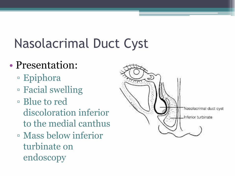
Nasolacrimal Duct Cyst
• Presentation:▫ Epiphora
▫ Facial swelling
▫ Blue to red discoloration inferior to the medial canthus
▫ Mass below inferior turbinate on endoscopy
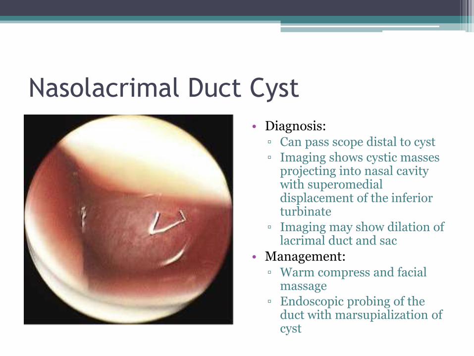
Nasolacrimal Duct Cyst
• Diagnosis:▫ Can pass scope distal to cyst
▫ Imaging shows cystic masses projecting into nasal cavity with superomedialdisplacement of the inferior turbinate
▫ Imaging may show dilation of lacrimal duct and sac
• Management:▫ Warm compress and facial
massage
▫ Endoscopic probing of the duct with marsupialization of cyst
Thornwaldt Cyst
• Named for Gustav Ludwig Thornwaldt▫ 1885 – series of 26 cases
• Embryology:▫ Congenital cyst in nasopharynx▫ Results from a communication between the
notochord and the nasopharyngeal endoderm▫ Incidence - 3-4% (slight male predilection)▫ Diagnosed in 2nd and 3rd decades of life▫ Triggered by infection, inflammation, complicated
adenoidectomy
Yuca K, Varsak YK. Thornwaldt’s Cyst. Eur J Gen Med 2012; 9(Suppl 1) 26-29
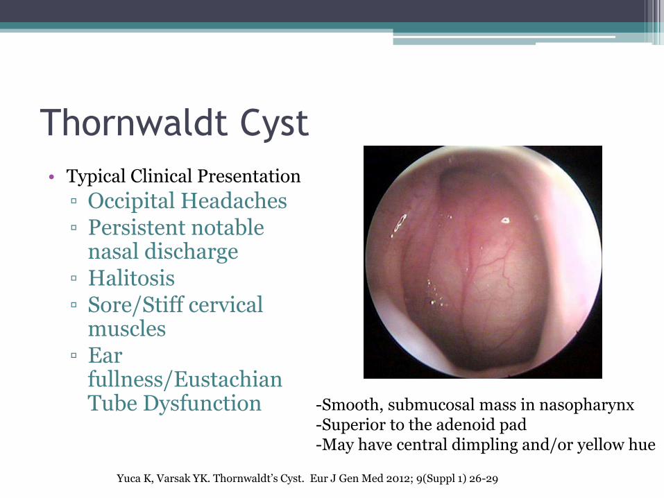
Thornwaldt Cyst
• Typical Clinical Presentation
▫ Occipital Headaches▫ Persistent notable
nasal discharge▫ Halitosis▫ Sore/Stiff cervical
muscles▫ Ear
fullness/Eustachian Tube Dysfunction -Smooth, submucosal mass in nasopharynx
-Superior to the adenoid pad-May have central dimpling and/or yellow hue
Yuca K, Varsak YK. Thornwaldt’s Cyst. Eur J Gen Med 2012; 9(Suppl 1) 26-29
Thornwaldt Cyst
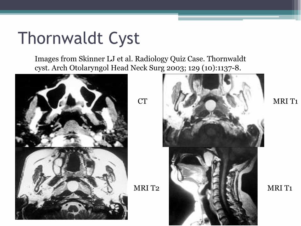
CT MRI T1
MRI T2 MRI T1
Images from Skinner LJ et al. Radiology Quiz Case. Thornwaldtcyst. Arch Otolaryngol Head Neck Surg 2003; 129 (10):1137-8.
Thornwaldt Cyst
• Work-Up:▫ History + Physical Exam + Imaging
• Differential Diagnosis:▫ Rathke pouch cyst▫ Adenoid Retention cyst▫ Sphenoid sinus mucocele▫ Nasopharyngeal CA
• Treatment:▫ Observation if asymptomatic▫ Marsupialization or resection
Yuca K, Varsak YK. Thornwaldt’s Cyst. Eur J Gen Med 2012; 9(Suppl 1) 26-29

Neonatal Nasal Tumors
• Congenital Tumors – 2-14 cases per 100,000 births
• Differential Diagnosis:
▫ Teratoma
▫ Hamartoma
▫ Rhabdomyosarcoma
▫ Hemangioma
▫ Neurofibroma
▫ Lymphatic Malformations
Nasal Obstruction and Masses
• Neonates are obligate nasal breathers
• Radiological findings help provide diagnosis
• MRI preferred over CT especially for intracranial assessment