Congenital epulis: A rare benign tumour - e-mjm.org · histomorphology. In congenital epulis,...
2
300 Med J Malaysia Vol 71 No 5 October 2016 SUMMARY Congenital epulis is a rare benign pedunculated tumour of the oral cavity arising from the alveolar ridges. It is usually detected in newborns and can be successfully resected surgically. We report a case of a newborn baby who had a 5x3x3cm pedunculated lobar mass arising from the upper alveolar ridge. KEY WORDS: Congenital epulis; newborn; benign tumour INTRODUCTION Congenital epulis is a rare benign tumour arising from the alveolar ridges of newborns. They are mainly composed of mesenchymal tissue and are usually surgically resected. They can pose quite a surprise to clinicians’ when presented at childbirth, as it is usually quite large and difficult to detect through prenatal ultrasound. 1 In large tumours, it can cause problems with swallowing, breastfeeding and respiratory compromise. Treatment is generally straightforward, the recovery period is unremarkable and the risk of recurrence is low. 2 CASE REPORT An otherwise healthy newborn girl was referred immediately after birth for a large pedunculated mass protruding from her oral cavity. The baby was born at term via caesarean section weighing 2.7kg and was breathing spontaneously with no distress. She had normal antenatal scans. Physical examination revealed a 5x3x3cm pedunculated mass arising just right of midline of the upper alveolar ridge. The mass was not obstructing the airway but did interfere with breastfeeding (Figure 1a). There were no dysmorphic features of note. As there were no other medical or congenital problems detected, a decision was made to excise this mass before allowing the patient to be discharged home. This was performed under local anesthesia in the operating theatre. After injecting 0.5% Marcain (with 1:100000 adrenaline) into the stalk, the lesion was ligated with a 2/0 silk suture and the mass was excised via monopolar diarthermy (Figure 1b). No feeding vessel was noted in the stalk. Hemorrhage was minimal. Postoperative recovery was uneventful. The patient was breastfeeding immediately after and discharged the next day. Follow-up at two weeks revealed a pristine looking alveolar bed with no evidence of the stalk. The patient was noted to be feeding well, thriving and gaining weight at the six-month mark. Histological examination revealed an encapsulated lesion covered with squamous epithelium macroscopically (Figure 2a). Microscopically there was evidence of granular eosinophilic and basophilic cytoplasm centrally located nuclei consistent with findings of a congenital epulis (Figure 2b). DISCUSSION Congenital epulis was first decribed by Neumann in 1871 and is a rare benign tumour of the jaw that occurs exclusively in neonates. Epulis literally means “of the gums” in greek and the tumour usually arises from the gingiva of the future site of the maxillary canine or the lateral incisors, but the unerupted teeth are not involved. 2 The lesion occurs in females more than males with a ratio of 10:1 and is more common in the maxilla than the mandible (3:1). 3 It usually presents as a pedicled smooth lobulated tumour with a firm consistency over the future site of the canine-incisor area.1 Congenital epulis is reported to be an isolated finding without associated congenital abnormalities. 4 It appears sporadically with no familial tendency and has an incidence rate of about 6 per million. 2 Diagnosis/differentials Differential diagnosis depends on site, size and velocity of growth. It is broad and based on clinical perspective only. These diagnoses could include congenital malformations, such as cephalocele and dermoid, as well as benign and malignant neoplasms, such as lymphatic malformation and rhabdomyosarcoma or non-specific soft tissue masses such as a fibroma or a vascular malformation or other developmental anomalies such as granular cell tumours (GCTs) or neuroectodermal tumours of infancy. 1, 4 Imaging The tumour can be diagnosed by prenatal ultrasound or with the more superior magnetic resonance imaging (MRI) as early as at 26 weeks of gestation or in third trimester of Congenital epulis: A rare benign tumour Danny Kit Chung Wong, MBChB 1 , Roszalina Ramli, FFDRCS 2 , Muhaizan Wan Mustaffa, MPath 3 , Primuharsa Putra Sabir Husin Athar, MSurg 4 1 ORL-HNS Registrar, KPJ Healthcare University College, Negeri Sembilan, Malaysia, 2 Department of Oral and Maxillofacial Surgery, Faculty of Dentistry, Universiti Kebangsaan Malaysia, Kuala Lumpur, Malaysia, 3 Consultant Pathologist, KPJ Lablink, Kuala Lumpur, Malaysia, 4 ORL-HNS Consultant, KPJ Seremban Specialist Hospital/ KPJ Healthcare University College, Negeri Sembilan, Malaysia CASE REPORT This article was accepted: 1 July 2016 Corresponding Author: Primuharsa Putra Sabir Husin Athar, Consultant ENT-Head & Neck Surgeon, Ear, Nose & Throat-Head & Neck Consultant Clinic, KPJ Seremban Specialist Hospital, Jalan Toman 1, Kemayan Square, 70200 Seremban, Negeri Sembilan Darul Khusus, Malaysia Email: [email protected]
Transcript of Congenital epulis: A rare benign tumour - e-mjm.org · histomorphology. In congenital epulis,...

300 Med J Malaysia Vol 71 No 5 October 2016
SUMMARYCongenital epulis is a rare benign pedunculated tumour ofthe oral cavity arising from the alveolar ridges. It is usuallydetected in newborns and can be successfully resectedsurgically. We report a case of a newborn baby who had a5x3x3cm pedunculated lobar mass arising from the upperalveolar ridge.
KEY WORDS:Congenital epulis; newborn; benign tumour
INTRODUCTIONCongenital epulis is a rare benign tumour arising from thealveolar ridges of newborns. They are mainly composed ofmesenchymal tissue and are usually surgically resected. Theycan pose quite a surprise to clinicians’ when presented atchildbirth, as it is usually quite large and difficult to detectthrough prenatal ultrasound.1 In large tumours, it can causeproblems with swallowing, breastfeeding and respiratorycompromise. Treatment is generally straightforward, therecovery period is unremarkable and the risk of recurrence islow.2
CASE REPORTAn otherwise healthy newborn girl was referred immediatelyafter birth for a large pedunculated mass protruding from heroral cavity. The baby was born at term via caesarean sectionweighing 2.7kg and was breathing spontaneously with nodistress. She had normal antenatal scans.
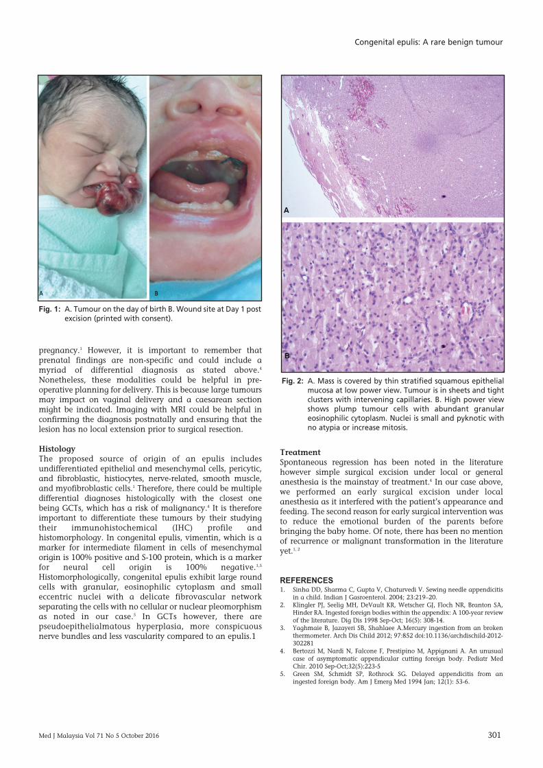
Physical examination revealed a 5x3x3cm pedunculatedmass arising just right of midline of the upper alveolar ridge.The mass was not obstructing the airway but did interferewith breastfeeding (Figure 1a). There were no dysmorphicfeatures of note.
As there were no other medical or congenital problemsdetected, a decision was made to excise this mass beforeallowing the patient to be discharged home. This wasperformed under local anesthesia in the operating theatre.After injecting 0.5% Marcain (with 1:100000 adrenaline) intothe stalk, the lesion was ligated with a 2/0 silk suture and themass was excised via monopolar diarthermy (Figure 1b). Nofeeding vessel was noted in the stalk. Hemorrhage wasminimal. Postoperative recovery was uneventful. The patient
was breastfeeding immediately after and discharged the nextday.
Follow-up at two weeks revealed a pristine looking alveolarbed with no evidence of the stalk. The patient was noted to befeeding well, thriving and gaining weight at the six-monthmark.
Histological examination revealed an encapsulated lesioncovered with squamous epithelium macroscopically (Figure2a). Microscopically there was evidence of granulareosinophilic and basophilic cytoplasm centrally locatednuclei consistent with findings of a congenital epulis (Figure2b).
DISCUSSIONCongenital epulis was first decribed by Neumann in 1871and is a rare benign tumour of the jaw that occurs exclusivelyin neonates. Epulis literally means “of the gums” in greekand the tumour usually arises from the gingiva of the futuresite of the maxillary canine or the lateral incisors, but theunerupted teeth are not involved. 2 The lesion occurs infemales more than males with a ratio of 10:1 and is morecommon in the maxilla than the mandible (3:1).3 It usuallypresents as a pedicled smooth lobulated tumour with a firmconsistency over the future site of the canine-incisor area.1Congenital epulis is reported to be an isolated findingwithout associated congenital abnormalities.4 It appearssporadically with no familial tendency and has an incidencerate of about 6 per million.2
Diagnosis/differentialsDifferential diagnosis depends on site, size and velocity ofgrowth. It is broad and based on clinical perspective only.These diagnoses could include congenital malformations,such as cephalocele and dermoid, as well as benign andmalignant neoplasms, such as lymphatic malformation andrhabdomyosarcoma or non-specific soft tissue masses such asa fibroma or a vascular malformation or otherdevelopmental anomalies such as granular cell tumours(GCTs) or neuroectodermal tumours of infancy.1, 4
ImagingThe tumour can be diagnosed by prenatal ultrasound or withthe more superior magnetic resonance imaging (MRI) asearly as at 26 weeks of gestation or in third trimester of
Congenital epulis: A rare benign tumour
Danny Kit Chung Wong, MBChB1, Roszalina Ramli, FFDRCS2, Muhaizan Wan Mustaffa, MPath3, PrimuharsaPutra Sabir Husin Athar, MSurg4
1ORL-HNS Registrar, KPJ Healthcare University College, Negeri Sembilan, Malaysia, 2Department of Oral and MaxillofacialSurgery, Faculty of Dentistry, Universiti Kebangsaan Malaysia, Kuala Lumpur, Malaysia, 3Consultant Pathologist, KPJ Lablink,Kuala Lumpur, Malaysia, 4ORL-HNS Consultant, KPJ Seremban Specialist Hospital/ KPJ Healthcare University College, NegeriSembilan, Malaysia
CASE REPORT
This article was accepted: 1 July 2016Corresponding Author: Primuharsa Putra Sabir Husin Athar, Consultant ENT-Head & Neck Surgeon, Ear, Nose & Throat-Head & Neck Consultant Clinic,KPJ Seremban Specialist Hospital, Jalan Toman 1, Kemayan Square, 70200 Seremban, Negeri Sembilan Darul Khusus, Malaysia Email: [email protected]
18-Congenital00033_3-PRIMARY.qxd 1/4/17 2:01 PM Page 300
Congenital epulis: A rare benign tumour
Med J Malaysia Vol 71 No 5 October 2016 301
pregnancy.1 However, it is important to remember thatprenatal findings are non-specific and could include amyriad of differential diagnosis as stated above.4
Nonetheless, these modalities could be helpful in pre-operative planning for delivery. This is because large tumoursmay impact on vaginal delivery and a caesarean sectionmight be indicated. Imaging with MRI could be helpful inconfirming the diagnosis postnatally and ensuring that thelesion has no local extension prior to surgical resection.
HistologyThe proposed source of origin of an epulis includesundifferentiated epithelial and mesenchymal cells, pericytic,and fibroblastic, histiocytes, nerve-related, smooth muscle,and myofibroblastic cells.1 Therefore, there could be multipledifferential diagnoses histologically with the closest onebeing GCTs, which has a risk of malignancy.4 It is thereforeimportant to differentiate these tumours by their studyingtheir immunohistochemical (IHC) profile andhistomorphology. In congenital epulis, vimentin, which is amarker for intermediate filament in cells of mesenchymalorigin is 100% positive and S-100 protein, which is a markerfor neural cell origin is 100% negative.1,5
Histomorphologically, congenital epulis exhibit large roundcells with granular, eosinophilic cytoplasm and smalleccentric nuclei with a delicate fibrovascular networkseparating the cells with no cellular or nuclear pleomorphismas noted in our case.5 In GCTs however, there arepseudoepithelialmatous hyperplasia, more conspicuousnerve bundles and less vascularity compared to an epulis.1
TreatmentSpontaneous regression has been noted in the literaturehowever simple surgical excision under local or generalanesthesia is the mainstay of treatment.4 In our case above,we performed an early surgical excision under localanesthesia as it interfered with the patient’s appearance andfeeding. The second reason for early surgical intervention wasto reduce the emotional burden of the parents beforebringing the baby home. Of note, there has been no mentionof recurrence or malignant transformation in the literatureyet.1, 2
REFERENCES1. Sinha DD, Sharma C, Gupta V, Chaturvedi V. Sewing needle appendicitis
in a child. Indian J Gasroenterol. 2004; 23:219–20. 2. Klingler PJ, Seelig MH, DeVault KR, Wetscher GJ, Floch NR, Branton SA,
Hinder RA. Ingested foreign bodies within the appendix: A 100-year reviewof the literature. Dig Dis 1998 Sep-Oct; 16(5): 308-14.
3. Yaghmaie B, Jazayeri SB, Shahlaee A.Mercury ingestion from an brokenthermometer. Arch Dis Child 2012; 97:852 doi:10.1136/archdischild-2012-302281
4. Bertozzi M, Nardi N, Falcone F, Prestipino M, Appignani A. An unusualcase of asymptomatic appendicular cutting foreign body. Pediatr MedChir. 2010 Sep-Oct;32(5):223-5
5. Green SM, Schmidt SP, Rothrock SG. Delayed appendicitis from aningested foreign body. Am J Emerg Med 1994 Jan; 12(1): 53-6.
Fig. 1: A. Tumour on the day of birth B. Wound site at Day 1 postexcision (printed with consent).
Fig. 2: A. Mass is covered by thin stratified squamous epithelialmucosa at low power view. Tumour is in sheets and tightclusters with intervening capillaries. B. High power viewshows plump tumour cells with abundant granulareosinophilic cytoplasm. Nuclei is small and pyknotic withno atypia or increase mitosis.
18-Congenital00033_3-PRIMARY.qxd 1/4/17 2:01 PM Page 301