Congenital and Acquired Immunodeficiency Diseases (not...
46
Congenital and Acquired Immunodeficiency Diseases (not HIV) May 6, 2009
Transcript of Congenital and Acquired Immunodeficiency Diseases (not...

Congenital and Acquired Immunodeficiency Diseases
(not HIV)May 6, 2009

IMMUNODEFICIENCY DISEASES
Defects in one or more components of the immune system can lead to serious and often fatal disorders collectively known as immunodeficiency diseases.
Primary immunodeficiency diseases - defects in genes for components of the immune system.
Secondary immunodeficiency diseases - due to factors that have an adverse impact on the immune system.

If primary immune deficiency diseases are genetic, why are some of these diseases first
diagnosed in adults?
• Compensating immune functionskeep serious problems from developing earlier.
OR• Slowly deteriorating immune
function, genetically determined, that does not become significant until later in life.

Patients with immunodeficiency diseases are most often recognized because of an increased susceptibility to infections.
1. Chronic/recurrent infections w/o other explanations.2. Infections with organisms of low virulence.3. Infections of unusual severity.
Immunodeficiency diseases may also present with non-infectious manifestations such as autoimmune diseases.

THERAPY
• Gamma globulin - fraction of blood that contains Igs. Pooled from 2,000 - 10,000 donors. Contains abs to many different ags.
• Bone marrow transplantation - requires a good match (sibling) - removed from pelvic bones
• Antibiotics - supportive• Cytokines• Gene therapy

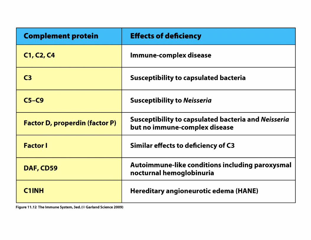
Complement deficiencies

Evaluation of the Components of the Human Immune System
• Differential cell counts/FACS analysis• B cell function
– In vivo: serum Ig levels, specific ab levels– In vitro: mitogen-induced ab production
• T cell function– In vivo: skin test– In vitro: T cell proliferation in response to
mitogens• Phagocytes: NBT test, intracellular killing of
bacteria• Complement: dilution of serum required to
lyse 50% of antibody-coated rbc or ELISA

SCID (Severe Combined Immunodeficiency Disease)/Defects in T
Cell Function• Defects in lymphoid development affecting T cells alone or with
B cells & NK cells.• Thymus does not develop. Few circulating T cells. Defective T
cell function - may extend to B cells & NK cells.• Usually presents in infancy.
– Failure to thrive– Fungal or viral infections - skin, mouth, and throat lesions -
pneumonia– Chronic diarrhea
• ~50% of cases are due to deficiency of the common gamma chain of the IL-2 receptor (“boy in the bubble”)

David Vetter (9/21/71-2/22/84)
• Lived in a plastic bubble - sterile environment - began 20 seconds after birth
• 1977 - NASA developed the Mobile Biological Isolation System - allowed David to venture outside the bubble
• 1983 - given bone marrow from sister - less than perfect match but expected to work with new methods using unmatched marrow
• Transplant seemed to work at first.• Transplant harbored EB virus - David died of
Burkitt’s lymphoma
1976 made-for-television movie starred John Travolta

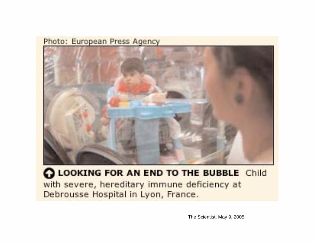
The Scientist, May 9, 2005

NK cells are present

Roitt et al, 19.9
ADA = adenosinedeaminaseT, B, NK cells
are affectedaccumulation
of’ adenosine
PNP = purine nucleosidephosphorylase
Treatment : bm transplant,PEG-ADA, gene therapy(retrovirus vectors)

• Wiskott -Aldrich syndrome (WAS)• Bare lymphocyte syndrome (MHC class II)• Bare lymphocyte syndrome (MHC class I)• ZAP-70 (signal transduction defect)• RAG mutations (cell development defect)

Wiskott Aldrich Syndrome• X-linked disease characterized by:
– eczema– thrombocytopenia – susceptibility to bacterial infection
• Defective gene encodes for CD43 (sialophorin) required for cytoskeletal reorganization - needed for T cells to deliver cytokines & signals - cell counts are normal
• Poor antibody response - gradual loss of humoral and CMI responses
www.uv.es

• Wiskott -Aldrich syndrome (WAS)• Bare lymphocyte syndrome (MHC class II)• Bare lymphocyte syndrome (MHC class I)• ZAP-70 (signal transduction defect)• RAG mutations (cell development defect)
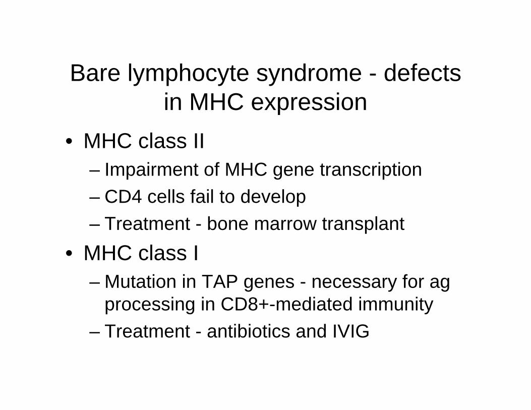
Bare lymphocyte syndrome - defects in MHC expression
• MHC class II– Impairment of MHC gene transcription– CD4 cells fail to develop– Treatment - bone marrow transplant
• MHC class I– Mutation in TAP genes - necessary for ag
processing in CD8+-mediated immunity– Treatment - antibiotics and IVIG

• Wiskott -Aldrich syndrome (WAS)• Bare lymphocyte syndrome (MHC class II)• Bare lymphocyte syndrome (MHC class I)• ZAP-70 (T cell signal transduction defect)• JAK3 (signal transduction defect)• RAG mutations (cell development defect) -
both T and B cells• Artemis - X-linked SCIDs with radiation
sensitivity

QuickTime™ and a decompressor
are needed to see this picture.
Cuvelier et al. Clin. Immunol. 131(2):179-188,2009
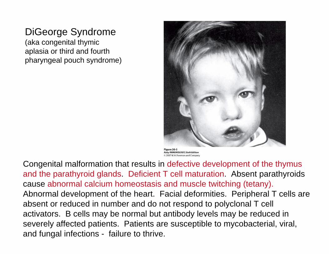
Congenital malformation that results in defective development of the thymusand the parathyroid glands. Deficient T cell maturation. Absent parathyroids cause abnormal calcium homeostasis and muscle twitching (tetany).Abnormal development of the heart. Facial deformities. Peripheral T cells are absent or reduced in number and do not respond to polyclonal T cell activators. B cells may be normal but antibody levels may be reduced in severely affected patients. Patients are susceptible to mycobacterial, viral, and fungal infections - failure to thrive.
DiGeorge Syndrome(aka congenital thymicaplasia or third and fourthpharyngeal pouch syndrome)

Humoral Deficiencies
X-linked agammaglobulinemiaX-linked hyper IgM syndromeSelective Ig deficienciesCVID


X-linked agammaglobulinemia(aka Bruton’s agammaglobulinemia)
• Characterized by: – Low levels or absence of gamma globulin in the
blood– Reduced or absent B cells in the peripheral blood
and lymphoid tissues– No germinal centers in lymph nodes– No plasma cells– Maturation, numbers and functions of T cells are
usually normal– Autoimmune diseases develop in ~20% of patients
- reason(s) unknown• Failure of B cells to mature beyond the pre-B
cell stage in the bone marrow because of mutations or deletions in the gene encoding B cell tyrosine kinase (Btk).

X-linked hyper-IgM syndrome (XHM)• Deficiency of IgG, IgA, and IgE - elevated
levels of IgM• Normal numbers of B cells• Both X-linked & acquired• Autoantibodies to PMNs, platelets, & rbc• Failure to produced germinal centers• Defect is in gene encoding CD40L (CD154)
required for B cell responses to T-dependent antigens. Class switching and memory B cells are not formed.

Selective Ig deficiencies
• IgA deficiency is most common– Symptoms range from unnoticed to various
problems including recurrent respiratory and urogenital tract infections
– Other problems: intestinal malabsorption, allergic disease, autoimmune disorders
– Some pts can substitute IgM for IgA as a mucosalantibody
• IgM deficiency rare autosomal recessive -severe infections, malignancies, autoimmune diseases
• IgG deficiency - rare - may be unnoticed till adulthood - treatment is Ig administration

Combined variable immunodeficiency disease (CVID)
• Often shows up later in life• Decrease in numbers of plasma cells -
therefore reduced serum levels of IgG, IgA, and often IgM - recurrent infections
• Diagnosis is made by exclusion of other causes for ab deficiency
• Some cases are sporadic but some are familial - may be due to genuine B cell defects presumably at the stage where B cells become plasma cells - mutations in TACl, a member of the TNFR family have been identified

Combined Deficiencies(defects of more than one lineage)
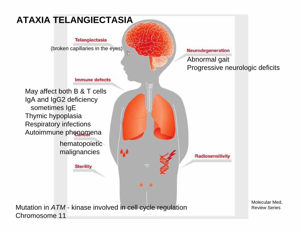
Molecular Med. Review SeriesMutation in ATM - kinase involved in cell cycle regulation
Chromosome 11
Abnormal gaitProgressive neurologic deficits
May affect both B & T cellsIgA and IgG2 deficiency
sometimes IgEThymic hypoplasiaRespiratory infectionsAutoimmune phenomena
hematopoietic malignancies
ATAXIA TELANGIECTASIA
(broken capillaries in the eyes)

Disorders of Myeloid Cells


Chronic granulomatous disease (CGD)
• X-linked (70%) and AR (30%) forms• Defect in pathway that produces hydrogen
peroxide and reactive products that killphagocytosed bacteria (missing or defective cytochrome b558) - also decrease in mononuclear cell ability to process & present antigen
• Excessive inflammatory reactions leading to gingivitis, swollen lymph nodes, and nonmalignant granulomas - also bacterial and fungal infections
• IFN-γ treatment has been successful - gene therapy is also promising

Necrotizing granulomatouslesions

Roitt et al, 19.14
NBT Test

Leukocyte Adhesion Deficiency -LAD1
imgt.cines.fr
Absence of ß2
Defective leukocyte migration toinflammatory sites
Defective T and NK cell cytotoxicityVariable disease phenotypes

Leukocyte Adhesion Deficiency - LAD 2Failure to convert GDP mannose to fucose - missing surface Sialyl Lewisx
which binds to P- and E-selectin on endotheliumCharacterized by delayed wound healing, chronic skin ulcers, periodontitis,mental retardation
www.mpi-muenster.mpg.de/ nvz/wilde.shtml
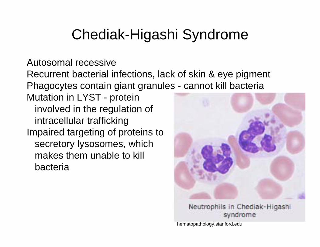
Chediak-Higashi Syndrome
hematopathology.stanford.edu
Autosomal recessiveRecurrent bacterial infections, lack of skin & eye pigmentPhagocytes contain giant granules - cannot kill bacteriaMutation in LYST - protein
involved in the regulation ofintracellular trafficking
Impaired targeting of proteins tosecretory lysosomes, whichmakes them unable to killbacteria
Roitt et al, 19.12
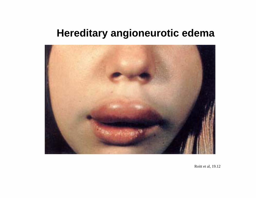
Hereditary angioneurotic edema

Recently discovered immunodeficiency diseases:
• UNC-93B - essential for the normal response to signaling through TLRs that respond to viral infection (TLR3, TLR7, TLR8, and TLR9). Deficiency results in reduced production of IFNα, IFNß, IFNλ, TNFα, IL-1ß, and IL-6 in response to stimulation.
• IFNγR - persistent mycobacterial infections• IPEX Syndrome - development of systemic autoimmunity
in first year of life - most common symptoms are watery diarrhea, eczema, insulin-dependent diabetes. FOXP3 is the only gene associated with the IPEX so far (~60%). Elevated IgE (other Igs are normal), autoantibodies to pancreatic islet ags, thyroid ags, small bowel mucosa. Autoimmune anemia, low platelets &/or PMNs

Secondary (acquired) immunodeficiency diseases
Protein-calorie malnutrition
Irradiation and chemotherapy for cancer
Cancer metastases to bone
Immunosuppressive drugs
Removal of spleen
Infection with HIV

Protein-calorie malnutrition• Increases susceptibility to infectious disease -
decreases all aspects of immune protection– Reduced production of IL-2 and IFN-γ and an
increased production of IL-4 and IL-10. Decrease in activation antigens (CD69 and CD25)
• Minerals and cofactors - transcriptional regulation of immune maturation
• Specific protein, vitamin and lipid needs• Increased demand for antioxidant protection
and tissue repair

Irradiation and chemotherapy
• Cytotoxic drugs enhance the susceptibility to infection– leukopenia (opportunistic infections) and thrombocytopenia– decreased cell-mediated immunity
• Neutropenia (leukemia, immunosuppression or irradiation) - patients develop gram-negative bacteremia from infections acquired through the mucous membranes or secondary to pneumonia
• Severely immunosuppressed patients, patients with Hodgkin's disease and HIV - depressed cellular immune mechanisms - serious infections with mycobacteria, Aspergillus, Candida, Cryptococcus,Histoplasma, Mucor, Nocardia, or Staphylococcus are frequent. Also Herpes zoster, cytomegalovirus, Pneumocystis, andToxoplasma infections
Cancer metastases to bone
• Competes with development of progenitor cells in the bone marrow.

Immunosuppressive drugs
• Immunosuppressive drugs - autoimmune diseases, prevent transplant rejection - interfere with lymphocytes function - not specific and suppress all of the immune system - leave the patient vulnerable to a variety of opportunistic infections and complications
• Example: corticosteroids inhibit the movement ofneutrophils, monocytes, and lymphocytes into inflammatory sites - patients have increased susceptibility to infection from both usual and unusual bacteria.

Removal of spleen• Functions of the spleen:
– Removal of unwanted elements from the blood– Major secondary organ of the immune system (DCs
trap antigens and present them to T cells).– Source of hematopoietic cells in cases of severe
anemia.– Can serve as a sequestering organ for blood
elements• Single major clinical manifestation: increased
susceptibility to disseminated infection with encapsulated bacteria (pneumococcus, meningococcus, Haemophilus influenzae) -probably due to the reduced filtering and antibody production