Conformationaldynamicsofthemolecular · PDF fileConformationaldynamicsofthemolecular...
27
Conformational dynamics of the molecular chaperone Hsp90 Kristin A. Krukenberg 1,3 , Timothy O. Street 1 , Laura A. Lavery 1 and David A. Agard 1,2 * 1 Department of Biochemistry and Biophysics, University of California, San Francisco, CA, USA 2 The Howard Hughes Medical Institute, University of California, San Francisco, CA, USA 3 Present address : Department of Systems Biology, Harvard Medical School, Boston, MA, USA Abstract. The ubiquitous molecular chaperone Hsp90 makes up 1–2 % of cytosolic proteins and is required for viability in eukaryotes. Hsp90 affects the folding and activation of a wide variety of substrate proteins including many involved in signaling and regulatory processes. Some of these substrates are implicated in cancer and other diseases, making Hsp90 an attractive drug target. Structural analyses have shown that Hsp90 is a highly dynamic and flexible molecule that can adopt a wide variety of structurally distinct states. One driving force for these rearrangements is the intrinsic ATPase activity of Hsp90, as seen with other chaperones. However, unlike other chaperones, studies have shown that the ATPase cycle of Hsp90 is not conformationally deterministic. That is, rather than dictating the conformational state, ATP binding and hydrolysis only shift the equilibria between a pre-existing set of conformational states. For bacterial, yeast and human Hsp90, there is a conserved three-state (apo–ATP–ADP) conformational cycle ; however ; the equilibria between states are species specific. In eukaryotes, cytosolic co-chaperones regulate the in vivo dynamic behavior of Hsp90 by shifting conformational equilibria and affecting the kinetics of structural changes and ATP hydrolysis. In this review, we discuss the structural and biochemical studies leading to our current understanding of the conformational dynamics of Hsp90, as well as the roles that nucleotide, co-chaperones, post-translational modification and substrates play. This view of Hsp90’s conformational dynamics was enabled by the use of multiple complementary structural methods including, crystallography, small-angle X-ray scattering (SAXS), electron microscopy, Fo ¨ rster resonance energy transfer (FRET) and NMR. Finally, we discuss the effects of Hsp90 inhibitors on conformation and the potential for developing small molecules that inhibit Hsp90 by disrupting the conformational dynamics. 1. Introduction 230 2. Architecture of Hsp90 231 3. Apo state flexibility of Hsp90 233 4. ATP-dependent conformational changes 235 5. The role of co-chaperones in regulating the conformational cycle of Hsp90 238 5.1. p23 arrests the ATPase cycle of Hsp90 240 5.2. Hop traps Hsp90 early in the conformational cycle 242 * Author for correspondence : D. A. Agard, Department of Biochemistry and Biophysics, University of California, San Francisco, CA 94158, USA. Tel. : 415-476-2521 ; Fax : 41-476-1902 ; Email : [email protected] Quarterly Reviews of Biophysics 44, 2 (2011), pp. 229–255. f Cambridge University Press 2011 229 doi:10.1017/S0033583510000314 Printed in the United States of America
Transcript of Conformationaldynamicsofthemolecular · PDF fileConformationaldynamicsofthemolecular...

Conformationaldynamicsof themolecularchaperoneHsp90
Kristin A. Krukenberg1,3, Timothy O. Street1, Laura A. Lavery1
and David A. Agard1,2*1 Department of Biochemistry and Biophysics, University of California, San Francisco, CA, USA2 The Howard Hughes Medical Institute, University of California, San Francisco, CA, USA3 Present address : Department of Systems Biology, Harvard Medical School, Boston, MA, USA
Abstract. The ubiquitous molecular chaperone Hsp90 makes up 1–2% of cytosolicproteins and is required for viability in eukaryotes. Hsp90 affects the folding and activation of awide variety of substrate proteins including many involved in signaling and regulatory processes.Some of these substrates are implicated in cancer and other diseases, making Hsp90 anattractive drug target. Structural analyses have shown that Hsp90 is a highly dynamic andflexible molecule that can adopt a wide variety of structurally distinct states. One driving forcefor these rearrangements is the intrinsic ATPase activity of Hsp90, as seen with otherchaperones. However, unlike other chaperones, studies have shown that the ATPase cycle ofHsp90 is not conformationally deterministic. That is, rather than dictating the conformationalstate, ATP binding and hydrolysis only shift the equilibria between a pre-existing set ofconformational states. For bacterial, yeast and human Hsp90, there is a conserved three-state(apo–ATP–ADP) conformational cycle ; however ; the equilibria between states are speciesspecific. In eukaryotes, cytosolic co-chaperones regulate the in vivo dynamic behavior of Hsp90by shifting conformational equilibria and affecting the kinetics of structural changes and ATPhydrolysis. In this review, we discuss the structural and biochemical studies leading to ourcurrent understanding of the conformational dynamics of Hsp90, as well as the roles thatnucleotide, co-chaperones, post-translational modification and substrates play. This view ofHsp90’s conformational dynamics was enabled by the use of multiple complementarystructural methods including, crystallography, small-angle X-ray scattering (SAXS), electronmicroscopy, Forster resonance energy transfer (FRET) and NMR. Finally, we discuss the effectsof Hsp90 inhibitors on conformation and the potential for developing small molecules thatinhibit Hsp90 by disrupting the conformational dynamics.
1. Introduction 230
2. Architecture of Hsp90 231
3. Apo state flexibility of Hsp90 233
4. ATP-dependent conformational changes 235
5. The role of co-chaperones in regulating the conformational cycle of Hsp90 238
5.1. p23 arrests the ATPase cycle of Hsp90 240
5.2. Hop traps Hsp90 early in the conformational cycle 242
* Author for correspondence : D. A. Agard, Department of Biochemistry and Biophysics, University ofCalifornia, San Francisco, CA 94158, USA.Tel. : 415-476-2521 ; Fax: 41-476-1902 ; Email : [email protected]
Quarterly Reviews of Biophysics 44, 2 (2011), pp. 229–255. f Cambridge University Press 2011 229doi:10.1017/S0033583510000314 Printed in the United States of America

5.3. p50, a kinase specific co-chaperone, also traps Hsp90 in an open conformation 243
5.4. Aha1 activates the ATPase of Hsp90 243
6. Client protein binding affects Hsp90 conformation 246
7. The effect of post-translational modifications on the conformational dynamics of
Hsp90 248
7.1. S-nitrosylation 248
7.2. Phosphorylation 248
7.3. Acetylation 248
8. Small molecules also shift the conformation of Hsp90 249
9. Concluding remarks 250
10. Acknowledgments 250
11. References 251
1. Introduction
Hsp90 is a highly conserved member of the class of proteins known as molecular chaperones,
which promote protein folding by reducing misfolding and aggregation. Well-known chaperones
such as Hsp60 (GroEL) and Hsp70 (DnaK) interact with unfolded polypeptide chains and
reduce misfolding through interactions with hydrophobic surfaces (Gomez-Puertas et al. 2004 ;
Kurt et al. 2006 ; Rudiger et al. 1997 ; Weissman et al. 1995). These chaperones undergo rounds of
ATP hydrolysis causing conformational changes that are coupled to substrate release into the
cytosol by Hsp70 or into the lumen of Hsp60 where proper folding can occur (Walter &
Buchner, 2002).
Similarly, Hsp90 can bind and hydrolyse ATP and is believed to interact with hydrophobic
surfaces on substrate (client) proteins. Indeed, like other molecular chaperones Hsp90 can sup-
press protein thermal aggregation in vitro ( Jakob et al. 1995 ; Wiech et al. 1992). However, in
eukaryotes Hsp90 plays a unique role among chaperones by its important regulatory e!ects.
These include the activation of kinases and steroid hormone receptors, the formation of
protein–protein complexes and e!ects on tra"cking and turnover, all of which involve diverse
complexes with clients, co-chaperones and binding partners of clients (Pearl & Prodromou,
2006). In addition to these interactions with specific client classes, Hsp90 also is involved in
general de-novo folding by receiving nascent unfolded proteins transferred from Hsp40/70
through bridging interactions with Hop (Hsp70/Hsp90 organizing protein). Hsp70 is known to
bind short linear hydrophobic peptides, and so the transfer to Hsp90 suggests that clients may
bind after early folding steps that sequester these exposed hydrophobic regions by the formation
of secondary and super-secondary structures. Indeed, Hsp90 is generally understood to act in the
late stages of folding ( Jakob et al. 1995).
The role of Hsp90 in late-stage folding is consistent with co-chaperone-recruited clients such
as kinases and steroid hormone receptors that likely have already achieved a largely folded or
almost fully folded conformation upon interacting with Hsp90 ( Jakob et al. 1995 ; McLaughlin
et al. 2002). Subsequent ATP-dependent conformational changes within Hsp90 are believed to
induce client conformational rearrangements that facilitate the binding of ligands or partner
230 K. A. Krukenberg et al.

proteins (Richter & Buchner, 2001 ; Young et al. 2001 ; Zhao et al. 2005). A list of in vivo identified
Hsp90 clients (http://www.picard.ch) indicate no shared sequence or structural characteristics,
and span a remarkable size range (e.g. a-synuclein to telomerase, 14–290 kDa (Falsone et al.
2009 ; Forsythe et al. 2001)). This client diversity immediately raises the question of how Hsp90
can achieve such broad specificity. As discussed in this review, Hsp90’s conformational flexibility
likely plays a central role.
Particular Hsp90 clients, such as p53, Cdk4, c-Src/v-Src and HER-2/HER-3, are oncogenic or
otherwise required for cell proliferation, making Hsp90 an attractive drug target (Neckers, 2007).
Inhibition of Hsp90 with small molecules such as geldanamycin (GA) and its derivatives has been
shown to reduce tumour growth, and several of these compounds are currently in clinical trials
(Chiosis et al. 2003 ; Neckers & Ivy, 2003 ; Solit et al. 2008 ; Workman, 2004). Hsp90 has also been
implicated in other diseases including Alzheimer’s (Dickey et al. 2008), vascular disease (Shah et al.
1999) and viral diseases (Geller et al. 2007). Although the role of Hsp90 in such diverse disease
processes makes it a promising drug target, the full utility of Hsp90 inhibitors is clearly limited by
the lack of molecular-level information about client binding and the role of ATP hydrolysis.
Hsp90 has been shown to be a highly flexible and dynamic molecule, and large conformational
changes are coupled to ATP binding and hydrolysis. All Hsp90 homologs studied so far exhibit
slow ATPase activity and this activity is essential for the correct functioning of Hsp90 in the cell.
The ATPase activity varies substantially between homologs (Table 1), but precise regulation of
the ATPase rate appears essential. Mutant Hsp90s with either a faster or slower ATPase rate
result in temperature-sensitive growth phenotypes and are unable to fully activate in vivo clients
such as the glucocorticoid receptor (GR) (Nathan & Lindquist, 1995 ; Prodromou et al. 2000).
Although it is clear that the ATPase function of Hsp90 plays an important role in recognizing
and activating client proteins, the conformational changes and determinants of client recognition
associated with Hsp90 function remain unclear. It is of tremendous importance to understand
the various means of regulating the dynamics and therefore function of Hsp90. The exact nature
of the conformations of Hsp90, their function and regulation have become an area of intense
study in recent years, and, as discussed below, much progress has been made in understanding
the conformational dynamics of Hsp90 and the role that these dynamics play in chaperone
function.
2. Architecture of Hsp90
Hsp90 is highly conserved from bacteria to eukaryotes with y50% sequence similarity between
Escherichia coli and humans. There are four human Hsp90 isoforms : two cytosolic (Hsp90a,
Table 1. Kinetic parameters of ATP hydrolysis for homologs of Hsp90 at 25 xC
Homolog Km (mM) ATPase rate (sx1)
Human Hsp90a 190 0!0009Yeast Hsp90b,f 100 0!02HtpGc,f 250 0!011Trap1d 15!4 0!0027Grp94e 92 0!006
Parameters were taken from the following : (a) McLaughlin et al. (2004), (b) Weikl et al. (2000), (c) Grafet al. (2009), (d) Leskovar et al. (2008), (e) Frey et al. (2007), (g) Cunningham et al. (2008) and (f) measured at30 xC.
Hsp90 conformational dynamics 231

Hsp90b) ; one mitochondrial (Trap1) ; and one specific to the endoplasmic reticulum (Grp94).
These isoforms all operate as obligate dimers consisting of three domains per monomer (Fig. 1).
The C-terminal domain (CTD, brown) is the site of dimerization, the middle domain (MD, green)
has been implicated in client binding, and ATP binds to the N-terminal domain (NTD, blue)
(Pearl & Prodromou, 2006). The nucleotide-binding pocket in the NTD is highly conserved
across species, and has been identified as related to the gyrase, Hsp90, histidine kinase, MutL
(GHKL) superfamily of ATPases (Bergerat et al. 1997 ; Dunbrack et al. 1997). As with other
GHKL superfamily proteins, binding and hydrolysis of nucleotide leads to local and global
conformational changes that result in closure by dimerization of the NTDs.
Within the domains of Hsp90 there are conserved elements that are functionally important in
the nucleotide cycle. These elements include the nucleotide lid (Fig. 1, purple region on NTD)
and the catalytic loop (cyan region on MD), which will be discussed in detail in the ‘ATP-
dependent conformational changes ’ section. The three individual domains of Hsp90 show high
levels of structural conservation but the domain boundaries are only weakly coupled, which
allows for large structural changes to occur by domain–domain rearrangements.
Despite the conserved domain architecture and key structural elements, there are important
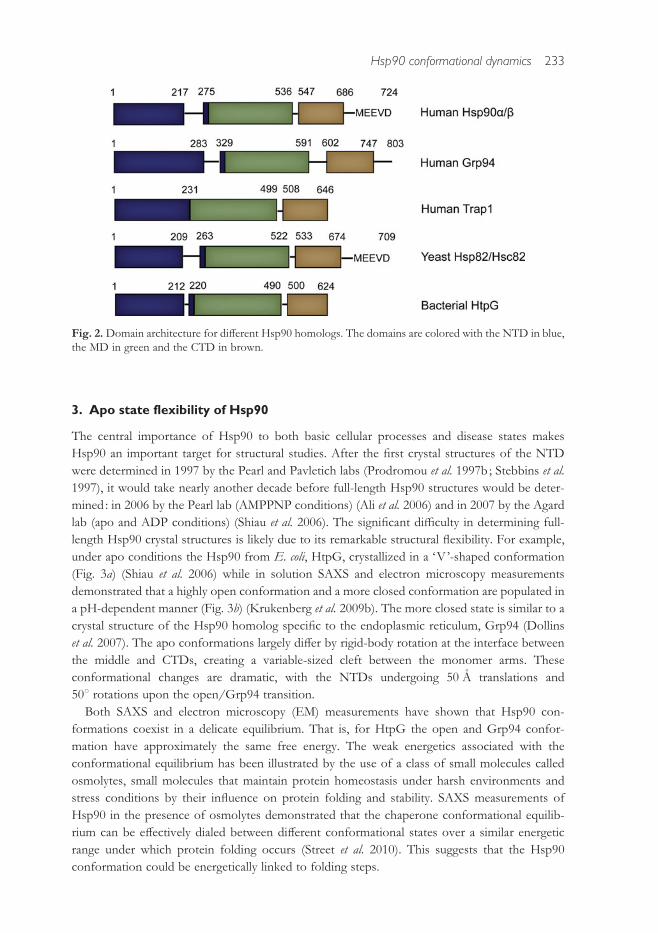
di!erences in the overall domain organization of the di!erent Hsp90s (Fig. 2). A repetitively
charged region (Fig. 1, red) that varies in length is present at the C-terminal end of the NTD in
yeast and cytosolic higher eukaryotic Hsp90s as well as in Grp94. This charged region is also
present in the bacterial protein HtpG, but it is significantly shorter. Although the charged region
is dispensable in yeast, it has been implicated in client binding (Scheibel et al. 1999). In addition
to the charged linker, the yeast and cytosolic higher eukaryotic Hsp90s also have a C-terminal
motif (MEEVD) that facilitates co-chaperone binding. The unique regulatory role of Hsp90 in
eukaryotes is likely due to the large number of co-chaperones that recruit specific classes of
clients (e.g. kinases via p50/Cdc37), modulate the Hsp90 ATPase activity (e.g. activation by
Aha1), aid in client folding (e.g. proline isomerization by cyclophilins) and maturation (p23).
Many of these co-chaperones contain tetratricopeptide repeat (TPR) domains that bind Hsp90
via the C-terminal MEEVD motif. Later we will discuss the role of co-chaperones on Hsp90
conformational dynamics.
Fig. 1. Hsp90 exists as a homodimer with three domains per monomer. The crystal structure of thebacterial homolog, HtpG (PDB code 2IOQ), is shown as both a cartoon representation and a surfacerepresentation. The domains of one monomer are colored blue (NTD), green (MD) and brown (CTD). Theother monomer is colored gray. The NTD binds nucleotide and contains a variable charged loop (red) andthe conserved lid region (purple), which is adjacent to the nucleotide-binding pocket. The MD contains theconserved catalytic loop (cyan) that is essential for ATP hydrolysis. The CTD provides the constitutivedimer interface.
232 K. A. Krukenberg et al.
3. Apo state flexibility of Hsp90
The central importance of Hsp90 to both basic cellular processes and disease states makes
Hsp90 an important target for structural studies. After the first crystal structures of the NTD
were determined in 1997 by the Pearl and Pavletich labs (Prodromou et al. 1997b ; Stebbins et al.
1997), it would take nearly another decade before full-length Hsp90 structures would be deter-
mined : in 2006 by the Pearl lab (AMPPNP conditions) (Ali et al. 2006) and in 2007 by the Agard
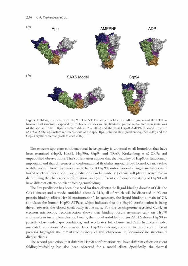
lab (apo and ADP conditions) (Shiau et al. 2006). The significant di"culty in determining full-
length Hsp90 crystal structures is likely due to its remarkable structural flexibility. For example,
under apo conditions the Hsp90 from E. coli, HtpG, crystallized in a ‘V’-shaped conformation
(Fig. 3a) (Shiau et al. 2006) while in solution SAXS and electron microscopy measurements
demonstrated that a highly open conformation and a more closed conformation are populated in
a pH-dependent manner (Fig. 3b) (Krukenberg et al. 2009b). The more closed state is similar to a
crystal structure of the Hsp90 homolog specific to the endoplasmic reticulum, Grp94 (Dollins
et al. 2007). The apo conformations largely di!er by rigid-body rotation at the interface between
the middle and CTDs, creating a variable-sized cleft between the monomer arms. These
conformational changes are dramatic, with the NTDs undergoing 50 A translations and
50x rotations upon the open/Grp94 transition.
Both SAXS and electron microscopy (EM) measurements have shown that Hsp90 con-
formations coexist in a delicate equilibrium. That is, for HtpG the open and Grp94 confor-
mation have approximately the same free energy. The weak energetics associated with the
conformational equilibrium has been illustrated by the use of a class of small molecules called
osmolytes, small molecules that maintain protein homeostasis under harsh environments and
stress conditions by their influence on protein folding and stability. SAXS measurements of
Hsp90 in the presence of osmolytes demonstrated that the chaperone conformational equilib-
rium can be e!ectively dialed between di!erent conformational states over a similar energetic
range under which protein folding occurs (Street et al. 2010). This suggests that the Hsp90
conformation could be energetically linked to folding steps.
Fig. 2.Domain architecture for di!erent Hsp90 homologs. The domains are colored with the NTD in blue,the MD in green and the CTD in brown.
Hsp90 conformational dynamics 233
The extreme apo state conformational heterogeneity is universal to all homologs that have
been examined (HtpG, Hsc82, Hsp90a, Grp94 and TRAP, Krukenberg et al. 2009a and
unpublished observations). This conservation implies that the flexibility of Hsp90 is functionally
important, and that di!erences in conformational flexibility among Hsp90 homologs may relate
to di!erences in how they interact with clients. If Hsp90 conformational changes are functionally
linked to client interactions, two predictions can be made : (1) clients will play an active role in
determining the chaperone conformation ; and (2) di!erent conformational states of Hsp90 will
have di!erent e!ects on client folding/misfolding.
The first prediction has been observed for three clients : the ligand-binding domain of GR; the
Cdk4 kinase ; and a model unfolded client D131D, all of which will be discussed in ‘Client
protein binding a!ects Hsp90 conformation’. In summary, the ligand-binding domain of GR
stimulates the human Hsp90 ATPase, which indicates that the Hsp90 conformation is being
driven towards the closed catalytically active state. For the co-chaperone-recruited Cdk4, an
electron microscopy reconstruction shows that binding occurs asymmetrically on Hsp90
and results in incomplete closure. Finally, the model unfolded protein D131D drives Hsp90 to
partially close under apo conditions, and accelerates full closure and ATP hydrolysis under
nucleotide conditions. As discussed later, Hsp90’s di!ering response to these very di!erent
proteins highlights the remarkable capacity of this chaperone to accommodate structurally
diverse clients.
The second prediction, that di!erent Hsp90 conformations will have di!erent e!ects on client
folding/misfolding has also been observed for a model client. Specifically, the thermal
(a)
(b)
Fig. 3. Full-length structures of Hsp90. The NTD is shown in blue, the MD in green and the CTD inbrown. In all structures, exposed hydrophobic surfaces are highlighted in purple. (a) Surface representationsof the apo and ADP HtpG structure (Shiau et al. 2006) and the yeast Hsp90 AMPPNP-bound structure(Ali et al. 2006). (b) Surface representations of the apo HtpG solution state (Krukenberg et al. 2008) and theGrp94 crystal structure (Dollins et al. 2007).
234 K. A. Krukenberg et al.

aggregation of citrate synthase can be strongly suppressed by HtpG in the Grp94 conformation
but only weakly suppressed in the open state (Krukenberg et al. 2009b). This observation was
enabled by identifying a mutation pair (H446E/K) that selectively populates the open/Grp94
conformations independent of pH.
These observations together support the idea that the conformational equilibrium of Hsp90
plays a central role in client interactions, and is possibly tuned to the client requirements of
each organism. The large structure of Hsp90 in combination with its conformational diversity
suggests that the chaperone can adopt a combinatorial set of binding surfaces and chaperone
geometries in order to interact with structurally diverse clients and large complexes that include
co-chaperones. As discussed next, the e!ect of nucleotide binding and hydrolysis adds a layer of
complexity, both structural and kinetic, to the Hsp90 conformational equilibrium.
4. ATP-dependent conformational changes
The transition to the closed ATP state involves three local structural changes that together
coordinate large-scale closure of the chaperone. ATP binding (1) restructures an N-terminal
helical lid region, (2) dramatically changes the NTD/MD orientation and (3) is associated with
the release of a b-strand, which is swapped across monomers stabilizing N-terminal dimeriza-
tion. These changes are discussed below.
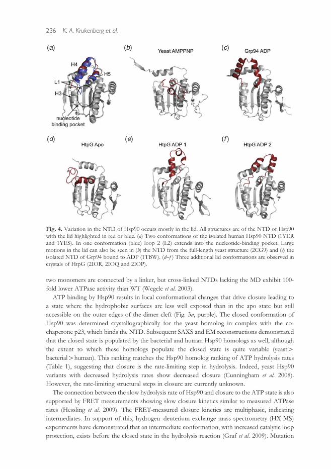
The lid region (Fig. 1, purple), a helix–loop–helix motif adjacent to the nucleotide-binding
pocket makes extensive NTD dimer contacts (Prodromou et al. 1997a ; Stebbins et al. 1997).
Crystal structures of isolated NTDs in the apo, AMPPNP and ADP bound states show that
nucleotide binding induces large structural changes in the lid (Fig. 4). Biochemical studies high-
light that the lid has a complex role in regulating NTD dimerization and ATP hydrolysis.
Specifically, removal of the lid abolishes all ATPase activity, but heterodimers lacking the lid on
only one NTD have substantially accelerated closure and ATPase hydrolysis rates (Hessling et al.
2009 ; Richter et al. 2006). The increased dimerization promoted by this heterodimer is coupled to
the first 24 amino acids of the N-terminus that form a swapped b-strand that stabilizes NTD
dimerization. Deletion of the N-terminal 24 residues confers flexibility to the lid suggesting that
nucleotide binding, lid restructuring and b-strand swapping are all coupled (Richter et al. 2006).
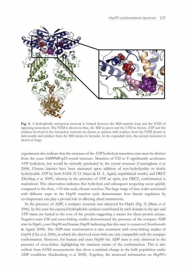
A second major structural change upon ATP binding is a repositioning of the NTD/MD
interface, which orients a highly conserved arginine (R380, in yeast numbering) that resides on
the MD catalytic loop (Fig. 1, cyan) to interact with the ATP c-phosphate. In the AMPPNP-
bound structure of Hsp90, R380 is the only residue contacting the c-phosphate, highlighting itsunique role in ATP binding and hydrolysis. The catalytic loop and arginine are both highly
conserved and appear essential for hydrolysis by Hsp90 alone and for the acceleration of
hydrolysis by the Aha1 co-chaperone (Meyer et al. 2003). The NTD/MD orientation has been
found to be variable in di!erent crystal structures, suggesting that rotational freedom allows for
the R380/c-phosphate alignment to be regulated by co-chaperone and client binding. The R380
residue on the catalytic loop forms a hydrophobic network with a loop (T22, V23 and Y24,
Hsp82 numbering) on the opposing monomer in the closed state, indicating cross-monomer
coupling of closure and hydrolysis (Fig. 5). Indeed, cross-monomer regulation of hydrolysis was
confirmed by examining the ATPase activity of a series of yeast Hsp90 heterodimers with
mutations in this network (Cunningham et al. 2008). The catalytic role of the MD and the
requirement for cross-monomer interactions in hydrolysis is further supported by truncation
experiments where Hsp90 deletion constructs lacking the CTD retain basal ATPase activity if the
Hsp90 conformational dynamics 235
two monomers are connected by a linker, but cross-linked NTDs lacking the MD exhibit 100-
fold lower ATPase activity than WT (Wegele et al. 2003).
ATP binding by Hsp90 results in local conformational changes that drive closure leading to
a state where the hydrophobic surfaces are less well exposed than in the apo state but still
accessible on the outer edges of the dimer cleft (Fig. 3a, purple). The closed conformation of
Hsp90 was determined crystallographically for the yeast homolog in complex with the co-
chaperone p23, which binds the NTD. Subsequent SAXS and EM reconstructions demonstrated
that the closed state is populated by the bacterial and human Hsp90 homologs as well, although
the extent to which these homologs populate the closed state is quite variable (yeast>bacterial>human). This ranking matches the Hsp90 homolog ranking of ATP hydrolysis rates
(Table 1), suggesting that closure is the rate-limiting step in hydrolysis. Indeed, yeast Hsp90
variants with decreased hydrolysis rates show decreased closure (Cunningham et al. 2008).
However, the rate-limiting structural steps in closure are currently unknown.
The connection between the slow hydrolysis rate of Hsp90 and closure to the ATP state is also
supported by FRET measurements showing slow closure kinetics similar to measured ATPase
rates (Hessling et al. 2009). The FRET-measured closure kinetics are multiphasic, indicating
intermediates. In support of this, hydrogen–deuterium exchange mass spectrometry (HX-MS)
experiments have demonstrated that an intermediate conformation, with increased catalytic loop
protection, exists before the closed state in the hydrolysis reaction (Graf et al. 2009). Mutation
(a) (b) (c)
(d) (e) (f )
Fig. 4. Variation in the NTD of Hsp90 occurs mostly in the lid. All structures are of the NTD of Hsp90with the lid highlighted in red or blue. (a) Two conformations of the isolated human Hsp90 NTD (1YERand 1YES). In one conformation (blue) loop 2 (L2) extends into the nucleotide-binding pocket. Largemotions in the lid can also be seen in (b) the NTD from the full-length yeast structure (2CG9) and (c) theisolated NTD of Grp94 bound to ADP (1TBW). (d–f ) Three additional lid conformations are observed incrystals of HtpG (2IOR, 2IOQ and 2IOP).
236 K. A. Krukenberg et al.
experiments also indicate that the structure of the ATP hydrolysis transition state must be distinct
from the yeast AMPPMP:p23 crystal structure. Mutation of T22 to Y significantly accelerates
ATP hydrolysis, but would be sterically precluded by the crystal structure (Cunningham et al.
2008). Closure kinetics have been measured upon addition of non-hydrolysable or slowly
hydrolysable ATP by both SAXS (T. O. Street & D. A. Agard, unpublished results) and FRET
(Hessling et al. 2009), whereas in the presence of ATP an open, low FRET, conformation is
maintained. This observation indicates that hydrolysis and subsequent reopening occur quickly
compared to the slow,y10 min scale, closure reaction. The large range of time scales associated
with di!erent steps in the Hsp90 reaction cycle demonstrates how kinetic regulation by
co-chaperones can play a pivotal role in a!ecting client interactions.
In the presence of ADP, a compact structure was observed for HtpG (Fig. 3) (Shiau et al.
2006). In this state the exposed hydrophobic surfaces contributed by each domain in the apo and
ATP states are buried in the core of the protein suggesting a means for client protein release.
Negative-stain EM and cross-linking studies demonstrated the presence of the compact ADP
state in HtpG, yeast Hsp90 and human Hsp90 indicating that this state is conserved (Southworth
& Agard, 2008). The ADP-state conformation is also consistent with cross-linking studies of
Grp94 (Chu et al. 2006), in which the observed cross-links are only compatible with the compact
conformation. However, for human and yeast Hsp90 the ADP state is only observed in the
presence of cross-linker, highlighting the transient nature of the conformation. This is also
evident from SAXS measurements that show a minimal change in the bulk population under
ADP conditions (Krukenberg et al. 2008). Together, the structural information on Hsp90’s
Fig. 5. A hydrophobic interaction network is formed between the MD catalytic loop and the NTD ofopposing monomers. The NTD is shown in blue, the MD in green and the CTD in brown. ATP and theresidues involved in the interaction network are shown as spheres with residues from the NTD shown indark purple and residues from the MD shown in lavender. In the expanded view, the second monomer isshown in beige.
Hsp90 conformational dynamics 237

conformational flexibility and nucleotide dependence map out the basic steps of a nucleotide
hydrolysis cycle (Fig. 6).
Molecular dynamics simulations of full-length Hsp90 found an increase in communication
e"ciency between residues in the NTD and CTD upon the addition of nucleotide (Morra et al.
2009). Interestingly, while both ATP- and ADP-bound Hsp90 show residues communicating
over a distance of 80 A, these studies suggest each nucleotide activates a di!erent set of com-
municating residues between the NTD and CTD as shown in Fig. 7. These di!erent pathways
may determine the di!erent conformations of Hsp90 in the presence of ATP or ADP; muta-
tional studies could be used to elucidate each network’s role in establishing Hsp90’s confor-
mation.
5. The role of co-chaperones in regulating the conformational cycle of Hsp90
In eukaryotes, Hsp90 is often found in large complexes with co-chaperones, some of which are
listed in Table 2. Co-chaperones provide a method for the direct recruitment of di!erent client
proteins to Hsp90, a means for regulating the Hsp90 conformational and ATP hydrolysis cycle,
and subsequently a level of control over client folding. A genomic comparison of Hsp90 co-
chaperones shows that many organisms have a unique repertoire of Hsp90 co-chaperones,
suggesting that specific co-chaperone combinations can tailor Hsp90’s activity to best suit the
client requirements for each particular organism ( Johnson & Brown, 2009). In this section we
discuss well-studied Hsp90 co-chaperones, their influence on Hsp90 conformational dynamics
and their role in kinetic regulation.
Fig. 6. The structures of Hsp90 suggest a nucleotide-dependent conformational cycle. In this cycle, thebinding of ATP shifts the conformation from an open structure (1) to an NTD-dimerized closed structure(3). The Grp94 crystal structure (2) may represent an intermediate along this path or non-catalytic con-formation. The hydrolysis of ADP causes a further compaction of the protein (4) and the release ofnucleotide restarts the conformational cycle.
238 K. A. Krukenberg et al.

A minimal set of necessary co-chaperones has been established for the progesterone receptor
(PR), the GR and the kinase Chk1 providing information about the role of di!erent co-
chaperones and about the specific requirements among client proteins. In vitro, GR requires only
Hsp70 and Hop in addition to Hsp90 for the reconstitution of steroid-binding activity (Arlander
et al. 2006; Dittmar & Pratt, 1997 ; Kosano et al. 1998). For the in vitro reconstitution of PR,
Hsp70, Hsp40, Hop and p23 are required in addition to Hsp90 (Kosano et al. 1998). The kinase
Chk1 also requires Hsp90, Hsp70, Hsp40 and Hop for maximal activation, but unlike PR, the
kinase-specific co-chaperone p50 (Cdc37) is also required while p23 is not (Arlander et al. 2006).
These di!erences in co-chaperone requirements may provide a mechanism for tuning the Hsp90
response.
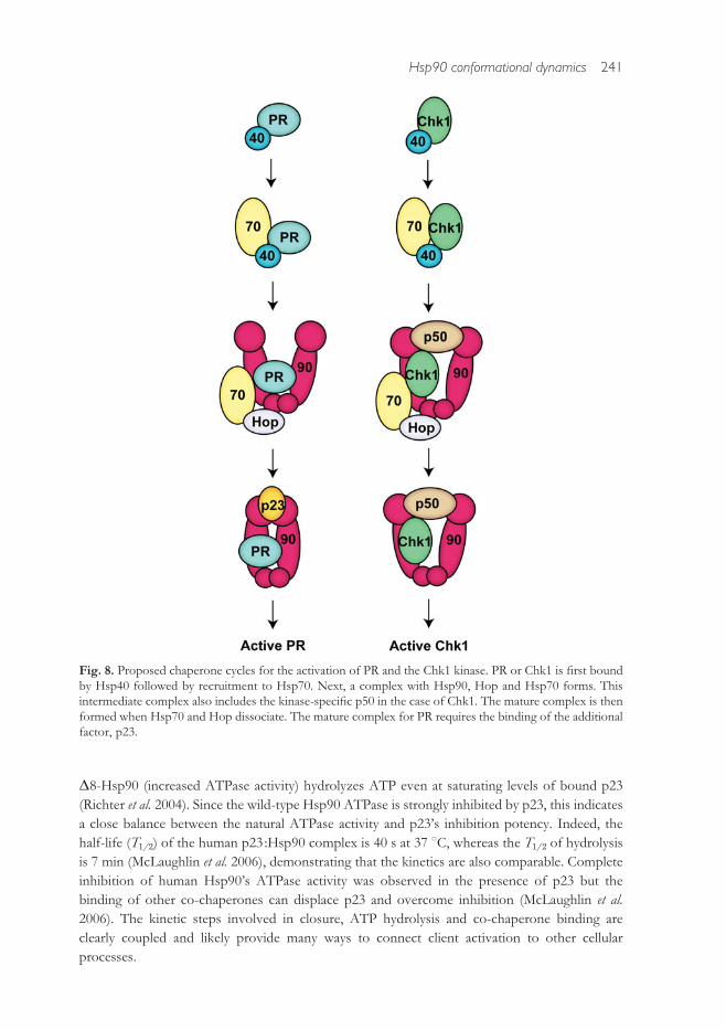
The reconstituted systems reveal that specific complexes are formed at di!erent stages
throughout the chaperone cycle (Fig. 8). Both Chk1 and PR bind Hsp40 to begin the cycle.
Hsp40 delivers the client to Hsp70 and the initial Hsp90 complex forms by client transfer from
Hsp70 facilitated by Hop which can simultaneously bind both Hsp70 and Hsp90 to stabilize a
ternary complex (Felts et al. 2007). In the case of Chk1, this initial complex also includes p50.
Hsp70 dissociates to form late-stage complexes. For Chk1, this includes the kinase, Hsp90 and
Fig. 7.Molecular dynamics simulations have shown di!erent long-range communication networks betweenthe NTD and CTD in the presence of ATP or ADP. Residues that participate in long-range communicationare shown as spheres on the yeast AMPPNP-bound crystal structure. The NTD is shown in blue, the MD inbeige and the CTD in green with the second monomer shown in gray. Long-range interactions observedonly in the presence of ATP are shown in purple. Interactions seen only in the presence of ADP are shownin orange. Interactions observed with both ATP and ADP are shown in dark gray.
Hsp90 conformational dynamics 239

p50. The steroid hormone receptors are in complex with Hsp90 and p23 at this stage. Activated
client proteins are then released from the late-stage Hsp90 complex (Felts et al. 2007 ; Hernandez
et al. 2002 ; Kosano et al. 1998; Smith, 1993).
The diagrams in Fig. 7 detail the minimal systems required for activation, but numerous other
co-chaperones have been identified and are believed to play important roles in the regulation of
the chaperone cycle in vivo (Table 2). For example, the immunophilin FKBP52 is found in
complex with Hsp90, GR/PR, and p23 in vivo ; FKBP52 also increases the hormone-binding
ability of GR and is essential for the correct activation of PR in vivo (Chadli et al. 2008; Riggs et al.
2003). To understand the operation of these large Hsp90/client/co-chaperone complexes, the
influence of each co-chaperone on Hsp90 needs to be known.
5.1 p23 arrests the ATPase cycle of Hsp90
Since it was first identified in complex with Hsp90 and unactivated PR (Smith et al. 1990), p23
has been shown to have enhanced/diminished binding to Hsp90 under ATP/ADP conditions
(Grenert et al. 1999 ; Johnson & Toft, 1995; Sullivan et al. 1997). Binding of p23 occurs primarily
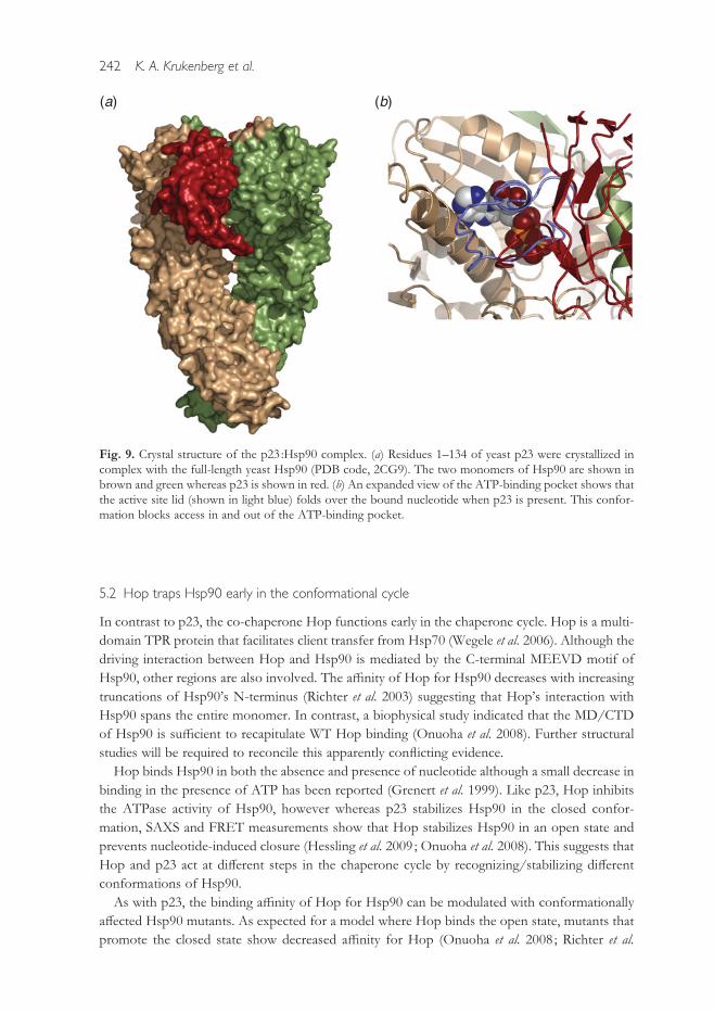
at the NTD with minor MD contacts (Fig. 9) (Ali et al. 2006 ; Martinez-Yamout et al. 2006). The
complex promotes and stabilizes a catalytically incompetent closed conformation (McLaughlin
et al. 2002, 2006 ; Richter et al. 2004 ; Siligardi et al. 2004). The a"nity of Hsp90 for p23 can be
tuned with Hsp90 mutants that either increase or decrease NTD dimerization (Richter et al.
2004 ; Siligardi et al. 2004). As expected, truncation of the NTD domain so that dimerization no
longer occurs abolishes the binding of p23.
Promoting the closed state of Hsp90 and inhibiting the ATPase activity has the functional
consequence of stabilizing client proteins in a complex with Hsp90 and p23. This has been
observed for both GR and PR (Dittmar & Pratt, 1997 ; Kosano et al. 1998). p23 may serve as a
molecular timer for the activation of client proteins ; once clients are activated p23 is released,
hydrolysis occurs and client proteins can dissociate. This timing regulation may occur by two
mechanisms : (1) slowed hydrolysis of ATP in the presence of p23 or (2) dissociation of p23
driven by the binding of other co-chaperones. There is evidence for both mechanisms. In yeast,
Table 2. Partial list of Hsp90 associated co-chaperones
Protein Function
Aha1 Stimulates ATPase activity*p50 Blocks ATPase activity and is thought to be kinase specific*Cpr6 TPR-containing and peptidy-prolyl-isomerase (Johnson et al. 2007)FKBP52 TPR-containing and peptidy-prolyl-isomerase (Riggs et al. 2003)GCUNC-45 TPR-containing that blocks progression of cycle (Chadli et al. 2006)Hop TPR-containing and sca!olds Hsp70-Hsp90 interaction*Hsp70 recruits clients to Hsp90*NudC CHORD-domain containing (Te et al. 2007)p23 Blocks ATPase activity and traps client proteins in complex with Hsp90*PP5 TPR-containing and phosphatase*Sse1 Part of Hsp90 complex in budding yeast (Liu et al. 1999)Tah1 TPR containing (Zhao et al. 2005)
For a list of additional co-chaperones see http://www.picard.ch/downloads/Hsp90interactors.pdf*References can be found in the text.
240 K. A. Krukenberg et al.
D8-Hsp90 (increased ATPase activity) hydrolyzes ATP even at saturating levels of bound p23
(Richter et al. 2004). Since the wild-type Hsp90 ATPase is strongly inhibited by p23, this indicates
a close balance between the natural ATPase activity and p23’s inhibition potency. Indeed, the
half-life (T1/2) of the human p23:Hsp90 complex is 40 s at 37 xC, whereas the T1/2 of hydrolysis
is 7 min (McLaughlin et al. 2006), demonstrating that the kinetics are also comparable. Complete
inhibition of human Hsp90’s ATPase activity was observed in the presence of p23 but the
binding of other co-chaperones can displace p23 and overcome inhibition (McLaughlin et al.
2006). The kinetic steps involved in closure, ATP hydrolysis and co-chaperone binding are
clearly coupled and likely provide many ways to connect client activation to other cellular
processes.
Fig. 8. Proposed chaperone cycles for the activation of PR and the Chk1 kinase. PR or Chk1 is first boundby Hsp40 followed by recruitment to Hsp70. Next, a complex with Hsp90, Hop and Hsp70 forms. Thisintermediate complex also includes the kinase-specific p50 in the case of Chk1. The mature complex is thenformed when Hsp70 and Hop dissociate. The mature complex for PR requires the binding of the additionalfactor, p23.
Hsp90 conformational dynamics 241
5.2 Hop traps Hsp90 early in the conformational cycle
In contrast to p23, the co-chaperone Hop functions early in the chaperone cycle. Hop is a multi-
domain TPR protein that facilitates client transfer from Hsp70 (Wegele et al. 2006). Although the
driving interaction between Hop and Hsp90 is mediated by the C-terminal MEEVD motif of
Hsp90, other regions are also involved. The a"nity of Hop for Hsp90 decreases with increasing
truncations of Hsp90’s N-terminus (Richter et al. 2003) suggesting that Hop’s interaction with
Hsp90 spans the entire monomer. In contrast, a biophysical study indicated that the MD/CTD
of Hsp90 is su"cient to recapitulate WT Hop binding (Onuoha et al. 2008). Further structural
studies will be required to reconcile this apparently conflicting evidence.
Hop binds Hsp90 in both the absence and presence of nucleotide although a small decrease in
binding in the presence of ATP has been reported (Grenert et al. 1999). Like p23, Hop inhibits
the ATPase activity of Hsp90, however whereas p23 stabilizes Hsp90 in the closed confor-
mation, SAXS and FRET measurements show that Hop stabilizes Hsp90 in an open state and
prevents nucleotide-induced closure (Hessling et al. 2009 ; Onuoha et al. 2008). This suggests that
Hop and p23 act at di!erent steps in the chaperone cycle by recognizing/stabilizing di!erent
conformations of Hsp90.
As with p23, the binding a"nity of Hop for Hsp90 can be modulated with conformationally
a!ected Hsp90 mutants. As expected for a model where Hop binds the open state, mutants that
promote the closed state show decreased a"nity for Hop (Onuoha et al. 2008; Richter et al.
(a) (b)
Fig. 9. Crystal structure of the p23 :Hsp90 complex. (a) Residues 1–134 of yeast p23 were crystallized incomplex with the full-length yeast Hsp90 (PDB code, 2CG9). The two monomers of Hsp90 are shown inbrown and green whereas p23 is shown in red. (b) An expanded view of the ATP-binding pocket shows thatthe active site lid (shown in light blue) folds over the bound nucleotide when p23 is present. This confor-mation blocks access in and out of the ATP-binding pocket.
242 K. A. Krukenberg et al.

2003). The inhibition by Hop can be reversed by the addition of other co-chaperones, such as
Cpr6 and PP5, two TPR-containing co-chaperones that compete with Hop for binding to Hsp90
(Prodromou et al. 1999). Co-chaperone competition for binding the MEEVD motif may be a
general mechanism for controlling the progression through di!erent phases of the Hsp90 con-
formational cycle. Whether this is a coordinated or stochastic process remains unclear.
5.3 p50, a kinase specific co-chaperone, also traps Hsp90 in an open conformation
The kinase-specific co-chaperone p50 also inhibits the ATPase activity and a!ects the con-
formational dynamics of Hsp90, but its mechanism di!ers from both Hop and p23
(Grammatikakis et al. 1999 ; Lee et al. 2002 ; Stepanova et al. 1996). Domain mapping indicates
that p50’s NTD is critical for binding kinases whereas the middle and CTDs interact with Hsp90
(Grammatikakis et al. 1999 ; Roe et al. 2004 ; Shao et al. 2003). Biochemical studies show that p50
binds to Hsp90 in a 1 :1 ratio with the interaction occurring largely at the NTD of Hsp90.
A crystal structure of the C-terminal region of p50 (residues 148–348) bound to the NTD of
Hsp90 shows a dimer for p50 but in this configuration the dimer interface is small and may arise
from crystal contacts (Roe et al. 2004). Indeed, biochemical studies suggest that p50 binds Hsp90
as a monomer because the KD for dimerization (80 mM) is higher than the KD for complex
formation (4 mM) (Zhang et al. 2004).
The crystal structure (CTD of p50 bound to NTD of Hsp90) indicates a mechanism for the
inhibition of Hsp90’s ATPase activity. Specifically, residue R167 of p50 points into the nucleo-
tide-binding pocket of Hsp90 and forms a hydrogen bond with the catalytic glutamate of Hsp90
(E33). Nucleotide can still bind, but the catalytic water is no longer coordinated for hydrolysis
(Roe et al. 2004). The crystal structure also reveals another potential means of inhibition in which
p50 blocks the conversion of Hsp90 from the open to the closed state. Specifically, when the
p50 :Hsp90 dimer is docked onto one NTD of the yeast Hsp90 AMPPNP-bound crystal struc-
ture, p50 sterically clashes with the second Hsp90 NTD (Fig. 10). When docked onto the full-
length apo crystal form of Hsp90, the p50 dimer fits into the cleft of the Hsp90 dimer potentially
stabilizing Hsp90 in an open conformation (Fig. 10c).
Two additional lines of evidence support the idea that p50 a!ects Hsp90 conformational
dynamics. First, biochemical studies using the Hsp90 mutants T22I and T101I support the
preference of p50 for the open conformation of Hsp90 (Millson et al. 2004). Second, for p50 to
bind to the full-length apo conformation a structural rearrangement must occur between the
NTD and MD of Hsp90 (Fig. 10c). Indeed, SAXS measurements of the p50:Hsp90 complex
indicate that complex formation causes a contraction in apo Hsp90 (Zhang et al. 2004). HX-MS
also shows changes in the protection of human Hsp90 in the presence of p50 consistent with
a conformational change (Phillips et al. 2007).
5.4 Aha1 activates the ATPase of Hsp90
Unlike the other co-chaperones discussed, Aha1 stimulates the ATPase activity of Hsp90 (Lotz
et al. 2003; Meyer et al. 2004 ; Panaretou et al. 2002). Aha1 binds mainly to the MD of Hsp90 and
this interaction is mediated by the N-terminal portion of Aha1 (N-Aha1) (Lotz et al. 2003 ; Meyer
et al. 2004 ; Siligardi et al. 2004). Recent NMR studies suggest that Aha1 also interacts with the
NTD of Hsp90 (Retzla! et al. 2010). N-Aha1 has been crystallized in complex with the MD
of Hsp90 (Meyer et al. 2004) and when aligned with either the closed structure or the open
Hsp90 conformational dynamics 243

structure, Aha1 binds to Hsp90 at the edge of the dimer cleft approximately 90x from the dimer
interface (Fig. 11), and NMR experiments with the full-length proteins suggest that Aha1 binds
to both NTDs of the Hsp90 dimer in the closed conformation (Retzla! et al. 2010). Gel filtration
and NMR measurements show that Aha1 competes with both Hop and p23 for binding to
Hsp90 (Harst et al. 2005 ; Martinez-Yamout et al. 2006), indicating that Aha1 can act at multiple
stages of the chaperone cycle to disrupt inhibition by other co-chaperones. Aha1 may displace
Hop from early complexes and p23 from mature complexes stimulating the ATPase activity and
allowing for the continuation of the chaperone cycle.
Aha1 stimulates the ATPase activity of Hsp90 by an asymmetric interaction, and Aha1 bound
to one Hsp90 monomer is able to activate both NTDs of the Hsp90 dimer (Retzla! et al. 2010).
(a)
(c)
(b)
Fig. 10. Crystal structure of p50 bound to Hsp90. (a) The C-terminal region of p50 (pink) and the NTD ofHsp90 (blue) form a heterotetramer with p50 forming a dimer between the two NTD domains of Hsp90.(b) p50 binding is incompatible with the dimerized NTDs of the Hsp90 closed state. The two NTDs ofHsp90 are shown in blue and beige and they are shown in the configuration seen in the yeast AMPPNP-bound crystal state. Docking of the p50 :Hsp90 crystal dimer onto one NTD of the closed state revealssteric clashes between p50 (red) and the second Hsp90 NTD (beige). (c) The p50:Hsp90 heterotetramerfrom A is aligned with the two NTDs from the apo bacterial crystal structure suggesting a mechanism forblocking the closure of Hsp90. One monomer of the apo structure is shown in green and the other in beige.For p50 to bind to this structure, the NTDs of the apo state would have to move as seen by the blue Hsp90NTDs.
244 K. A. Krukenberg et al.

This activation most likely occurs by the interaction of Aha1 with the Hsp90 NM domain
optimally positioning the MD catalytic loop for catalysis (Fig. 11). As discussed earlier, in the apo
state, the catalytic loop (and the critical R380) is positioned away from the NTD (Fig. 12b),
whereas in the closed state R380 contacts the c-phosphate of ATP (Figs 5 and 12 c). Even though
in the Aha1:Hsp90 complex parts of the catalytic loop are disordered, the orientation is much
more consistent with the loop conformation found in the closed state (Fig. 12). Furthermore, the
R380A mutant, which has a decreased ATPase rate, is not activated by Aha1 (Meyer et al. 2003)
implying that R380 is critical in the Aha1-mediated activation of Hsp90. Data showing that the
Hsp90 F349A mutant, located at the NM interface and potentially involved in interdomain
communication, is hypersensitive to Aha1 also support this conclusion (Meyer et al. 2003 ;
Siligardi et al. 2004).
Although it is clear from these studies that NTD dimerization and rearrangement of the
NM domain are important for Aha1’s e!ect on Hsp90, it is not clear as to whether Aha1
simply stabilizes the catalytic conformation once it forms or if binding of Aha1 results in a
(a)
(b)
Fig. 11. Aha1 binds Hsp90 on the edge of the cleft. By docking the N-Aha1:MD-Hsp90 complex on thefull-length apo (a) and ATP (b) crystal structures, the approximate orientation of Aha1 can be determined.Aha1 is colored in gray whereas one monomer of Hsp90 is green and the other monomer is beige.
Hsp90 conformational dynamics 245

conformational change in Hsp90 leading to catalysis. A recent FRET study suggests that Aha1
directly induces structural changes in yeast Hsp90. Aha1-binding results in the rapid acceleration
of conformational changes between the NTD and MD similar to the acceleration seen in the
lidless Hsp90 mutant, which shows increased NTD dimerization. The FRET results also suggest
that Aha1 binding allows Hsp90 to bypass the first conformational intermediate after nucleotide
binding leading to an accumulation of the second NTD dimerized intermediate instead (Hessling
et al. 2009).
6. Client protein binding affects Hsp90 conformation
The conformational dynamics of Hsp90 are likely involved in the folding and activation of client
proteins, and changes in Hsp90 likely confer structural changes upon the client protein itself. As
another level of control over the Hsp90 cycle, client proteins themselves may also a!ect the
conformation of Hsp90. Early indirect evidence for client proteins altering the conformation of
Hsp90 came from in vitro studies showing that the ligand-binding domain of GR stimulates the
human Hsp90 ATPase rate (McLaughlin et al. 2002) suggesting that GR shifts the population
of human Hsp90 towards the closed catalytic state. This correlates well with the observation that
human Hsp90 has a low basal ATPase rate because it does not significantly populate the closed
state. Furthermore, both Hop and p23 inhibit the GR-stimulated rate (McLaughlin et al. 2002),
as expected based on their e!ect on conformational dynamics as discussed above.
The clearest structural evidence for client binding influencing the conformation of Hsp90
comes from a 3D negative-stain EM reconstruction of the p50 :Hsp90 :Cdk4 complex (Vaughan
et al. 2006). The reconstruction shows an asymmetric complex with two Hsp90 monomers, one
p50 monomer and one Cdk4 kinase monomer. The Hsp90 dimer is in a conformation similar to
the closed AMPPNP-bound state but the NTDs are not dimerized. One NTD hinges backwards
away from the other and p50 binds between the two NTDs. Cdk4 interacts with the outer edge
(a) (b) (c)
Fig. 12. Aha1 binding to Hsp90 causes a rearrangement in the MD catalytic loop. (a) N-Aha1 (gray) wascrystallized in complex with the MD of Hsp90 (green). The catalytic loop, shown in red, is partiallydisordered in the crystal structure. (b) The catalytic loop in the apo full-length structure (beige) has morea-helical structure than in the Aha1:Hsp90 complex, and the catalytically required R380 (shown in sticks)forms contacts with the MD in the apo structure. (c) In the closed AMPPNP-bound crystal structure thecatalytic loop, shown in purple, orients R380 towards the NTD where it interacts with the c-phosphateof ATP. The loop structure in the Aha1:Hsp90 complex is closely related to the loop structure in theclosed state.
246 K. A. Krukenberg et al.

of the complex. The kinase likely contacts the Hsp90 MD with the C-lobe, and the N-lobe
contacts one Hsp90 NTD and p50. Previous studies, discussed above, indicate that p50 binds as
a 1 :1 complex with Hsp90 and this complex is locked in a conformation more closely related to
the apo crystal structure. It has been suggested that the kinase initially binds a dimer of p50 and
this complex is recruited to Hsp90 forming a symmetric complex. A structural rearrangement
then occurs displacing one p50 monomer and leading to the asymmetric complex observed by
EM (Fig. 13) (Vaughan et al. 2006).
Both GR and kinase clients are actively recruited to Hsp90 by co-chaperones, which adds a
layer of complexity to the chaperone/client interactions. Understanding how Hsp90 interacts
with just a client is crucial for building more complex models involving the additional involve-
ment of co-chaperones. However, trapping Hsp90 with a mostly unfolded protein presents
significant technical challenges. As a solution to this problem, Hsp90/client interactions have
been measured with a constitutively unfolded protein D131D, a well-studied fragment of
staphylococcal nuclease. While D131D is largely unfolded it does not aggregate even at high
concentrations. These properties have made it an ideal tool for investigating structural properties
of unfolded proteins (Shortle, 2002). Binding of this unfolded protein to Hsp90 drives large-scale
conformational changes. Specifically, under apo conditions Hsp90 adapts its conformation by
partially closing around D131D. In the presence of AMPPNP, D131D binds with increased
a"nity to Hsp90’s fully closed state, accelerates the open/closed transition and stimulates
ATP hydrolysis (Street and Agard, in preparation). Further studies will be required to see how
Hsp90 responds to di!erent unfolded proteins, and how co-chaperones modulate the Hsp90
response.
A central unanswered question concerns the physical state of Hsp90-bound clients ; currently
there is conflicting evidence. For example, studies of thermally denatured citrate synthase suggest
that Hsp90 interacts in early unfolding or the late stages of folding ( Jakob et al. 1995). In contrast,
NMR studies of a p53 mutant suggest that Hsp90 binds after complete client unfolding (Rudiger
et al. 2002). Additionally, D131D has significant residual structure despite being globally unfolded
(Alexandrescu et al. 1994; Alexandrescu & Shortle, 1994). More studies will be required to
determine the folding state of Hsp90-bound clients and which structural features are involved in
the binding.
Fig. 13. Proposed model for Hsp90:p50:Cdk4 complex formation. Initially, the kinase Cdk4 binds to adimer of p50. This complex is then recruited to a dimer of Hsp90 to form a symmetric complex.A conformational rearrangement then occurs and one monomer of p50 is displaced leading to the asym-metric complex observed by EM.
Hsp90 conformational dynamics 247
7. The effect of post-translational modifications on the conformationaldynamics of Hsp90
Emerging evidence indicates that post-translational modifications regulate specific client protein
interactions with Hsp90. Several modifications have been identified including S-nitrosylation,
phosphorylation, ubiqutination and acetylation (Blank et al. 2003 ; Wandinger et al. 2008). As
discussed below, these modifications likely a!ect multiple aspects of the Hsp90 conformational
cycle.
7.1 S-nitrosylation
S-nitrosylation is the covalent attachment of nitric oxide (NO) to a reactive cysteine. Although
the E. coli and yeast Hsp90s have no cysteines, human Hsp90 is S-nitrosylated at the CTD
(residue 597) by endothelial nitric oxide synthase (eNOS), resulting in reduced ATPase
activity (Martinez-Ruiz et al. 2005). Given that the site of S-nitrosylation is far from the Hsp90
nucleotide-binding pocket, this suggests an indirect mechanism for the ATPase reduction.
Indeed, FRET measurements demonstrate that S-nitrosylation at Cys597 leads to decreased
dimerization, suggesting that Hsp90’s ATPase activity can be regulated by tuning the monomer/
dimer equilibrium. eNOS is an Hsp90 client that is activated by the chaperone’s ATPase activity,
and so the S-nitrosylation of Hsp90 by eNOS may be a feedback mechanism to limit the
accumulation of activated eNOS (Martinez-Ruiz et al. 2005).
7.2 Phosphorylation
Phosphorylation of Hsp90 has di!ering consequences on client interactions depending on the
client protein involved and the site of phosphorylation. For example, phosphorylation at S225
and S254 in Hsp90 causes decreased a"nity for the aryl hydrocarbon receptor (AhR) (Ogiso et al.
2004), whereas phosphorylation at Y300 by c-Src is required for the binding of eNOS (Duval et al.
2007). Phosphorylation of Hsp90 can also regulate its cellular abundance. Swe1 phosphorylates
Hsp90 at Y24 in a cell-cycle-dependant manner, which consequently causes Hsp90 to be
ubiquitinated and degraded in the yeast cytosol (Mollapour et al. 2010). When mapped to the
closed structure, Y24 sits in the interface between the NTDs in a hydrophobic patch shown to be
essential for hydrolysis (Ali et al. 2006 ; Cunningham et al. 2008). Indeed, mimicking Y24 phos-
phorylation by mutagenesis leads to decreased ATPase activity and N-terminal dimerization
(Mollapour et al. 2010). Interestingly, this modification decreases Hsp90 activation of kinases but
does not a!ect steroid hormone receptor maturation. Together, these data suggest the phos-
phorylation of Hsp90 at Y24 leads to an altered conformational cycle with reduced closure and
ATP hydrolysis, and this change in dynamics a!ects the activation of specific clients.
7.3 Acetylation
Lysine acetylation of Hsp90 can disrupt co-chaperone and client interactions with Hsp90
(Kekatpure et al. 2009 ; Kovacs et al. 2005 ; Scroggins et al. 2007; Yu et al. 2002). HDAC6 can
de-acetylate Hsp90 (Kekatpure et al. 2009) and inhibition or knockdown of HDAC6 leads to
hyperacetylation (Yu et al. 2002). Hsp90 contains at least two acetylation sites and one site has
been mapped to K294 (Hsp82 numbering) at the junction of the NTD and MD (Scroggins et al.
248 K. A. Krukenberg et al.

2007). Given this location, it is likely that this modification will a!ect the NTD/MD orientation,
which, as discussed earlier, plays a key role in coordinating the catalytic loop with NTD-bound
ATP. More studies are needed to elucidate the conformational consequences of this modification
and their relationship to the chaperone cycle and client binding.
Although our knowledge of post-translational modifications as they apply to Hsp90 is still in
the early stages, it is clear that they play an important regulatory role. With more site-specific
information and biophysical studies, it should be possible to gain a more complete mechanistic
picture of post-translational modification-mediated regulation of the essential chaperone cycle
of Hsp90.
8. Small molecules also shift the conformation of Hsp90
The conformational dynamics of Hsp90 are essential for proper function and multiple layers of
regulation a!ect the conformation, suggesting that small molecules that a!ect Hsp90’s con-
formational dynamics may have clinical value. Currently, most Hsp90 inhibitors compete for
binding with ATP and were not designed to influence the conformation of Hsp90. There are,
however, indications that some inhibitors shift the conformation of Hsp90 to states that are
incapable of client protein binding.
As would be expected for small molecules occupying the nucleotide-binding pocket, inhibitor
binding causes local changes in the lid of Hsp90 (Immormino et al. 2004 ; Stebbins et al. 1997).
Binding of the inhibitor GA leads to a lid conformation of Hsp90 (Stebbins et al. 1997) similar to
the conformation seen in the isolated apo human Hsp90 NTD (Fig. 3a, red). This conformation
is unlike the lid state seen in the nucleotide-bound complexes where hydrophobic surfaces are
accessible for NTD dimerization. The local changes in the NTD induced by the inhibitor most
likely correlate with changes in the global structure of the Hsp90 dimer. The GA-bound lid
conformation suggests that GA and nucleotide have very di!erent e!ects on the overall structure
of Hsp90.
In vivo, the binding of GA to Hsp90 leads to the destabilization and degradation of client
proteins (Mimnaugh et al. 1996; Schulte et al. 1995 ; Whitesell et al. 1994). In most cases, desta-
bilization is due to the disruption of Hsp90 :client complexes as observed for the kinases v-Src
and Bcr-Abl (An et al. 2000). GA also disrupts the binding of p23 (An et al. 2000 ; Sullivan et al.
1997), indicating that GA also inhibits closure. GA increases the presence of Hop and Hsp70 in
pull-downs of Hsp90 (An et al. 2000 ; Sullivan et al. 1997). Although GA binds to an open
conformation of Hsp90, this conformation is distinct from the conformation of apo Hsp90. As
measured by SAXS, GA causes compaction of Hsp90 but not closure (Zhang et al. 2004).
Binding of inhibitors clearly a!ects the local structure of Hsp90, as HX-MS experiments have
shown that DMAG, a derivative of GA and radicicol, an inhibitor similar to GA, causes changes
in the protection of amide protons in all three domains of Hsp90 (Phillips et al. 2007). Radicicol
also increases the protease sensitivity of Hsp90 as compared to the apo protein (Nishiya et al.
2009).
Other small molecule inhibitors that function via alternative mechanisms may also cause
Hsp90 conformational changes. Novobiocin is a CTD-binding inhibitor (Marcu et al. 2000a, b ;
Soti et al. 2002) which, like GA and radicicol, inhibits the formation of Hsp90 complexes with
client or p23, but unlike GA it also reduces the a"nity of Hsp90 for TPR-containing co-
chaperones that bind to the CTD (Allan et al. 2006; Yun et al. 2004). Epigallocatechin gallate
(EGCG) the active ingredient found in green tea also directly binds to Hsp90 and acts as an AhR
Hsp90 conformational dynamics 249

antagonist. As opposed to disrupting the interaction of Hsp90 with AhR, EGCG functions by
stabilizing the interaction between Hsp90 and AhR (Palermo et al. 2005). A cyclic peptide,
Sansalvamide A, appears to bind at the NM domain interface, has no e!ect on ATPase activity,
and yet di!erentially alters client protein binding in vitro and client stabilization in vivo (Vasko et al.
2010). This demonstrates the potential of non-ATP directed inhibitors to show unique client
selectivity, presumably by allosteric modulation of Hsp90 conformational dynamics.
9. Concluding remarks
Structural dynamics play an important role in the function of Hsp90, and understanding the
complete conformational ensemble is imperative for defining Hsp90’s molecular mechanism.
Large conformational changes occur throughout the ATPase hydrolysis cycle, and the con-
formational state is not rigorously determined by the bound nucleotide. Instead nucleotide
binding shifts a conformational equilibrium between states that are also sampled in the absence
of nucleotide. In vivo, Hsp90 will also sample nucleotide-bound and -unbound states simul-
taneously due to the weak interaction with ATP. For the cytosolic eukaryotic Hsp90s, co-
chaperones regulate the conformational state of Hsp90 by shifting the equilibrium, and the finely
tuned regulation is essential for proper chaperone function. Other levels of regulation exist in the
form of post-translational modifications such as S-nitrosylation, phosphorylation and acety-
lation, but the exact nature of these e!ects remains poorly understood. Other modifications will
also require investigation to determine their importance in the regulation of Hsp90. Disrupting
the conformational dynamics of Hsp90 provides an attractive target for new therapeutics for the
treatment of cancer and potentially vascular disease and Alzheimer’s, and inhibitors currently in
clinical trials show signs of altering the conformation of Hsp90 and thereby disrupting the
binding of client proteins.
Although a much clearer picture of the mechanism of Hsp90 has emerged over the past few
years, many outstanding questions require investigation. To begin, the nature of the interaction
of client proteins with Hsp90 is largely uncharacterized, and it is essential to understand how
client protein binding a!ects the conformation of Hsp90 and vice versa. This information is key
to the understanding of how Hsp90’s conformational changes relate to the activation of client
proteins. Another open question is the origin of the energy required for the remodeling of client
protein. Because the ATPase activity is low and not strongly coupled to the conformational
changes that occur, it is unclear from where the force originates to drive the conformational
changes in Hsp90 and any consequent changes that occur in the client protein. Also, important
di!erences exist between di!erent homologs and each homolog has a uniquely determined
balance between conformational states and transition kinetics. Understanding how the equilibria
are established and the functional consequences for shifting the equilibria will provide critical
information about the molecular mechanism of Hsp90. Ultimately, a more detailed under-
standing of Hsp90, its dynamics and interactions will pave the way for the development of more
targeted therapeutics.
10. Acknowledgments
We would like to thank Ulrike Bottcher and Daniel Southworth for their helpful discussions and
critical reading of the manuscript. Funding has been provided by HHMI.
250 K. A. Krukenberg et al.
11. References
ALEXANDRESCU, A. T., ABEYGUNAWARDANA, C. & SHORTLE,
D. (1994). Structure and dynamics of a denatured 131-
residue fragment of staphylococcal nuclease: a hetero-
nuclear NMR study. Biochemistry 33, 1063–1072.
ALEXANDRESCU, A. T. & SHORTLE, D. (1994). Backbone
dynamics of a highly disordered 131 residue fragment of
staphylococcal nuclease. Journal of Molecular Biology 242,
527–546.
ALI, M. M., ROE, S. M., VAUGHAN, C. K., MEYER, P.,
PANARETOU, B., PIPER, P. W., PRODROMOU, C. &
PEARL, L. H. (2006). Crystal structure of an Hsp90-
nucleotide-p23/Sba1 closed chaperone complex.Nature
440, 1013–1017.
ALLAN, R. K., MOK, D., WARD, B. K. & RATAJCZAK, T.
(2006). Modulation of chaperone function and cocha-
perone interaction by novobiocin in the C-terminal do-
main of Hsp90: evidence that coumarin antibiotics
disrupt Hsp90 dimerization. Journal of Biological Chemistry
281, 7161–7171.
AN, W. G., SCHULTE, T. W. & NECKERS, L. M. (2000). The
heat shock protein 90 antagonist geldanamycin alters
chaperone association with p210bcr-abl and v-src pro-
teins before their degradation by the proteasome. Cell
Growth and Di!erentiation 11, 355–360.
ARLANDER, S. J., FELTS, S. J., WAGNER, J. M., STENSGARD,
B., TOFT, D. O. & KARNITZ, L. M. (2006). Chaperoning
checkpoint kinase 1 (Chk1), an Hsp90 client, with pur-
ified chaperones. Journal of Biological Chemistry 281,
2989–2998.
BERGERAT, A., DE MASSY, B., GADELLE, D., VAROUTAS,
P. C., NICOLAS, A. & FORTERRE, P. (1997). An atypical
topoisomerase II from Archaea with implications for
meiotic recombination. Nature 386, 414–417.
BLANK, M., MANDEL, M., KEISARI, Y., MERUELO, D. &
LAVIE, G (2003). Enhanced ubiquitinylation of heat
shock protein 90 as a potential mechanism for mitotic
cell death in cancer cells induced with hypericin. Cancer
Res 63, 8241–8247.
CHADLI, A., BRUINSMA, E. S., STENSGARD, B. & TOFT, D.
(2008). Analysis of Hsp90 cochaperone interactions
reveals a novel mechanism for TPR protein recognition.
Biochemistry 47, 2850–2857.
CHADLI, A., GRAHAM, J. D., ABEL, M. G., JACKSON, T. A.,
GORDON, D. F., WOOD, W. M., FELTS, S. J., HORWITZ,
K. B. & TOFT, D. (2006). GCUNC-45 is a novel regu-
lator for the progesterone receptor/hsp90 chaperoning
pathway. Molecular and Cellular Biology 26, 1722–1730.
CHIOSIS, G., LUCAS, B., HUEZO, H., SOLIT, D., BASSO, A. &
ROSEN, N. (2003). Development of purine-sca!old
small molecule inhibitors of Hsp90. Current Cancer Drug
Targets 3, 371–376.
CHU, F., MAYNARD, J. C., CHIOSIS, G., NICCHITTA, C. V. &
BURLINGAME, A. L. (2006). Identification of novel quat-
ernary domain interactions in the Hsp90 chaperone,
GRP94. Protein Science 15, 1260–1269.
CUNNINGHAM, C. N., KRUKENBERG, K. A. & AGARD, D. A.
(2008). Intra- and intermonomer interactions are re-
quired to synergistically facilitate ATP hydrolysis in
Hsp90. Journal of Biological Chemistry 283, 21170–21178.
DICKEY, C. A., KOREN, J., ZHANG, Y. J., XU, Y. F., JINWAL,
U. K., BIRNBAUM, M. J., MONKS, B., SUN, M., CHENG,
J. Q., PATTERSON, C., BAILEY, R. M., DUNMORE, J.,
SORESH, S., LEON, C., MORGAN, D. & PETRUCELLI, L.
(2008). Akt and CHIP coregulate tau degradation
through coordinated interactions. Proceedings of the
National Academy of Sciences, USA 105, 3622–3627.
DITTMAR, K. D. & PRATT, W. B. (1997). Folding of the
glucocorticoid receptor by the reconstituted Hsp90-
based chaperone machinery. The initial hsp90.
p60.hsp70-dependent step is su"cient for creating the
steroid binding conformation. Journal of Biological
Chemistry 272, 13047–13054.
DOLLINS, D. E., WARREN, J. J., IMMORMINO, R. M. &
GEWIRTH, D. T. (2007). Structures of GRP94-nucleotide
complexes reveal mechanistic di!erences between the
hsp90 chaperones. Molecular Cell 28, 41–56.
DUNBRACK, JR, R. L., GERLOFF, D. L., BOWER, M., CHEN,
X., LICHTARGE, O. & COHEN, F. E. (1997). Meeting re-
view: the second meeting on the critical assessment of
techniques for protein structure prediction (CASP2),
Asilomar, California, December 13–16, 1996. Folding
and Design 2, R27–R42.
DUVAL, M., LE BOEUF, F., HUOT, J. & GRATTON, J. P.
(2007). Src-mediated phosphorylation of Hsp90 in re-
sponse to vascular endothelial growth factor (VEGF) is
required for VEGF receptor-2 signaling to endothelial
NO synthase. Mol Biol Cell 18, 4659–4668.
FALSONE, S. F., KUNGL, A. J., REK, A., CAPPAI, R. &
ZANGGER, K. (2009). The molecular chaperone Hsp90
modulates intermediate steps of amyloid assembly of
the Parkinson-related protein alpha-synuclein. Journal of
Biological Chemistry 284, 31190–31199.
FELTS, S. J., KARNITZ, L. M. & TOFT, D. O. (2007).
Functioning of the Hsp90 machine in chaperoning
checkpoint kinase I (Chk1) and the progesterone re-
ceptor (PR). Cell Stress and Chaperones 12, 353–363.
FORSYTHE, H. L., JARVIS, J. L., TURNER, J. W., ELMORE,
L. W. & HOLT, S. E. (2001). Stable association of hsp90
and p23, but Not hsp70, with active human telomerase.
Journal of Biological Chemistry 276, 15571–15574.
FREY, S., LESKOVAR, A., REINSTEIN, J. & BUCHNER, J. (2007).
The ATPase cycle of the endoplasmic chaperone
Grp94. J Biol Chem 282, 35612–35620.
GELLER, R., VIGNUZZI, M., ANDINO, R. & FRYDMAN, J.
(2007). Evolutionary constraints on chaperone-medi-
ated folding provide an antiviral approach refractory to
development of drug resistance. Genes Development 21,
195–205.
GOMEZ-PUERTAS, P., MARTIN-BENITO, J., CARRASCOSA,
J. L., WILLISON, K. R. & VALPUESTA, J. M. (2004). The
Hsp90 conformational dynamics 251

substrate recognition mechanisms in chaperonins.
Journal of Molecular Recognition 17, 85–94.
GRAF, C., STANKIEWICZ, M., KRAMER, G. & MAYER, M. P.
(2009). Spatially and kinetically resolved changes in the
conformational dynamics of the Hsp90 chaperone
machine. EMBO Journal 28, 602–613.
GRAMMATIKAKIS, N., LIN, J. H., GRAMMATIKAKIS, A.,
TSICHLIS, P. N. & COCHRAN, B. H. (1999). p50(cdc37)
acting in concert with Hsp90 is required for Raf-1
function. Molecular and Cellular Biology 19, 1661–1672.
GRENERT, J. P., JOHNSON, B. D. & TOFT, D. O. (1999). The
importance of ATP binding and hydrolysis by hsp90 in
formation and function of protein heterocomplexes.
Journal of Biological Chemistry 274, 17525–17533.
HARST, A., LIN, H. & OBERMANN, W. M. (2005). Aha1
competes with Hop, p50 and p23 for binding to the
molecular chaperone Hsp90 and contributes to kinase
and hormone receptor activation. Biochemical Journal
387, 789–796.
HERNANDEZ, M. P., CHADLI, A. & TOFT, D. O. (2002).
HSP40 binding is the first step in the HSP90 chaper-
oning pathway for the progesterone receptor. Journal of
Biological Chemistry 277, 11873–11881.
HESSLING, M., RICHTER, K. & BUCHNER, J. (2009).
Dissection of the ATP-induced conformational cycle of
the molecular chaperone Hsp90. Nature Structural and
Molecular Biology 16, 287–293.
IMMORMINO, R. M., DOLLINS, D. E., SHAFFER, P. L.,
SOLDANO, K. L., WALKER, M. A. & GEWIRTH, D. T.
(2004). Ligand-induced conformational shift in the N-
terminal domain of GRP94, an Hsp90 chaperone.
Journal of Biological Chemistry 279, 46162–46171.
JAKOB, U., LILIE, H., MEYER, I. & BUCHNER, J. (1995).
Transient interaction of Hsp90 with early unfolding
intermediates of citrate synthase. Implications for
heat shock in vivo. Journal of Biological Chemistry 270,
7288–7294.
JOHNSON, J. L. & BROWN, C. (2009). Plasticity of the Hsp90
chaperone machine in divergent eukaryotic organisms.
Cell Stress and Chaperones 14, 83–94.
JOHNSON, J. L., HALAS, A. & FLOM, G. (2007). Nucleotide-
dependent interaction of saccharomyces cerevisiae
Hsp90 with the cochaperone proteins Sti1, Cpr6, and
Sba1. Molecular and Cellular Biology 27, 768–776.
JOHNSON, J. L. & TOFT, D. O. (1995). Binding of p23 and
hsp90 during assembly with the progesterone receptor.
Molecular Endocrinology 9, 670–678.
KEKATPURE, V. D., DANNENBERG, A. J. & SUBBARAMAIAH,
K. (2009). HDAC6 modulates Hsp90 chaperone activity
and regulates activation of ary1 hydrocarbon receptor
signaling. J Biol Chem 284, 7436–7445.
KOSANO, H., STENSGARD, B., CHARLESWORTH, M. C.,
MCMAHON, N. & TOFT, D. (1998). The assembly of
progesterone receptor-hsp90 complexes using purified
proteins. Journal of Biological Chemistry 273, 32973–32979.
KOVACS, J. J., MURPHY, P. J., GAILLARD, S., ZHAO, X., WU,
J. T., NICCHITTA, C. V., YOSHIDA, M., TOFT, D. O.,
PRATT, W. B. & YAO, T. P. (2005). HDAC6 regulates
Hsp90 acetylation and chaperone-dependent activation
of glucocorticoid receptor. Mol Cell 18, 601–607.
KRUKENBERG, K. A., BOTTCHER, U. M., SOUTHWORTH,
D. R. & AGARD, D. A. (2009a). Grp94, the endoplasmic
reticulum Hsp90, has a similar solution conformation to
cytosolic Hsp90 in the absence of nucleotide. Protein
Science 18, 1815–1827.
KRUKENBERG, K. A., FORSTER, F., RICE, L. M., SALI, A. &
AGARD, D. A. (2008). Multiple conformations of E. coli
Hsp90 in solution: insights into the conformational
dynamics of Hsp90. Structure 16, 755–765.
KRUKENBERG, K. A., SOUTHWORTH, D. R., STREET, T. O. &
AGARD, D. A. (2009b). pH-dependent conformational
changes in bacterial Hsp90 reveal a Grp94-like confor-
mation at pH 6 that is highly active in suppression of
citrate synthase aggregation. J Mol Biol 390, 278–291.
KURT, N., RAJAGOPALAN, S. & CAVAGNERO, S. (2006).
E!ect of hsp70 chaperone on the folding andmisfolding
of polypeptides modeling an elongating protein chain.
Journal of Molecular Biology 355, 809–820.
LEE, P., RAO, J., FLISS, A., YANG, E., GARRETT, S. &
CAPLAN, A. J. (2002). The Cdc37 protein kinase-binding
domain is su"cient for protein kinase activity and cell
viability. Journal of Cell Biology 159, 1051–1059.
LESKOVAR, A., WEGELE, H., WERBECK, N. D., BUCHNER, J.
& REINSTEIN, J. (2008). The ATPase cycle of the
mitochondrial Hsp90 analog Trap 1. J Biol Chem 283,
11677–11688.
LIU, X. D., MORANO, K. A. & THIELE, D. J. (1999).
The yeast Hsp110 family member, Sse1, is an Hsp90
cochaperone. Journal of Biological Chemistry 274,
26654–26660.
LOTZ, G. P., LIN, H., HARST, A. & OBERMANN, W. M.
(2003). Aha1 binds to the middle domain of Hsp90,
contributes to client protein activation, and stimulates
the ATPase activity of the molecular chaperone. Journal
of Biological Chemistry 278, 17228–17235.
MARCU, M. G., CHADLI, A., BOUHOUCHE, I., CATELLI, M. &
NECKERS, L. M. (2000a). The heat shock protein 90
antagonist novobiocin interacts with a previously
unrecognized ATP-binding domain in the carboxyl
terminus of the chaperone. Journal of Biological Chemistry
275, 37181–37186.
MARCU, M. G., SCHULTE, T. W. & NECKERS, L. (2000b).
Novobiocin and related coumarins and depletion of
heat shock protein 90-dependent signaling proteins.
Journal of National Cancer Institute 92, 242–248.
MARTINEZ-RUIZ, A., VILLANUEVA, L., GONZALEZ DE
ORDUNA, C., LOPEZ-FERRER, D., HIGUERAS, M. A.,
TARIN, C., RODRIGUEZ-CRESPO, I., VAZQUEZ, J. & LAMAS,
S. (2005). S-nitrosylation of Hsp90 promotes the inhi-
bition of its ATPase and endothelial nitric oxide
252 K. A. Krukenberg et al.
synthase regulatory activities. Proc Natl Acad Sci U S A
102, 8525–8530.
MARTINEZ-YAMOUT, M. A., VENKITAKRISHNAN, R. P.,
PREECE, N. E., KROON, G., WRIGHT, P. E. & DYSON,
H. J. (2006). Localization of sites of interaction between
p23 and Hsp90 in solution. Journal of Biological Chemistry
281, 14457–14464.
MCLAUGHLIN, S. H., SMITH, H. W. & JACKSON, S. E. (2002).
Stimulation of the weak ATPase activity of human
hsp90 by a client protein. Journal of Molecular Biology 315,
787–798.
MCLAUGHLIN, S. H., VENTOURAS, L. A., LOBBEZOO, B. &
JACKSON, S. E. (2004). Independent ATPase activity of
hsp90 subunits creates a flexible assembly platform.
J Mol Biol 344, 813–826.
MCLAUGHLIN, S. H., SOBOTT, F., YAO, Z. P., ZHANG, W.,
NIELSEN, P. R., GROSSMANN, J. G., LAUE, E. D.,
ROBINSON, C. V. & JACKSON, S. E. (2006). The co-
chaperone p23 arrests the Hsp90 ATPase cycle to trap
client proteins. Journal of Molecular Biology 356, 746–758.
MEYER, P., PRODROMOU, C., HU, B., VAUGHAN, C., ROE,
S. M., PANARETOU, B., PIPER, P. W. & PEARL, L. H.
(2003). Structural and functional analysis of the middle
segment of hsp90: implications for ATP hydrolysis and
client protein and cochaperone interactions. Molecular
Cell 11, 647–658.
MEYER, P., PRODROMOU, C., LIAO, C., HU, B., MARK ROE,
S., VAUGHAN, C. K., VLASIC, I., PANARETOU, B., PIPER,
P. W. & PEARL, L. H. (2004). Structural basis for re-
cruitment of the ATPase activator Aha1 to the Hsp90
chaperone machinery. EMBO Journal 23, 511–519.
MILLSON, S. H., TRUMAN, A. W., WOLFRAM, F., KING, V.,
PANARETOU, B., PRODROMOU, C., PEARL, L. H. & PIPER,
P. W. (2004). Investigating the protein-protein interac-
tions of the yeast Hsp90 chaperone system by two-
hybrid analysis : potential uses and limitations of this
approach. Cell Stress and Chaperones 9, 359–368.
MIMNAUGH, E. G., CHAVANY, C. & NECKERS, L. (1996).
Polyubiquitination and proteasomal degradation of the
p185c-erbB-2 receptor protein-tyrosine kinase induced
by geldanamycin. Journal of Biological Chemistry 271,
22796–22801.
MOLLAPOUR, M., TSUTSUMI, S., DONNELLY, A. C., BEEBE,
K., TOKITA, M. J., LEE, M. J., LEE, S., MORRA, G.,
BOURBOULIA, D., SCROGGINS, B. T., COLOMBO, G.,
BLAGG, B. S., PANARETOU, B., STETLER-STEVENSON,
W. G., TREPEL, J. B., PIPER, P. W., PRODROMOU, C.,
PEARL, L. H. & NECKERS, L. (2010). Swe1Wee1-
dependent tyrosine phosphorylation of Hsp90 regulates
distinct facets of chaperone function. Mol Cell 37,
333–343.
MORRA, G., VERKHIVKER, G. & COLOMBO, G. (2009).
Modeling signal propagation mechanisms and ligand-
based conformational dynamics of the Hsp90 molecular
chaperone full-length dimer. PLoS Computational Biology
5, e1000323.
NATHAN, D. F. & LINDQUIST, S. (1995). Mutational analysis
of Hsp90 function: interactions with a steroid receptor
and a protein kinase. Molecular and Cellular Biology 15,
3917–3925.
NECKERS, L. (2007). Heat shock protein 90 : the cancer
chaperone. Journal of Bioscience 32, 517–530.
NECKERS, L. & IVY, S. P. (2003). Heat shock protein 90.
Current Opinion in Oncology 15, 419–524.
NISHIYA, Y., SHIBATA, K., SAITO, S., YANO, K., ONEYAMA,
C., NAKANO, H. & SHARMA, S. V. (2009). Drug-target
identification from total cellular lysate by drug-induced
conformational changes. Analytical Biochemistry 385,
314–320.
OGISO, H., KAGI, N., MATSUMOTO, E., NISHIMOTO, M.,
ARAI, R., SHIROUZU, M., MIMURA, J., FUJII-KURIYAMA,
Y. & YOKOYAMA, S.. (2004). Phosphorylation analysis
of 90 kDa heat shock protein within the cytosolic aryl-
hydrocarbon receptor complex. Biochemistry 43, 15510–
15519.
ONUOHA, S. C., COULSTOCK, E. T., GROSSMANN, J. G. &
JACKSON, S. E. (2008). Structural studies on the co-
chaperone Hop and its complexes with Hsp90. Journal of
Molecular Biology 379, 732–744.
PALERMO, C. M., WESTLAKE, C. A. & GASIEWICZ, T. A.
(2005). Epigallocatechin gallate inhibits aryl hydro-
carbon receptor gene transcription through an indirect
mechanism involving binding to a 90 kDa heat shock
protein. Biochemistry 44, 5041–5052.
PANARETOU, B., SILIGARDI, G., MEYER, P., MALONEY, A.,
SULLIVAN, J. K., SINGH, S., MILLSON, S. H., CLARKE,
P. A., NAABY-HANSEN, S., STEIN, R., CRAMER, R.,
MOLLAPOUR, M., WORKMAN, P., PIPER, P. W., PEARL,
L. H. & PRODROMOU, C. (2002). Activation of the
ATPase activity of hsp90 by the stress-regulated co-
chaperone aha1. Molecular Cell 10, 1307–1318.
PEARL, L. H. & PRODROMOU, C. (2006). Structure and
mechanism of the Hsp90 molecular chaperone
machinery. Annual Review of Biochemistry 75, 271–294.
PHILLIPS, J. J., YAO, Z. P., ZHANG, W., MCLAUGHLIN, S.,
LAUE, E. D., ROBINSON, C. V. & JACKSON, S. E. (2007).
Conformational dynamics of the molecular chaperone
Hsp90 in complexes with a co-chaperone and anti-
cancer drugs. Journal of Molecular Biology 372, 1189–1203.
PRODROMOU, C., PANARETOU, B., CHOHAN, S., SILIGARDI,
G., O’BRIEN, R., LADBURY, J. E., ROE, S. M., PIPER, P. W.
& PEARL, L. H. (2000). The ATPase cycle of Hsp90
drives a molecular ‘clamp’ via transient dimerization of
the N-terminal domains. EMBO Journal 19, 4383–4392.
PRODROMOU, C., ROE, S. M., O’BRIEN, R., LADBURY, J. E.,
PIPER, P. W. & PEARL, L. H. (1997a). Identification and
structural characterization of the ATP/ADP-binding
site in the Hsp90 molecular chaperone. Cell 90, 65–75.
PRODROMOU, C., ROE, S. M., PIPER, P. W. & PEARL, L. H.
(1997b). A molecular clamp in the crystal structure of
the N-terminal domain of the yeast Hsp90 chaperone.
Nature Structural Biology 4, 477–482.
Hsp90 conformational dynamics 253

PRODROMOU, C., SILIGARDI, G., O’BRIEN, R., WOOLFSON,
D. N., REGAN, L., PANARETOU, B., LADBURY, J. E., PIPER,
P. W. & PEARL, L. H. (1999). Regulation of Hsp90
ATPase activity by tetratricopeptide repeat (TPR)-
domain co-chaperones. EMBO Journal 18, 754–762.
RETZLAFF, M., HAGN, F., MITSCHKE, L., HESSLING, M.,
GUGEL, F., KESSLER, H., RICHTER, K. & BUCHNER, J.
(2010). Asymmetric activation of the hsp90 dimer by its
cochaperone aha1. Molecular Cell 37, 344–354.
RICHTER, K. & BUCHNER, J. (2001). Hsp90: chaperoning
signal transduction. Journal of Cellular Physiology 188,
281–290.
RICHTER, K., MOSER, S., HAGN, F., FRIEDRICH, R., HAINZL,
O., HELLER, M., SCHLEE, S., KESSLER, H., REINSTEIN, J.
& BUCHNER, J. (2006). Intrinsic inhibition of the Hsp90
ATPase activity. Journal of Biological Chemistry 281,
11301–11311.
RICHTER, K., MUSCHLER, P., HAINZL, O., REINSTEIN, J. &
BUCHNER, J. (2003). Sti1 is a non-competitive inhibitor
of the Hsp90 ATPase. Binding prevents the N-terminal
dimerization reaction during the atpase cycle. Journal of
Biological Chemistry 278, 10328–10333.
RICHTER, K., WALTER, S. & BUCHNER, J. (2004). The Co-
chaperone Sba1 connects the ATPase reaction of
Hsp90 to the progression of the chaperone cycle. Journal
of Molecular Biology 342, 1403–1413.
RIGGS, D. L., ROBERTS, P. J., CHIRILLO, S. C., CHEUNG-
FLYNN, J., PRAPAPANICH, V., RATAJCZAK, T., GABER, R.,
PICARD, D. & SMITH, D. F. (2003). The Hsp90-binding
peptidylprolyl isomerase FKBP52 potentiates gluco-
corticoid signaling in vivo. EMBO Journal 22, 1158–1167.
ROE, S. M., ALI, M. M., MEYER, P., VAUGHAN, C. K.,
PANARETOU, B., PIPER, P. W., PRODROMOU, C. & PEARL,
L. H. (2004). The mechanism of Hsp90 regulation by
the protein kinase-specific cochaperone p50(cdc37). Cell
116, 87–98.
RUDIGER, S., FREUND, S. M., VEPRINTSEV, D. B. & FERSHT,
A. R. (2002). CRINEPT-TROSY NMR reveals p53
core domain bound in an unfolded form to the
chaperone Hsp90. Proceedings of the National Academy of
Sciences, USA 99, 11085–11090.
RUDIGER, S., GERMEROTH, L., SCHNEIDER-MERGENER, J. &
BUKAU, B. (1997). Substrate specificity of the DnaK
chaperone determined by screening cellulose-bound
peptide libraries. EMBO Journal 16, 1501–1507.
SCHEIBEL, T., SIEGMUND, H. I., JAENICKE, R., GANZ, P.,
LILIE, H. & BUCHNER, J. (1999). The charged region of
Hsp90 modulates the function of the N-terminal do-
main. Proceedings of the National Academy of Sciences, USA
96, 1297–1302.
SCHULTE, T. W., BLAGOSKLONNY, M. V., INGUI, C. &
NECKERS, L. (1995). Disruption of the Raf-1-Hsp90
molecular complex results in destabilization of Raf-1
and loss of Raf-1-Ras association. Journal of Biological
Chemistry 270, 24585–24588.
SCROGGINS, B. T., ROBZYK, K., WANG, D., MARCU, M. G.,
TSUTSUMI, S., BEEBE, K., COTTER, R. J., FELTS, S., TOFT,
D., KARNITZ, L., ROSEN, N. & NECKERS, L. (2007). An
acetylation site in the middle domain of Hsp90 regulates
chaperone function. Mol Cell 25, 151–159.
SHAH, V., WIEST, R., GARCIA-CARDENA, G., CADELINA, G.,
GROSZMANN, R. J. & SESSA, W. C. (1999). Hsp90 regu-
lation of endothelial nitric oxide synthase contributes to
vascular control in portal hypertension. American Journal
of Physiology 277, G463–G468.
SHAO, J., IRWIN, A., HARTSON, S. D. & MATTS, R. L. (2003).
Functional dissection of cdc37: characterization of
domain structure and amino acid residues critical for
protein kinase binding. Biochemistry 42, 12577–12588.
SHIAU, A. K., HARRIS, S. F., SOUTHWORTH, D. R. & AGARD,
D. A. (2006). Structural Analysis of E. coli hsp90 reveals
dramatic nucleotide-dependent conformational re-
arrangements. Cell 127, 329–340.
SHORTLE, D. (2002). The expanded denatured state : an
ensemble of conformations trapped in a locally encoded
topological space. Advances in Protein Chemistry 62, 1–23.
SILIGARDI, G., HU, B., PANARETOU, B., PIPER, P. W., PEARL,
L. H. & PRODROMOU, C. (2004). Co-chaperone regu-
lation of conformational switching in the Hsp90
ATPase cycle. Journal of Biological Chemistry 279, 51989–
51998.
SMITH, D. F. (1993). Dynamics of heat shock protein
90-progesterone receptor binding and the disactivation
loop model for steroid receptor complexes. Molecular
Endocrinology 7, 1418–1429.
SMITH, D. F., FABER, L. E. & TOFT, D. O. (1990).
Purification of unactivated progesterone receptor and
identification of novel receptor-associated proteins.
Journal of Biological Chemistry 265, 3996–4003.
SOLIT, D. B., OSMAN, I., POLSKY, D., PANAGEAS, K. S.,
DAUD, A., GOYDOS, J. S., TEITCHER, J., WOLCHOK, J. D.,
GERMINO, F. J., KROWN, S. E., COIT, D., ROSEN, N. &
CHAPMAN, P. B. (2008). Phase II trial of 17-allylamino-
17-demethoxygeldanamycin in patients with metastatic
melanoma. Clinical Cancer Research 14, 8302–8307.
SOTI, C., RACZ, A. & CSERMELY, P. (2002). A nucleotide-
dependent molecular switch controls ATP binding at
the C-terminal domain of Hsp90. N-terminal nucleotide
binding unmasks a C-terminal binding pocket. Journal of
Biological Chemistry 277, 7066–7075.
SOUTHWORTH, D. R. & AGARD, D. A. (2008). Species-
dependent ensembles of conserved conformational
states define the Hsp90 chaperone ATPase cycle.
Molecular Cell 32, 631–640.
STEBBINS, C. E., RUSSO, A. A., SCHNEIDER, C., ROSEN, N.,
HARTL, F. U. & PAVLETICH, N. P. (1997). Crystal struc-
ture of an Hsp90-geldanamycin complex: targeting of a
protein chaperone by an antitumor agent. Cell 89,
239–250.
STEPANOVA, L., LENG, X., PARKER, S. B. & HARPER,
J. W. (1996). Mammalian p50Cdc37 is a protein
254 K. A. Krukenberg et al.

kinase-targeting subunit of Hsp90 that binds and stabi-
lizes Cdk4. Genes and Development 10, 1491–1502.
STREET, T. O., KRUKENBERG, K. A., ROSGEN, J., BOLEN,
D. W. & AGARD, D. A. (2010). Osmolyte-induced con-
formational changes in the Hsp90 molecular chaperone.
Protein Science 19, 57–65.
SULLIVAN, W., STENSGARD, B., CAUCUTT, G., BARTHA, B.,
MCMAHON, N., ALNEMRI, E. S., LITWACK, G. & TOFT, D.
(1997). Nucleotides and two functional states of hsp90.
Journal of Biological Chemistry 272, 8007–8012.
TE, J., JIA, L., ROGERS, J., MILLER, A. & HARTSON, S. D.
(2007). Novel subunits of the mammalian Hsp90 signal
transduction chaperone. Journal of Proteome Research 6,
1963–1973.
VASKO, R. C., RODRIGUEZ, R. A., CUNNINGHAM, C. N.,
ARDI, V. C., AGARD, D. A. & MCALPINE, S. R. (2010).
Mechanistic studies of sansalvamide a-amide : an allos-
teric modulator of Hsp90. ACS Medicinal Chemistry
Letters 1, 4–8.
VAUGHAN, C. K., GOHLKE, U., SOBOTT, F., GOOD, V. M.,
ALI, M. M., PRODROMOU, C., ROBINSON, C. V., SAIBIL,
H. R. & PEARL, L. H. (2006). Structure of an Hsp90-
Cdc37-Cdk4 complex. Molecular Cell 23, 697–707.
WALTER, S. & BUCHNER, J. (2002). Molecular chaper-
ones – cellular machines for protein folding. Angewandte
Chem (International Edition in English) 41, 1098–1113.
WANDINGER, S. K., RICHSTER, K. & BUCHNER, J. (2008).
The Hsp90 chaperone machinery. J Biol Chem 283,
18473–18477.
WEGELE, H., MUSCHLER, P., BUNCK, M., REINSTEIN, J. &
BUCHNER, J. (2003). Dissection of the contribution of
individual domains to the ATPase mechanism of
Hsp90. Journal of Biological Chemistry 278, 39303–39310.
WEGELE, H., WANDINGER, S. K., SCHMID, A. B., REINSTEIN,
J. & BUCHNER, J. (2006). Substrate transfer from the
chaperone Hsp70 to Hsp90. Journal of Molecular Biology
356, 802–811.
WEISSMAN, J. S., HOHL, C. M., KOVALENKO, O., KASHI, Y.,
CHEN, S., BRAIG, K., SAIBIL, H. R., FENTON, W. A. &
HORWICH, A. L. (1995). Mechanism of GroEL action:
productive release of polypeptide from a sequestered
position under GroES. Cell 83, 577–587.
WHITESELL, L., MIMNAUGH, E. G., DE COSTA, B., MYERS,
C. E. & NECKERS, L. M. (1994). Inhibition of heat shock
protein HSP90-pp60v-src heteroprotein complex for-
mation by benzoquinone ansamycins: essential role for
stress proteins in oncogenic transformation. Proceedings
of the National Academy of Sciences, USA 91, 8324–8328.
WEIKL, T., MUSCHLER, P., RICHTER, K., VEIT, T., REINSTEIN,
J. & BUCHNER, J. (2000). C-terminal regions of Hsp90
are important for trapping the nucleotide during the
ATPase cycle. J Mol Biol 303, 583–592.
WIECH, H., BUCHNER, J., ZIMMERMANN, R. & JAKOB, U.
(1992). Hsp90 chaperones protein folding in vitro.Nature
358, 169–170.
WORKMAN, P. (2004). Combinatorial attack on multistep
oncogenesis by inhibiting the Hsp90 molecular chaper-
one. Cancer Letters 206, 149–157.
YOUNG, J. C., MOAREFI, I. & HARTL, F. U. (2001). Hsp90: a
specialized but essential protein-folding tool. Journal of
Cell Biology 154, 267–273.
YU, X., GUO, Z. S., MARCU, M. G., NECKERS, L., NGUYEN,
D. M., CHEN, G. A. & SCHRUMP, D. S. (2002). Modu-
lation of p53, ErbB1, ErbB2, and Raf-1 expression in
lung cancer cells by depsipeptide FR901228. J Natl
Cancer Inst 94, 504–513.
YUN, B. G., HUANG, W., LEACH, N., HARTSON, S. D. &
MATTS, R. L. (2004). Novobiocin induces a distinct
conformation of hsp90 and alters hsp90-cochaperone-
client interactions. Biochemistry 43, 8217–8229.
ZHANG, W., HIRSHBERG, M., MCLAUGHLIN, S. H., LAZAR,
G. A., GROSSMANN, J. G., NIELSEN, P. R., SOBOTT, F.,
ROBINSON, C. V., JACKSON, S. E. & LAUE, E. D. (2004).
Biochemical and structural studies of the interaction of
Cdc37 with Hsp90. Journal of Molecular Biology 340,
891–907.
ZHAO, R., DAVEY, M., HSU, Y. C., KAPLANEK, P., TONG, A.,
PARSONS, A. B., KROGAN, N., CAGNEY, G., MAI, D.,
GREENBLATT, J., BOONE, C., EMILI, A. & HOURY, W. A.
(2005). Navigating the chaperone network: an integrat-
ive map of physical and genetic interactions mediated by
the hsp90 chaperone. Cell 120, 715–727.
Hsp90 conformational dynamics 255