Conditions of the Stomach (II)
66
Conditions of the Stomach (II) R2 Kanyarat Olarachin, M.D.
Transcript of Conditions of the Stomach (II)

Conditions of the Stomach (II)
R2 Kanyarat Olarachin, M.D.

Table of Contents
05
06
03
04
01
02
Pyloric duplication
Antral web
Hypertrophic Pyloric stenosis
Pyloric atresia
07
08
Gastric perforation in newborn
Congenital microgastria
Gastric volvulus
Peptic ulcer

Gastric volvulus

● Rare● Potentially life-threatening condition● 21% of cases are neonates in recent studies● Associated with diaphragmatic defect● Older children : associated with neurodevelopmental delay
and splenic abnormalities
● Infancy : acute complete volvulus● Older children : chronic, partial volvulus
Introduction

● Primary gastric volvulus● Laxity of the gastric ligaments
● Secondary gastric volvulus● Paraesophageal hernia or diaphragmatic hernia
Introduction

● Abnormal rotation of one part of the stomach● 180 – 360 degree● Anterior direction
● Organoaxial (54%)● Mesenteroaxial (41%)● Combined (2%)
Etiology

Pathogenesis

● About two-third of children with gastric volvulus has diaphragm eventration or herniation (paraesophageal, posterolateral, Morgagni)● Upward displacement of the transverse colon● Greater curve of the stomach was pulled up into the
expanded left upper quadrant
● Other rare causes of gastric volvulus● Abnormal band or adhesion● Rectal atresia -> overdistention of the transverse colon● Congenital absence or left lobe liver resection● Congenital deficiency of the gastrocolic omentum● Asplenic syndrome● Post-operative complication in older children
Pathogenesis

● Adults and older children : Borchardt triad● Unproductive retching● Acute localized epigastric distention● Inability to pass nasogastric tube
● Neonates● Persistent regurgitation● Hematemesis, anemia● Failure to thrive● Chest infection, wheeze● Successful passing NG tube cannot ruled out
Clinical features

● Plain abdominal radiograph● Distend stomach in abnormal position
Diagnosis

● Acute gastric volvulus need appropriate resuscitation and urgent surgery
● Gastric decompression pre-operatively● Nasogastric suction● Needle aspiration might perform before manipulating a
tensely dilated stomach and reducing the volvulus
● Abdominal approach is recommended● Can identify associated gastrointestinal tract anomaly● Accurate diaphragmatic repair
Treatment

● Gastrostomy● Fixation● Route for feeding
● Stamm gastrostomy● 10- or 12- French gauge
Malecot catheter● Secured by a double-purse
string absorbable sutures
Treatment

● Anterior gastropexy● Added if patients don’t have
diaphragmatic defect
● Suture the greater curve of the omentum to the parietal peritoneum and the undersurface of the diaphragm with non-absorbable sutures
Treatment

● Endoscopic assisted percutaneous anterior gastropexy : Chronic mesenteroaxial volvulus in older children
● Laparoscopic anterior gastropexy :Older children with isolated gastric volvulus
● Fundoplication :● Might be needed if patients have gross
gastroesophageal reflux● Crural repair alone might be sufficient
● Gastric volvulus due to a wandering spleenSplenopexy alone might be sufficient
Treatment

● Prolonged gastric ileus● Pyloric ischemia● Gastric outlet obstruction● Gastric necrosis ● Gastric perforation
● Mortality rate● 7.1% in acute gastric volvulus● 27% in chronic gastric volvulus● Untreated : mortality rate up to 80%
Complication

Acute and chronicpeptic ulcer

● Primary peptic ulcer disease● H. pylori infection
● Secondary peptic ulcer disease● Excessive acid production● Stress● Other conditions : e.g. Eosinophilic gastroenteritis, etc.● Drug-related : NSAIDS, Aspirin, Ethanol
Epidemiology
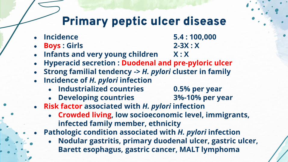
● Incidence 5.4 : 100,000● Boys : Girls 2-3X : X● Infants and very young children X : X● Hyperacid secretion : Duodenal and pre-pyloric ulcer● Strong familial tendency -> H. pylori cluster in family● Incidence of H. pylori infection
● Industrialized countries 0.5% per year● Developing countries 3%-10% per year
● Risk factor associated with H. pylori infection ● Crowded living, low socioeconomic level, immigrants,
infected family member, ethnicity● Pathologic condition associated with H. pylori infection
● Nodular gastritis, primary duodenal ulcer, gastric ulcer, Barett esophagus, gastric cancer, MALT lymphoma
Primary peptic ulcer disease

● Stress ulcers 80%● Associated with critical illness, major trauma● Multiple superficial mucosal erosions at fundus of stomach● Causative factors
● Decrease mucosal blood flow● Disruption of the protective mucosal barrier● Intraluminal acidity
● Drug- and chemical-induced ulcer : resemble stress ulcers● Cushing ulcer : overstimulation of the Vagus nerve due to
increase intracranial pressure -> increase acid output● single, deep ulcer -> prone to perforation
Secondary peptic ulcer disease
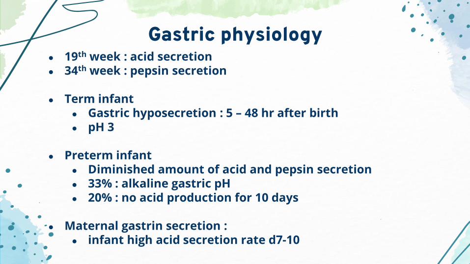
● 19th week : acid secretion● 34th week : pepsin secretion
● Term infant● Gastric hyposecretion : 5 – 48 hr after birth ● pH 3
● Preterm infant● Diminished amount of acid and pepsin secretion● 33% : alkaline gastric pH● 20% : no acid production for 10 days
● Maternal gastrin secretion : ● infant high acid secretion rate d7-10
Gastric physiology

Pathophysiology
Aggressive factorsVascular injury : decrease microcirculation
Cancer chemotherapeutic agentsAspirinNSAIDs
Infectious agent : CMV, Herpes virusIncrease systemic stress
Increase pepsin secretionH. pylori
Defensive factorsMucosal circulation : adequate microcirculationEpithelial cell turnoverIncrease bicarbonate secretionInhibit gastric acid secretion Anti-inflammatory drugsPreserve vascular CMV flow/microcirculationRestore epithelial cell surface catecholaminesMucous layer : glycoprotein, glycocalyxBicarbonate layer : pH gradientImmunoglobulins : IgG, IgA
Primary peptic ulcer
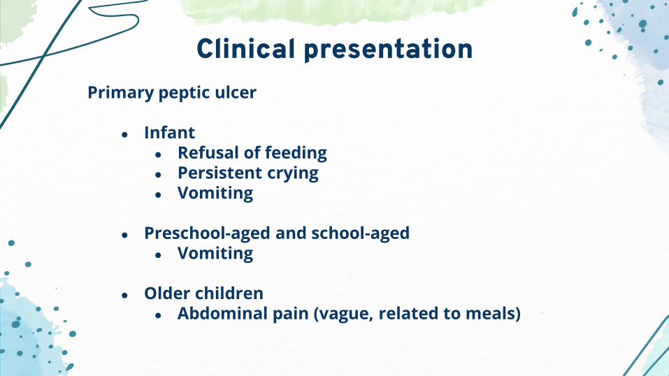
● Infant ● Refusal of feeding● Persistent crying● Vomiting
● Preschool-aged and school-aged● Vomiting
● Older children● Abdominal pain (vague, related to meals)
Clinical presentation

Secondary peptic ulcer
● Acute onset● Upper gastrointestinal hemorrhage (92%)● Vomiting● Perforation
Clinical presentation

● Clinical presentation● GI bleeding● Dysphagia● Persistent vomiting● Abdominal pain
● Gold standard : EGD (85%) ● Angiography : useful in locating a bleeding ulcer if rate of
bleeding is at least 0.5 mL/min
Diagnosis

● H. pylori infection● Invasive
● EGD + biopsy : ● nodularity in the antrum (specific but not sensitive)● Urease activity
● Non-invasive● H. pylori specific IgG● Urea breath test (> 2 yrs old)● Stool antigen test
Diagnosis

● Medical treatment● Antacids● H2 receptor antagonists
● Other agents● Selective anticholinergic● Proton-pump inhibitors● Cytoprotective agents● Anti-infective agents
Treatment


● Neutralize acid secretion● Heal peptic ulcer● Dosage : 0.5 mL/kg
● 1 hr ac, 3 hr pc, hs● Side effects
● Diarrhea● Constipation
Antacid

● Cimetidine ● Dosage 20-40 mg/kg/day● Antiandrogen side effects
● Ranitidine● Dosage 6-9 mg/kg/day oral, 2-4 mg/kg/day IV
H2 receptor antagonist

● Inhibit the stimulation of gastric acid secretion at the final common pathway
● Omeprazole● Dosage 1 mg/kg/day, max 20 mg/day● Side effects : headache, nausea, abdominal pain
● Lansoprazole● Dosage 0.5 mg/kg/day up to 30 kg● 30 mg daily if BW > 30 kg
Proton pump inhibitor

● Sucralfate● Negative charge of sulfated disaccharide adheres to
positive protein charge of the injured mucosa● Stimulated mucous production and prostaglandin
synthesis● Dosage 40-80 mg/kg/day● Side effects : constipation
● Prostaglandin E : misoprostol, enprostil, arbaprostil● Blocking production of cyclic AMP, stimulation of HCO3-,
increase mucosal blood flow● Few data in children use
Cytoprotective agent

H. Pylori associated disease
Medication Dose Duration
Amoxicillin 50 mg/kg/day 14 days bid
Clarithromycin 15 mg/kg/day 14 days bid
Proton pump inhibitor 1 mg/kg/day 1 month bid
Amoxicillin 50 mg/kg/day 14 days bid
Metronidazole 20 mg/kg/day 14 days bid
Proton pump inhibitor 1 mg/kg/day 1 month bid
Clarithromycin 15 mg/kg/day 14 days bid
Metronidazole 20 mg/kg/day 14 days bid
Proton pump inhibitor 1 mg/kg/day 1 month bid

● Reserved for peptic ulcer disease with complication● Perforation● Bleeding● Obstruction ● Intractable pain
● Prefer vagotomy and pyloroplasty > gastric resection
Surgical treatment

● Bleeding or perforated ulcer in the first 1-2 wk of life● Hypersecretion of acid caused by maternal gastrin● Often respond to OG decompression, lavage
● If surgical intervention is needed, ● Perforation : simplest method● Bleeding : simple suture ligation of the ulcer bed
● Chronic partial gastric outlet obstruction secondary to a congenital problem can result in peptic ulcer -> correct obstruction
Surgical treatment

● Hypergastrinemia● Relatively rare in children● Diagnosis
● Large gastric rugal fold● Duodenal dilatation● Edema of the small bowel mucosa
● Confirmed by elevated serum gastrin, calcium infusion test
● Treatment● Total gastrectomy if cannot completely resect primary
tumor● MEN1 can be present in 25% of cases
Zollinger-Ellison syndrome

● Prevention is the best● Medication : H2 receptor antagonists and PPI
● Major UGIB or recurrent UGIB : EGD● Therapeutic injection : hypertonic saline, epinephrine,
ethanol● Cauterization : heater probe, bipolar coagulation, laser● Stand-by surgery
● Massive hemorrhage : need operation ● blood loss in 24 hr
● < 2 yr : = total estimate blood volume● > 2 yr : = half of total estimate blood volume
(80mL/kg)
Stress ulcer

● Treat underlying cause● Simple surgical procedure : plication at perforation site,
oversewing of the bleeding point● Vagotomy and pyloroplasty : not interfere growth and
development
Stress ulcer

Gastric perforation in the newborn

● Rare condition in newborn● Incidence 1 in 2900 live births● 10-15% of all GI perforation in neonates and children● Male : Female -> inconclusive● Mortality 25%-50% in most case series ● Can occur in full-term, premature, and SGA neonates
Introduction

History
First demonstrated GI spontaneous
perforation
Leger et al.First successful repair of a
neonatal gastric perforation Lloyd
Multiple predisposing factors(selective circulatory ischemia)
1950
1969
1926
Siebold

Etiology
Spontaneous
Ischemic
Traumatic
Physiologic stress- Prematurity- Asphyxia- Sepsis- NEC
Redistribution of blood flowCausing microvascular injury
- Pneumatic distention : mask ventilation, PPV- Gastric intubation
Theories - Congenital absence of gastric muscle
- Forced exerted during vaginal delivery- Pneumatic distention : perforate at
fundus, ischemic change
Recent studyDeficiency of tyrosine kinase receptor
C-KIT+ mast cell and a lack of C-KIT+
interstitial cell of Cajal
Impair immunity andabnormal motility

● Idiopathic (Spontaneous)● Perinatal stress● Iatrogenic● Medication
Causes of neonatal gastric perforation

● Perinatal stress● Hypoxia● Asphyxia● Anatomic defect● Distal obstruction● Tracheoesophageal fistula● Congenital deficiency of gastric muscle
Causes of neonatal gastric perforation

● Iatrogenic● Nasogastric tube● Aggressive bag ventilation● Cardiopulmonary resuscitation● Positive pressure ventilation● Unintentional perforation during surgery (VP shunt)● Vaginal delivery
Causes of neonatal gastric perforation

● Medication● Indomethacin● Corticosteroids
Causes of neonatal gastric perforation

● Often occurs at DOL 3-5 (within the first 7 days)
● Presentation
Clinical presentation
Feeding intolerance Emesis contains Rapid blood abdominal distention

● Respiratory distress● Hemodynamic instability● Signs of shock
● Hypothermia● Cyanosis● Poor peripheral perfusion● Low urine output
● Physical examination● Abdomen rapidly tense and tender (peritoneal irritation)● Subcutaneous emphysema at the abdominal wall or
pneumoscrotum
Signs and symptoms

● Common site of perforation● Greater curvature● Posterior perforation into the lesser sac : insidious cause
(Difficult to diagnose)
● Increase risk in pregnancy with complicationAbruptio placentaePlacenta previaAmnionitisDelivered by emergency caesarian section
Signs and symptoms

● Clinical history + physical examination + radiographic studies
● Finding in plain abdominal radiographs● 90% non-visualized stomach● Pneumoperitoneum● Subcutaneous emphysema● Pneumoscrotum● OG/NG outside the confines of the stomach● Pneumatosis intestinalis : NEC (co-exist)
● Laboratory investigation● Complete blood count - Blood cultures● Arterial blood gas - Electrolyte profile
Diagnosis


● Vomiting and abdominal distention● Hirschsprung’s disease● Intestinal atresia● Meconium ileus● Meconium plug syndrome● Imperforate anus● Perforated viscus● NEC● Midgut volvulus
Differential diagnosis

● Cardiovascular collapse● Sepsis● Pneumothorax● Cardiac dysfunction● Intraventricular hemorrhage● NEC● Perforated hollow viscous organ● Malrotation with midgut volvulus
Differential diagnosis

● Early recognition and prompt treatment● Respiratory distress from marked abdominal distention
● Require intubation and ventilatory support● Broad-spectrum antibiotics● Fluid resuscitation, blood transfusion● OG/NG carefully passed and placed on low intermittent
suction for gastric decompression
● Paracentesis with IV canula : lifesaving when abdomen overly distend and interfere ventilation
Perioperative care

● Upper abdominal transverse incision● Dissect through the rectus muscle layer by
layer until the peritoneum is entered● The umbilical vein is divided● Peritoneal fluid and debris are evaluated and
sent for cultures (aerobic, anaerobic, fungi)
Surgical technique

● Explore site of perforation● Mostly spontaneous gastric perforation :
along greater curvature● Duodenal ulcer :
anterior wall or near the pyloroduodenal junction● Gastric ulcer :
along lesser curvature near the antral-fundic junction
● If cannot find the perforated site, carefully explore the EG junction, duodenum, small bowel and colon
● Open lesser sac and inspected for contamination andlesion at posterior wall of stomach
Surgical technique

● Debrided non-viable tissue around the perforated site
● Closed defect in one or two layers +/- omentalpatch
● Extensive perforation or necrosis may requiresub-total or total gastrectomy● If the greater curve is extensively
necrosed -> resection● If the antrum is extensively necrosed
-> Billroth I
● Reconstruction could be performed in stable infants
Surgical technique

● In total gastrectomy cases● Transverse colonic interposition● Roux-en-Y esophago-jejunal
anastomosis● Hunt-Lawrence pouch
reconstruction
● Staged surgery in unstable patients● Performed several weeks later when
the patient’s condition has improved
Reconstruction technique
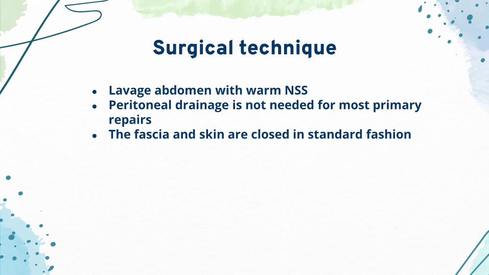
● Lavage abdomen with warm NSS● Peritoneal drainage is not needed for most primary
repairs● The fascia and skin are closed in standard fashion
Surgical technique

● Continue broad spectrum antibiotics until …● WBC and PMN within normal range● Evidence of bowel and gastric function
returns● Clear and low volume OG content
● Gastric acid suppression therapy● TPN● Continue gastric decompression● NPO until the patient has stabilized● May obtain a contrast study before starting
enteral feeding
Post-operative care

● Isolated gastric perforation survival 75-80%● Poor outcome associated with multiple-organ dysfunction,
sepsis, immature immunologic function● High morbidity and mortality in infants with gastric necrosis
with extensive NEC
Outcomes

Congenital microgastria

Rare congenital anomaly of the caudal part of foregut● Small, tubular stomach, megaesophagus, incomplete gastric
rotation, normal mucosa● Associated anomalies : VACTERL association
- GI : Non-rotation of midgut with duodenal band with asplenia, absence of gallbladder- Skeletal : micrognathia, radial and ulnar hypoplasia, vertebral anomalies, oligodactyly, and hypoplastic nail (rare : anophthalmia)- CVS : single atrium, single ventricle, total anomalous pulmonary venous return into the portal vein
Embryology and pathology

● Prenatal ultrasonographyPolyhydramniosSmall stomach
● Dilated esophagus with ill-defined E-G junction
● Vomiting● Aspiration and pneumonia● Bacterial overgrowth and blind-looplike syndrome
Failure to thriveDiarrhea Normal Schilling test
Clinical presentation

● Upper GI study● EGD : confusing results● If patients have GERD
ManometryEsophageal pH
● If patients have diarrhea and malabsorption
Intestinal absorption studies
Diagnosis

● Medical treatment- Continuous or night-time OG feeding- If GERD develops, start prokinetic agents and acid-reducing therapy- Complication of GERDS may require NJ feeding orsurgical jejunostomy tube feeding
● If stomach fails to enlarge, several authors recommended create a double-row jejunal reservoir (Hunt-Lawrence pouch)
Treatment

CREDITS: This presentation template was createdby Slidesgo, including icons by Flaticon,
infographics & images by Freepik
Thank you for your attention
Please keep this slide for attribution