Conditional ectopic expression of C/EBPP in NIH-3T3...
15
Conditional ectopic expression of C/EBPP in NIH-3T3 cells induces PPAR7 and stimulates adipogenesis Zhidan Wu/'^ Yuhong Xie/'^ Nancy L.R. Bucher,^ and Stephen R. Farmer^* Departments of 'Biochemistry and ^Pathology, Boston University School of Medicine, Boston, Massachusetts 02118 USA; ^Department of Chemistry, Boston University, Boston, Massachusetts 02215 USA Activation of adipogenesis in 3T3 preadipocytes by exposure to the adipogenic inducers dexamethasone, methylisobutyixanthine, insulin, and fetal bovine serum is accompanied by a transient burst of C/EBPp protein expression that precedes the induction of the fat gene program. In this study we have investigated the role of C/EBPp in initiating the adipogenic program by overexpressing C/EBPp in multipotential NIH-3T3 fibroblasts. Conditional ectopic expression of C/EBPp was accomplished by using an artificial transcriptional regulatory system based on the Escherichia coli tetracycline repressor to generate a stable cell line, p2, that expresses C/EBPp mRNA and protein in a tightly controlled tetracycline dose-dependent manner. Induction of C/EBPp DNA-binding activity in NIH-3T3 P2 cells exposed to dexamethasone in the presence of insulin and fetal bovine serum activates the expression of an adipocyte-specific nuclear hormone receptor, PPAR7, that stimulates the conversion of these fibroblasts into committed preadipocytes. Either ectopic expression of C/EBPp or treatment with dexamethasone alone is incapable of inducing PPARy expression, but when present together, they have a synergistic effect on the adipogenic program. Exposure of these stimulated cells to a PPAR activator 5,8,11,14-eicosatetraynoic acid (ETYA) results in the accumulation of fat droplets and expression of the adipocyte-enriched genes aP2 and glycerol phosphate dehydrogenase (GPD). The number of PZ cells that can differentiate into adipocytes is related to the concentration of tetracycline and, therefore, the amount of the exogenous C/EBPp protein expressed. C/EBPp can induce PPARy mRNA in the absence of ETYA; however, expression of aP2 mRNA and maximum fat deposition is dependent on the PPAR activator. Our results suggest that enhanced expression of C/EBPp converts multipotential mesenchymal precursor cells into preadipocytes that respond to adipogenic inducers, including dexamethasone and PPAR activators to differentiate into adipocytes. [Key Words: NIH-3T3 fibroblasts; adipogenesis; CCAAT/enhancer-binding protein, C/EBPP; peroxisome proliferator activated receptor, PPAR7] Received May 31, 1995; revised version accepted August 10, 1995. The differentiation of multipotential stem cells into functionally distinct cell types within different tissues involves the activation of a regulatory network of nu- clear factors and the cessation of cell proliferation. Sig- nificant progress toward an understanding of these elab- orate processes has been made by investigators studying the in vitro differentiation of mesenchymal cells into fat and muscle. In both cases, families of transcription factors have been identified that play a direct role in regulating the expression of tissue-restricted genes in the different cell types (Tapscott and Weintraub 1991; Vas- seur-Cognet and Lane 1993). The advances made in un- derstanding adipogenesis have stemmed from the isola- tion and characterization of the 3T3-L1 and 3T3-F442A preadipocyte cell lines that were originally derived from 'Corresponding author. mouse fibroblasts (Green and Kehinde 1974, 1975, 1976). In the case of 3T3-L1 cells, differentiation is induced upon exposure of a confluent population of cells to fetal bovine serum (FBS), dexamethasone (DEX), methylisobu- tyixanthine (MIX), and insulin. Differentiation of 3T3- F442A cells, however, requires only exposure of conflu- ent cells to FBS and insulin. In both cases, the differen- tiation is marked by a major change in cell morphology that includes the accumulation of large lipid droplets and the induction of a program of adipocyte-specific genes. Regulation of this program of gene expression has re- cently been shown to involve two families of transcrip- tion factors, the CCAAT/enhancer-binding proteins (C/ EBPs) and peroxisome proliferator-activated receptors (PPARs). C/EBPs are members of a diverse group of nu- clear factors that contain a leucine zipper domain re- quired for dimer formation and a basic DNA-binding do- 2350 GENES & DEVELOPMENT 9:2350-2363 © 1995 by Cold Spring Harbor Laboratory Press ISSN 0890-9369/95 $5.00 Cold Spring Harbor Laboratory Press on May 14, 2018 - Published by genesdev.cshlp.org Downloaded from
Transcript of Conditional ectopic expression of C/EBPP in NIH-3T3...

Conditional ectopic expression of C/EBPP in NIH-3T3 cells induces PPAR7 and stimulates adipogenesis Zhidan W u / ' ^ Yuhong X i e / ' ^ Nancy L.R. Bucher,^ and Stephen R. Farmer^*
Departments of 'Biochemistry and ^Pathology, Boston University School of Medicine, Boston, Massachusetts 02118 USA; ^Department of Chemistry, Boston University, Boston, Massachusetts 02215 USA
Activation of adipogenesis in 3T3 preadipocytes by exposure to the adipogenic inducers dexamethasone, methylisobutyixanthine, insulin, and fetal bovine serum is accompanied by a transient burst of C/EBPp protein expression that precedes the induction of the fat gene program. In this study we have investigated the role of C/EBPp in initiating the adipogenic program by overexpressing C/EBPp in multipotential NIH-3T3 fibroblasts. Conditional ectopic expression of C/EBPp was accomplished by using an artificial transcriptional regulatory system based on the Escherichia coli tetracycline repressor to generate a stable cell line, p2, that expresses C/EBPp mRNA and protein in a tightly controlled tetracycline dose-dependent manner. Induction of C/EBPp DNA-binding activity in NIH-3T3 P2 cells exposed to dexamethasone in the presence of insulin and fetal bovine serum activates the expression of an adipocyte-specific nuclear hormone receptor, PPAR7, that stimulates the conversion of these fibroblasts into committed preadipocytes. Either ectopic expression of C/EBPp or treatment with dexamethasone alone is incapable of inducing PPARy expression, but when present together, they have a synergistic effect on the adipogenic program. Exposure of these stimulated cells to a PPAR activator 5,8,11,14-eicosatetraynoic acid (ETYA) results in the accumulation of fat droplets and expression of the adipocyte-enriched genes aP2 and glycerol phosphate dehydrogenase (GPD). The number of PZ cells that can differentiate into adipocytes is related to the concentration of tetracycline and, therefore, the amount of the exogenous C/EBPp protein expressed. C/EBPp can induce PPARy mRNA in the absence of ETYA; however, expression of aP2 mRNA and maximum fat deposition is dependent on the PPAR activator. Our results suggest that enhanced expression of C/EBPp converts multipotential mesenchymal precursor cells into preadipocytes that respond to adipogenic inducers, including dexamethasone and PPAR activators to differentiate into adipocytes.
[Key Words: NIH-3T3 fibroblasts; adipogenesis; CCAAT/enhancer-binding protein, C/EBPP; peroxisome proliferator activated receptor, PPAR7]
Received May 31, 1995; revised version accepted August 10, 1995.
The differentiation of multipotential stem cells into functionally distinct cell types within different tissues involves the activation of a regulatory network of nuclear factors and the cessation of cell proliferation. Significant progress toward an understanding of these elaborate processes has been made by investigators studying the in vitro differentiation of mesenchymal cells into fat and muscle. In both cases, families of transcription factors have been identified that play a direct role in regulating the expression of tissue-restricted genes in the different cell types (Tapscott and Weintraub 1991; Vas-seur-Cognet and Lane 1993). The advances made in understanding adipogenesis have stemmed from the isolation and characterization of the 3T3-L1 and 3T3-F442A preadipocyte cell lines that were originally derived from
'Corresponding author.
mouse fibroblasts (Green and Kehinde 1974, 1975, 1976). In the case of 3T3-L1 cells, differentiation is induced upon exposure of a confluent population of cells to fetal bovine serum (FBS), dexamethasone (DEX), methylisobutyixanthine (MIX), and insulin. Differentiation of 3T3-F442A cells, however, requires only exposure of confluent cells to FBS and insulin. In both cases, the differentiation is marked by a major change in cell morphology that includes the accumulation of large lipid droplets and the induction of a program of adipocyte-specific genes.
Regulation of this program of gene expression has recently been shown to involve two families of transcription factors, the CCAAT/enhancer-binding proteins (C/ EBPs) and peroxisome proliferator-activated receptors (PPARs). C/EBPs are members of a diverse group of nuclear factors that contain a leucine zipper domain required for dimer formation and a basic DNA-binding do-
2350 GENES & DEVELOPMENT 9:2350-2363 © 1995 by Cold Spring Harbor Laboratory Press ISSN 0890-9369/95 $5.00
Cold Spring Harbor Laboratory Press on May 14, 2018 - Published by genesdev.cshlp.orgDownloaded from
C/EBPp induces adipogenesis in mouse fibroblasts
main that binds to regulatory elements in the promoters and/or enhancers of target genes. Expression of the C/EBP genes is regulated in such a way that three of the proteins (a,(3, and 8) are restricted to a limited number of tissues, most notably fat and liver (Cao et al. 1991; Williams et al. 1991). All three C/EBPs readily form het-erodimers with one another, exhibit similar DNA-bind-ing activities, and are capable of trails-activating reporter genes that contain C/EBP regulatory elements within their promoters (Cao et al. 1991). The pattern of expression of these genes during in vitro adipogenesis is consistent with a role for each protein in controlling the sequential activation of the adipogenic gene program (Cao et al. 1991). Recent studies suggest that C/EBPa has a direct role to play in establishing the quiescent, terminally differentiated state of adipocytes (Samuelsson et al. 1991; Umek et al. 1991; Lin and Lane 1994), and it can also stimulate adipogenesis when overexpressed in various mouse fibroblast cell lines (Freytag et al. 1994). Whether C/EBPa is responsible for initiating adipogenesis is questionable, however, because its expression occurs relatively late in the differentiation process (Vasseur-Cognet and Lane 1993).
A search for an adipocyte-specific transcription factor responsible for activating the adipogenic gene program led Spiegelman and co-workers (Tontonoz et al. 1994a) to identify a tissue-restricted member of the PPAR family of nuclear hormone receptors, PPAR72. As is the case with the C/EBPs, the PPAR proteins consist of a family of related proteins that are expressed to varying extents in different tissues (Kliewer et al. 1994). PPARa and PPAR7 are produced along with C/EBPa in tissues involved in energy metabolism. PPARot is expressed abundantly in liver (Isseman and Green 1990; Dreyer et al. 1992; Zhu et al. 1993), whereas PPAR7 expression is most prominent in adipose tissue (Tontonoz et al. 1994a). PPAR8 and NUCl are both expressed at low levels in many tissues (Schmidt et al. 1992; Chen et al. 1993; Khewer et al. 1994). PPARs are activated by a structurally diverse group of compounds, the peroxisome proliferators, which include the fibrate class of hypolipidemic drugs, herbicides, phthalate plasticizers, and long-chain fatty acids (Dreyer et al. 1992; Gottlicher et al. 1992; Keller et al. 1993). Activation of these receptors by specific peroxisome proliferators in target tissues such as liver and fat results in the expression of a variety of genes that encode proteins and enzymes involved in regulating lipid metabolism (Dreyer et al. 1992; Keller et al. 1993; Tontonoz et al. 1994b). Activation of PPARa in the liver and kidney causes a dramatic increase in the size and number of peroxisomes, thereby enhancing the capacity of these tissues to metabolize long-chain fatty acids through an increased production of enzymes that function in the p-oxidation cycle (Dreyer et al. 1992). The high level of PPAR7 expression in adipose tissue and its early induction during the differentiation of 3T3-L1 and 3T3-F442A preadipocytes suggest that this factor may initiate fat gene expression (Chawla et al. 1994; Tontonoz et al. 1994a). A recent study demonstrates that retroviral expression of PPAR72 in NIH-3T3 mouse fi
broblasts promotes adipocyte differentiation in a manner that is dependent upon the concentration of PPAR activators in the culture medium (Tontonoz et al. 1994b). Furthermore, the extent of this differentiation is enhanced severalfold when C/EBPa is expressed together with PPAR72, suggesting that both factors cooperate to induce the expression of adipocyte-specific genes. In fact, at least two genes expressed in fat, aP2 and PEPCK, contain regulatory elements within their promoters or enhancers that respond to trans-activation by C/EBPa and PPAR7 (Christy et al. 1989; Park et al. 1990; Tontonoz et al. 1994a, 1995).
It appears, therefore, that PPAR7 and C/EBPa are both capable of inducing fat-specific gene expression in mul-tipotential precursor cells such as NIH-3T3 fibroblasts, but it is not known whether one of these factors is responsible for inducing the expression of the other. It is unlikely that C/EBPa regulates PPAR7, because it is expressed at least 1 day after the induction of PPAR7 during the differentiation of 3T3 preadipocytes (Tontonoz et al. 1994b). It is more likely that PPAR7 regulates C/EBPa. If this is the situation, then an important question is. Which transcription factors activate PPAR7? The early transient activation of C/EBPp and C/EBP8 in 3T3-Ll cells in response to DEX and MIX (Cao et al. 1991) suggests that these factors may provide this important function of activating PPAR7. To address this question, we have developed an inducible expression system to facilitate the production of different amounts of C/EBPs in NIH-3T3 cells in response to an extracellular regulator. During the final stages of this work, a study by McK-night and co-workers (Yeh et al. 1995) demonstrated that retrovirus-mediated overcxpression of C/EBPP in NIH-3T3 cells can induce adipogenesis in the presence of adipogenic inducers. This observation was in conflict with earlier investigations by Freytag et al. (1994) who reported that C/EBPp was not capable of converting fibroblasts into fat cells. In the present study we demonstrate that conditional ectopic expression of C/EBPp in NIH-3T3 cells activates the synthesis of PPAR7 mRNA and subsequent exposure of these cells to PPAR activators stimulates their conversion into adipocytes.
Results
Conditional expression of C/EBPfS using the TET activator expression system
Previous investigations have shown that PPAR7 and C/EBPp mRNAs are induced severalfold during the initial period following the differentiation of 3T3 preadipocytes into adipocytes (Cao et al. 1991; Tontonoz et al. 1994a). It appears, however, that the peak of expression of C/EBPp protein precedes the activation and accumulation of PPAR7 mRNA. These data are consistent with our hypothesis that C/EBPp is involved in activating PPAR7 expression during fat cell differentiation. To test this idea, we established a stable line of NIH-3T3 cells that produce controlled amounts of C/EBP|3 protein from an inducible expression vector, and we have deter-
GENES & DEVELOPMENT 2351
Cold Spring Harbor Laboratory Press on May 14, 2018 - Published by genesdev.cshlp.orgDownloaded from
Wu et al.
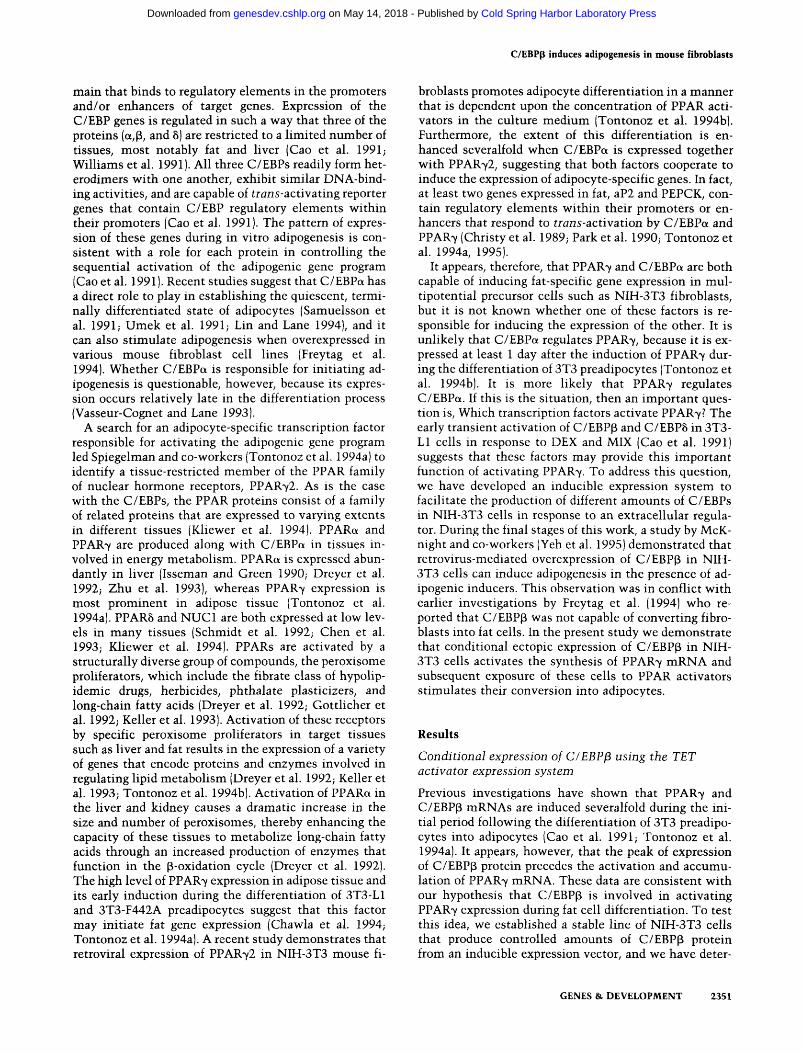
mined that C/EBPp can stimulate adipogenesis by inducing PPAR7 expression. To conditionally express C/EBPp proteins in NIH-3T3 cells, we employed an artificial transcriptional regulatory system developed by Gossen and Bujard (1992) that uses the bacterial tetracy-cline-resistant operator/repressor (Materials and methods). C/EBP(3 cDNA was subcloned into the expression vector, pUHDlO-3, downstream of a synthetic promoter composed of tandem repeats of the tetracycline operator and a CMV promoter. Activation of this minimal promoter is dependent on coexpression of an artificial trans-activator (tTA) composed of the tetracycline repressor fused to the activation domain of the herpes simplex virus transcriptional activator VP16. The presence of tetracycline prevents the binding of this trans-activator to the operator and, therefore, inhibits the expression of C/EBPp mRNA (Fig. lA). The plasmid, pUHD15-l, expressing tTA was transfected together with the C/EBPp expression vector and a neomycin-resistant plasmid. Conditional expression of C/EBP(3 mRNA in stably transfected cells was assessed by Northern blot analysis of total RNA isolated from G418-resistant colonies cultured in the presence or absence of tetracycline. One clone, referred to as ^2, which produced enhanced levels of C/EBPp mRNA in response to culture medium lacking tetracycline, was selected for further analysis. Cell lines expressing abundant quantities of tTA trans-activator alone were selected as controls.
The Northern blot analysis in Figure IB shows a modest level of C/EBPp mRNA expression in cells grown in the presence of tetracycline (lane 1). Removal of the drug results in a gradual but extensive induction of the exogenous mRNA over a 48-hr period. The size of this mRNA is larger than the endogenous species as shown by its migration to a slightly higher position in the gel. This is attributable to the transcription of part of the expression vector that contains the SV40 polyadenylation element giving rise to an additional —100 nucleotides in the exogenous mRNA. Addition of tetracycline to these stimulated cells results in a rapid decrease of C/EBPp mRNA levels within the initial 4 hr after treatment, indicating that the exogenous C/EBPp mRNA species is very short lived. Western blot analysis of total cell ly-sates obtained from |i2 cells maintained for several days in the presence or absence of tetracycline was performed with an anti-C/EBP(i antibody. Figure IC shows that (i2 cells grown in the presence of tetracycline and the NIH-3T3 cells expressing the tet activator alone (tTA cells) contained detectable quantities of the 32-kD C/EBPp (LAP) polypeptide but significantly lower amounts of the 21-kD C/EBPp (UP) protein. Culture of the p2 cells in the absence of tetracycline resulted in an increase by severalfold of both C/EBPp polypeptides, indicating that the ectopically expressed C/EBPp mRNA (see Fig. lA) is translated correctly in this 3T3 fibroblast clone.
PCMV tTA colEl bla pUHDlS-l
-tetracycline
Tet-O-C/EBPp
tctR-VPI6 fusion protein
+ tetracycline
tetO-PCMV C/EBPpcDNA SV4()-pA •IIIIIIIIIIIMIIM lllllllllllllll B-ggggga
COlEl
- tetracycline
C/EBPp polypeptides
bla
C Tetracycline
C/EBP(3^ (LAP)
C/EBPp^ (LIP)
NL tTA p2 p2 - - + -
B I
Remove Tetracycline Add Tetracycline
.C/EBPp '"mRNA
Figure 1. Inducible expression of C/EBPp in NIH-3T3 cells. [A] An illustration of the tetracycline activator expression system. [B] Kinetics of activation or inhibition of C/EBPp mRNA expression in ^2 cells in response to removal or addition of tetracycline. p2 cells were maintained in growth medium containing 1 M-g/nil of tetracycline. When the cells reached —50% confluence (time 0 hr, lane 1), tetracycline was removed and the cultures were incubated for the indicated times (0-48 hr, lanes 2-6). At 48 hr, cells were reexposed to the antibiotic (1 txg/ml) and cul
tured for an additional time period as shown (lanes 7-12). Twenty micrograms of total RNA isolated from each time point was analyzed by Northern blot hybridization for expression of the C/EBPp mRNA. (C) Western blot analysis of C/EBPp protein expression in 32 cells in the presence or absence of tetracycline for 48 hr. The tTA cells were maintained in tetracycline-free medium. Forty micrograms of nuclear protein extracted from both cell lines was examined by Western blot analysis using anti-C/EBPp antibody as described in Materials and methods. (NL) Normal adult rat liver nuclear proteins.
TimeHr->-Oh -8 -12 -24 -36 -48 +4 +8 +12+24+36+48
9ttt0|ti«ttt 8 9 10 11 12
2352 GENES & DEVELOPMENT
Cold Spring Harbor Laboratory Press on May 14, 2018 - Published by genesdev.cshlp.orgDownloaded from
C/EBPp induces adipogenesis in mouse fibroblasts
The DNA-binding activity of C/EBP polypeptides in 3T3-L1 pieadipocytes and NIH-3T3 fibroblasts
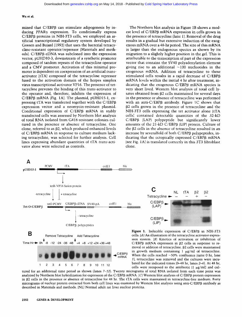
Preadipocytes have been shown to express at least three isoforms of C/EBP, a, (3, and b, at specific times during their differentiation into adipocytes. Each of these C/EBP polypeptides can associate with the others to form homo- and heterodimeric complexes that bind to DNA with different affinities (Cao et al. 1991). To identify the complexes expressed during the early phase of preadipocyte differentiation and to compare them with C/EBP complexes in the p2 cells, we performed a series of gel mobility supershift assays. Figure 2A (lane 1) shows that 3T3-L1 preadipocytes exposed to the adipo-genic inducers DEX, MIX, insulin, and FBS for 2 days result in the expression of a heterogeneous group of C/EBP complexes that are capable of binding to a consensus C/EBP binding site. At 4 days following induction of differentiation, the abundance of these complexes decreases quite significantly (lane 7). Supershift analysis with antibodies against the three C/EBP isoforms, a, p, and 5, shows that the major C/EBP binding activity ex
pressed at day 2 consists of complexes containing C/EBP(B (Fig. 2A, lane 3). Other reports have demonstrated that C/EBP8 polypeptides are also abundantly expressed at this stage of the differentiation, but the super-shift assay shown in lane 4 suggests that these proteins do not constitute a significant proportion of the total DNA-binding activity of the C/EBP complexes. The data in lane 2 shows a low but detectable level of C/EBPa binding activity at 2 days post induction. It is worth noting that the anti-C/EBPp antibody supershifts almost all of the C/EBP complexes (lane 3) except two low-abundance species that appear to correspond to 88 ho-modimers (the lower species) and a a homodimers (the upper species). Specifically, the lower species in lane 3 can be supershifted when the 2-day nuclear extracts are treated with anti-C/EBPp and anti-C/EBP8 antibodies together (lane 5), and the upper species is shifted when anti-C/EBPa antibody is included with the other two antibodies (lane 6). By day 4 of differentiation, the binding activity of both the C/EBPp and C/EBP8 complexes drop severalfold (cf. lane 9 with lane 3 and lane 10 with lane 4). In fact, it appears that the total C/EBP binding
C/EBP Antibody
C/EBP Complexes
DAY 4
a P 5 a/p p/5 a/
• - a p 5 p/5 p/5 - ex p 5 a/p p/5
WW W ^ ^
:Supershifted Complexes
1 2 3 4 5 6 7 8 9 10 11 12
B C/EBP Antibody
C/EBP Complexes
NL p2 + tetracycline p2 - tetracycline tJA-T
, ^ Supershifted Complexes
H^lMg^^WMi
Figure 2. Analysis of C/EBP DNA-binding activity in 3T3-L1 preadipocytes, 32, and tTA fibroblasts. {A) 3T3-L1 cells were cultured and induced to differentiate as described in Materials and methods. Nuclear proteins were extracted at day 2 and day 4 following exposure to differentiation medium. Ten micrograms of each extract was subjected to EMSA using a ''^P-labeled C/EBP oh-gonucleotide as probe. For the supershift assay, each extract was incubated with the appropriate antibodies (anti-C/EBPa, anti-C/EBPp, anti-C/ EBP8, or the combinations of these three as indicated) for 2 hr at room temperature prior to addition of the radiolabeled probe. (B) The fibroblast cell lines (p2 and tTA) were cultured in the presence or absence of tetracycline as described in the legend to Fig. 1. Nuclear proteins were extracted, and equal amounts (10 |xg) of each extract were subjected to EMSA as described in A.
GENES fit DEVELOPMENT 2353
Cold Spring Harbor Laboratory Press on May 14, 2018 - Published by genesdev.cshlp.orgDownloaded from
Wu et al.
activity expressed at this stage of adipogenesis is accounted for by complexes primarily involving C/EBPa (lane 8). Figure 2B shows that nuclear extracts isolated from NIH-3T3 cells, either |32 cells maintained in tetracycline (lane 3) or fibroblasts transfected with the tTA expression vector alone (lane 11), have low but measurable levels of C/EBP binding activity when compared with differentiating preadipocytes (Fig. 2A) or normal rat liver (Fig. 2B, lane 1). Most of this activity in the 3T3 cells is attributable to C/EBPp polypeptides (Fig. 2B, lanes 5,12). Culture of the (32 cells in the absence of tetracycline for 48 hr results in a severalfold induction of total C/EBP binding activity (Fig. 2B, lane 7), which can be attributed to the overexpressed C/EBPp proteins because the anti-C/EBPp antibody supershifts almost all of the C/EBP complexes produced in these cells (lane 9). There is very little C/EBP6 DNA-binding activity and no C/EBPa activity.
Conditional expression of C/EBPp in NIH-3T3 cells stimulates adipogenic conversion
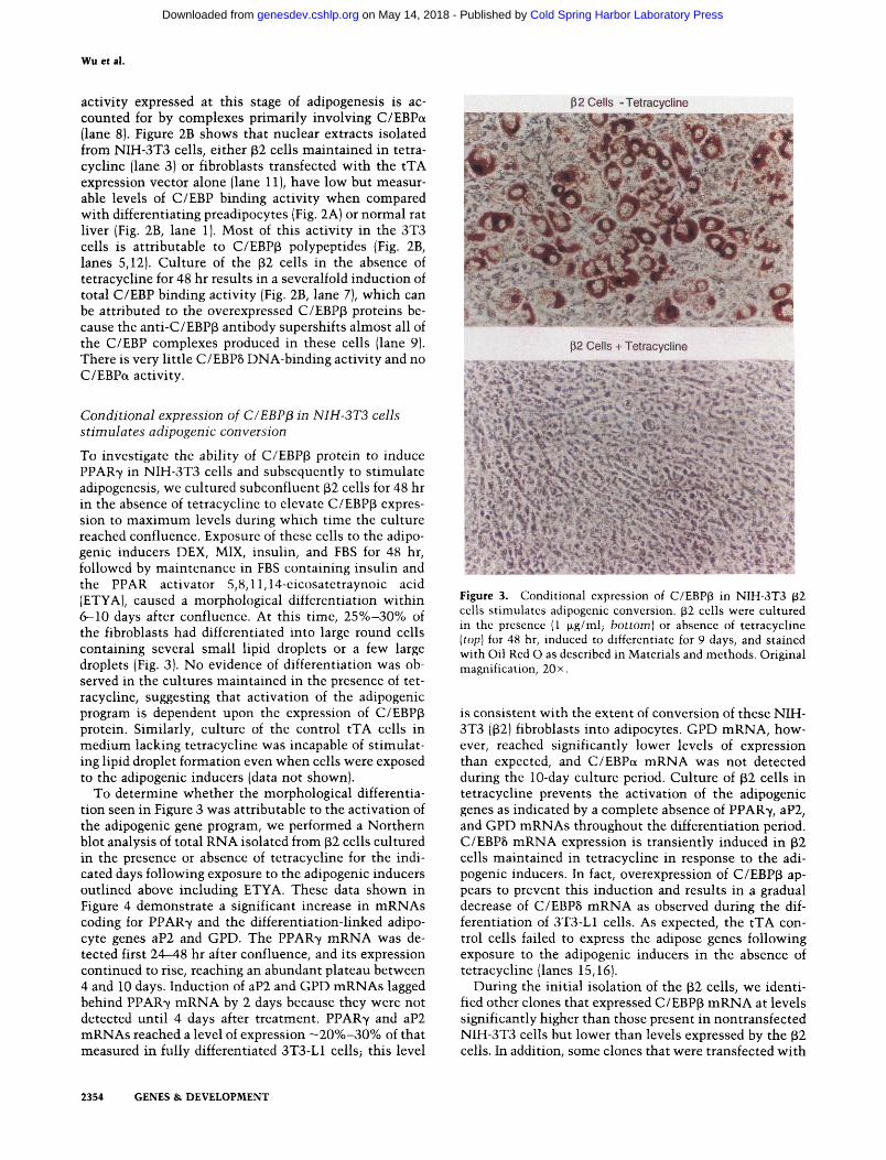
To investigate the ability of C/EBP(3 protein to induce PPAR7 in NIH-3T3 cells and subsequently to stimulate adipogenesis, we cultured subconfluent p2 cells for 48 hr in the absence of tetracycline to elevate C/EBPp expression to maximum levels during which time the culture reached confluence. Exposure of these cells to the adipogenic inducers DEX, MIX, insulin, and FBS for 48 hr, followed by maintenance in FBS containing insulin and the PPAR activator 5,8,11,14-cicosatetraynoic acid (ETYA), caused a morphological differentiation within 6-10 days after confluence. At this time, 25%-30% of the fibroblasts had differentiated into large round cells containing several small lipid droplets or a few large droplets (Fig. 3). No evidence of differentiation was observed in the cultures maintained in the presence of tetracycline, suggesting that activation of the adipogenic program is dependent upon the expression of C/EBPp protein. Similarly, culture of the control tTA cells in medium lacking tetracycline was incapable of stimulating lipid droplet formation even when cells were exposed to the adipogenic inducers (data not shown).
To determine whether the morphological differentiation seen in Figure 3 was attributable to the activation of the adipogenic gene program, we performed a Northern blot analysis of total RNA isolated from (32 cells cultured in the presence or absence of tetracycline for the indicated days following exposure to the adipogenic inducers outlined above including ETYA. These data shown in Figure 4 demonstrate a significant increase in mRNAs coding for PPAR7 and the differentiation-linked adipocyte genes aP2 and GPD. The PPAR7 mRNA was detected first 24-48 hr after confluence, and its expression continued to rise, reaching an abundant plateau between 4 and 10 days. Induction of aP2 and GPD mRNAs lagged behind PPAR7 mRNA by 2 days because they were not detected until 4 days after treatment. PPAR7 and aP2 mRNAs reached a level of expression ~20%-30% of that measured in fully differentiated 3T3-L1 cells; this level
P2Cells -Tetracycline
p2 Cells + Tetracycline
£ »*
' '*• V- e - '« ^ • -5. \ A . t ^
Figure 3. Conditional expression of C/EBPp in NIH-3T3 (32 cells stimulates adipogenic conversion. (i2 cells were cultured in the presence (1 fJLg/ml; bottom) or absence of tetracycline [top] for 48 hr, induced to differentiate for 9 days, and stained with Oil Red O as described in Materials and methods. Original magnification, 20 x.
is consistent with the extent of conversion of these NIH-3T3 02) fibroblasts into adipocytes. GPD mRNA, however, reached significantly lower levels of expression than expected, and C/EBPa mRNA was not detected during the 10-day culture period. Culture of p2 cells in tetracycline prevents the activation of the adipogenic genes as indicated by a complete absence of PPAR7, aP2, and GPD mRNAs throughout the differentiation period. C/EBP6 mRNA expression is transiently induced in p2 cells maintained in tetracycline in response to the adipogenic inducers. In fact, overexpression of C/EBPp appears to prevent this induction and results in a gradual decrease of C/EBP8 mRNA as observed during the differentiation of 3T3-L1 cells. As expected, the tTA control cells failed to express the adipose genes following exposure to the adipogenic inducers in the absence of tetracycline (lanes 15,16).
During the initial isolation of the p2 cells, we identified other clones that expressed C/EBP(3 mRNA at levels significantly higher than those present in nontransfected NIH-3T3 cells but lower than levels expressed by the (B2 cells. In addition, some clones that were transfected with
2354 GENES & DEVELOPMENT
Cold Spring Harbor Laboratory Press on May 14, 2018 - Published by genesdev.cshlp.orgDownloaded from
C/EBPp induces adipogenesis in mouse fibroblasts
P 2 +Tetracycline , P2 - Tetracycline ,tTA-T,L1
Day -2 0 1 2 4 6 7 I 0 1 2 4 6 7 10| 0 101
PPAR7
C/EBP8 ! • • • • • • • • • • • • • -
1 2 3 4 5 6 7 8 9 10 11 12 13 14 15 16 17
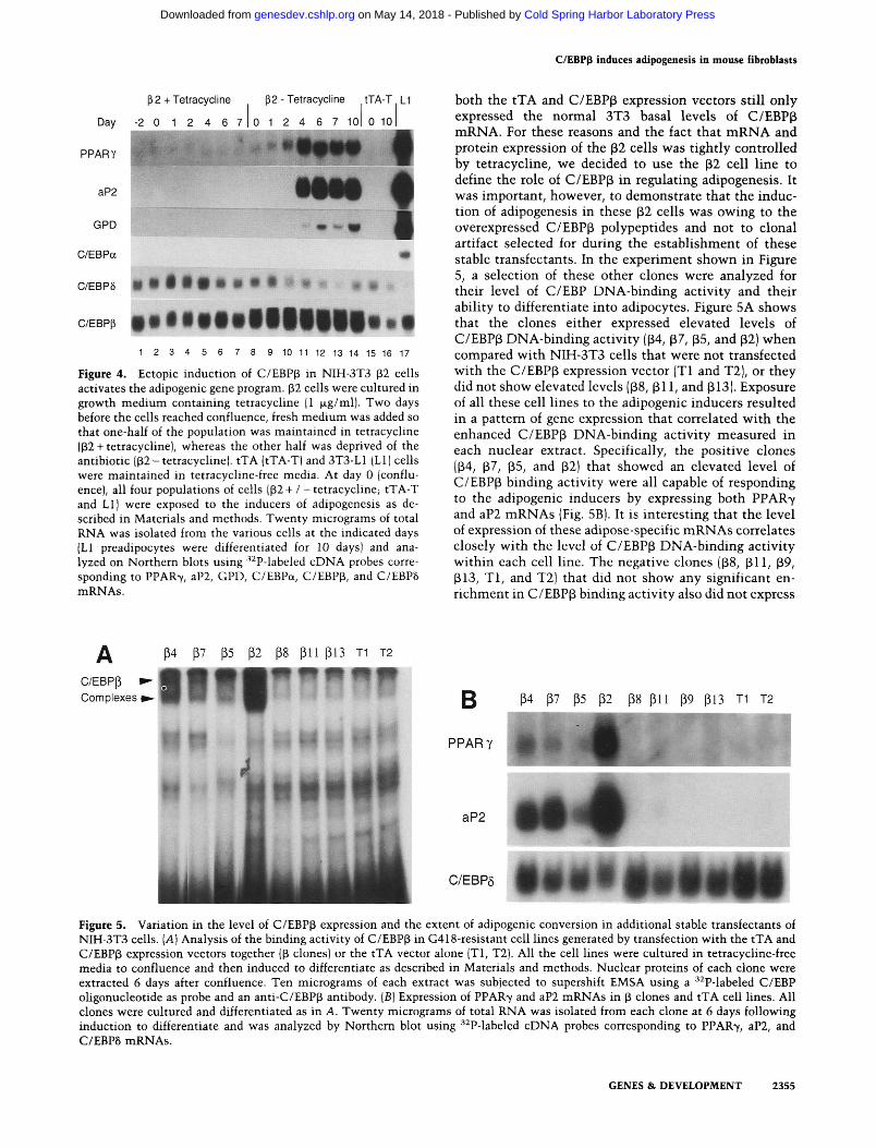
Figure 4. Ectopic induction of C/EBPp in NIH-3T3 (32 cells activates the adipogenic gene program. P2 cells were cultured in growth medium containing tetracycline |1 fxg/ml). Two days before the cells reached confluence, fresh medium was added so that one-half of the population was maintained in tetracycline (P2 +tetracycline), whereas the other half was deprived of the antibiotic (p2-tetracychne!. tTA (tTA-T) and 3T3-L1 (LI) cells were maintained in tetracycline-free media. At day 0 (confluence), all four populations of cells 02 + / -tetracycline; tTA-T and LI) were exposed to the inducers of adipogenesis as described in Materials and methods. Twenty micrograms of total RNA was isolated from the various cells at the indicated days (LI preadipocytes were differentiated for 10 days) and analyzed on Northern blots using '•^P-labeled cDNA probes corresponding to PPAR7, aP2, GPD, C/EBPa, C/EBPp, and C/EBP8 mRNAs.
both the tTA and C/EBPp expression vectors still only expressed the normal 3T3 basal levels of C/EBPp mRNA. For these reasons and the fact that mRNA and protein expression of the p2 cells was tightly controlled by tetracycline, we decided to use the P2 cell line to define the role of C/EBPp in regulating adipogenesis. It was important, however, to demonstrate that the induction of adipogenesis in these p2 cells was owing to the overexpressed C/EBPp polypeptides and not to clonal artifact selected for during the establishment of these stable transfectants. In the experiment shown in Figure 5, a selection of these other clones were analyzed for their level of C/EBP DNA-binding activity and their ability to differentiate into adipocytes. Figure 5A shows that the clones either expressed elevated levels of C/EBPp DNA-binding activity ((34, (37, (35, and p2) when compared with NIH-3T3 cells that were not transfected with the C/EBPp expression vector (Tl and T2), or they did not show elevated levels ((38, p i 1, and ^13). Exposure of all these cell lines to the adipogenic inducers resulted in a pattern of gene expression that correlated with the enhanced C/EBPp DNA-binding activity measured in each nuclear extract. Specifically, the positive clones (p4, p7, (35, and ^2) that showed an elevated level of C/EBP(3 binding activity were all capable of responding to the adipogenic inducers by expressing both PPAR7 and aP2 mRNAs (Fig. 5B). It is interesting that the level of expression of these adipose-specific mRNAs correlates closely with the level of C/EBP(3 DNA-binding activity within each cell line. The negative clones ((38, (311, P9, 313, T l , and T2) that did not show any significant enrichment in C/EBPp binding activity also did not express
C/EBP(3 I Complexes
(34 (37 (35 (32 pS p i l p l3 Tl T2
lllinnnni M ^ U M M M M M iW P H ^ ^ W i 1P| ^^P W •_
B P4 [37 p5 (32 (38 p l l P9 pl3 T1 T2
PPARY
aP2
C/EBP5
Figure 5. Variation in the level of C/EBPp expression and the extent of adipogenic conversion in additional stable transfectants of NIH-3T3 cells. [A] Analysis of the binding activity of C/EBP(3 in G418-resistant cell lines generated by transfection with the tTA and C/EBPp expression vectors together (p clones) or the tTA vector alone (Tl, T2). All the cell lines were cultured in tetracycline-free media to confluence and then induced to differentiate as described in Materials and methods. Nuclear proteins of each clone were extracted 6 days after confluence. Ten micrograms of each extract was subjected to supershift EMSA using a ^^P-labeled C/EBP oligonucleotide as probe and an anti-C/EBPp antibody. [B] Expression of PPAR7 and aP2 mRNAs in p clones and tTA cell lines. All clones were cultured and differentiated as in A. Twenty micrograms of total RNA was isolated from each clone at 6 days following induction to differentiate and was analyzed by Northern blot using ^^P-labeled cDNA probes corresponding to PPAR-y, aP2, and C/EBP8 mRNAs.
GENES & DEVELOPMENT 2355
Cold Spring Harbor Laboratory Press on May 14, 2018 - Published by genesdev.cshlp.orgDownloaded from
Wu et al.
the adipogenic gene program when exposed to differentiation medium.
The extent of the differentiation of NIH-3T3 cells into adipocytes is dependent on the amount of C/EBPp protein
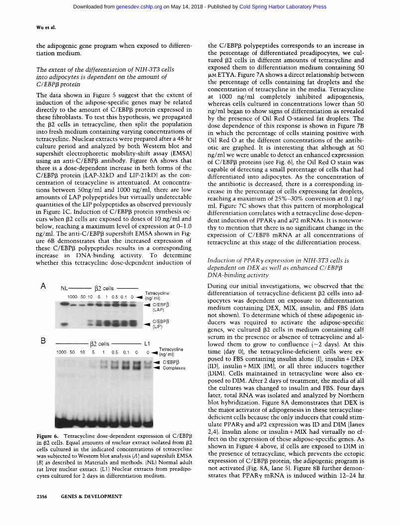
The data shown in Figure 5 suggest that the extent of induction of the adipose-specific genes may be related directly to the amount of C/EBPp protein expressed in these fibroblasts. To test this hypothesis, we propagated the p2 cells in tetracycline, then split the population into fresh medium containing varying concentrations of tetracycline. Nuclear extracts were prepared after a 48-hr culture period and analyzed by both Western blot and supershift electrophoretic mobility-shift assay (EMSA) using an anti-C/EBPp antibody. Figure 6A shows that there is a dose-dependent increase in both forms of the C/EBPp protein (LAP-32kD and LIP-21kD) as the concentration of tetracycline is attentuated. At concentrations between 50ng/ml and 1000 ng/ml, there are low amounts of LAP polypeptides but virtually undetectable quantities of the LIP polypeptides as observed previously in Figure IC. Induction of C/EBPp protein synthesis occurs when p2 cells are exposed to doses of 10 ng/ml and below, reaching a maximum level of expression at O-1.0 ng/ml. The anti-C/EBPp supershift EMSA shown in Figure 6B demonstrates that the increased expression of these C/EBPp polypeptides results in a corresponding increase in DNA-binding activity. To determine whether this tetracycline dose-dependent induction of
NL (32 cells
1000 50 10 5 1 0.5 0.1 0 Tetracycline (ng/ ml)
& -^ C/EBPP (LAP)
C/EBPP • * (LIP)
B (32 cells
1000 50 10 5 1 0.5 0,1
LI
0
BBV^:: Tetracycline (ng/ ml)
C/EBPP Complexes
Figure 6. Tetracycline dose-dependent expression of C/EBPp in p2 cells. Equal amounts of nuclear extract isolated from 32 cells cultured in the indicated concentrations of tetracycline was subjected to Western blot analysis [A] and supershift EMSA (B) as described in Materials and methods. (NL) Normal adult rat liver nuclear extract. (LI) Nuclear extracts from preadipo-cytes cultured for 2 days in differentiation medium.
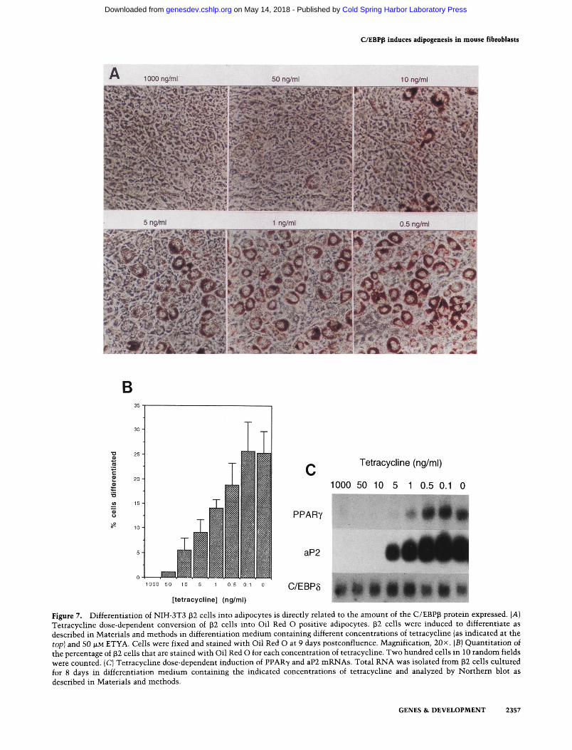
the C/EBPp polypeptides corresponds to an increase in the percentage of differentiated preadipocytes, we cultured p2 cells in different amounts of tetracycline and exposed them to differentiation medium containing 50 |XM ETYA. Figure 7A shows a direct relationship between the percentage of cells containing fat droplets and the concentration of tetracycline in the media. Tetracycline at 1000 ng/ml completely inhibited adipogenesis, whereas cells cultured in concentrations lower than 50 ng/ml began to show signs of differentiation as revealed by the presence of Oil Red O-stained fat droplets. The dose dependence of this response is shown in Figure 7B in which the percentage of cells staining positive with Oil Red O at the different concentrations of the antibiotic are graphed. It is interesting that although at 50 ng/ml we were unable to detect an enhanced expresssion of C/EBPp proteins (see Fig. 6), the Oil Red O stain was capable of detecting a small percentage of cells that had differentiated into adipocytes. As the concentration of the antibiotic is decreased, there is a corresponding increase in the percentage of cells expressing fat droplets, reaching a maximum of 25%-30% conversion at 0.1 ng/ ml. Figure 7C shows that this pattern of morphological differentiation correlates with a tetracycline dose-dependent induction of PPAR7 and aP2 mRNAs. It is noteworthy to mention that there is no significant change in the expression of C/EBP8 mRNA at all concentrations of tetracycline at this stage of the differentiation process.
Induction of PPARy expression in NIH-3T3 cells is dependent on DEX as well as enhanced C/EBPp DNA-binding activity
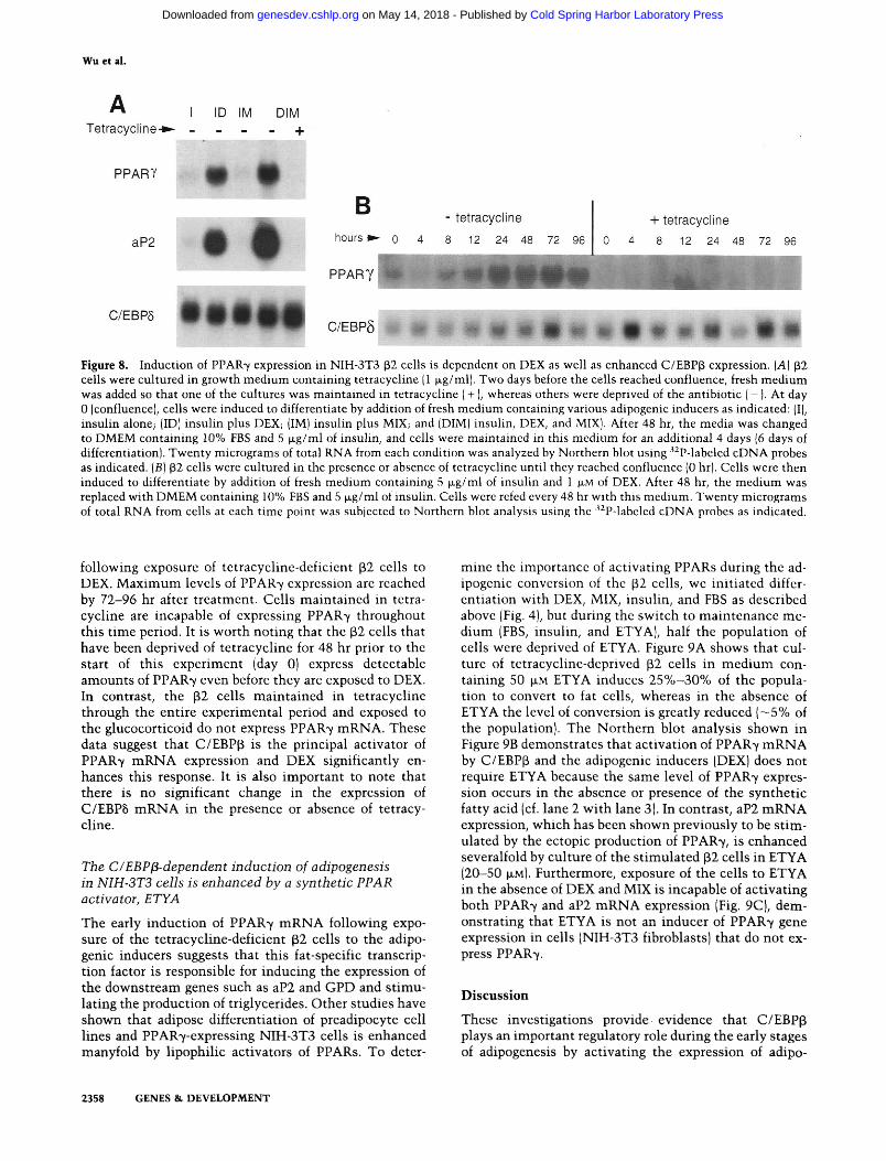
During our initial investigations, we observed that the differentiation of tetracycline-deficient (32 cells into adipocytes was dependent on exposure to differentiation medium containing DEX, MIX, insulin, and FBS (data not shown). To determine which of these adipogenic inducers was required to activate the adipose-specific genes, we cultured p2 cells in medium containing calf serum in the presence or absence of tetracycline and allowed them to grow to confluence (~2 days). At this t ime (day 0), the tetracycline-deficient cells were exposed to FBS containing insulin alone (I), insulin -I- DEX (ID), insulin-I-MIX (IM), or all three inducers together (DIM). Cells maintained in tetracycline were also exposed to DIM. After 2 days of treatment, the media of all the cultures was changed to insulin and FBS. Four days later, total RNA was isolated and analyzed by Northern blot hybridization. Figure 8A demonstrates that DEX is the major activator of adipogenesis in these tetracycline-deficient cells because the only inducers that could stimulate PPAR7 and aP2 expression was ID and DIM (lanes 2,4). Insulin alone or insulin-t-MIX had virtually no effect on the expression of these adipose-specific genes. As shown in Figure 4 above, if cells are exposed to DIM in the presence of tetracycline, which prevents the ectopic expression of C/EBP|i protein, the adipogenic program is not activated (Fig. 8A, lane 5). Figure 8B further demonstrates that PPAR7 mRNA is induced within 12-24 hr
2356 GENES & DEVELOPMENT
Cold Spring Harbor Laboratory Press on May 14, 2018 - Published by genesdev.cshlp.orgDownloaded from
C/EBPp induces adipogenesis in mouse fibroblasts
1000ng/ml
5 ng/ml
50 ng/ml 10 ng/ml
1 ng/ml 0.5 ng/ml
B
Tetracycline (ng/ml)
1000 50 10 5 1 0.5 0.1 0
1000 50 10 5 1 0.5 0.1
[tetracycline] (ng/ml)
PPARy
aP2
C/EBP8
Figure 7. Differentiation of NIH-3T3 32 cells into adipocytes is directly related to the amount of the C/EBPp protein expressed. [A] Tetracychne dose-dependent conversion of p2 cells into Oil Red O positive adipocytes. p2 cells were induced to differentiate as described in Materials and methods in differentiation medium containing different concentrations of tetracycline (as indicated at the top] and 50 M-M E T Y A . Cells were fixed and stained with Oil Red O at 9 days postconfluence. Magnification, 20 x. (B) Quantitation of the percentage of (32 cells that are stained with Oil Red O for each concentration of tetracychne. Two hundred cells in 10 random fields were counted. (C) Tetracycline dose-dependent induction of PPAR7 and aP2 mRNAs. Total RNA was isolated from p2 cells cultured for 8 days in differentiation medium containing the indicated concentrations of tetracycline and analyzed by Northern blot as described in Materials and methods.
GENES & DEVELOPMENT 2357
Cold Spring Harbor Laboratory Press on May 14, 2018 - Published by genesdev.cshlp.orgDownloaded from
Wu et al.
A Tetracycline*-
1 ID IM DIM . - - - +
PPARY • •
aP2 • •
B - tetracycline
hours » • 0 4 8 12 24 48 72 96
PPARY
+ tetracycline
0 4 8 12 24 48 72 96
C/EBP6 C/EBP5 • gi li' 9 ' 9 9 f l | A f l | A # # f l | i i ^ f l | 9
Figure 8. Induction of PPAR'Y expression in NIH-3T3 (32 cells is dependent on DEX as well as enhanced C/EBPp expression. (A) (32 cells were cultured in growth medium containing tetracycline |1 fig/ml). Two days before the cells reached confluence, fresh medium was added so that one of the cultures was maintained in tetracycline (+ ), whereas others were deprived of the antibiotic (-). At day 0 (confluence), cells were induced to differentiate by addition of fresh medium containing various adipogenic inducers as indicated: (I), insulin alone; (ID) insulin plus DEX; (IM) insulin plus MIX; and (DIM) insulin, DEX, and MIX). After 48 hr, the media was changed to DMEM containing 10% FBS and 5 |xg/ml of insulin, and cells were maintained in this medium for an additional 4 days (6 days of differentiation). Twenty micrograms of total RNA from each condition was analyzed by Northern blot using '^P-labeled cDNA probes as indicated. (B) p2 cells were cultured in the presence or absence of tetracycline until they reached confluence (0 hr). Cells were then induced to differentiate by addition of fresh medium containing 5 |xg/ml of insulin and 1 |xM of DEX. After 48 hr, the medium was replaced with DMEM containing 10% FBS and 5 |xg/ml of insulin. Cells were rcfed every 48 hr with this medium. Twenty micrograms of total RNA from cells at each time point was subjected to Northern blot analysis using the • P-labeled cDNA probes as indicated.
follow^ing exposure of tetracycline-deficicnt (i2 cells to DEX. Maximum levels of PPAR7 expression are reached by 72-96 hr after treatment. Cells maintained in tetracycline are incapable of expressing PPAR7 throughout this time period. It is worth noting that the ^2 cells that have been deprived of tetracycline for 48 hr prior to the start of this experiment (day 0) express detectable amounts of PPAR-y even before they are exposed to DEX. In contrast^ the p2 cells maintained in tetracycline through the entire experimental period and exposed to the glucocorticoid do not express PPAR7 mRNA. These data suggest that C/EBP(3 is the principal activator of PPAR'v mRNA expression and DEX significantly enhances this response. It is also important to note that there is no significant change in the expression of C/EBP5 mRNA in the presence or absence of tetracycline.
The C/EBP ^-dependent induction of adipogenesis in NIH-3T3 cells is enhanced by a synthetic PPAR activator, ETYA
The early induction of PPAR7 mRNA foUov^ ing exposure of the tetracycline-deficient p2 cells to the adipogenic inducers suggests that this fat-specific transcription factor is responsible for inducing the expression of the downstream genes such as aP2 and GPD and stimulating the production of triglycerides. Other studies have shown that adipose differentiation of preadipocyte cell lines and PPAR7-expressing NIH-3T3 cells is enhanced manyfold by lipophilic activators of PPARs. To deter
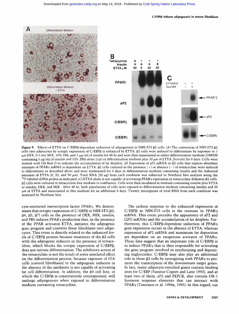
mine the importance of activating PPARs during the adipogenic conversion of the p2 cells, we initiated differentiation with DEX, MIX, insulin, and FBS as described above (Fig. 4), but during the switch to maintenance medium (FBS, insulin, and ETYA), half the population of cells were deprived of ETYA. Figure 9A shows that culture of tetracycline-deprived p2 cells in medium containing 50 \xM ETYA induces 25%-30% of the population to convert to fat cells, whereas in the absence of ETYA the level of conversion is greatly reduced (—5% of the population). The Northern blot analysis shown in Figure 9B demonstrates that activation of PPAR7 mRNA by C/EBPp and the adipogenic inducers (DEX) does not require ETYA because the same level of PPAR7 expression occurs in the absence or presence of the synthetic fatty acid (cf. lane 2 with lane 3). In contrast, aP2 mRNA expression, which has been shown previously to be stimulated by the ectopic production of PPAR7, is enhanced severalfold by culture of the stimulated P2 cells in ETYA (20-50 JJLM). Furthermore, exposure of the cells to ETYA in the absence of DEX and MIX is incapable of activating both PPAR7 and aP2 mRNA expression (Fig. 9C], demonstrating that ETYA is not an inducer of PPAR7 gene expression in cells (NIH-3T3 fibroblasts) that do not express PPAR7.
Discussion
These investigations provide evidence that C/EBPp plays an important regulatory role during the early stages of adipogenesis by activating the expression of adipo-
2358 GENES & DEVELOPMENT
Cold Spring Harbor Laboratory Press on May 14, 2018 - Published by genesdev.cshlp.orgDownloaded from
C/EBPp induces adipogenesis in mouse fibroblasts
Differentiation Medium
Differentiation Medium + ETYA
B Tetracycline + - - -
Insulin + + + + ETYAixM 50 0 20 50
PPARY ETY A/INS Dex/MIX
aP2
C/EBPp
•N * •III V
C/EBP5 HI m *
PPARY
•aP2
•C/EBP5
Figure 9. Effects of ETYA on C/EBP(3-dependent induction of adipogenesis in NIH-3T3 32 cells. [A] The conversion of NIH-3T3 (32 cells into adipocytes by ectopic expression of C/EBPp is enhanced by ETYA. p2 cells were induced to differentiate by exposure to 1 (JLM DEX, 0.5 mM MIX, 10% FBS, and 5 Jl.g/ml of insulin for 48 hr and were then maintained in either differentiation medium (DMEM containing 5 |xg/ml of insulin and 10% FBS) alone [top] or differentiation medium plus 50 p,M of ETYA [bottom) for 9 days. Cells were stained with Oil Red O to indicate the accumulation of fat droplets. [B] Expression of aP2 mRNA in ^2 cells that express abundant amounts of PPAR7 mRNA is dependent on ETYA. ^2 cells cultured in the presence ( + ) or absence ( - ) of tetracycline were induced to differentiate as described above and were maintained for 4 days in differentiation medium containing insulin and the indicated amounts of ETYA (0, 20, and 50 JJLM). Total RNA (20 |jLg| from each condition was subjected to Northern blot analysis using the ''^P-labeled cDNA probes as indicated. (C) ETYA alone is not capable of activating PPAR7 expression in tetracycline-deficient p2 cells. p2 cells were cultured in tetracycline-free medium to confluence. Cells were then incubated in medium containing insulin plus ETYA or insulin, DEX, and MIX . After 48 hr, both populations of cells were exposed to differentiation medium containing insulin and 50 JJLM of ETYA and maintained in this medium for an additional 4 days. Twenty micrograms of total RNA from each condition was analyzed by Northern blot.
cyte-restricted transcription factor PPAR7. We demonstrate that ectopic expression of C/EBPp in NIH-3T3 (32, P4, (35, (i7) cells in the presence of DEX, MIX, insulin, and FBS induces PPAR7 production that, in the presence of the PPAR activator ETYA, initiates the adipogenic gene program and converts these fibroblasts into adipocytes. This event is directly related to the enhanced levels of C/EBP(3 protein because treatment of the ^2 cells with the adipogenic inducers in the presence of tetracycline, which blocks the ectopic expression of C/EBP(3, does not initiate differentiation. The inhibitory action of the tetracycline is not the result of some unrelated effect on the differentiation process, because exposure of tTA cells (control fibroblasts) to the adipogenic inducers in the absence of the antibiotic is not capable of activating fat cell differentiation. In addition, the (i4 cell line, in which the C/EBP(i is constitutively overexpressed, will undergo adipogenesis when exposed to differentiation medium containing tetracycline.
The earliest response to the enhanced expression of C/EBPp in NIH-3T3 cells is the increase in PPAR7 mRNA. This event precedes the appearence of aP2 and GPD mRNAs and the accumulation of fat droplets. Furthermore, this C/EBP(i-dependent induction of PPAR7 gene expression occurs in the absence of ETYA, whereas expression of aP2 mRNA and maximum fat deposition are dependent on an exogenous activator of PPAR7. These data suggest that an important role of C/EBPp is to induce PPAR7 that is then responsible for activating the gene program involved in synthesizing and depositing triglycerides. C/EBP^ may also play an additional role in these p2 cells by synergizing with PPAR7 to promote the transcription of the downstream target genes. In fact, many adipocyte-enriched genes contain binding sites for C/EBP (Vasseur-Cognet and Lane 1993), and at least two of them, aP2 and PEPCK, also contain DR-1 hormone response elements that can interact with PPAR7 (Tontonoz et al. 1994a, 1995). In this regard, our
GENES & DEVELOPMENT 2359
Cold Spring Harbor Laboratory Press on May 14, 2018 - Published by genesdev.cshlp.orgDownloaded from

Wu et al.
preliminary data (not shown) suggest that C/EBPp may function to maintain expression of the adipocyte genes at the later stages of the differentiation process because addition of tetracycline to (32 cells (to inhibit C/EBPp) after PPAR7 has reached a maximum level of expression (day 4) reduces aP2 expression.
Enhanced expression of C/EBPp alone is not sufficient to activate adipogenesis in the NIH-3T3 p2 cell line because exposure to DEX in the presence of insulin and PBS is also required. What role DEX and the other inducers is playing is not known, but information may be gained from investigations on the mechanisms that regulate the early phase of 3T3-L1 preadipocyte differentiation. Studies by Cao et al. (1991) show that DEX enhances C/EBP5 mRNA expression, whereas MIX is responsible for inducing C/EBPp mRNA production in the preadipocytes. It is unlikely that these effectors are involved in stimulating either C/EBPp or C/EBP8 expression in the NIH-3T3 P2 cells because C/EBPp, of course, is activated by removal of tetracycline, whereas C/EBP8 appears to decrease somewhat soon after the differentiation process has commenced (Fig. 4). These observations suggest that DEX must be fulfilling a function other than enhancing expression of the C/EBP8 proteins during the differentiation of the p cells. Other investigations (Lemberger et al. 1994) have demonstrated that glucocorticoids can activate PPARa gene expression in primary cultures of rat hepatocytes, suggesting that the PPAR genes may contain glucocorticoid response elements in their promoters and/or enhancers. The role of insulin and FBS in promoting adipogenesis in both 3T3-L1 and p2 cells is unclear; one possibility is that they promote the translation of the induced adipogenic mRNAs.
The mechanism by which C/EBPp induces PPAR7 mRNA is not known, although, on the basis of studies analyzing the regulation of other C/EBP-responsive genes, it seems reasonable to propose that PPAR7 gene transcription is likewise regulated by binding of various C/EBPs to corresponding binding sites within the promoter and/or enhancer of the PPAR7 gene. In fact, we have shown that ectopic expression of C/EBPa in NIH-3T3 cells can also induce expression of the PPAR7 gene and stimulate adipogenesis (Z. Wu et al., unpubl.). The data shown in Figure 2 suggest that the C/EBP complexes responsible for inducing PPAR-Y expression and stimulating adipogenesis in the 3T3-L1 and (32 cells consist primarily of C/EBPp homodimers.
It is possible that the forced expression of C/EBPp homodimers in the NIH-3T3 cells and the hormonal induction of these and other complexes in 3T3-L1 cells (Fig. 2A) result in somewhat different programs of gene expression. In this regard, it is interesting that C/EBPa gene expression is not induced by C/EBPp in the p2 cells. This observation is consistent with that of McKnight and co-workers (Yeh et al. 1995) who have recently found that constitutive, retrovirus expression of C/EBPp in NIH-3T3 cells induces fat droplet accumulation and aP2 expression without enhancing C/EBPa expression. The promoter of the mouse C/EBPa gene contains a C/EBP regulatory element that is capable of ac
tivating C/EBPa transcription in transfection studies in which C/EBPa and C/EBPp expression vectors are coex-pressed along with C/EBPa promoter/CAT reporter gene constructs (Christy et al. 1991; Rana et al. 1995). It appears, therefore, that C/EBPp may be necessary but not sufficient to induce C/EBPa transcription during adipogenesis. Other factors may include C/EBP6 that, as discussed above, appears to contribute only minimally to the formation of functional C/EBP complexes during the early stages of the differentiation of (32 cells into adipocytes. In addition, the sustained high levels of C/EBPp homodimers expressed in the differentiating p2 cells may in some way prevent the activation of C/EBPa transcription even though other fat-enriched genes that contain C/EBP-binding sites are activated. In fact, during the normal differentiation of 3T3-L1 cells there is a several-fold decrease in the abundance of the C/EBP(3 proteins prior to the induction of C/EBPa gene expression (Cao et al. 1991; Fig. 2A). The C/EBP(3 polypeptide referred to as LIP, which is a potent inhibitor of C/EBPp (LAP) activity (Descombes and Schibler 1991), is also abundantly expressed in (32 cells following removal of tetracycline (Fig. IC). It is possible, therefore, that LIP attenuates the activity of LAP during the induction of adipogenesis in (32 cells by reducing LAP's ability to trans-activate some of the adipocyte-enriched genes including C/EBPa. This may be one reason why only 25%-30% of the p2 cell population differentiate into adipocytes. C/EBPa may also be regulated by PPAR7, and the C/EBPa gene may resemble other downstream genes such as aP2 and PEPCK by possessing DR-1 hormone response elements. If this is the case, then it is conceivable that activation of C/EBPa by PPAR7 may depend on a PPAR activator that is more potent than the ETYA used in these studies.
As stated at the outset, 3T3 preadipocyte cell lines were originally derived from mouse fibroblasts by selecting colonies that could differentiate into fat cells when exposed to the appropriate medium (Green and Kehinde 1974, 1975, 1976). An important question in light of the present studies is. Which molecular mechanisms are responsible for committing multipotential fibroblast cells to the adipogenic lineage? It is very apparent that NIH-3T3 mouse fibroblasts cannot simply be induced to differentiate into adipocytes by addition of DEX, MIX, insulin, and FBS, suggesting that the signaling pathways that respond to these effectors do not converge on fat-specific genes in these nonadipogenic cells. Our data show that enhancing the production of C/EBPp polypeptides can convert a fibroblast into a preadipocyte when exposed to these adipogenic inducers and suggests that the mechanism responsible for commitment to the adipogenic lineage involves the induction of C/EBPp gene transcription and translation. It is interesting that elevation of cAMP levels by treatment with MIX in 3T3-L1 preadipocytes causes a severalfold increase in C/EBPp gene expression and, therefore, facilitates induction of PPAR7 mRNA. Similar treatment of normal, untrans-fected NIH-3T3 cells with MIX has little or no effect on C/EBPp production, and the cells are incapable of converting into adipocytes. It is important to note that 3T3-
2360 GENES & DEVELOPMENT
Cold Spring Harbor Laboratory Press on May 14, 2018 - Published by genesdev.cshlp.orgDownloaded from
C/EBPp induces adipogenesis in mouse fibroblasts
F442A preadipocytes do not require MIX or DEX to differentiate into adipocytes. It is possible that these cells are slightly further along in the differentiation process than 3T3-L1 cells, and, therefore, they may constitu-tively express abundant amounts of C/EBPp and C/EBP8 mRNAs. They may then only require insulin and/or FBS to enhance synthesis of the corresponding proteins to activate the fat gene program including PPAR7 expression. It is unclear whether the mechanisms responsible for inducing C/EBPp expression in preadipocytes are specific to the adipogenic lineage. The mechanisms that activate PPAR7 expression appear not to be adipocyte specific because they can operate in NIH-3T3 cells, unless induction of C/EBPp proteins in these fibroblasts converts them to preadipocytes prior to the increase in PPAR7 expression. An additional component of the commitment process may include the production and/or utilization of PPAR activators. It is interesting that differentiation of (32 fibroblasts into adipocytes after the induction of PPAR7 by C/EBPP requires stimulation v^ith ETYA to presumably activate transcription of genes containing PPAR response elements. In contrast, 3T3-L1 and 3T3-F442A preadipocytes do not require any additional stimulation from PPAR activators to differentiate into fat cells other than the fatty acids present in the serum (Chav^^la and Lazar 1994). It is possible that these preadipocytes synthesize a specific ligand for PPAR7 and that this event is adipocyte specific and contributes to the acquisition of the adipogenic lineage.
In light of the observations made in this study, it is interesting to compare this regulatory network of adipogenic transcription factors with those expressed in myogenic cells. In the muscle cells, there are several nuclear regulatory proteins including MyoD, Myf 5, Myogenin, Mrf 4, and myocyte enhancer factor 2 (MEF2) that are all involved in regulating skeletal muscle gene expression. Many of the genes coding for these myogenic factors can be trans-activated by the other family members, giving rise to a complex regulatory circuit that controls the expression of muscle-specific genes during myogenesis (Ed-mondson et al. 1992; Weintraub 1993; Olson and Klein 1994). A similar situation may exist in adipocytes in which PPARs and C/EBPs may represent two arms of a network of regulatory factors that can trans-activate each other and, in so doing, amplify the expression of the downstream adipocyte-enriched genes.
Materials and methods
Plasmid constructions
tTA (pUHD15-l) and tetracycline operator-CMV promoter expression (pUHDlO-3) plasmids were obtained from H. Bujard of ZFMB, University of Heidelberg, Germany (Gossen and Bujard 1992). The MSV-C/EBPp plasmid was provided by S. McKnight of Tularik, Inc., South San Francisco, California. To construct a tTA-dependent C/EBPp expression vector, the 1.5-kb C/EBPp cDNA from an EcoRl-BamHl double digest of the MSV-C/ EBPp plasmid was inserted into the EcoRl-BamHl sites within the polylinker region of pUHDlO-3. Transcription of C/EBPp mRNA from the resultant plasmid, Tet-O-C/EBPp, is driven by
the CMV promoter containing seven repeats of the tet operator upstream of the C/EBPp cDNA in response to coexpression of tTA from the pUHD15-l plasmid.
Cell culture and transfections
3T3-L1 preadipocytes were maintained in growth medium consisting of Dulbecco's modified Eagle medium (DMEM; GIBCO BRL, Life Technologies, Inc., Gaithersburg, MD) containing 10% normal calf serum (Intergen, Co., Purchase, NY) and were induced to differentiate as described previously by Student et al. (1980). This involved growing cells to confluence that was considered day 0 of the differentiation program. At this stage, cells were exposed to fresh DMEM containing 10% FBS, 1 JJLM DEX, 0.5 mM MIX, and 10 |JLg/ml of insulin for 48 hr to initiate adipogenesis. The medium was then replaced with DMEM containing 10% FBS and 2.5 fjig/ml of insulin, and cells were refed every other day. The NIH-3T3 mouse fibroblasts were maintained in DMEM with 10% FBS. To estabUsh an inducible C/EBPp expression cell line, the fibroblasts were transfected with pUHD 15-1 (tTA), Tet-O-C/EBPp and pSV2neo with lipo-fectAMINE following the protocol provided by the supplier (GIBCO BRL). Briefly, a mixture of 10 )xg of Tet-O-C/EBPp, 10 jjLg of pUHD15-l, 1 Jig of pSV2neo plasmids, and 40 p.1 of lipo-fectAMINE in 1.6 ml of DMEM was incubated at room temperature for 30 min and then diluted to 8 ml with DMEM. Cells (1.5x10'") per 100-mm dish were washed twice with DMEM, and the diluted DNA-lipid mixture was added. After 6 hr of incubation at 37°C, the cells were refed with DMEM supplemented with 10% FBS and 1 ^.g/ml of tetracycline and were cultured for an additional 36 hr before 0.4 mg/ml of G418 was added. The transfected cells were selected in DMEM containing 0.4 mg/ml of G418 for 10 to 14 days, and the resistant clones were subsequently isolated and propagated into cell lines. The stable cell lines were maintained in DMEM containing 10% FBS, 1 |JLg/ml tetracycline, and 0.2 mg/ml of G418 and were screened by Northern blot analysis to identify a clone ((32) that expressed abundant amounts of C/EBPp mRNA in response to withdrawal of tetracycline.
To differentiate the p series of stable transfectants into adipocytes, cells were grown to —50% confluence in DMEM/10% FBS containing 1 jjig/ml of tetracycline. To activate C/EBPp expression, tetracycline was removed from the culture media, and the cells were allowed to grow for an additional 48 hr by which time they had reached confluence. At this stage, referred to as day 0, adipogenesis was induced by addition of fresh medium containing 1 |xm of DEX, 0.5 mM of MIX, and 5 M-g/ml of insulin. After 48 hr, the medium was changed to DMEM containing 10% FBS, 5 jJLg/ml of insulin, and 50 |JLM of ETYA or as indicated. The cells were refed every 2 days. To confirm the appearance of adipocyte morphology, especially the accumulated fat droplets in the cytoplasts, the cells were fixed by 10% formaldehyde in phosphate-buffered saline and stained with Oil Red O (Preece 1972).
RNA analysis
Total RNA was isolated from the NIH-3T3 p2 and 3T3-L1 cells by the procedure of Chomczynski and Sacchi (1987). Cells were washed twice with ice-cold phosphate-buffered saline and lysed with Solution D (4M guanidinium thiocyanate, 25 mM sodium citrate at pH 7.0, 0.5% Sarkosyl, 0.1 M 2-mercaptoethanol), and the lysate was extracted with acidic phenol/chloroform; this was followed by isopropanol precipitation at - 20°C overnight. Twenty micrograms of each RNA sample was analyzed by Northern blot hybridization as described previously (Bond and
GENES & DEVELOPMENT 2361
Cold Spring Harbor Laboratory Press on May 14, 2018 - Published by genesdev.cshlp.orgDownloaded from
Wu et al.
Farmer 1983), except the RNA was blotted onto Hybond-N nylon membrane (Amersham). The probes were labeled by random priming using the Klenow fragment of DNA polymerase I (New England Biolabs, Inc., Beverley, MA) and [a^^P]dCTP(DuPont, NEN Research Products, Boston, MA). cDNAs used as probes were as follows: rat C/EBPa, C/EBPp, and C/EBP8 (Cao et al. 1991), 422/aP2 (Spiegelman et al. 1983; Bernlohr et al. 1984), glycerol phosphate dehydrogenase (GPD) (Dobson et al. 1987), and PPAR-Y (Aperlo et al. 1995).
Western blot (immunoblot) analysis
Nuclei were isolated from normal liver hepatocytes, 3T3-L1, and NIH-3T3 ^2 cells following a procedure described previously (Rana et al. 1994). The cells were washed twice with ice-cold phosphate-buffered saline and lysed with NP-40 lysis buffer (10 mM Tris-HCl at pH 7.4, 10 mM NaCl, 3 mM MgClj, and 0.5% NP-40). The cell lysates were then transferred to a conical tube and incubated on ice for 15 min, and nuclei were pelleted by centrifugation at 1000 g for 5 min at 4°C. The pellets were washed again with lysis buffer, resuspended in nuclear suspension buffer (50 mM Tris-HCl at pH 7.5, 10 mM magnesium acetate, 40% glycerol, 1 mM dithiothreitol (DTT)|, and stored at - 80°C until ready for use. To extract nuclear proteins, the stored nuclei were thawed on ice and harvested by centrifugation at 1000 g for 10 min; protein extraction buffer (0.4 M NaCl, 5 mM EDTA, 10 mM sodium HEPES at pH 7.5, 0.2 mM PMSF, and 1 mM DTT) was added to the nuclear pellet and incubated on ice for 20 min with occasional mixing. The nuclear protein supernant was recovered by centrifugation for 15 min and stored at - 80°C after addition of glycerol to a final concentration of 15%. The protein concentration was determined using Bio-Rad protein assay kit (Bio-Rad Laboratories, Hercules, CA). Fourty micrograms of nuclear protein was fractionated by 12% SDS-polyacrylamide gel electrophoresis (SDS-PAGE) and electropho-retically transferred to nitrocellulose membrane (Schleicher &. Schuell, Inc., Keene, NH). Membranes were incubated in TBST (25 mM Tris-HCl at pH B.O, 125 mM NaCl, and 0 .1% Tween 20) containing 10% nonfat dry milk (Carnation Co., Los Angeles, CA) to block nonspecific binding. The blots were then incubated with primary antibody, a polyclonal anti-C/EBP^ antiserum (1:1000 dilution; Santa Cruz Biotechnology, Inc., Santa Cruz, CA) in blocking solution (TBST containing 5% milk) for 1 hr at room temperature. After being washed three times with the blocking solution, the blots were incubated with a secondary antibody, horseradish peroxidase-conjugated rabbit antimo-use IgG (1:1000 dilution; Sigma Chemical Co., St Louis, MO) for 1 hr. After a final wash with TBST, the blots were developed by the enhanced chemiluminescence system (Amersham Life Sciences, Arlington, IL) and visualized by exposure to autoradiography film.
EMSA
DNA-binding assays were performed as described previously (Rana et al. 1994). Ten micrograms of nuclear extract was incubated with 3 |jLg of poly[d(I-C)], 2 \il of carrier mix (50 mM MgCli and 340 mM KCl), and delta buffer (0.1 mM EDTA, 40 mM KCl, 25 mM HEPES at pH 7.6, 8% Ficoll, and 1 mM DTT) at 4°C for 15 min. The ['^^P]dCTP-radiolabeled double-stranded oligonucleotide (50,000 cpm) was added to the reaction mixture and incubated for 30 min at 4°C. For the super shift assay, the appropriate antibody was added to the nuclear extract and incubated at room temperature for 1.5 hr before the probe was added. The anti-C/EBPa, C/EBPp and C/EBP8 antibodies were obtained from Santa Cruz Biotechnology. Binding of the oligonu
cleotide to specific proteins was determined by fractionating the nuclear proteins through a nondenaturing 6% polyacryl-amide gel at 150-200 V for 3-5 hr at 4 ^ in TBE buffer (0.09 M Tris-borate, 0.002 M EDTA at pH 8.0). The gel was dried at 80°C for 1 hr before exposure to autoradiography. The sequence of the C/EBP oligonucletide used in this study is 5'-gatccGCGT-TGCGCCACGATG-3' (Christy et al. 1991). The double-stranded oligonucleotide was end-labeled by T4 DNA polynucleotide kinase (New England Biolabs) and ['v-'^^PlATP (Du Pont).
A c k n o w l e d g m e n t s
We are grateful to Johan Auwerx, Hermann Bujard, Deborah Dobson, Steven McKnight and Jacqueline Stephens for providing cDNA probes and expression vectors. We also thank Deborah Dobson and Jacqueline Stephens for helpful discussions and advice on culturing 3T3 preadipocytes. This work was funded by U.S. Public Health Service grants DK45048 and CA39099 from the National Institutes of Health.
The publication costs of this article were defrayed in part by payment of page charges. This article must therefore be hereby marked "advertisement" in accordance with 18 USC section 1734 solely to indicate this fact.
Note added in proof
Zhu et al. [PTOC. Natl. Acad. Sci. 92: 7921-7925, 1995) have recently demonstrated that the promoter of the mouse PPAR-^ gene contains two C/EBP sites.
References
Aperlo, C , P. Pognonec, J. Auwerx, and K.E. Boulokos. 1995. Isolation and characterization of the human peroxisome pro-liferator activated receptor, hPPAR^, a member of the nuclear hormone receptor superfamily. Gene 162: 297-302.
Bernlohr, D.A., C.W. Angus, M.D. Lane, M.A. Bolanowski, and T.J.J. Kelly. 1984. Expression of specific mRNAs during adipose differentiation: Identification of an mRNA encoding a homologue of myelin P2 protein. Proc. Natl. Acad. Sci. 81:5468-5472.
Bond, J.F. and S.R. Farmer. 1983. Regulation of tubulin and actin mRNA production in rat brain: Expression of a new p-tubu-lin mRNA with development. Moi. Cell. Biol. 3: 1333-1342.
Cao, Z., R.M. Umek, and S.L. McKnight. 1991. Regulated expression of three C/EBP isoforms during adipose conversion of 3T3-L1 cells. Genes &. Dev. 5: 1538-1552.
Chawla, A. and M.A. Lazar. 1994. Peroxisome proliferator and retinoid signalling pathways co-regulate preadipocyte phe-notype and survival. Pioc. Natl. Acad. Sci. 91: 1786-1790.
Chawla, A., E.J. Schwartz, D.D. Dimaculangan, and M.A. Lazar. 1994. Peroxisome proliferator-activated receptor (PPAR) gamma: Adipose-predominant expression and induction early in adipocyte differentiation. Endocrinology 135: 798-800.
Chen, F., S.W. Law, and B.W. O'Malley. 1993. Identification of two mPPAR related receptors and evidence for the existence of five subfamily members. Biochem. Biophys. Res. Com-mun. 196: 671-677.
Chomczynski, P. and N. Sacchi. 1987. Single-step method of RNA isolation by acid guanidinium thiocyanate-phenol-chloroform extraction. Anal. Biochem. 162: 156-159.
Christy, R.J., V.W. Yang, J.M. Ntambi, D.E. Getman, W.H. Landschulz, A.D. Friedman, Y. Nakabeppu, J.T. Kelly, and
2362 GENES & DEVELOPMENT
Cold Spring Harbor Laboratory Press on May 14, 2018 - Published by genesdev.cshlp.orgDownloaded from
C/EBPp induces adipogenesis in mouse fibroblasts
M.D. Lane. 1989. Differentiation-induced gene expression in 3T3-L1 preadipocytes:CCAAT/enhancer binding protein interacts with and activates the promoters of two adipocyte-specific genes. Genes &. Dev. 3: 1323-1335.
Christy, R.J., K.H. Kaestner, D.E. Geiman, and M.D. Lane. 1991. CCAAT-Enhancer binding protein gene promoter: Binding of nuclear factors during differentiation of 3T3-L1 preadipo-cytes. Proc. Natl. Acad. Sci. 88: 2593-2597.
Descombes, P. and U. Schibler. 1991. A liver-enriched transcriptional activator protein, LAP, and a transcriptional inhibitory protein, LIP, are translated from the same mRNA. Cell 67: 569-579.
Dobson, D.E., D.L. Groves, and B.M. Spiegelman. 1987. Nucleotide sequence and hormonal regulation of mouse glycerophosphate dehydrogenase mRNA during adipocyte and muscle cell differentiation. /. Biol. Chem. 262: 1804-1809.
Dreyer, C , G. Krey, H. Keller, F. Givel, G. Helftenbein, and W. Wahli. 1992. Control of the peroxisomal p-oxidation pathway by a novel family of nuclear hormone receptors. Cell 68: 879-887.
Edmondson, D.G., T.C. Cheng, P. Cserjesi, T. Chakraborty, and E.N. Olson. 1992. Analysis of the myogenin promoter reveals an indirect pathway for positive autoregulation mediated by the muscle-specific enhancer factor MEF-2. Mol. Cell. Biol. 12: 3665-3677.
Freytag, S.O., D.L.Paielli, and J.D. Gilbert. 1994. Ectopic expression of the CCAAT/enhancer-binding protein a promotes the adipogenic program in a variety of mouse fibroblastic cells. Genes &. Dev. 8: 1654-1663.
Gossen, M. and H. Bujard. 1992. Tight control of gene expression in mammalian cells by tetracycline-responsive promoters. Proc. Natl. Acad. Sci. 89: 5547-5551.
GottUcher, M., E. Widmark, Q. Li, and J.A. Gustafsson. 1992. Fatty acids activate a chimera of the clofibric acid-activated receptor and the glucocorticoid receptor. Proc. Natl. Acad. Sci. 89: 4653-4657.
Green, H. and O. Kehinde. 1974. Sublines of mouse 3T3 cells that accumulate lipid. Cell 1: 113-116.
. 1975. An established cell line and its differentiation in culture 11. Factors affecting the adipose conversion. Cell 5: 19-27.
1976. Spontaneous heritable changes leading to increased adipose conversion of 3T3 cells. Cell 7: 105-113.
Isseman, I. and S. Green. 1990. Activation of a member of the steroid hormone receptor superfamily by peroxisome prolif-erators. Nature 347: 645-649.
Keller, H., C. Dreyer, J. Medin, A. Mahfoudi, K. Ozato, and W. Wahli. 1993. Fatty acids and retinoids control lipid metabolism through activation of peroxisome proliferator-activated receptor-retinoid X receptor heterodimers. Proc. Natl. Acad. Sci. 90: 2160-2164.
Kliewer, S.A., B.M. Forman, B. Blumberg, E.S. Ong, U. Borgm-eyer, D.J. Mangelsdorf, K. Umesono, and R.M. Evans. 1994. Differential expression and activation of a family of murine peroxisome proliferator-activated receptors. Proc. Natl. Acad. Sci. 91 : 7355-7359.
Lemberger, T., B. Staels, R. Saladin, B. Desvergne, J. Auwerx, and W. Wahli. 1994. Regulation of the peroxisome proliferator-activated receptor a gene by glucocorticoids. /. Biol. Chem. 269: 24527-24530.
Lin, F.-T. and M.D. Lane. 1994. CCAAT/enhancer binding protein a is sufficient to initiate the 3T3-L1 adipocyte program. Proc. Natl. Acad. Sci. 91 : 8757-8761.
Olson, E.N. and W.H. Klein. 1994. bHLH factors in muscle development: Dead lines and commitments, what to leave in and what to leave out. Genes & Dev. 8: 1-8.
Park, E.A., W.J. Roesler, J. Liu, D.J. Klemm, A.L. Gumey, J.D. Thatcher, J. Schuman, A. Friedman, and R.W. Fianson. 1990. The role of the CAAT/enhancer-binding protein in the transcriptional regulation of the gene for phosphoenolpyruvate carboxykinase (GTP). Mol. Cell. Biol. 10: 6264-6272.
Preece, A. 1972. Manual for histologic technicians, p. 260. Little, Brown, and Co., Boston, MA.
Rana, B., D. Mischoulon, Y. Xie, N.L.R. Bucher, and S.R. Farmer. 1994. Cell-extracellular matrix interactions can regulate the switch between growth and differentiation in rat hepatocytes: Reciprocal expression of C/EBPa and immediate-early growth response transcription factors. Mol. Cell. Biol. 14: 5858-5869.
Rana, B., Y. Xie, D. Mischoulon, N.L.R. Bucher, and S.R. Farmer. 1995. The DNA binding activity of C/EBP transcription factors is regulated in the G l phase of the hepatocyte cell cycle. /. Biol. Chem. 270: 18123-18132.
Samuelsson, L., K. Stromberg, K. Vikman, G. Bjursell, and S. Enerback. 1991. The CCAAT/enhancer binding protein and its role in adipocyte differentiation: Evidence for direct involvement in terminal adipocyte development. EMBO f. 10: 3787-3793.
Schmidt, A., N. Endo, S.J. Rutledge, R. Vogel, D. Shinar, and G.A. Rodan. 1992. Identification of a new member of the steroid hormone receptor superfamily that is activated by a peroxisome proliferator and fatty acid. Mol. Endocrinol. 6:1634-1641.
Spiegelman, B.M., M. Frank, and H. Green. 1983. Molecular cloning of mRNA from 3T3 adipocytes. /. Biol. Chem. 258:10083-10089.
Student, A.K., R.Y. Hsu, and M.D. Lane. 1980. Induction of fatty acid synthetase synthesis in differentiating 3T3-L1 preadi-pocytes. /. Biol. Chem. 255: 4745-4750.
Tapscott, S.J. and H. Weintraub. 1991. MyoD and the regulation of myogenesis by helix-loop-helix proteins. /. Clin. Invest. 87: 1133-1138.
Tontonoz, P., E. Hu, R.A. Graves, A.I. Budavari, and B.M. Spiegelman. 1994a. mPPAR^: Tissue-specific regulator of an adipocyte enhancer. Genes &. Dev. 8: 1224—1234.
Tontonoz, P., E. Hu, and B.M. Spiegelman. 1994b. Stimulation of adipogenesis in fibroblasts by PPAR^, a lipid-activated transcription factor. Cell 79: 1147-1156.
Tontonoz, P., E. Hu, J. Devine, E.G. Beale, and B.M. Spiegelman. 1995. PPAR72 regulates adipose expression of the phosphoenolpyruvate carboxykinase gene. Mol. Cell. Biol. 15:351-357.
Umek, R.M., A.D. Friedman, and S.L. McKnight. 1991. CCAAT/enhancer binding protein: A component of a differentiation switch. Science 251: 288-292.
Vasseur-Cognet, M. and M.D. Lane. 1993. Trans-acting factors involved in adipogenic differentiation. Curr. Opin. Genet. Dev. 3: 238-245.
Weintraub, H. 1993. The MyoD family and myogenesis: Redundancy, networks, and thresholds. Cell 75: 1241-1244.
Williams, S.C, C.A. Cantwell, and P.F. Johnson. 1991. A family of C/EBP-related proteins capable of forming covalently linked leucine zipper dimers in vitro. Genes &. Dev. 5: 1553-1567.
Yeh, W.-C, Z. Cao, M. Classon, and S.L. McKnight. 1995. Cascade regulation of terminal adipocyte differentiation by three members of the C/EBP family of leucine zipper proteins. Genes & Dev. 9: 168-181.
Zhu, Y., K. Alvares, Q. Huang, M.S. Rao, and J.K. Reddy. 1993. Cloning a new member of the peroxisome proliferator-activated receptor gene family from mouse liver. /. Biol. Chem. 268:26817-26820.
GENES & DEVELOPMENT 2363
Cold Spring Harbor Laboratory Press on May 14, 2018 - Published by genesdev.cshlp.orgDownloaded from
10.1101/gad.9.19.2350Access the most recent version at doi: 9:1995, Genes Dev.
Z Wu, Y Xie, N L Bucher, et al. induces PPAR gamma and stimulates adipogenesis.Conditional ectopic expression of C/EBP beta in NIH-3T3 cells
References
http://genesdev.cshlp.org/content/9/19/2350.full.html#ref-list-1
This article cites 43 articles, 26 of which can be accessed free at:
License
ServiceEmail Alerting
click here.right corner of the article or
Receive free email alerts when new articles cite this article - sign up in the box at the top
Copyright © Cold Spring Harbor Laboratory Press
Cold Spring Harbor Laboratory Press on May 14, 2018 - Published by genesdev.cshlp.orgDownloaded from