Synthesis and Molecular Recognition Studies of Pyrrole Sulfonamides
Supporting Information
Condensation of cyclohexanediones with pyrrole under acidic conditions: Unusual
products and interesting structural features
Sanjeev P. Mahanta,† Pradeepta K. Panda*
,†
†School of Chemistry, University of Hyderabad, Hyderabad, 500 046, India
Email: [email protected]; [email protected]
Electronic Supplementary Material (ESI) for RSC AdvancesThis journal is © The Royal Society of Chemistry 2013
Instrumentation and reagents:
NMR spectra were recorded on a Bruker Avance-400 MHz FT NMR spectrometer at room temperature.
Mass spectral determinations were carried out by Shimadzu-LCMS-2010 mass spectrometer and
elemental analyses were obtained through Thermo Finnigan Flash EA 1112 analyzer. Melting points were
determined on MR-Vis+ visual melting point range apparatus from LABINDIA instruments private
limited. IR spectra were recorded on a JASCO-FT-IR model 5300 and NICOLET 5700 FT-IR
spectrometer.
Crystallographic data for Compound-4 (polymorph-1, polymorph-2), Compound-5, Compound-6,
Compound-7, and Compound-8 was collected on Oxford Gemini A Ultra diffractometer with dual source.
Mo-Kα (λ = 0.71073 Å) radiation was used to collect the X-ray reflections of the crystal. Data reduction
was performed using CrysAlisPro
171.33.55 software.S1
Structures were solved and refined using
SHELXL-97S2
with anisotropic displacement parameters for non-H atoms. Hydrogen atoms on N were
located from the Fourier map in all of the crystal structures. All C–H atoms were fixed geometrically.
Empirical absorption correction was done using spherical harmonics, implemented in SCALE3
ABSPACK scaling algorithm. A check of the final CIF file using PLATONS3
did not show any missed
symmetry.
Crystallographic data for compound-2 were collected on BRUKER SMART-APEX CCD diffractometer.
Mo−Kα (λ = 0.71073 Å) radiation was used to collect X-ray reflections on the single crystal. Data
reduction was performed using Bruker SAINTS4
software. Intensities for absorption were corrected using
SADABSS5
and refined using SHELXL-97S2
with anisotropic displacement parameters for non-H atoms.
Hydrogen atoms on O and N were experimentally located in difference electron density maps. All C−H
atoms were fixed geometrically using HFIX command in SHELX-TL. A check of the final CIF file using
PLATONS3
did not show any missed symmetry.
Crystallographic data (excluding the structure factor) for structures compound-2, compound-4
(polymorph-1 and polymorph-2), compound-5, compound-6, Compound-7 and Compound-8 in this
paper have been deposited in the Cambridge Crystallographic Data Centre as supplementary publication
Electronic Supplementary Material (ESI) for RSC AdvancesThis journal is © The Royal Society of Chemistry 2013
number CCDC 903469-903473, 914173 and 914174. Copies of the data can be obtained free of charge on
application to CCDC, 12 Union Road, Cambridge CB2 1EZ, UK (Fax: +44(0)-1223-336033 or e-mail:
S1 Oxford Diffraction (2008). CrysAlis CCD and CrysAlis RED. Versions 1.171.33.55. Oxford
Diffraction Ltd, Yarnton, Oxfordshire, England.
S2 Sheldrick, G. M.; SHELXS-97 and SHELXL-97, Programs for the Solution and Refinement of Crystal
Structures, University of Göttingen, Germany, 1997.
S3 (a) Spek, A. L.; PLATON, A Multipurpose Crystallographic Tool, Utrecht University, Utrecht, The
Netherlands, 2002; (b) Spek, A. L. J. Appl. Cryst. 2003, 36, 7-13.
S4 SAINT, version 6.45 /8/6/03, Bruker AXS, 2003.
S5 Sheldrick, G. M.; SADABS, Program for Empirical Absorption Correction of Area Detector Data,
University of Göttingen, Germany, 1997.
Electronic Supplementary Material (ESI) for RSC AdvancesThis journal is © The Royal Society of Chemistry 2013
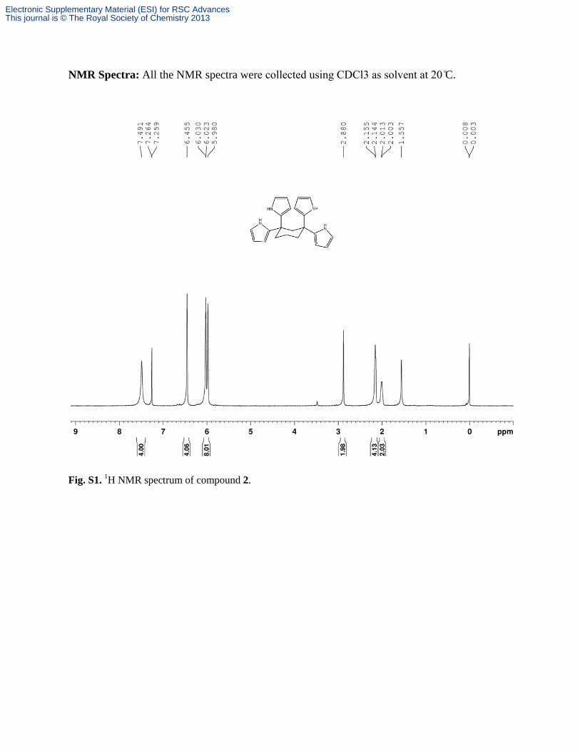
NMR Spectra: All the NMR spectra were collected using C Cl as solvent at 0 C.
Fig. S1. 1H NMR spectrum of compound 2.
Electronic Supplementary Material (ESI) for RSC AdvancesThis journal is © The Royal Society of Chemistry 2013

Fig. S2. 13
C NMR spectrum of compound 2.
Electronic Supplementary Material (ESI) for RSC AdvancesThis journal is © The Royal Society of Chemistry 2013
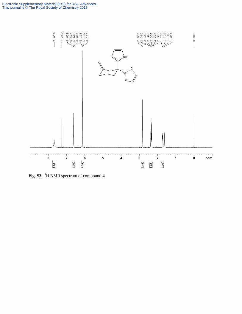
Fig. S3. 1H NMR spectrum of compound 4.
Electronic Supplementary Material (ESI) for RSC AdvancesThis journal is © The Royal Society of Chemistry 2013
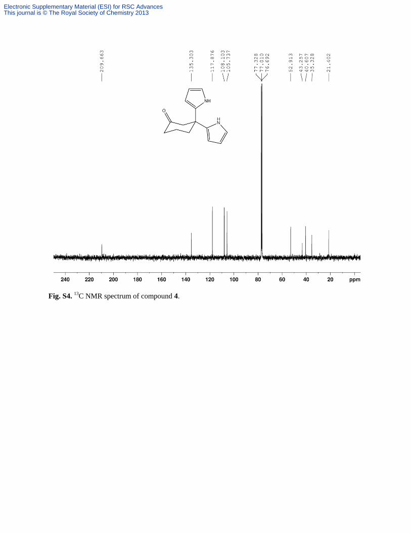
Fig. S4. 13
C NMR spectrum of compound 4.
Electronic Supplementary Material (ESI) for RSC AdvancesThis journal is © The Royal Society of Chemistry 2013
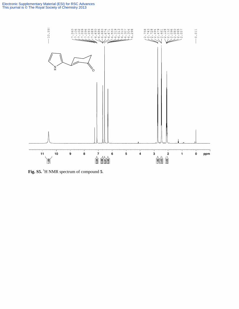
Fig. S5. 1H NMR spectrum of compound 5.
Electronic Supplementary Material (ESI) for RSC AdvancesThis journal is © The Royal Society of Chemistry 2013

Fig. S6. 13
C NMR spectrum of compound 5.
Electronic Supplementary Material (ESI) for RSC AdvancesThis journal is © The Royal Society of Chemistry 2013
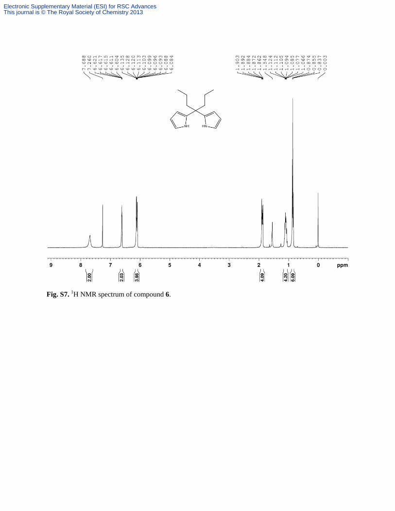
Fig. S7. 1H NMR spectrum of compound 6.
Electronic Supplementary Material (ESI) for RSC AdvancesThis journal is © The Royal Society of Chemistry 2013
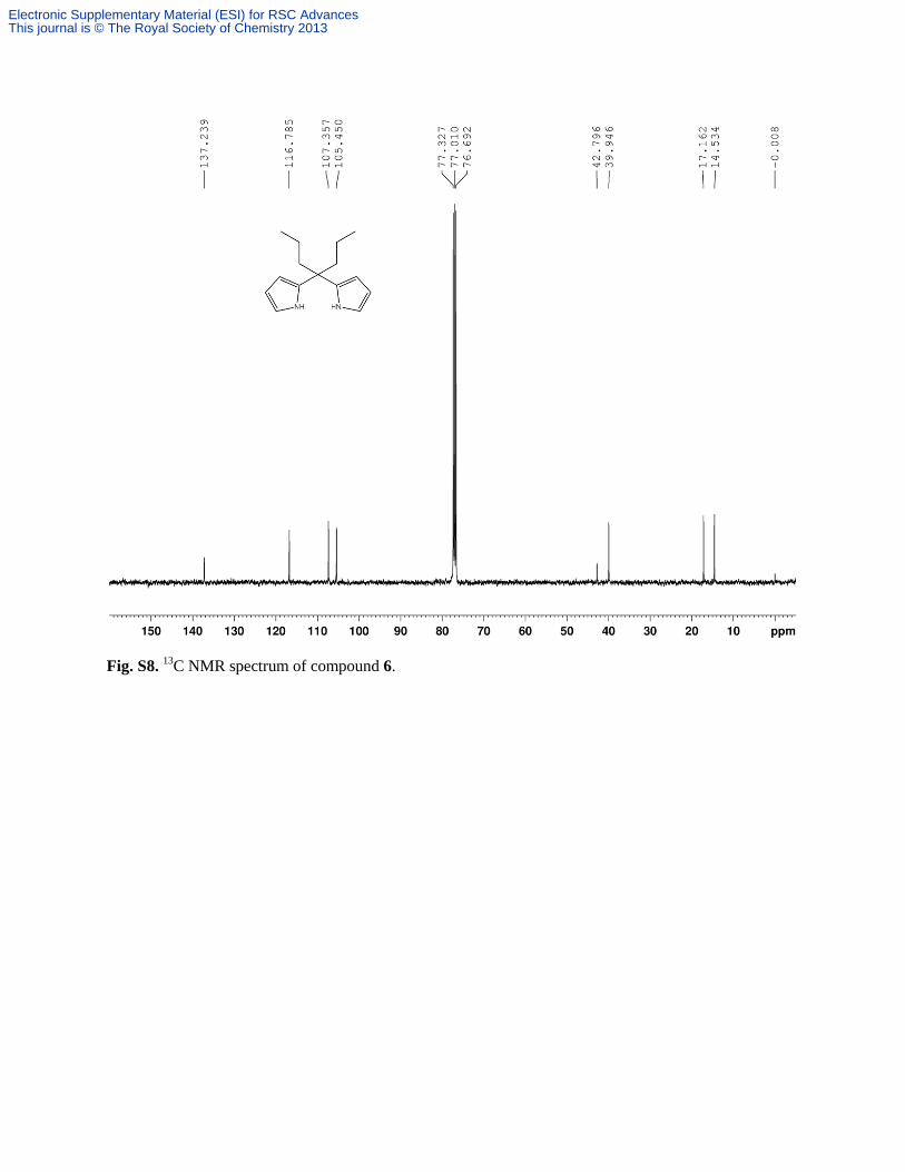
Fig. S8. 13
C NMR spectrum of compound 6.
Electronic Supplementary Material (ESI) for RSC AdvancesThis journal is © The Royal Society of Chemistry 2013
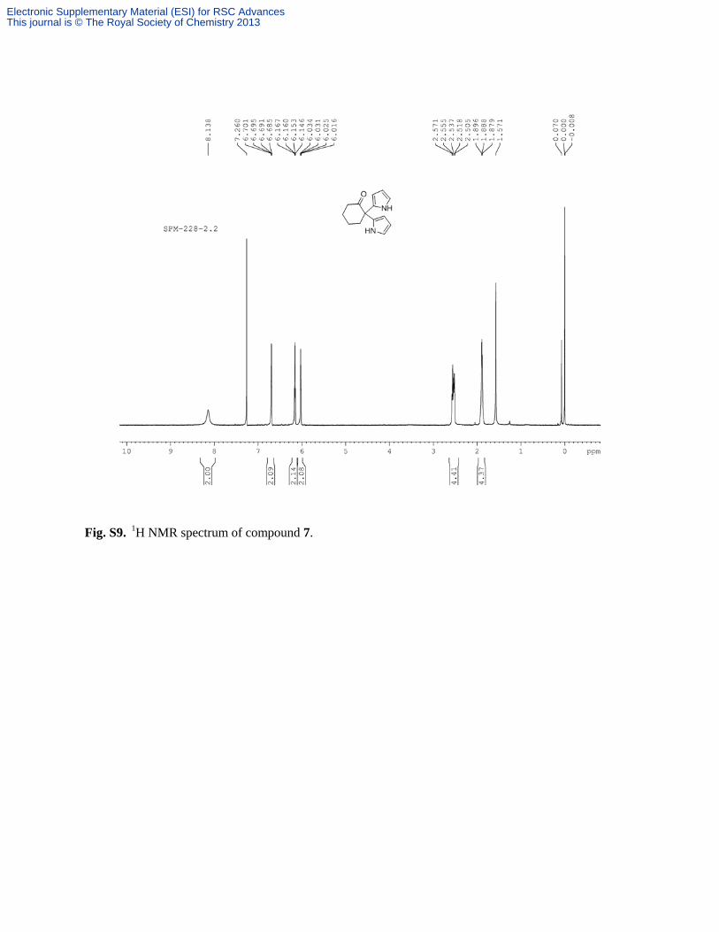
Fig. S9. 1H NMR spectrum of compound 7.
Electronic Supplementary Material (ESI) for RSC AdvancesThis journal is © The Royal Society of Chemistry 2013
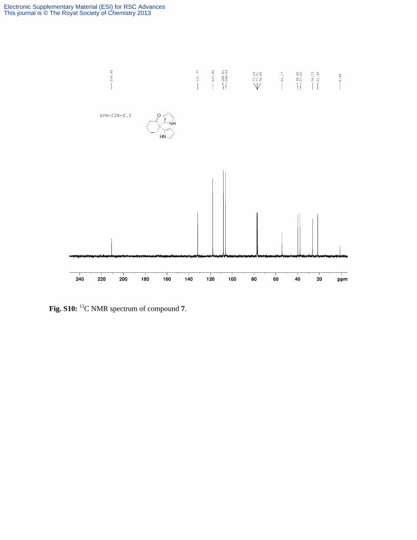
Fig. S10: 13
C NMR spectrum of compound 7.
Electronic Supplementary Material (ESI) for RSC AdvancesThis journal is © The Royal Society of Chemistry 2013
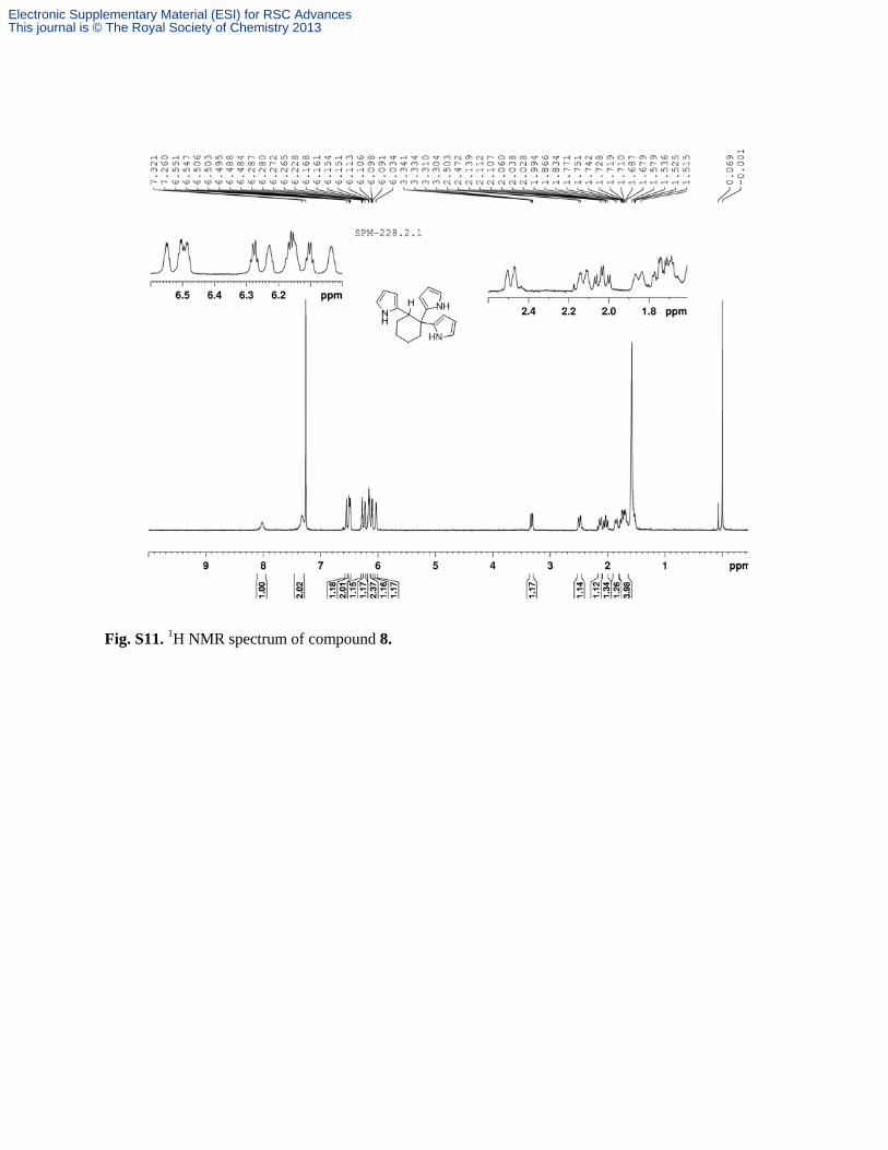
Fig. S11. 1H NMR spectrum of compound 8.
Electronic Supplementary Material (ESI) for RSC AdvancesThis journal is © The Royal Society of Chemistry 2013
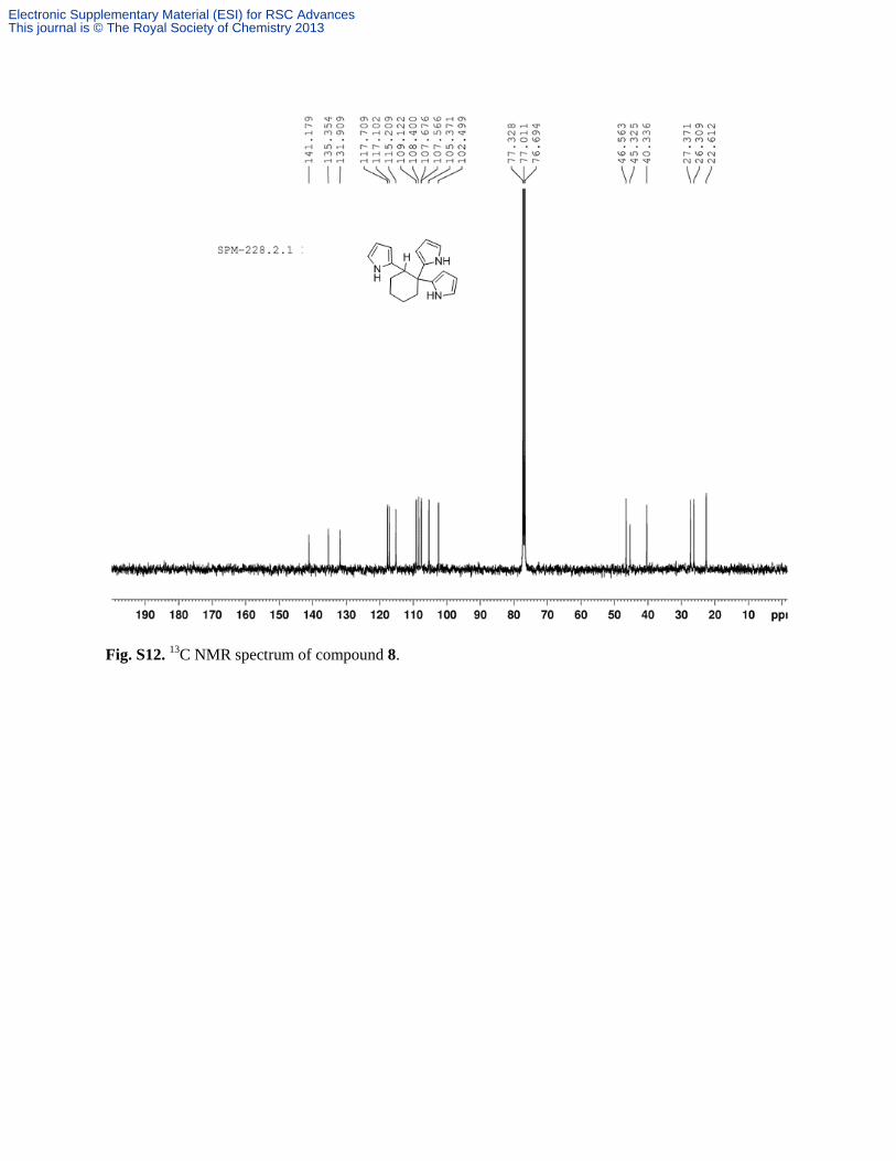
Fig. S12. 13
C NMR spectrum of compound 8.
Electronic Supplementary Material (ESI) for RSC AdvancesThis journal is © The Royal Society of Chemistry 2013
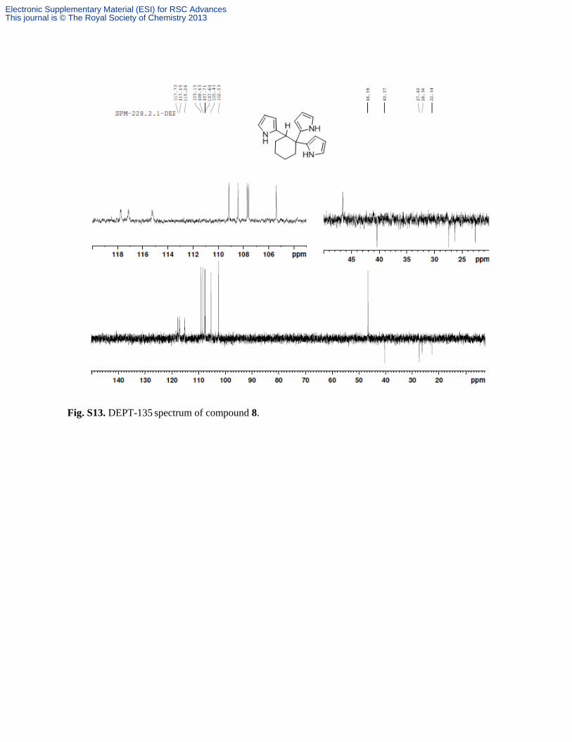
Fig. S13. DEPT-135 spectrum of compound 8.
Electronic Supplementary Material (ESI) for RSC AdvancesThis journal is © The Royal Society of Chemistry 2013
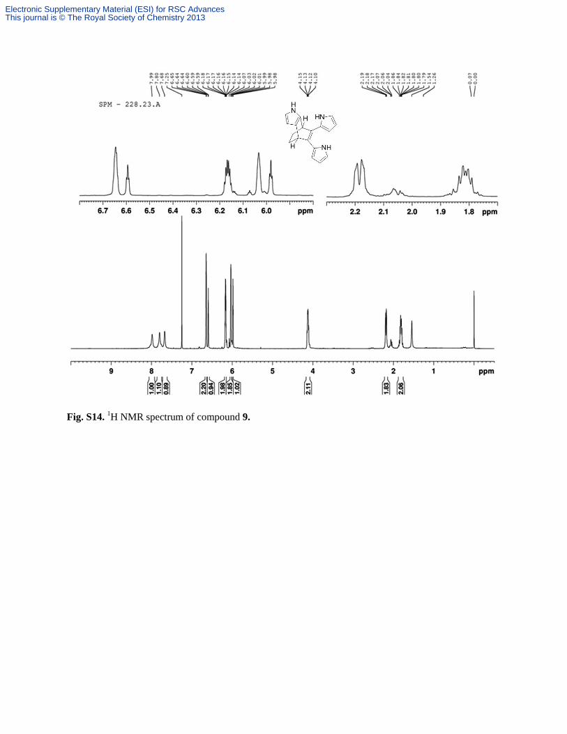
Fig. S14. 1H NMR spectrum of compound 9.
Electronic Supplementary Material (ESI) for RSC AdvancesThis journal is © The Royal Society of Chemistry 2013
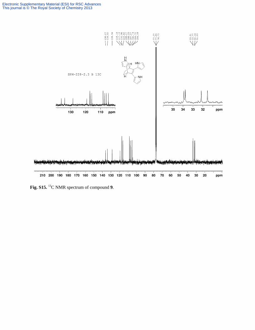
Fig. S15. 13
C NMR spectrum of compound 9.
Electronic Supplementary Material (ESI) for RSC AdvancesThis journal is © The Royal Society of Chemistry 2013
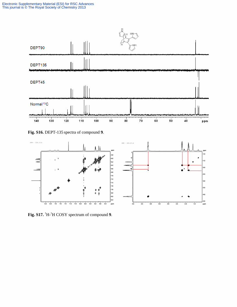
Fig. S16. DEPT-135 spectra of compound 9.
Fig. S17. 1H-
1H COSY spectrum of compound 9.
Electronic Supplementary Material (ESI) for RSC AdvancesThis journal is © The Royal Society of Chemistry 2013
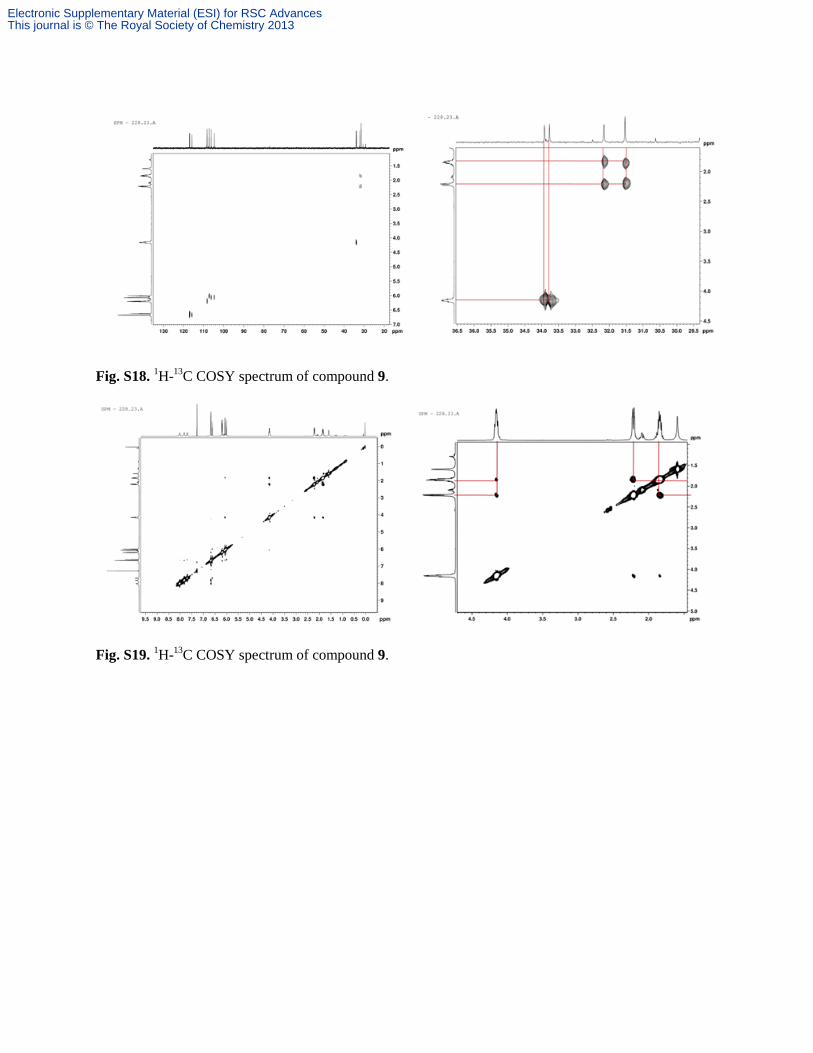
Fig. S18. 1H-
13C COSY spectrum of compound 9.
Fig. S19. 1H-
13C COSY spectrum of compound 9.
Electronic Supplementary Material (ESI) for RSC AdvancesThis journal is © The Royal Society of Chemistry 2013
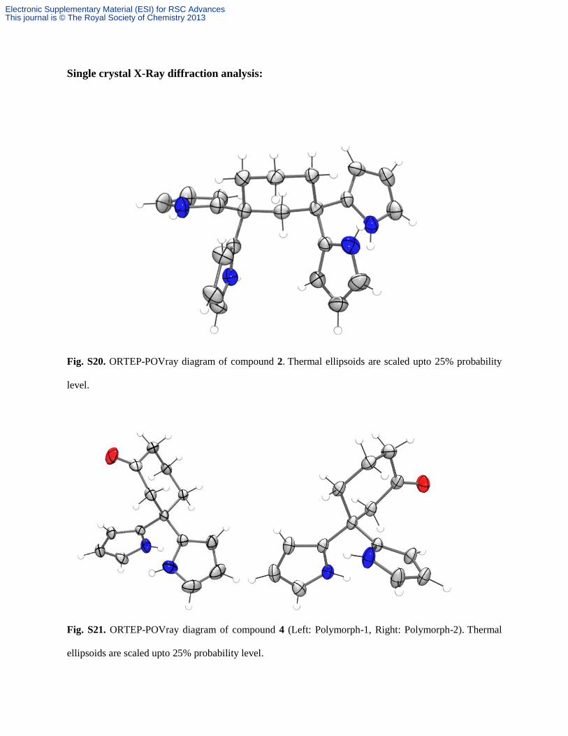
Single crystal X-Ray diffraction analysis:
Fig. S20. ORTEP-POVray diagram of compound 2. Thermal ellipsoids are scaled upto 25% probability
level.
Fig. S21. ORTEP-POVray diagram of compound 4 (Left: Polymorph-1, Right: Polymorph-2). Thermal
ellipsoids are scaled upto 25% probability level.
Electronic Supplementary Material (ESI) for RSC AdvancesThis journal is © The Royal Society of Chemistry 2013
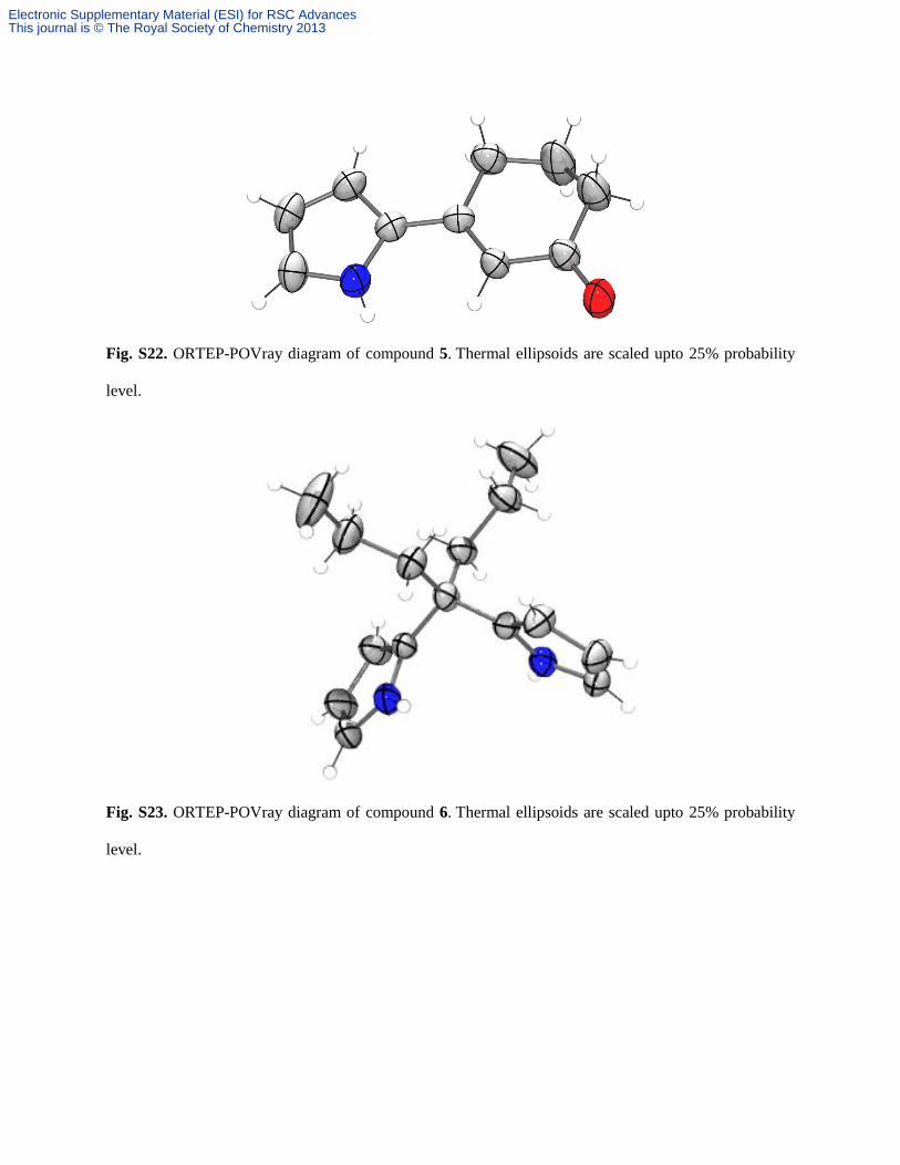
Fig. S22. ORTEP-POVray diagram of compound 5. Thermal ellipsoids are scaled upto 25% probability
level.
Fig. S23. ORTEP-POVray diagram of compound 6. Thermal ellipsoids are scaled upto 25% probability
level.
Electronic Supplementary Material (ESI) for RSC AdvancesThis journal is © The Royal Society of Chemistry 2013
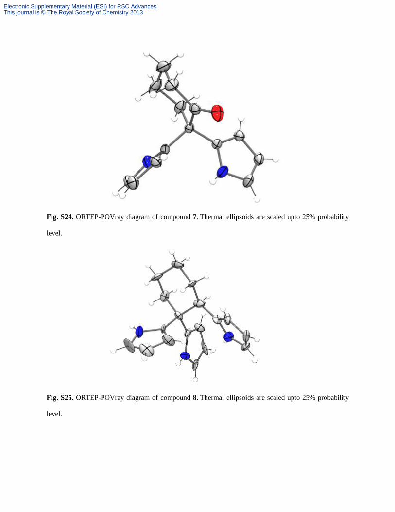
Fig. S24. ORTEP-POVray diagram of compound 7. Thermal ellipsoids are scaled upto 25% probability
level.
Fig. S25. ORTEP-POVray diagram of compound 8. Thermal ellipsoids are scaled upto 25% probability
level.
Electronic Supplementary Material (ESI) for RSC AdvancesThis journal is © The Royal Society of Chemistry 2013

Author’s comment on IUCR check .cif alert:
Compond 2:
RINTA01_ALERT_3_A The value of Rint is greater than 0.25 Rint given 0.397
PLAT020_ALERT_3_A The value of Rint is greater than 0.12 .........0.397
PLAT026_ALERT_3_A Ratio Observed / Unique Reflections too Low ....25 Perc.
Compound 4:
Polymorph-1:
PLAT026_ALERT_3_A Ratio Observed/Unique Reflections too Low.... 28 Perc.
PLAT222_ALERT_3_A Large Non-Solvent H Uiso(max)/Uiso(min) .. 10.0 Ratio
Polymorph-2:
PLAT029_ALERT_3_A diffrn_measured_fraction_theta_full Low ....... 0.868
Compound 5:
REFLT03_ALERT_3_A Reflection count < 85% complete (theta max?)
From the CIF: diffrn_reflns_theta_max 28.99
From the CIF: diffrn_reflns_theta_full 28.99
From the CIF: reflns_number_total 1942
TEST2: Reflns within _diffrn_reflns_theta_max
Count of symmetry unique reflns 2294
Completeness (_total/calc) 84.66%
PLAT029_ALERT_3_A _diffrn_measured_fraction_theta_full Low ....... 0.847
Compound 6:
PLAT029_ALERT_3_A _diffrn_measured_fraction_theta_full Low .......0.865
Compound 7:
PLAT029_ALERT_3_A diffrn_measured_fraction_theta_full Low ....... 0.854
Compound 8:
Electronic Supplementary Material (ESI) for RSC AdvancesThis journal is © The Royal Society of Chemistry 2013
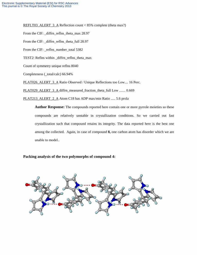
REFLT03_ALERT_3_A Reflection count < 85% complete (theta max?)
From the CIF: _diffrn_reflns_theta_max 28.97
From the CIF: _diffrn_reflns_theta_full 28.97
From the CIF: _reflns_number_total 5382
TEST2: Reflns within _diffrn_reflns_theta_max
Count of symmetry unique reflns 8040
Completeness (_total/calc) 66.94%
PLAT026_ALERT_3_A Ratio Observed / Unique Reflections too Low.... 16 Perc.
PLAT029_ALERT_3_A diffrn_measured_fraction_theta_full Low ....... 0.669
PLAT213_ALERT_2_A Atom C18 has ADP max/min Ratio ..... 5.6 prola
Author Response: The compounds reported here contain one or more pyrrole moieties so these
compounds are relatively unstable in crystallization conditions. So we carried out fast
crystallization such that compound retains its integrity. The data reported here is the best one
among the collected. Again, in case of compound 8, one carbon atom has disorder which we are
unable to model..
Packing analysis of the two polymorphs of compound 4:
Electronic Supplementary Material (ESI) for RSC AdvancesThis journal is © The Royal Society of Chemistry 2013
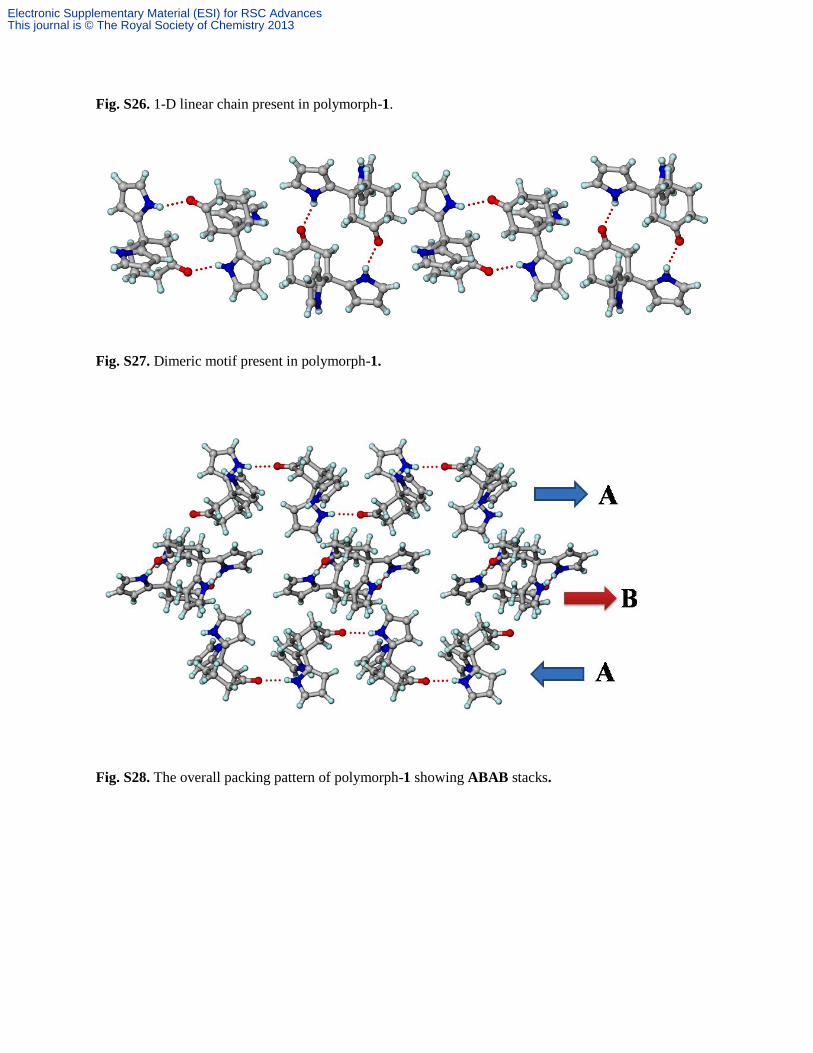
Fig. S26. 1-D linear chain present in polymorph-1.
Fig. S27. Dimeric motif present in polymorph-1.
Fig. S28. The overall packing pattern of polymorph-1 showing ABAB stacks.
Electronic Supplementary Material (ESI) for RSC AdvancesThis journal is © The Royal Society of Chemistry 2013
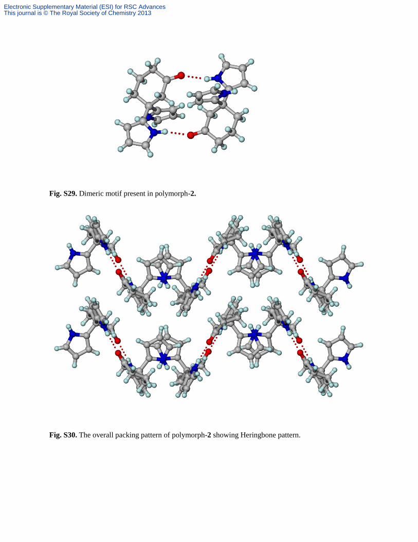
Fig. S29. Dimeric motif present in polymorph-2.
Fig. S30. The overall packing pattern of polymorph-2 showing Heringbone pattern.
Electronic Supplementary Material (ESI) for RSC AdvancesThis journal is © The Royal Society of Chemistry 2013