Condensation of Adhesion Domains in Biological and Bio-Mimetic...
101
Condensation of Adhesion Domains in Biological and Bio-Mimetic Membranes Thesis submitted in partial fulfillment of the requirements for the degree of DOCTOR OF PHILOSOPHY by Nadiv Dharan Submitted to the Senate of Ben-Gurion University of the Negev July 24, 2017 Be’er-Sheva
Transcript of Condensation of Adhesion Domains in Biological and Bio-Mimetic...
Condensation of Adhesion Domains in
Biological and Bio-Mimetic Membranes
Thesis submitted in partial fulfillment
of the requirements for the degree of
DOCTOR OF PHILOSOPHY
by
Nadiv Dharan
Submitted to the Senate of Ben-Gurion University of the Negev
July 24, 2017
Be’er-Sheva

Condensation of Adhesion Domains in
Biological and Bio-Mimetic Membranes
Thesis submitted in partial fulfillment
of the requirements for the degree of
DOCTOR OF PHILOSOPHY
by
Nadiv Dharan
Submitted to the Senate of Ben-Gurion University of the Negev
Approved by the advisor:
Approved by the Dean of the Kreitman School of Advanced Graduate Studies:
July 24, 2017
Be’er-Sheva

This work was carried out under the supervision of Prof. Oded Farago
In the Department of Biomedical Engineering
Faculty of Engineering

Research-Student’s Affidavit when Submitting the Doctoral
Thesis for Judgment
I Nadiv Dharan, whose signature appears below, hereby declare that (Please mark the ap-
propriate statements):
X I have written this Thesis by myself, except for the help and guidance offered by my
Thesis Advisors.
X The scientific materials included in this Thesis are products of my own research, culled
from the period during which I was a research student.
This Thesis incorporates research materials produced in cooperation with others, ex-
cluding the technical help commonly received during experimental work. Therefore, I am
attaching another affidavit stating the contributions made by myself and the other partici-
pants in this research, which has been approved by them and submitted with their approval.
Date: July 24, 2017 Student’s name: Nadiv Dharan Signature:

Acknowledgments
First and foremost, I wish to sincerely thank my supervisor Prof. Oded Farago for the in-
valuable guidance and support over the past years, which enabled me to obtain a deeper
appreciation for biophysical phenomena, greatly expanded my theoretical skills, and stimu-
lated me to pursue scientific research.
I thank the Negev-Faran scholarship for providing the necessary financial support for this
work.
I would like to thank my colleagues and friends Mr. Noam Weil and Dr. Yotam Yosef Avital
for the stimulating discussions we had over the years and the help throughout this project.
I would like to expresses my utmost gratitude to my family for the great support they have
provided over the years.
Finally, a special thanks to my beloved wife and best friend Bettina, who never stops ex-
pressing her unconditional love and full support, and has continuously encouraged me to
follow my passions.

Abstract
Lipid bilayers are ubiquitous in biology, and constitute natural barriers separating the
outer environment from the inner content of cells and intracellular organelles. Among nu-
merous other proteins with diverse biological functions, the plasma membrane is equipped
with adhesion proteins that bind the membrane to the extracellular matrix and/or neighbor-
ing cell membranes. Adhesion bonds often aggregate on the surface of the membrane and
form macroscopically large adhesion domains containing hundreds to thousands of bonds,
which provide greater mechanical stability to cells and also promotes signaling cues for var-
ious biological purposes. The formation of adhesion clusters is, thus, fundamental to life as
it regulates numerous important biological processes, such as tissue formation, cell migra-
tion and intercellular communication. One important thermodynamic mechanism facilitat-
ing adhesion cluster formation is the membrane-mediated potential of mean force (PMF)
between adhesion bonds, which originates from the membrane’s thermal undulations and
elastic curvature energy. The aggregation of adhesion bonds that bind a membrane to an-
other surface enhances the membrane’s thermal roughness and reduces its curvature energy;
the membrane-mediated PMF is essentially the associated free energy gained. In this work,
we employ statistical-mechanical methods, conduct molecular computer simulations, and use
novel mean-field calculations to investigate the influence of membrane-mediated interactions
on the condensation transition of adhesion bonds in supported biomimetic and biological
membranes.
The entropic membrane-mediated mechanism for aggregation of adhesion bonds is in-
vestigated by conducting molecular simulations of a solvent-free coarse-grained model for
supported lipid bilayers. Our simulation results corroborate the conclusions drawn from
previous theoretical studies, and show that the fluctuation-induced PMF is too weak to pro-
mote condensation on its own; nevertheless, it greatly facilitates aggregation by partially
compensating for the loss of mixing entropy, and effectively reducing the temperature by a
factor of 2-3. The influence of thermal fluctuations on the condensation transition is further
examined by simulating membranes exhibiting reduced or enhanced thermal undulations, by
subjecting them to physical confinement or negative surface tension, respectively. Our results
reveal that while the condensation transition is significantly shifted for confined membranes,
1
the impact of negative tension is negligible. Nevertheless, once adhesion domains form, a
negative tension may result in strong membrane buckling and the formation of elongated
adhesion domains.
In contrast to the long-range fluctuation-induced interactions, the curvature-induced
PMF spans over a correlation length, ξγ, which for biological membranes is typically in the
range of several tens of nanometers. Our investigation of adhesion domain formation driven
by the curvature-mediated mechanism relies on a novel mean-field approach, in which the
elastic energy of the membrane is numerically estimated for various random distributions
of bonds. We obtain an empirical expression for the system’s free energy, from which we
derive the phase diagram of the system. Our analysis reveals that the typical membrane
deformations caused by adhesion bonds in biological systems may lead to the formation of
adhesion domains with semi-dilute densities of the order of ∼ ξ−2γ . The conclusions drawn
from our analysis are further examined in relation to the important biological system of
the immunological synapse (IS). This special cellular junction forms between T cells and
antigen-presenting cells as part of the immune response, and is established by two types of
adhesion bonds, namely TCR-pMHC and LFA1-ICAM1. The IS is characterized by a unique
molecular pattern where the TCR-pMHC form a central cluster at the contact area, while
LFA1-ICAM1 adhesion bonds accumulate around it. We locate the system in the two-phase
region of our mean-field phase diagram, and identify the IS as a semi-dilute domain with
roughly 100 bonds per µm2, in line with experimental observations.
While our investigation finds that passive (thermodynamic) membrane-mediate mech-
anisms may be crucially important for the aggregation of the IS, the formation of the TCR-
pMHC cluster at the center of the contact area can be only explained by a symmetry breaking
mechanism. A widely accepted source for this symmetry breaking is the active cytoskeletal
processes originating from actin retrograde flow and dynein-mediated directed transport. To
further investigate the interplay between passive and active mechanisms in IS formation, we
present and simulate an implicit-membrane lattice-gas model, where the curvature-mediated
PMF and the active cytoskeletal-based forces are introduced via simple potentials. The
spatio-temporal evolution of the lattice simulations is found to be astonishingly similar to
the signature features of the IS formation process. Specifically, we observe that small TCR-
pMHC microclusters are initially formed at the periphery of the contact region, and then
2

migrate (while continuing to grow in size) to the center of the contact area, where they accu-
mulate into a quasi-circular domain. Moreover, we find that this process is completed within
a biologically relevant timescale of ∼ 30 minutes. Our simulation results, thus, reveal the
important role played by the membrane-mediated interactions in regulating the rate of the
IS formation process. Explicitly, membrane elasticity facilitates the formation of long-lived
TCR-pMHC peripheral microclusters, which are important for T cell activation, thereby al-
lowing sustained signaling over tens of minutes prior to the formation of the central domain,
where these signals are terminated.
Keywords: membrane elasticity, thermal undulations, cell junctions, adhesion domains,
lattice-gas, coarse-grained, Monte Carlo simulations, condensation transition, mean-field the-
ory, immunological synapse, active and passive forces.
3
Contents
1 Introduction 6
1.1 Statistical mechanics of fluctuating membranes . . . . . . . . . . . . . . . . . . . . . . . . . . 7
1.2 Membrane-mediated interactions between adhesion bonds . . . . . . . . . . . . . . . . . . . . 9
1.2.1 The Helfrich regime . . . . . . . . . . . . . . . . . . . . . . . . . . . . . . . . . . . . . 12
1.2.1.1 Entropic attachment penalty of a single adhesion bond . . . . . . . . . . . . 12
1.2.1.2 Two-body fluctuation-induced attraction . . . . . . . . . . . . . . . . . . . . 14
1.2.1.3 Many-body fluctuation-induced PMF . . . . . . . . . . . . . . . . . . . . . . 14
1.2.2 The van der Waals regime . . . . . . . . . . . . . . . . . . . . . . . . . . . . . . . . . . 18
1.2.2.1 Deformation energy of membranes with a single adhesion bond . . . . . . . . 19
1.2.2.2 Pairwise curvature-induced attraction . . . . . . . . . . . . . . . . . . . . . . 22
1.2.2.3 Many-body curvature-induced PMF . . . . . . . . . . . . . . . . . . . . . . . 23
1.3 Outline of the thesis . . . . . . . . . . . . . . . . . . . . . . . . . . . . . . . . . . . . . . . . . 24
2 Molecular simulations of membranes in the Helfrich regime 26
2.1 Introduction . . . . . . . . . . . . . . . . . . . . . . . . . . . . . . . . . . . . . . . . . . . . . . 26
2.2 Comparison with the Weil-Farago model . . . . . . . . . . . . . . . . . . . . . . . . . . . . . . 27
2.3 Adhesion domain formation in stressed and confined membranes . . . . . . . . . . . . . . . . 32
2.3.1 Membranes under physical confinement . . . . . . . . . . . . . . . . . . . . . . . . . . 33
2.3.2 Membranes under negative surface tension . . . . . . . . . . . . . . . . . . . . . . . . . 34
2.4 Summary . . . . . . . . . . . . . . . . . . . . . . . . . . . . . . . . . . . . . . . . . . . . . . . 40
3 Formation of semi-dilute adhesion domains in the van der Waals regime 43
3.1 Introdcution . . . . . . . . . . . . . . . . . . . . . . . . . . . . . . . . . . . . . . . . . . . . . . 43
3.2 Mean-field theory . . . . . . . . . . . . . . . . . . . . . . . . . . . . . . . . . . . . . . . . . . . 44
3.2.1 Energy calculations . . . . . . . . . . . . . . . . . . . . . . . . . . . . . . . . . . . . . . 45
3.2.2 Phase diagram . . . . . . . . . . . . . . . . . . . . . . . . . . . . . . . . . . . . . . . . 49
3.3 Semi-dilute domains . . . . . . . . . . . . . . . . . . . . . . . . . . . . . . . . . . . . . . . . . 50
3.4 Summary . . . . . . . . . . . . . . . . . . . . . . . . . . . . . . . . . . . . . . . . . . . . . . . 53
4
4 Passive and active mechanisms in the formation of the immunological synapse 55
4.1 Introduction . . . . . . . . . . . . . . . . . . . . . . . . . . . . . . . . . . . . . . . . . . . . . . 55
4.2 Model and simulations . . . . . . . . . . . . . . . . . . . . . . . . . . . . . . . . . . . . . . . . 58
4.2.1 Nearest-neighbor approximation . . . . . . . . . . . . . . . . . . . . . . . . . . . . . . 58
4.2.2 Lattice-gas model . . . . . . . . . . . . . . . . . . . . . . . . . . . . . . . . . . . . . . . 60
4.2.3 Monte Carlo simulations . . . . . . . . . . . . . . . . . . . . . . . . . . . . . . . . . . . 62
4.2.4 Active cytoskeleton forces . . . . . . . . . . . . . . . . . . . . . . . . . . . . . . . . . . 62
4.3 Results . . . . . . . . . . . . . . . . . . . . . . . . . . . . . . . . . . . . . . . . . . . . . . . . . 64
4.4 Summary . . . . . . . . . . . . . . . . . . . . . . . . . . . . . . . . . . . . . . . . . . . . . . . 69
5 Conclusion 72
Appendix 77
A 78
5

Chapter 1
Introduction
Lipid membranes serve as a physical barrier that separates the interior of the cell from its
outer environment. They are typically arranged as a bilayer of a number of lipids, such
as phospholipids, glycolipids and cholesterol [1]. Similar to the plasma membrane that
surrounds the cells, several intracellular organelles such as the mitochondria, the nucleus
and the endoplasmic reticulum are also compartmentalized by a lipid membrane. One of
the most important roles of lipid bilayers is to regulate the bidirectional flow of ions and
other molecules into and out of the cell [2]. Biological membranes are also embedded with
numerous types of proteins, which exhibit lateral diffusion in the membrane’s plane [3, 4].
Cell adhesion molecules constitute a special class of membrane proteins that enable the
attachment of the membrane to various elements, such as the extracellular matrix (ECM),
the cytoskeleton and neighboring cell membranes. The adhesion process is mediated by
the formation of specific receptor-ligand bonds that anchor the membrane to the adhesive
element. Cell adhesion is crucial for the proper function of individual cells and the organism
as a whole, and plays a vital role in many biological processes, including cell migration [5],
tissue morphogenesis [6], intercellular communication [7], and T cell activation as part of the
immune response [8].
One common feature of cell adhesion is the ability of the adhesion bonds to aggre-
gate and form large adhesion domains. For instance, connexon protein complexes aggregate
into gap junction plaques that bind opposing cell membranes. Gap junctions directly con-
nect the cytoplasms of two neighboring cells and regulate the exchange of ions and small
6

molecules, thereby regulating intercellular communication [9]. Likewise, proteins of the in-
tegrin family aggregate into clusters called focal adhesion, which physically link the ECM
to the cell cytoskeleton. This mechanical linkage not only provides strong anchoring of the
cell to the ECM, but also enables the cell to generate traction forces necessary for cellular
locomotion [10]. In addition, focal adhesions act as mechano-sensors and promote biological
signals that regulate cell growth, survival, proliferation and differentiation [11–14]. Finally,
cadherin proteins cluster into adherens junctions that bind neighboring cells within tissues.
The formation of this type of adhesion domains is vital for proper tissue organization and
architecture maintenance in developing and adult organisms [15,16]. Given the enormous im-
portance of adhesion clusters in biological systems, it is essential to acquire a comprehensive
understanding of the biophysical principles governing the formation of these structures.
Due to the highly complexed nature of the cell membrane, several experimental studies
have developed model systems containing the key components of biological membranes to
gain insights into the adhesion process. These models include lipid bilayers deposited onto
solid substrates and thin polymer-coated supports [17, 18]. When incorporated with spe-
cific adhesion proteins, such supported membranes may serve as targets for the binding of
synthetic ligand coated vesicles or liposomes [19, 20]. These biomimetic systems are able to
elucidate many aspects of cell adhesion, such as the effect of density and strength of ligand-
receptor bonds on adhesion efficiency [21], as well as estimation of adhesion bonds binding
affinity [22]. Supported membranes also constitute new potential applications in designing
biosensor nano-devices [23].
1.1 Statistical mechanics of fluctuating membranes
The elastic behavior of lipid bilayers is traditionally studied in the framework of the Helfrich
effective surface Hamiltonian [24] for two-dimensional manifolds with local principle curva-
tures c1 and c2. The system Hamiltonian is described in terms of the bending energy of the
membrane, and is given by
H =
∫dS
{1
2κ (c1 + c2 − c0)2 + κc1c2
}, (1.1)
7
where the integration is taken over the entire surface of the membrane. In eq. (1.1), κ is
the bending modulus of the membrane, κ is the saddle-splay (Gaussian) modulus, and c0
is the spontaneous curvature. If one assumes that the two leaflets of the bilayer membrane
have similar lipid compositions, the spontaneous curvature can be simply set to c0 = 0.
For fluctuating membranes that do not change their topology, the surface integral over the
Gaussian curvature term would simply result in a constant, which has no effect on the
underlying physics of the system and, thus, can be ignored. A useful way to parametrize
the membrane surface is the Monge gauge. Within this parametrization, the membrane is
represented by a height function h(r) measured relative to a reference plane, where r = (x, y)
is the two-dimensional position vector. Assuming the small gradient approximation, which
essentially states that the membrane does not fluctuate considerably (and, in particular,
does not develop “overhangs”), one can re-express the membrane curvature (c1 + c2) and
the area differential element (dS) in terms of the membrane’s height function. Keeping only
quadratic terms in h, one obtains the following simplified version of the effective Helfrich
Hamiltonian
H =
∫AP
1
2κ(∇2h
)2d2r, (1.2)
where, in this case, the integration is taken over the projected area of the membrane, AP.
Eq. (1.2) is the most commonly used form of the Helfrich energy in the literature of membrane
biophysics.
The quadratic nature of eq. (1.2) gives rise to a harmonic theory, in which membrane
undulations can be viewed as a collection of independent harmonic oscillators. This is done
by using the Fourier representation of the height profile
h(r) =
(l
L
)2∑q
hq exp (−iq · r) (1.3)
q =2π
L(n1, n2) ; ni=1,2 =
−L2l, . . . ,
L
2l
where q is the two dimensional wave vector, l is a microscopic cutoff lengthscale of the order
of the membrane thickness (≈ 5nm), and L is the linear size of the membrane such that
8
L2 = AP. In Fourier space the Helfrich Hamiltonian (1.2) reads
H =l4
L2
∑q
1
2κq4
∣∣h2q
∣∣ , (1.4)
where q = |q|. From eq. (1.4) we observe that the different Fourier modes decouple; thus,
each mode constitutes an independent fluctuation degree of freedom of the membrane. For
quadratic energy functionals, the equipartition theorem implies that each fluctuation mode
contributes an average of kBT/2 to the system energy, where kB is the Boltzmann constant
and T denotes the system temperature. Thus, the fluctuation spectrum of the membrane
obeys
〈∣∣h2
q
∣∣〉 =L2kBT
l4κq4. (1.5)
Eq. (1.5) provides an incredibly useful method for estimating the bending modulus κ of the
membrane, by measuring the fluctuation spectrum using flicker spectroscopy [25] or computer
simulations [26]. Fitting the data to eq. (1.5) results in typical values of the bending rigidity
that lie within the range of κ ≈ 10− 50kBT [27].
One can also derive an expression for the mean square height fluctuations of the mem-
brane
∆20 = 〈h(r)2〉 =
(l
L
)4∑q
〈∣∣h2
q
∣∣〉 ≈ kBT
16π3κL2, (1.6)
which gives the “thermal roughness” of the membrane. Note that the mean square fluctua-
tions of a free bilayer are proportional to the system temperature as one would expect, but
diverge with the system size L.
1.2 Membrane-mediated interactions between adhesion
bonds
The clustering process of membrane adhesion bonds at the surface of membranes requires
attractive interactions between them to overcome their mixing entropy. These may originate
9

from direct electrostatic and Van der Waals interactions [28], or effective forces stemming
from cytoskeleton remodeling and the action of motor proteins that actively translocate and
redistribute the adhesion bonds [29]. Another interesting mechanism that can facilitate ad-
hesion domain formation is related to the thermodynamics of the membrane itself, which can
induce effective forces between the adhesion proteins [30]. Membrane-mediated interactions
between adhesion bonds originate from two interrelated mechanisms operating in concert.
The first mechanism is related to the suppression of membrane thermal fluctuations by the
presence of adhesion bonds, which locally fix the membrane’s height [31]. The resulting
loss in the membrane’s fluctuation entropy can be partially mitigated if the adhesion bonds
aggregate into a single domain, which allows the membrane to fluctuate more freely. The
second mechanism stems from the local membrane deformations that are imposed by the
adhesion bonds. These membrane distortions are minimized once the bonds reside in close
proximity to each other, which can greatly relieve the mechanical stress of the system [32].
Thus, membrane curvature and thermal fluctuations induce an effective attractive potential
of mean force (PMF) between the adhesion bonds, which may trigger their aggregation.
The non-specific membrane-mediated interactions have also been studied in relation to con-
densation of trans-membrane proteins (membrane “inclusions”) [33–35], and in the broader
context of “Casimir-like” interactions in condensed matter [36].
The main challenge in analyzing and deriving expressions for membrane-mediated in-
teractions arises from their many-body nature [37], i.e., their non-trivial dependence on the
spatial distribution of the adhesion bonds. In a system with a large number of bonds, the
PMF acting between each pair of bonds depends on the locations of all of the other bonds
and, therefore, cannot be expressed as a simple sum of pairwise interactions. To illustrate
this complexity, consider the two configurations schematically depicted in Fig. 1.1. In the
first configuration [Fig. 1.1(a)], the membrane is attached by two adhesion bonds separated
by a distance r0, whereas in the second one [Fig. 1.1(b)] the membrane is attached by a
single bond and another small cluster of three bonds, with the same distance r0 between
them. It is readily apparent that the spectrum of thermal undulations, as well as the degree
of membrane curvature, is quite similar in both scenarios and, therefore, the membrane-
mediated PMFs are expected to be roughly the same in both cases. In other words, the
membrane-mediated force exerted on the single bond in Fig. 1.1(a) by the second bond is
10
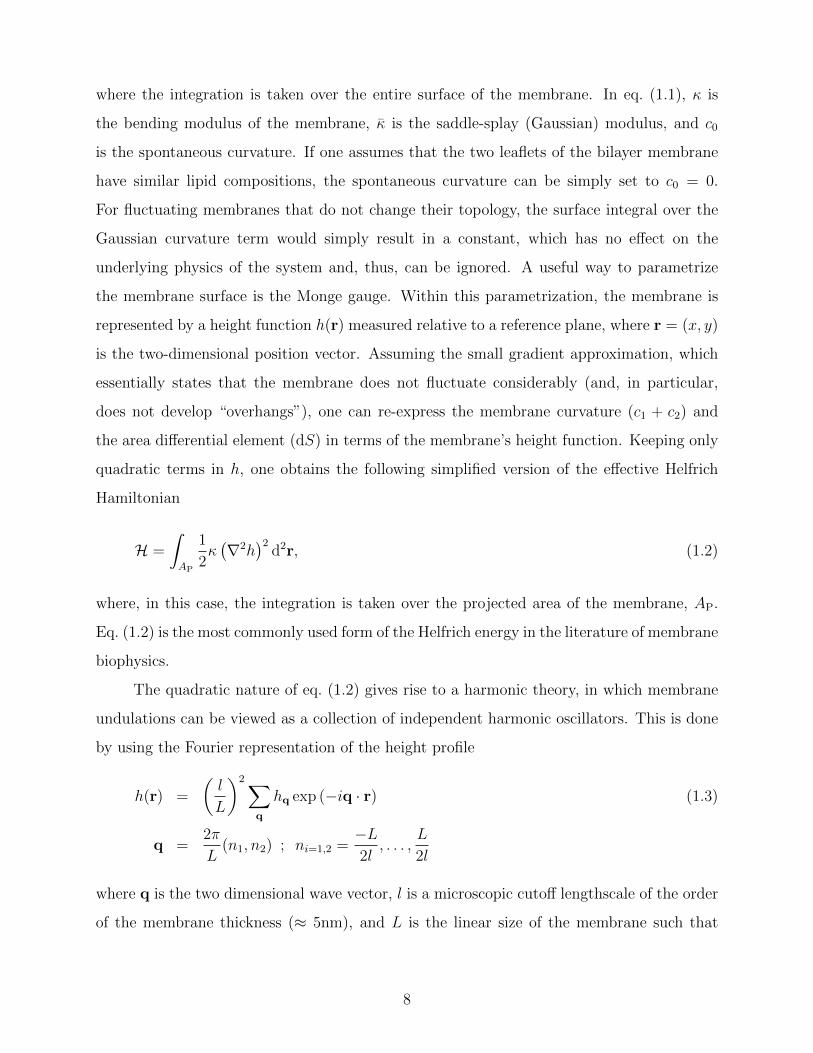
similar to that exerted on the single bond in Fig. 1.1(b) by the three bond cluster, and not
three times smaller.
(a) (b)
Figure 1.1: Schematics of a membrane bound to a surface by (a) two isolated adhesionbonds, and (b) a single adhesion bond and a small cluster of three bonds. The separationbetween the two bonds in (a) and between the single bond and the cluster in (b) is identicaland equals r0. The membrane-mediated PMF is similar in both cases, which illustrates itsmany-body nature..
Several theoretical studies have been devoted to characterizing and deriving expressions
for the membrane-mediated PMF between adhesion bonds. In a seminal work by Bruinsma,
Goulain and Pincus, the aggregation of gap junctions was investigated by considering two
opposing tensionless membranes with bending moduli 2κ that are connected to each other
at several points [38]. The system Hamiltonian is given by
H =
∫AP
{1
2κ(∇2h
)2+ V (h)
}d2r, (1.7)
where the effective potential, V (h), stands for the various interactions between the two mem-
branes, and h is the distance between them. A similar Hamiltonian describes the energy of
a single supported membrane with bending rigidity κ and a flat underlying surface. For sup-
ported membranes in model systems, a number of factors can contribute to V (h), including
short-range van der Waals attraction and excluded volume repulsion, double layer forces,
and effective repulsion due to thermal collisions between the membrane and the underlying
surface [39]. In the context of the living cell, confinement effects may arise from the ECM,
the cytoskeleton and the glycocalyx coating of the cell, which can all be clumped into this
effective non-specific potential.
Two important regimes for V (h) have been considered, corresponding to different
membrane-surface interactions. The first regime is termed the Helfrich regime, and deals
with membranes exhibiting large thermal fluctuations. In this regime, V (h) is an effective
11

repulsive potential (per unit area) arising from thermal collisions between the membrane
and the surface. The second regime, termed the van der Waals regime, describes membranes
with large bending moduli, such that the thermal undulations are small. In this case, the
curvature of the membrane dominates the system and, thus, V (h) can be replaced by a
Lennard-Jones type of potential. In the following sections, we review results for the thermo-
dynamic behavior of membranes in the Helfrich and the van der Waals regimes, from which
insights can be drawn into the fluctuation- and curvature-mediated interactions, respectively.
1.2.1 The Helfrich regime
1.2.1.1 Entropic attachment penalty of a single adhesion bond
An interesting feature of membranes attached to a surface at a single point is that the
fluctuation spectrum remains unaffected when compared to a free (unbound) membrane [40].
This rather surprising property can be understood from the fact that the membrane energy
is invariant under vertical translations and, thus, one can always position the underlying
adhesive surface at the global minimum of the membrane. That the attachment of the
membrane to the surface at a single point eliminates its horizontal degrees of freedom with
respect to a free membrane implies that the configurational space of the attached membrane
occupies a fraction l2/L2 of that of the free membrane, where l2 is roughly the area occupied
by the single adhesion bond. Therefore, the ratio between the partition functions of these
two systems satisfies Z/Zfree = l2/L2. Since the free energy of the attached membrane is
simply given by F1 = −kBT ln (Z/Zfree), the free energy cost associated with attaching a
membrane by a single adhesion bond is given by
F1 = 2kBT ln
(L
l
). (1.8)
Eq. (1.8) can also be derived by employing a different approach. Helfrich showed that thermal
collisions between the membrane and the underlying surface result in an effective repulsive
potential per unit area that scales with the height h of the membrane above the surface
12

as [41]
Vrep(h) ∼ (kBT )2
κh2. (1.9)
Using this form in eq. (1.7) gives
H =
∫AP
{1
2κ(∇2h
)2+ C
(kBT )2
κh2
}d2r, (1.10)
where C is an unknown numerical constant. Minimizing eq. (1.10) with respect to h results
in the membrane’s average height profile that grows linearly with the distance r from the
adhesion point (see also Fig. 1.2) according to
〈h(r)〉 ∼ r
√kBT
κ. (1.11)
Substituting eq. (1.11) in eq. (1.9) results in the effective fluctuation-induced repulsion be-
tween the membrane and the surface as a function of r
Vrep(r) ∼ kBT
r2. (1.12)
The free energy penalty associated with the attachment of the membrane by the single bond
can now be derived by integrating eq. (1.12) over the membrane’s projected area (excluding
a region of size l around the adhesion point), yielding
F1 =
∫AP
V (r)dr '∫ L
l
2πrkBT
r2dr = 2πkBT ln
(L
l
). (1.13)
One readily finds that in order to reconcile eq.(1.13) with eq. (1.8), the scaling behavior of
eq. (1.12) can be replaced by the exact form
Vrep(r) =kBT
πr2. (1.14)
13

Figure 1.2: Schematics of the mean height profile of a membrane tethered by a single adhesionbond to a surface in the Helfrich regime. The solid line represents the linear growth of themean height with the distance r from the bond. The dashed curve represents thermalundulations around the average profile.
1.2.1.2 Two-body fluctuation-induced attraction
The effective steric repulsion proposed by Helfrich, which stems from the thermal collisions
between the membrane and the surface, may be used to analyze the fluctuation-induced
interactions between a pair of membrane adhesion bonds. This is done by considering a
membrane attached to the surface at a single adhesion point and the probability density,
p [h(r) = 0], that the membrane collides with the surface at a distance r away from it [31].
Since this probability density function is directly related to the rate of collision between
the membrane and the surface, it should display the same scaling behavior with r as does
V (r) (1.14), namely p [h(r) = 0] ∼ 1/r2. If one thinks of the collision point as the location of
the second adhesion bond, then the pair correlation function between the two adhesion bonds
should also scales as g(r) ∼ 1/r2, which immediately gives the pair fluctuation-induced PMF
Φ2(r) ≡ −kBT ln [g(r)] = 2kBT ln(r). (1.15)
This shows that the fluctuation-induced pair PMF is a long-range (infinitely ranged) attrac-
tive potential which, remarkably, is independent of the bending modulus κ. This result has
also been verified by computer simulations of coarse-grained bilayer membranes [42].
1.2.1.3 Many-body fluctuation-induced PMF
The study of the clustering process of adhesion bonds traditionally uses a lattice model [43–
45], in which the membrane is discretized into patches that may or may not contain adhesion
molecules that bind (via receptor-ligand bonds) the membrane to an underlying surface.
14

Such models constitute discrete versions of the Helfrich continuum surface model of lipid
bilayers. Thus, each lattice site is characterized by two variables si and hi. The former
parameter characterizes the distribution of adhesion bonds, where si = 1 corresponds to
a membrane segment that is connected to the surface and si = 0 to a segment which is
free to fluctuate. The latter parameter, hi, represents the local height of the membrane.
Analyzing the aggregation behavior of the adhesion bonds by means of computer simulations
requires sampling over different distributions of lattice sites, as well as over different height
conformations. This may become a computationally expensive task in simulations of large
systems. It is, therefore, desirable to develop a model that integrates out the degrees of
freedom associated with the height fluctuations and, instead, assigns a potential of mean force
between the lattice adhesion sites. Apart from computational simplicity, another advantage
of this approach is that it makes possible a direct comparison with the well-investigated two-
dimensional (2D) lattice-gas model and, thus, highlights the role played by the membrane-
mediated interactions in the aggregation process1.
Such a lattice model was recently proposed by Weil and Farago (WF) [46], which
combines two attractive energy terms:
HWF = −ε∑〈i,j〉
sisj +∑i
Vi(1− si). (1.16)
The first term constitutes the conventional lattice-gas model, where the sum runs over all
pairs of nearest-neighbor lattice sites. The energy ε > 0 gained for each pair of nearest-
neighbor occupied sites accounts for all the interactions between the adhesion bonds other
than the membrane-mediated PMF. The latter potential is represented by the second term
in eq. (1.16) which, quite unusually, involves summation over the empty sites alone. The
energy of each empty site measures the amount of free energy lost due to the suppression of
the thermal height fluctuations of the corresponding membrane segment. Weil and Farago
conjectured that this free energy penalty depends on the distance of the segment from the
nearest adhesion bond dmini , i.e., the distance to the nearest occupied site. This assump-
tion is based on the idea that each of the adhesion bonds restricts the membrane thermal
1Note that an opposite approach is taken in refs. [43, 44], where the positional degrees of freedom si areintegrated out by using the mean-field solution of the 2D lattice-gas model. This introduces an effectivemembrane-surface interaction energy term in the Helfrich Hamiltonian that depends on the local hi.
15

fluctuations mainly in its own vicinity. In other words, the local suppression of thermal un-
dulations is essentially determined by the nearest adhesion bond, while the effect of the other
more distant bonds is effectively screened out. The energy penalty term, Vi, in eq. (1.16)
associated with the (empty) site i is given by
Vi =kBT
π
(l
dmini
)2
, (1.17)
which generalizes eq. (1.14) for the free energy density at a distance r from a single isolated
adhesion bond. In eq. (1.17), l is the lattice constant (which should be of the order of a few
nanometers – comparable to the thickness of the membrane), and dmini measures the distance
of site i to the nearest occupied site. Note that, in general, dmini depends on the distribution
of all the occupied sites; therefore, the second term in eq. (1.16) represents a many-body
PMF between the adhesion bonds. This potential is attractive because most of the entropy
is lost at the proximity of the occupied lattice sites, where dmini is small. The many-body
fluctuation-induced PMF can be evaluated by: (i) constructing a Voronoi diagram for the
distribution of adhesion bonds, (ii) calculating the attachment free energy penalty within
each Voronoi cell by integrating eq. (1.14) over the area of the given Voronoi cell, and (iii)
adding up the free energy contributions of all the Voronoi cells. Thus, the many-body PMF
is given by
ΦN =N∑i=1
∫Ai
kBT
πr2dr, (1.18)
where within each Voronoi cell i with area Ai the distance r is measured from the adhesion
bond located inside the cell.
To study the aggregation behavior of adhesion bonds in membranes, Monte Carlo
simulations of the WF model were conducted on a triangular lattice and were compared to
simulation results of the standard lattice-gas model with only nearest-neighbor interactions,
i.e., in the absence of the second term in eq. (1.16). In both sets of simulations, the system
exhibited a first order condensation transition at a certain threshold value ε = εc. The
simulation results revealed that the transition value, εc, of the WF model is smaller than the
corresponding value of the standard lattice-gas model at the same density of bonds, typically
16
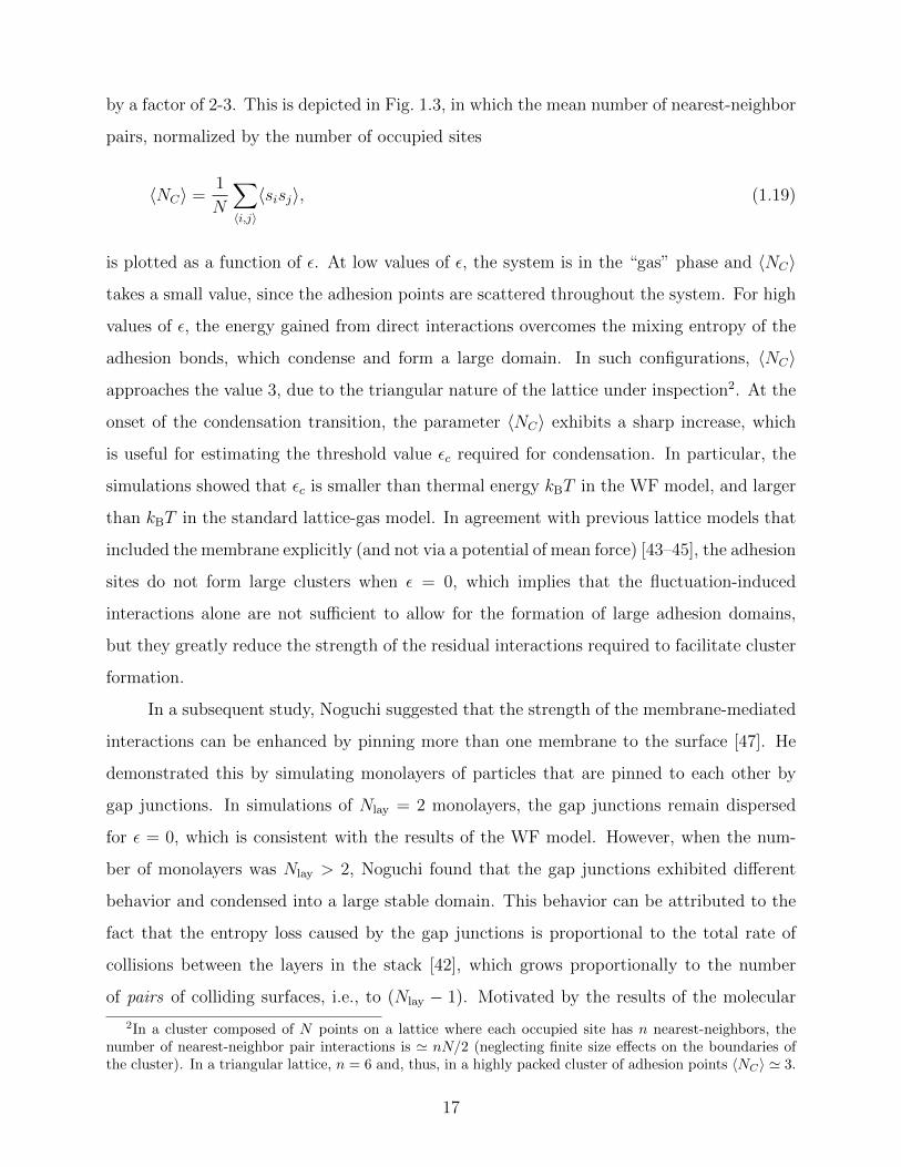
by a factor of 2-3. This is depicted in Fig. 1.3, in which the mean number of nearest-neighbor
pairs, normalized by the number of occupied sites
〈NC〉 =1
N
∑〈i,j〉
〈sisj〉, (1.19)
is plotted as a function of ε. At low values of ε, the system is in the “gas” phase and 〈NC〉
takes a small value, since the adhesion points are scattered throughout the system. For high
values of ε, the energy gained from direct interactions overcomes the mixing entropy of the
adhesion bonds, which condense and form a large domain. In such configurations, 〈NC〉
approaches the value 3, due to the triangular nature of the lattice under inspection2. At the
onset of the condensation transition, the parameter 〈NC〉 exhibits a sharp increase, which
is useful for estimating the threshold value εc required for condensation. In particular, the
simulations showed that εc is smaller than thermal energy kBT in the WF model, and larger
than kBT in the standard lattice-gas model. In agreement with previous lattice models that
included the membrane explicitly (and not via a potential of mean force) [43–45], the adhesion
sites do not form large clusters when ε = 0, which implies that the fluctuation-induced
interactions alone are not sufficient to allow for the formation of large adhesion domains,
but they greatly reduce the strength of the residual interactions required to facilitate cluster
formation.
In a subsequent study, Noguchi suggested that the strength of the membrane-mediated
interactions can be enhanced by pinning more than one membrane to the surface [47]. He
demonstrated this by simulating monolayers of particles that are pinned to each other by
gap junctions. In simulations of Nlay = 2 monolayers, the gap junctions remain dispersed
for ε = 0, which is consistent with the results of the WF model. However, when the num-
ber of monolayers was Nlay > 2, Noguchi found that the gap junctions exhibited different
behavior and condensed into a large stable domain. This behavior can be attributed to the
fact that the entropy loss caused by the gap junctions is proportional to the total rate of
collisions between the layers in the stack [42], which grows proportionally to the number
of pairs of colliding surfaces, i.e., to (Nlay − 1). Motivated by the results of the molecular
2In a cluster composed of N points on a lattice where each occupied site has n nearest-neighbors, thenumber of nearest-neighbor pair interactions is ' nN/2 (neglecting finite size effects on the boundaries ofthe cluster). In a triangular lattice, n = 6 and, thus, in a highly packed cluster of adhesion points 〈NC〉 ' 3.
17

0.0 0.5 1.0 1.5 2.0 2.5ǫ/kBT
0.0
0.5
1.0
1.5
2.0
2.5
3.0
⟨ NC
⟩
Figure 1.3: Condensation transition curves obtained from Monte Carlo simulations of theWeil-Farago model and the standard lattice-gas model of system with a particle densityof φ = 0.1. The average number of nearest-neighbor pairs per occupied site is plotted asa function of the short-range interaction parameter ε, for the Weil-Farago model (circles)and for the standard lattice-gas model (squares). The left and right vertical dashed linesare located at εc ' 0.65kBT and εc ' 1.3kBT , respectively, and mark the condensationtransition. The solid lines serve as a guide to the reader’s eyes.
simulations, Noguchi also simulated the WF lattice model with a free energy term which is
simply (Nlay − 1) times larger than Vi given by eq. (1.17). The WF model yielded results in
very good agreement with the molecular simulations.
1.2.2 The van der Waals regime
In membranes where the thermal undulations are small, the system’s free energy is dominated
by the bending energy of the membrane. In this case, one may consider the membrane-
surface interactions V (h) to take the form of a Lennard-Jones type of potential. For small
deviations from the potential’s minimum, which for simplicity can be set to h = 0, a harmonic
approximation for V (h) can be assumed. Hence, the effective Helfrich Hamiltonian of a
tensionless membrane becomes
H =1
2
∫AP
{κ(∇2h
)2+ γh2
}d2r, (1.20)
18

where γ = ∂2V/∂h2|h=0 denotes the strength of the harmonic confining potential, which
acts to suppress the thermal fluctuations of the membrane. The influence of the harmonic
potential on the membrane can be appreciated by considering the spectrum of thermal
fluctuations, which is now given by
〈∣∣h2
q
∣∣〉 =L2kBT
l4 (κq4 + γ). (1.21)
Comparing eq. (1.21) with eq. (1.5), it is clear that the harmonic confining potential strongly
suppresses the amplitudes of long wavelength (with small q) undulation modes satisfying
κq4 � γ, whereas the fluctuation spectrum is essentially unaffected at much smaller length
scales. To put it differently, eq. (1.21) introduces the characteristic length scale
ξγ =
(κ
γ
)1/4
, (1.22)
which sets the crossover between two regimes. On length scales r � ξγ the membrane’s
height profile is governed by the bending energy, whereas the r � ξγ regime is dominated by
the harmonic confining potential. The parameter ξγ also gives the typical length scale over
which membrane shape undulations are correlated (see Appendix A). From eq. (1.21), one
can also derive the thermal roughness of the membrane in the van der Waals regime
∆2 = 〈h(r)2〉 =
(l
L
)4∑q
〈∣∣h2
q
∣∣〉 ≈ kBT
8√κγ
=kBT
8κξ2γ, (1.23)
which can be compared to eq. (1.6) to see that the harmonic confinement eliminates the
dependence on the system size. Instead, the mean square fluctuations now depend on the
length scale ξγ.
1.2.2.1 Deformation energy of membranes with a single adhesion bond
The mechanical equilibrium state of a membrane that is not connected to a surface by
adhesion bonds is that of flat bilayer at h = 0. This ground state minimizes the confinement
energy and, in addition, is characterized by zero curvature energy. What happens when a
single adhesion protein locally “pulls” the membrane away from this equilibrium height and
19
attaches it to an adhesive surface located at h = h0 6= 0? One way to answer this question is
to find the height function h(r) that minimizes the Helfrich Hamiltonian, under the constraint
that at the location of the adhesion bond (which can be set to r = 0) the membrane height
is fixed at h(0) = h0 where the adhesive surface resides. The equilibrium (mean) height
profile is the solution of the corresponding Euler-Lagrange differential equation, which for
the Hamiltonian (1.20) is given by the biharmonic equation [38]
∇4h+h
ξ4γ
= 0 (1.24)
with the following boundary conditions (BCs)
h(0) = h0
h(r →∞) = 0
∂h
∂r
∣∣∣∣r=0
= 0
∂h
∂r
∣∣∣∣r→∞
= 0
. (1.25)
The solution of eq. (1.24) subject to the BCs (1.25) is
h(r) = − 4
πh0 kei
(r
ξγ
), (1.26)
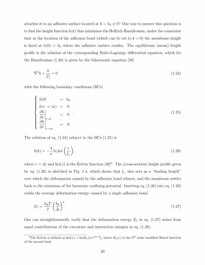
where r = |r| and kei(x) is the Kelvin function [48]3. The (cross-section) height profile given
by eq. (1.26) is sketched in Fig. 1.4, which shows that ξγ also acts as a “healing length”
over which the deformation caused by the adhesion bond relaxes, and the membrane settles
back to the minimum of the harmonic confining potential. Inserting eq. (1.26) into eq. (1.20)
yields the average deformation energy caused by a single adhesion bond
E1 =kBT
2
(h0
∆
)2
. (1.27)
One can straightforwardly verify that the deformation energy E1 in eq. (1.27) arises from
equal contributions of the curvature and interaction energies in eq. (1.20).
3The Kelvin is defined as kei(x) = ImK0
(xei3π/4
), where K0(z) is the 0th order modified Bessel function
of the second kind.
20
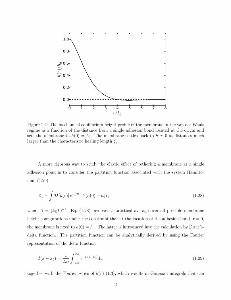
0 1 2 3 4 5 6 7 8r/ξγ
0.0
0.2
0.4
0.6
0.8
1.0
h(r)/h0
Figure 1.4: The mechanical equilibrium height profile of the membrane in the van der Waalsregime as a function of the distance from a single adhesion bond located at the origin andsets the membrane to h(0) = h0. The membrane settles back to h ' 0 at distances muchlarger than the characteristic healing length ξγ.
A more rigorous way to study the elastic effect of tethering a membrane at a single
adhesion point is to consider the partition function associated with the system Hamilto-
nian (1.20)
Z1 =
∫D [h(r)] e−βH · δ (h(0)− h0) , (1.28)
where β = (kBT )−1. Eq. (1.28) involves a statistical average over all possible membrane
height configurations under the constraint that at the location of the adhesion bond, r = 0,
the membrane is fixed to h(0) = h0. The latter is introduced into the calculation by Dirac’s-
delta function. The partition function can be analytically derived by using the Fourier
representation of the delta function
δ(x− x0) =1
2πi
∫ i∞
−i∞e−w(x−x0)dw, (1.29)
together with the Fourier series of h(r) (1.3), which results in Gaussian integrals that can
21
be readily evaluated. It follows that Z1 has the form
Z1 =Z0√2π∆2
exp
{−kBT
2
(h0
∆
)2}, (1.30)
where Z0 is the partition function of a membrane in the absence of adhesion bonds. The
associated free energy reads4
F1 = −kBT ln
(Z1
Z0
)=kBT
2
(h0
∆
)2
+kBT
2ln(2π∆2
)(1.31)
From eq. (1.31) we identify the ground state energy as the first term on the r.h.s. in line
with eq. (1.27), and the second term measures the entropy of thermal fluctuations around the
mean profile. Interestingly, the entropic component in eq. (1.31) is found to be independent
of the deformation h0.
1.2.2.2 Pairwise curvature-induced attraction
Pulling the membrane by a second adhesion point, separated by a distance r from the first
adhesion bond at r = 0 can be introduced into the statistical mechanical analysis by a
second delta function representing the additional height constraint imposed by the second
bond. The partition function of a membrane with two adhesion bonds now reads
Z2(r) =
∫D [h(r)] e−βH · δ (h(0)− h0) δ (h(r)− h0) . (1.32)
The associated free energy is given by
Φ2(r) = −kBT ln
(Z2
Z0
)=
2E1
1− 4πkei
(r
ξγ
) +kBT
2ln
{4π2∆4
[1−
(4
π
)2
kei2(r
ξγ
)]}, (1.33)
4In the second term on the r.h.s. of eq. (1.31), the thermal roughness ∆ should be measured in units ofthe relevant de Broglie wavelength ∆ → ∆/ΛdB. The same holds true for the second term on the r.h.s. ofeqs. (1.33) and (1.37) below.
22
where E1 is given by eq. (1.27). The pair PMF is composed of a deformation energy con-
tribution and an entropic component, which are represented by the first and second terms
on the r.h.s. of eq. (1.33), respectively. The former represents the curvature-induced inter-
actions between the pair of adhesion bonds, while the latter gives the fluctuation-induced
interactions. Note that both terms describe a short-range pair attraction that spans over a
typical range of ξγ, unlike the Helfrich regime where the pairwise fluctuation-induced PMF
is infinitely long-range. This is a direct result of the harmonic confinement potential in-
troduced in the van der Waals regime which suppresses the long wavelength undulations,
whereas in the Helfrich regime, the amplitudes of thermal fluctuations continue to grow with
the wavelength.
1.2.2.3 Many-body curvature-induced PMF
For a system with N ≥ 3 adhesion bonds, the attachment between the surface and the
membrane can be incorporated by a set of height constraints satisfying h({ri}Ni=1
)= h0,
where the bonds are positioned at {ri}Ni=1 and h0 is the height of the surface. Thus, the
partition function of such systems reads
ZN =
∫D [h(r)] e−βH ·
N∏i=1
δ (h(ri)− h0) . (1.34)
The partition function ZN can be evaluated by: (i) using the Fourier representations of
the height function and the Dirac-delta functions, (ii) applying N Hubbard-Stratonovich
transformations, and (iii) evaluating the resulting Gaussian integrals [45,49–51]. This leads
to the following expression
ZN =Z0
(2π∆2)N/2√
det Mexp
{−1
2
(h0
∆
)2 N∑i,j=1
(M−1
)ij
}, (1.35)
where Z0 is the partition function corresponding to the Hamiltonian (1.20), with V (h) =
12γh2 and without adhesion bonds (N = 0). The coupling matrix M appearing in eq. (1.35)
23
is given by,
Mij =2kBT
Ap∆2
∑q
cos [q · (ri − rj)]
κq4 + γ' − 4
πkei
(|ri − rj|ξγ
). (1.36)
For a given distribution of adhesion bonds, the PMF is given by the free energy
ΦN
({ri}Ni=1
)= −kBT ln
(ZNZ0
)(1.37)
=kBT
2
[(h0
∆
)2 N∑i,j=1
(M−1
)ij
+ ln((
2π∆2)N
det M)]
.
The first term on the r.h.s. of eq. (1.37) gives the energy of the height function that mini-
mizes the Hamiltonian (1.20) with the harmonic potential, subject to the height constraints
imposed by the bonds. The second term is the entropic contribution due to the thermal
undulations around this profile [51]. Note that the energetic and the entropic components
in the free energy decouple in this model, which follows from the quadratic nature of the
Hamiltonian in q-space. Also note that both terms in eq. (1.37) depend on the elements of
the matrix Mij (1.36) in a non-linear manner, which is a mathematical manifestation of the
many-body nature of the membrane-mediated PMF.
An interesting observation was made by Speck, Reister and Seifert [50], who argued
that the model depicted in eq. (1.37) belongs to the two dimensional Ising universality class
(see detailed discussion in Appendix A). Furthermore, if the typical spacing between the
adhesion bonds is much larger than the healing length ξγ (i.e., for dilute systems), the
model can be mapped onto a lattice-gas with nearest-neighbor interactions. By estimating
the effective interaction parameter between adhesion bonds occupying neighboring sites, the
authors of ref. [50] were able to draw the phase diagram of the system and estimate the
critical temperature below which clusters appear.
1.3 Outline of the thesis
This thesis is organized as follows. In chapter 2, we analyze the aggregation behavior of ad-
hesion bonds in the Helfrich regime by conducting computer simulations of a coarse-grained
model for supported lipid bilayers. We find the conditions under which the condensation
24
transition occurs and compare our results to the predictions of the WF lattice model. The
role of the fluctuation-induced interactions in adhesion cluster formation is further studied by
simulating membranes subjected to a physical confinement or to a negative surface tension
that, respectively, feature reduced or enhanced thermal undulations. In chapter 3, we investi-
gate adhesion domain formation in the van der Waals regime. We present a novel mean-field
theory for the free energy of the system, and determine the condensation transition from the
phase diagram of the system. In chapter 4, we study the role of the curvature-induced at-
traction in a specific biological process, which is the formation of the immunological synapse
– a specialized cellular junction that forms between a T cell and an antigen-presenting cell
as part of the immune response. This process is driven by both passive (thermodynamic)
and active (ATP-driven) forces, and the ideas developed in chapter 3 are used to study their
respective roles in the formation of this unique biological pattern. Chapter 5 covers the
concluding remarks.
25

Chapter 2
Molecular simulations of membranes
in the Helfrich regime
2.1 Introduction
Various models have been proposed over the years in the attempt to describe and study the
behavior and the biophysical properties of lipid bilayers. Typically, different models vary
in the resolution by which the describe the system, which directly relates to the time and
length scales that can be explored within the framework. For instance, continuum models
(such as the Helfrich elasticity model of an infinitely fluctuating manifold – see section 1.1)
can be used to study properties of membranes on macroscopic length scales that can reach
several micrometers [52]. Such models are applicable to membranes with lateral dimensions
that greatly exceed its thickness, but are too crude to describe processes on smaller length
scales. On the other hand, high resolution fully-atomistic models display great chemical
specificity, but can be used to study small systems of a few tens of nanometers in size over
timescales of about a 100-1000 nanoseconds [53,54]. Since many cellular phenomena (includ-
ing membrane adhesion) occur on larger length- and time-scales, a variety of coarse-grained
models have evolved [55–58], which constitute a certain compromise between the fully atom-
istic and continuum approaches. Within the coarse-grained approach, it is assumed that the
detailed atomic nature of the system has a relatively small impact on the mesoscopic behav-
ior under investigation and, hence, a number of atoms are grouped together and treated as
26
single beads. Depending on the degree of coarsening, coarse-grained models are extremely
advantageous in terms of the computation time, allowing one to simulate larger systems and
longer processes than those typically explored on atomistic scales. The simulation results can
be compared to continuum theoretical models, while retaining a reasonable molecular de-
tail. Notably, several solvent-free models have been presented, in which the water molecules
surrounding the membrane are excluded from the simulations. Instead, the hydrophobic
effect is introduced via effective interactions between the beads that enable self-assembly
and maintain membrane integrity [59,60]. Such implicit-solvent models substantially reduce
the CPU time required for equilibrating the system and, concomitantly, allow simulations
of substantially larger systems for increasingly larger durations. In this chapter we employ
an implicit-solvent coarse-grained membrane model to study the formation of membrane
adhesion clusters under several external constraints.
2.2 Comparison with the Weil-Farago model
We begin by testing the validity and accuracy of the WF model for condensation of adhe-
sion bonds. To this end, we use the coarse-grained model proposed by Cooke and Deserno,
in which lipids are modeled as trimmers consisting of one hydrophilic (head) and two hy-
drophobic (tail) beads [61]. This model is less coarse-grained than the one used is ref. [47]
and, thus, gives a better representation of lipid membranes which are simulated as bilayers
rather than monolayers. A flat plate, which cannot be intersected by the lipids, was placed
underneath the lower monolayer at z = 0, and the attachment of the membrane to the
surface was established by restricting N head beads from the lower monolayer to z = 0 and
allowing them to move only in-plane. We conducted Monte Carlo (MC) simulations with
periodic boundary conditions of a bilayer comprising of 2Nl = 2000 lipids (where Nl denotes
the number of lipids per monolayer) at different densities of adhesive lipids, φ = N/Nl. A
slight change in the Cooke-Deserno model was made where, for pairs of adhesive head beads,
the pair potential was switched from head-head to tail-tail. While the former pair potential
is purely repulsive, the latter also includes a cosine potential well whose depth can be tuned
(see eq. (4) in ref. [61]). This attractive part of the pair potential plays the same role played
by the standard lattice-gas term in eq. (1.16), with ε denoting the interaction energy between
27

nearest-neighbor occupied sites. By setting the depth of the potential well in the molecular
model to ε, and by simulating the WF lattice model with same value of ε, one can directly
compare the two models to each other.
The molecular simulations of the Cooke-Deserno model, which were conducted at zero
surface tension, consist of several types of MC moves, including translation of beads, rotation
of lipids, and changes in the cross-sectional projected area of the membrane, which are
accepted according to the standard Metropolis criterion [62]. To achieve equilibration within
a reasonable computing time, two additional move types were also performed. The first
move type resolves the problem arising from the slow changes in the amplitudes of the long
wavelength bending modes [63]. It involves a collective change in the heights of all the lipids,
allowing acceleration and rapid relaxation of these modes. The other process limiting the
approach to equilibrium is the slow diffusion of the lipids, especially those pinned to the
surface and serve as the adhesion bonds. In order to speed up the aggregation of adhesion
domains, one needs to allow the adhesion bonds to “jump” across the membrane. This
is accomplished by the second move type, in which two lipids simultaneously experience
opposite vertical translations: the free lipid whose head resides closest to the surface is
brought down and attached to the surface, while a randomly chosen pinned lipid is lifted
and released [42].
We simulated membranes with different concentrations φ of adhesion bonds, and for
different values of ε (measured in units of the thermal energy kBT ). Snapshots of equilibrium
configurations corresponding to ε = 0.4kBT and ε = 1.2kBT are shown, respectively, in
Figs. 2.1(A) and Fig. 2.1(B). The concentration in both cases is φ = 0.2. The distinction
between the two configurations is clear: In (A) the adhesion bonds are scattered across
the membrane in relatively small clusters, while in (B) they are assembled into one big
aggregate. The transition between the gas and the condensed phases of adhesion bonds
displayed in Figs. 2.1(A) and (B), respectively, occurs at intermediate values of ε. In order
to characterize the transition value ε = εc, we compute the non-discrete analogue for 〈NC〉 in
the WF model. In the molecular simulations, this is achieved by measuring the interaction
energy between the adhesive beads, and normalizing it by Nε. The condensation transition
value εc is empirically defined via the equality 〈NC〉 = 1.5. In Fig. 2.2 we plot 〈NC〉 as
a function of ε, the (maximum) strength of the pair interaction, for φ = 0.05 (A) and
28

(A) (B)
Figure 2.1: Bottom view of a membrane with concentration of adhesion bonds φ = 0.2 for(A) ε = 0.4kBT and (B) ε = 1.2kBT . The head and tail beads of the lipids are colored ingrey and blue, respectively, while the adhesive beads are colored in red. In (A), ε < εc andthe adhesion bonds are in the gas phase. In (B), ε > εc and the adhesion bonds condenseand form a single cluster..
φ = 0.1 (B). The simulation results, which are plotted in solid squares (with the dashed
line serving as a guide to the eye), suggest that the transition between the phases is of first
order. The parameter 〈NC〉 steeply increases around εc ≈ 0.7kBT from a low value reflecting
the dispersed distribution of adhesion bonds in the gas phase where the number of pair
interactions is small, to a high value characterizing a big cluster where the bonds are closely
packed and experience a large number of pair interactions. Also plotted in Fig. 2.2 are the
results of lattice simulations of the WF model for identical values of φ and for various values
of ε (solid circles with solid line serving as a guide to the eye). The agreement between the
molecular simulations and the lattice simulations of the WF model is very good. The lattice
model predicts a very similar value of εc ≈ 0.7kBT (for both simulated concentrations), and
gives very similar values of 〈NC〉 in the gas phase (ε < εc).
A slight discrepancy between the molecular and lattice simulation is observed in the
condensed phase for ε > εc, where the WF model appears to give higher values for 〈NC〉. This
deviation between the results of the lattice and continuum molecular models is anticipated
considering the nature of the models. In the former, the sites are organized on a perfect
triangular lattice, and the energy assigned to every pair of nearest-neighbor occupied sites is
29

(A) (B)
Figure 2.2: The dimensionless (normalized by ε) average energy of direct interactions betweenthe adhesion bond, per bond, as function of the pair interaction energy ε. Results forφ = 0.05 and φ = 0.1 are shown in (A) and (B), respectively. Solid squares and circlesdenote the results of the molecular simulations and of the Weil-Farago 2D lattice simulations,respectively. The solid and dashed lines are guides to the eye.
exactly ε. In the latter, on the other hand, the bonds within each cluster do not necessarily
have a long-range positional order [see, e.g., the snapshot in Fig. 2.1(B)], and ε denotes the
depth of the interaction well. The actual strength of the interaction is expected to be lower
than ε in the continuum molecular model, which explains why it gives lower values of 〈NC〉
than in the lattice simulations.
At even higher values of ε, the close agreement between the lattice and the molecu-
lar simulations is regained. This occurs due to another phase transition that the clusters
undergo, from disordered liquid-like structures into more ordered organizations such as the
one displayed in Fig. 2.3(A) for φ = 0.2 and ε = 3.4kBT . This phase transition can be
understood within the framework of the KTHNY theory, which proposes the formation of a
two dimensional hexatic phase with a quasi-long range hexagonal (orientational) order [64].
This transition is characterized by the bond orientational order parameter
ψ6j =1
Nj
Nj∑k=1
ei6θkj , (2.1)
where the sum runs over the nearest-neighbor bonds k to a given bond j (whose identity
is determined by Voronoi tessellation), and θkj is the angle between the line connecting the
pair of bonds j and k and some fixed axis. Averaging over all the bonds within the cluster
30

(A) (B)
(C) (D)
(A) (B)
(C) (D)
Figure 2.3: The molecular simulation results for a membrane with φ = 0.2 for ε > εc.(A)Snapshot of an equilibrium configuration with ε = 3.4kBT , depicting an adhesion domainorganized in the hexatic phase. Color coding as in Fig. 2.1. (B) The mean bond orientationalorder parameter 〈Φ6〉 as a function of the pair interaction energy ε. The transition into thehexatic phase occurs around εh ≈ 1.9kBT where a sudden increase in 〈Φ6〉 is observed. (C)The mean square displacement of the adhesion bonds vs. the simulation time for differentvalues of ε. The slope of each curve is a measure for the self diffusion coefficient of theadhesion bonds within the cluster D. The results for ε/kBT = 1, 1.8, 2 are marked byarrows. (D) The dimensionless average energy of direct interactions between the adhesionbond (normalized per bond) as function of the pair interaction energy ε. A jump in 〈NC〉 isobserved around εh.
yields the global orientational order parameter
Φ6 =
∣∣∣∣∣ 1
N
N∑j=1
ψ6j
∣∣∣∣∣ . (2.2)
Another quantity undergoing rapid variations at the transition is the self-diffusion coefficient
of the bonds (relative to the diffusion of their center of mass), defined by
D = limt→∞
1
4Nt
N∑i=1
⟨[(~ri(t)− ~rcm(t))− (~ri(t = 0)− ~rcm(t = 0))]2
⟩(2.3)
≡ limt→∞
⟨(∆r′)2⟩
4t,
31
where ~ri(t) and ~rcm(t) denote, respectively, the position of adhesion bond i and of the center
of mass of the cluster at time t (measured in MC time units), and 〈· · · 〉 denotes statistical
average. The transition into the hexatic phase is characterized by (i) an increase in Φ6,
associated with the emergence of orientational order, and (ii) a sharp decrease in D, reflecting
a lower mobility of the bonds. In Fig. 2.3(B), we plot our results for 〈Φ6〉, as a function of ε
for φ = 0.2. In Fig. 2.3(C) the mean squared displacement of the adhesion bonds (measured
in units of σ2, where σ is the diameter of the beads1) is plotted versus the simulation
time (measured in MC time units), with the curves, from top to bottom, corresponding
to increasingly higher values of ε. [Each curve in Fig. 2.3(C) corresponds to a data point
in Fig. 2.3(B)]. The curves display a linear increase in 〈(∆r′)2〉 with t, and the slope of
each curve is proportional to D. Both Figs. 2.3(B) and (C) indicate that the transition
from disordered-liquid into an ordered-hexatic structure occurs at around ε = εh ≈ 1.9kBT .
Another evidence for the fluid to hexatic transition is also observed in Fig 2.3(D), showing
a “jump” in 〈NC〉 between ε = 1.8kBT and ε = 2.0kBT . Note that the values of 〈NC〉 in the
hexatic phase is higher than three, which is the maximum possible value in simulations of
the WF model on a triangular lattice. This feature is related to the form of the attractive
tail-tail pair potential in the molecular simulations whose cut-off range was set to slightly
less that 2.5σ. This implies that, in a closely packed cluster, each adhesion bond weakly
interacts with its next- and next-next-nearest neighbors, which explains why 〈NC〉 becomes
larger than three.
2.3 Adhesion domain formation in stressed and con-
fined membranes
Thus far, we have focused on the aggregation behavior of adhesion bonds in tensionless sup-
ported membranes. We now wish to extend our investigations to lipid bilayers subjected to
a physical confinement and lateral negative tensions. The former constraint acts to suppress
the long wavelength thermal undulations, while the latter amplifies their amplitudes (and, for
very large negative values, can even lead to instability). It follows that for systems subjected
1In the Cooke-Deserno model, σ is also the range of the head-head repulsive potential [61].
32
to confinement or negative tension, one should expect that the resulting fluctuation-induced
PMF between the adhesion bonds becomes weaker or stronger, respectively. Therefore, such
constraints alter the magnitude of the direct short-range attraction required for condensa-
tion. In other words, one may presume that physical confinement may hinder the formation
of adhesion domains, and that the transition threshold value, εc, into the condensed state
grows with the degree of confinement. Likewise, one can also anticipate that a negative
tension would cause a reduction in εc. It is especially interesting to examine whether εc
can decrease to zero, in which case the adhesion domains will form without an additional
short-range potential, i.e., on purely entropic grounds.
In order to address the above issues, we conducted Monte Carlo simulations of a lipid
bilayer using the Cooke-Deserno implicit-solvent coarse-grained model [61]. The details of
the simulations are similar to those presented in section 2.2. Our simulations for confined
membranes included an additional impermeable surface placed above the upper monolayer
at z = zconf . Simulations of membranes under constant mechanical surface tension τ were
carried out according to the method described in ref. [66]. Similarly to the strategy presented
in section 2.2, we followed the aggregation behavior by measuring the average energy of
direct pairwise interactions between adhesive beads, normalized per bond and expressed in
dimensionless units by dividing it by the potential strength ε, to obtain the typical number
of contacts per bonds 〈NC〉. As will be shown further on, our results suggest that physical
confinement might have a very strong impact on εc, unlike the application of a negative
surface tension, which may lead to buckled configurations where we observe the formation
of elongated adhesion domains close to the transition point.
2.3.1 Membranes under physical confinement
We first start with our simulation results for non-stressed membranes confined between the
supporting underlying surface (on which the adhesive beads reside) and a second impene-
trable upper surface. The former is located at z = 0, just underneath the tips of the head
beads of the lower leaflet, while the latter is placed at z = zconf ≥ 6σ. The degree of con-
finement increases when zconf decreases which, in turn, would lead to stronger suppression
of the membrane thermal fluctuations and a shift in the transition threshold εc to larger
33
values. This trend is demonstrated in Fig. 2.4, showing our simulation results for zconf = 6σ,
7.5σ, and 9σ in supported membranes with φ = 0.1 (A) and φ = 0.2 (B). For zconf = 9σ, our
results for 〈NC〉 (green diamonds) match perfectly with the results obtained for non-confined
membranes (black circles). This means that the rate of collisions between the membrane
and the upper surface is negligibly small and, therefore, it does not affect the fluctuation
spectrum. Lowering the confining surface by a distance equal to the size of a bead and a
half to zconf = 7.5σ has a more noticeable effect on membrane thermal undulations, which
leads to an increase in the condensation transition value from εc ' 0.65kBT for non-confined
membranes at φ = 0.1 to εc ' 0.8kBT . As mentioned in section 2.2, εc is found by the
condition 〈NC〉 ' 1.5. For φ = 0.2, the shift is smaller, from εc ' 0.6kBT to εc ' 0.7kBT .
When the upper surface is further lowered to zconf = 6σ, it touches the tips of the head
beads in the upper leaflet, as the thickness of the bilayer is equal to the size of six beads.
A confining surface located at zconf = 6σ completely suppresses thermal undulations, and
eliminates the fluctuation-mediated interactions between the adhesion bonds. Under these
conditions, the threshold value for aggregation increases to εc ' 1.2kBT at φ = 0.1, and
εc ' 1kBT for φ = 0.2. These values are approximately twice larger than the corresponding
values found when no upper plate exists (εc ' 0.65kBT and εc ' 0.6kBT for φ = 0.1 and
φ = 0.2, respectively), which is in accord with the conclusions of refs. [46, 47], that the
entropic gain of aggregation compensates for, roughly, half of the loss in mixing entropy of
the adhesion bonds.
2.3.2 Membranes under negative surface tension
We next aim to address the implications of applying a negative surface tension. A constant
mechanical surface tension introduces an additional energy term to the system Hamilto-
nian, proportional to the surface area of the membrane. In the Monge gauge, the system
Hamiltonian is given by
H =
∫ [1
2κ(∇2h
)2+ τ
(~∇h)2]d2r, (2.4)
34

(B)
(A)
Figure 2.4: The dimensionless energy 〈NC〉 in membranes confined by a surface located atzconf = 9σ (green diamonds), zconf = 7.5σ (blue triangles) and zconf = 6σ (red squares), asa function of ε, for (A) φ = 0.1, and (B) φ = 0.2. Results for non-confined membranes aredenoted by black circles. The lines serve as a guide to the reader’s eye. The statistical errorsare comparable to the size of the symbols.
where τ denotes the surface tension. The fluctuation spectrum corresponding to eq. (2.4)
now reads
〈∣∣h2
q
∣∣〉 =L2kBT
l4 (κq4 + τq2), (2.5)
which can be compared with eq. (1.5) for non-stressed membranes. On length scales larger
than the characteristic lengthscale ξτ ∼√κ/τ , the imposed surface tension dominates the
thermal fluctuations of the membrane, while fluctuation modes with q � 2π/ξτ remain
largely unaffected by it. In general, a negative tension imposed on the membrane leads to a
reduction in its projected area, AP, and amplifies the long wavelength bending modes. Hence,
the fluctuation-induced attraction between the adhesion bonds is expected to be stronger
35

(A) (B)
(C)
Figure 2.5: The dimensionless energy 〈NC〉 in membranes under surface tension τ =−0.24kBT/σ
2, as a function of ε for (A) φ = 0.1, and (B) φ = 0.2. The results are plottedin red squares, and are compared with the results for tensionless membranes (τ = 0) thatare depicted by black open circles. (C) The mean projected area per lipid as a function of ε,for φ = 0.1 (black) and φ = 0.2 (red). Circles denote the results for tensionless membranes,while the results for τ = −0.24kBT/σ
2 are shown in squares. The lines serve as guides tothe eye.
which, in turn, implies that the threshold for condensation εc should become smaller than
in tensionless membranes. To test this hypothesis, we simulated the membrane under a
negative tension of τ = −0.24kBT/σ2. We performed two sets of independent MC simula-
tions, one starting from a random distribution of adhesion bonds, and another where initially
the adhesion bonds were organized in one large cluster. The system was equillibrated until
configurations originating from these two distinct initial conditions achieved similar charac-
teristics. Fig. 2.5 shows our results for 〈NC〉 as a function of ε for φ = 0.1 (A) and φ = 0.2
(B). Contrary to our expectation to observe a reduction in εc, the data for τ = −0.24kBT/σ2
appears almost identical to the results of the tensionless case, with εc ' 0.6kBT for both val-
36
ues of φ, and seems to suggest that a negative tension has a minor impact on the aggregation
process.
The negative tension, however, does have an impact on the shape of membranes. Freely
fluctuating bilayers assume buckled configurations at negative tensions larger (in absolute
value) than τc ' −4π2κ/AP [67, 68] 2. In this study, non-stressed systems are character-
ized by AP/N ' 1.33σ2 [see Fig. 2.5(C)] which, for the bending modulus of the present
model membrane κ ' 8 kBT [63], gives τc ' −0.24kBT/σ2. In ref. [69], a similar model
membrane consisting of the same number of lipids was simulated and, indeed, for τ = τc
the membrane appeared quite buckled. In supported membranes, however, the emergence
of buckled configurations occurs only in membranes with large adhesion domains (i.e., for
ε & εc). Fig. 2.6 shows typical equilibrium configuration for φ = 0.1 with ε = 0 (A), 0.6kBT
(B), and 1.0kBT (C). Each configuration is shown both in side and bottom views (lower and
upper panels, respectively). When the attractive potential is set to ε = 0, the distribution of
the adhesion bonds is scattered and the membrane remains fairly flat. This indicates that
the mixing entropy of the bonds dominates the fluctuation entropy of the bilayer, despite the
imposed negative tension [see Fig. 2.6(A)]. For ε = 1.0kBT , the short-range pair interactions
between the bonds lead to their aggregation. Once the bonds condense, their influence on
the thermal behavior of the membrane is greatly weakened, and strong bending undulations
appear [see Fig. 2.6(C)]. Close to the condensation transition, at ε = 0.6kBT , the system ex-
hibits some interesting features: The amplitude of one of the two longest wavelength bending
modes [with wavevector ~q(1,0) = (2π/√AP)(1, 0), or ~q(0,1) = (2π/
√AP)(0, 1)] grows consider-
ably, and the membrane assumes an anisotropic buckled configuration. The adhesion bonds
are concentrated throughout the minimum of the dominating bending mode, forming an
elongated domain (“stripe”) [see Fig. 2.6(B)]. These observed characteristics represent an
intricate balance between the driving forces that govern the thermodynamic behavior of the
system. Under negative tension, the system benefits from a reduction in the projected area,
leading to a decrease in the Gibbs free energy. The membrane, however, is quite incompress-
ible and, thus, the reduction in AP must be accompanied by an increase in the area stored in
2This value for the critical tension for buckling, τc, stems from the amplitude divergence of the longestFourier mode with q0 = 2π/L. Looking at eq. (2.5), the amplitude of this mode diverges when κq40 +τq20 ' 0,which is satisfied for a negative tension of τ = τc ' −κq20 = −4π2κ/AP.
37
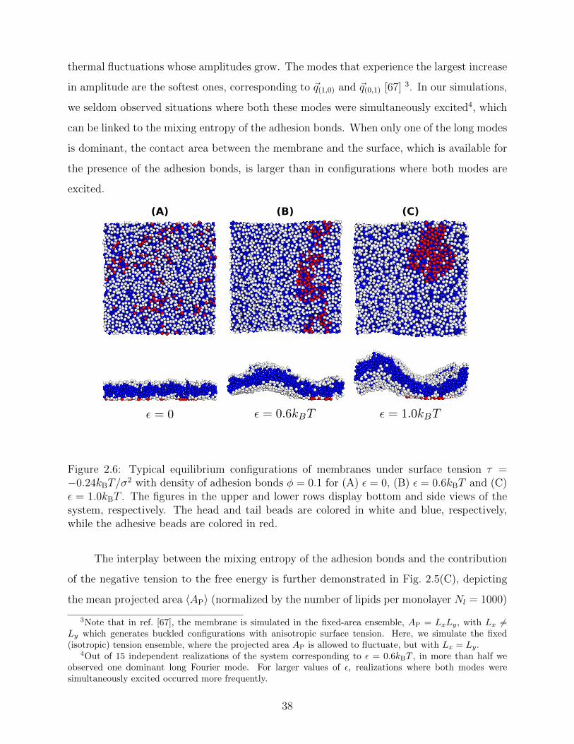
thermal fluctuations whose amplitudes grow. The modes that experience the largest increase
in amplitude are the softest ones, corresponding to ~q(1,0) and ~q(0,1) [67] 3. In our simulations,
we seldom observed situations where both these modes were simultaneously excited4, which
can be linked to the mixing entropy of the adhesion bonds. When only one of the long modes
is dominant, the contact area between the membrane and the surface, which is available for
the presence of the adhesion bonds, is larger than in configurations where both modes are
excited.
(A) (B) (C)
Figure 2.6: Typical equilibrium configurations of membranes under surface tension τ =−0.24kBT/σ
2 with density of adhesion bonds φ = 0.1 for (A) ε = 0, (B) ε = 0.6kBT and (C)ε = 1.0kBT . The figures in the upper and lower rows display bottom and side views of thesystem, respectively. The head and tail beads are colored in white and blue, respectively,while the adhesive beads are colored in red.
The interplay between the mixing entropy of the adhesion bonds and the contribution
of the negative tension to the free energy is further demonstrated in Fig. 2.5(C), depicting
the mean projected area 〈AP〉 (normalized by the number of lipids per monolayer Nl = 1000)
3Note that in ref. [67], the membrane is simulated in the fixed-area ensemble, AP = LxLy, with Lx 6=Ly which generates buckled configurations with anisotropic surface tension. Here, we simulate the fixed(isotropic) tension ensemble, where the projected area AP is allowed to fluctuate, but with Lx = Ly.
4Out of 15 independent realizations of the system corresponding to ε = 0.6kBT , in more than half weobserved one dominant long Fourier mode. For larger values of ε, realizations where both modes weresimultaneously excited occurred more frequently.
38
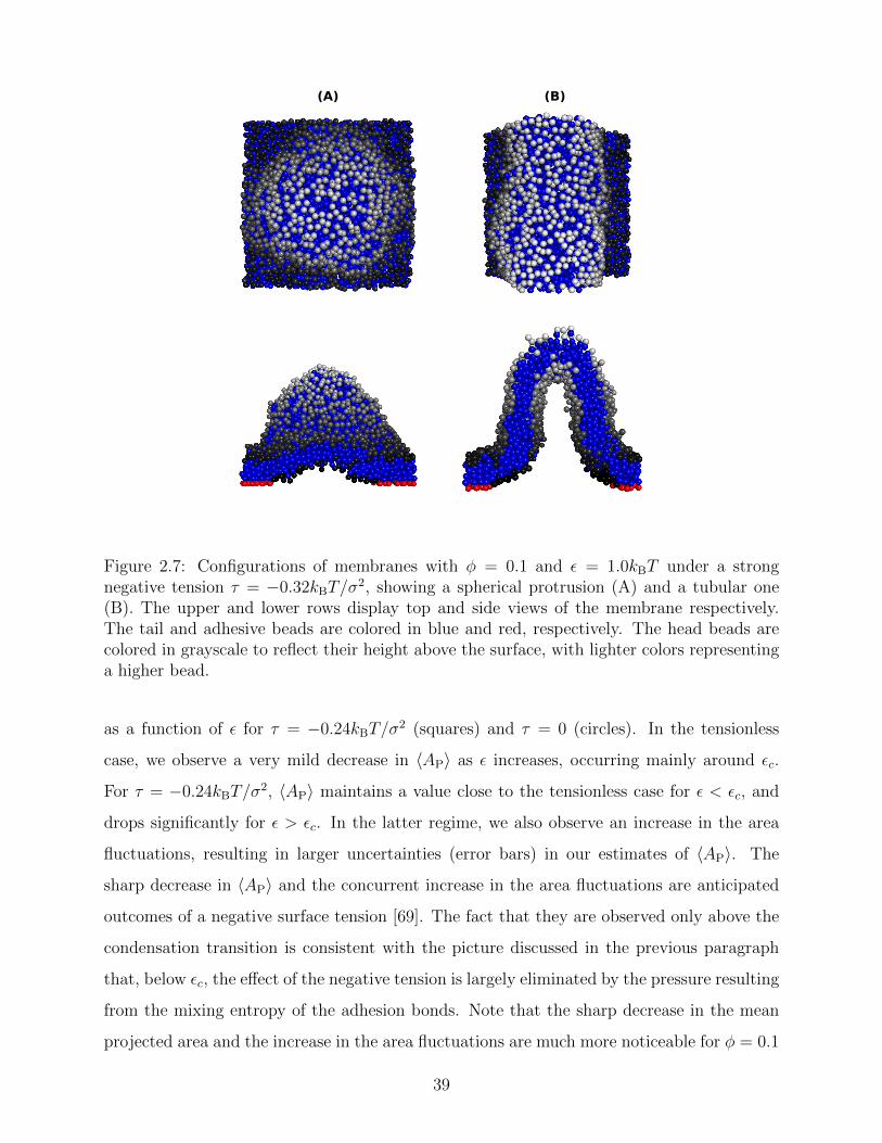
(A) (B)
Figure 2.7: Configurations of membranes with φ = 0.1 and ε = 1.0kBT under a strongnegative tension τ = −0.32kBT/σ
2, showing a spherical protrusion (A) and a tubular one(B). The upper and lower rows display top and side views of the membrane respectively.The tail and adhesive beads are colored in blue and red, respectively. The head beads arecolored in grayscale to reflect their height above the surface, with lighter colors representinga higher bead.
as a function of ε for τ = −0.24kBT/σ2 (squares) and τ = 0 (circles). In the tensionless
case, we observe a very mild decrease in 〈AP〉 as ε increases, occurring mainly around εc.
For τ = −0.24kBT/σ2, 〈AP〉 maintains a value close to the tensionless case for ε < εc, and
drops significantly for ε > εc. In the latter regime, we also observe an increase in the area
fluctuations, resulting in larger uncertainties (error bars) in our estimates of 〈AP〉. The
sharp decrease in 〈AP〉 and the concurrent increase in the area fluctuations are anticipated
outcomes of a negative surface tension [69]. The fact that they are observed only above the
condensation transition is consistent with the picture discussed in the previous paragraph
that, below εc, the effect of the negative tension is largely eliminated by the pressure resulting
from the mixing entropy of the adhesion bonds. Note that the sharp decrease in the mean
projected area and the increase in the area fluctuations are much more noticeable for φ = 0.1
39

than for φ = 0.2. This is to be expected because the smaller φ, the smaller the restrictions
imposed by the adhesion bonds on large thermal undulations, which are directly coupled to
the projected area by the highly incompressible character of the membrane.
Applying an even stronger negative tension causes the membrane to lose its mechan-
ical stability. This is demonstrated in Fig. 2.7, showing snapshots in top and side views of
membranes with φ = 0.1 and ε = 1.0kBT subjected to τ = −0.32kBT/σ2. In these snap-
shots, the head beads are colored in grayscale, with lighter colors indicating beads located
higher above the underlying surface. The application of a strong negative tension causes
the supported membrane to develop large protrusions with either spherical [Fig. 2.7(A)] or
tubular [Fig. 2.7(B)] shapes. The adhesion bonds (which are colored in red, and are only
partially visible) are concentrated in the periphery of the protrusion, where the membrane
is in contact with the underlying surface. We note that the observed protrusions tend to
evolve slowly and, therefore, the snapshots shown in Fig. 2.7 may not represent true equilib-
rium structures. On the other hand, we also note that very similar equilibrium structures,
featuring spherical and tubular protrusions, have been recently observed in an experimental
study where supported lipid bilayers were subjected to lateral compression [70].
2.4 Summary
In this chapter, we used solvent-free coarse-grained molecular simulations to study the for-
mation of adhesion domains in supported membranes. We have focused on the Helfrich
regime, in which the membrane’s thermal undulations are substantial, which results in fre-
quent collisions with the underlying supporting surface. Theses constitute the entropic source
of the fluctuation-mediated attractive PMF between the adhesion bonds, which may drive
their aggregation into large domains in order to reduce the entropic free energy penalty.
Our molecular simulations are found to be in excellent agreement with the recently pro-
posed Weil-Farago (WF) lattice model. Both the molecular and lattice simulations of the
WF model show that adhesion domain formation requires an additional direct attraction
between the bonds of strength ε > 0. In addition, results from the molecular simulations
show that the transition into the condensed phase occurs at values of ε = εc extremely
similar to those predicted by the WF model. Our simulation results for physically confined
40

membranes reveal that placing the membrane between two impenetrable plates significantly
alters the condensation point and requires a stronger direct attraction between the bonds.
This stems directly from the fact that the thermal fluctuations of confined membranes are
strongly suppressed, which weakens the fluctuation-induced PMF. In the most extreme case,
where the membrane is completely flattened between the two plates, the thermal fluctuations
are entirely eliminated. This reduces the problem to the phase transition in the standard
lattice gas model, and results in a roughly two-fold increase in the value of εc, which is in
accord with the conclusions of the WF model. The implication of the fluctuation-induced
shift in εc is that the fluctuation entropy gained by the condensation of the adhesion bonds
compensates for about half of the mixing entropy that is lost in the transition. In other
words, the fluctuation-mediated attraction effectively renormalizes the system temperature
to about half its value. Thus, while the fluctuation-induced PMF is too weak to promote
adhesion domain formation purely on entropic grounds, it greatly facilitates the conditions
required for the aggregation of adhesion bonds.
The excellent agreement between the molecular simulations presented here and the WF
model lands great credibility to the main idea of the WF model, which is the notion that
each adhesion bond suppresses the thermal fluctuations mainly in its immediate vicinity.
When studying the system via a lattice model, therefore, one can associate the fluctuation-
induced PMF between the bonds with free energies assigned to the empty sites of the lattice.
The empty sites represent the fluctuating segments of the supported membrane, and the free
energy assigned to each site measures the free energy loss due to the local restrictions imposed
on the membrane’s thermal undulations. This free energy penalty mainly depends on the
distance, dmin, between an empty site and the closest occupied site (representing an adhesion
bond).
We also presented simulations of membranes under negative surface tension which,
presumably, fluctuate more strongly than tensionless membranes and, therefore, should ex-
hibit stronger membrane-mediated effects. Surprisingly, we find that the application of a
negative tension has a very minor effect on the condensation transition. Nevertheless, once
the adhesion bonds are aggregated into a large domain (i.e., for ε > εc), the negative tension
affects the shape of the membrane and causes it to buckle. Below εc, the adhesion bonds
are scattered across the membrane, which prevents the formation of buckled configurations.
41

Close to εc, we observed both membrane buckling and the formation of elongated adhesion
stripes. Such configurations emerge from a delicate interplay between the mixing entropy
of the adhesion bonds, the short-range residual potential, and the applied negative tension.
Finally, under a very strong negative tension, we observe tubular and spherical structures
protruding out of the membrane’s plane, which indicates that the system is at the onset of
mechanical instability.
42

Chapter 3
Formation of semi-dilute adhesion
domains in the van der Waals regime
3.1 Introdcution
In this chapter, we focus our attention on the van der Walls regime, in which membranes
experience small thermal undulations such that the bending energy dominates the system’s
free energy. Such situations are frequent in biology as the plasma membrane fluctuations
are quite confined due to the presence of the ECM, the underlying cytoskeleton and the
glycocalyx coating of the cell. This regime can be studied via the effective Helfrich Hamilto-
nian given by eq. (1.20), which accounts for the membrane-surroundings interactions via an
additional non-specific harmonic confining potential V [h (r)] = γh2/2 that prevents strong
membrane fluctuations, and limits the thermal roughness of the membrane to 〈h2〉 = ∆2.
A single adhesion bond that locally fixes the membrane to h = h0 pulls the membrane
away from the minimum of the harmonic potential and leads to an elastic deformation,
which relaxes after a typical healing length ξγ = (κ/γ)1/4 (see Fig. 1.4). In a many-body
system with a multiple number of adhesion bonds, this curvature energy induces an effective
attractive PMF in order to minimize the elastic deformations. As discussed in section 1.2.2.3,
obtaining the curvature-induced PMF requires a full statistical average over the membrane’s
height degrees of freedom, under the height constraints imposed by the bonds. While an
analytical expression for the PMF was previously introduced [see eq. (1.37)], a satisfactory
43

description of the thermodynamic behavior of the system is still lacking. This is attributed
to the highly complicated task of further tracing over the positional degrees of freedom of
the adhesion bonds, which is required in order to characterize the condensation transition.
Here, we employ a different strategy and derive the phase diagram of the system for a
wide range of healing lengths, ξγ, and adhesion bonds densities, φ. Our investigation relies
on a novel mean-field treatment of the system’s free energy. We obtain the spinodal and
binodal curves and locate the critical temperature of the system, Tc, above which adhesion
domains do not form. As shown below, results for different systems exhibit data collapse
when (∆/h0)2 ∼ T/Tc is plotted as a function of the rescaled density ξ2γφ. Interestingly,
we find that the critical point is located at extremely low densities, which is linked to the
many-body membrane-mediated PMF. Therefore, close to critically, a phase coexistence is
found between two extremely dilute phases, while dense domains form only for T � Tc, i.e.,
when each bond deforms the membrane considerably.
3.2 Mean-field theory
The membrane-mediated PMF in the van der Waals regime, ΦN , as written in eq. (1.37),
corresponds to a system with a given spatial distribution of N fixed adhesion bonds. The
thermodynamics of a system with N mobile bonds is characterized by the free energy F ,
which depends on the bond density φ = aN/AP, where a is a microscopic unit area for which
0 ≤ φ ≤ 1. The free energy F (and, thus, the phase diagram) can be derived from the
corresponding partition function F (φ) = −kBT lnZ, where
Z = Tr{ri}
[e−ΦN({ri}Ni=1)/kBT
], (3.1)
is obtained by integrating out the translational degrees of freedom of the bonds at a given
density φ. Since the exact calculation of the partition function is out of reach, we invoke
a simpler mean-field approach. Within a mean field approximation, the free energy can be
written as
aF
AP
= kBT [φ lnφ+ (1− φ) ln(1− φ)] + φ
⟨ΦN
N
⟩MF
, (3.2)
44

where the first term on the r.h.s. accounts for the mixing entropy of the bonds, and the
second term represents a mean-field estimation of ΦN .
We recall that, here, we are interested in the van der Waals regime, which is charac-
terized by small thermal roughness ∆ [see eq. (1.23)]. Following previous studies [38, 50],
we will also make the assumption that each adhesion bond causes a deformation h0 signif-
icantly larger than ∆. This allows us to drop the second term on the r.h.s. of eq. (1.37)
accounting for the entropic contribution of the thermal fluctuations to the PMF in the van
der Waals regime. Thus, the curvature-mediated PMF can be expressed just by the first
term representing the elastic energy of the ground state
ΦN
({r}Ni=1
)' kBT
2
(h0
∆
)2 N∑i,j=1
(M−1
)ij, (3.3)
with the coupling matrix M, whose entries are given in eq. (1.36). The elastic energy (3.3)
can be estimated by considering a lattice of adhesion bonds with spacing r ∼√aφ−0.5, which
gives an energy landscape that depends on the ratio r/ξγ. This approach yields good an-
alytical expressions for the elastic energy only in the limits r/ξγ � 1 and r/ξγ � 1 [38];
however, it fails to capture the correct thermodynamic behavior at the intermediate regime
r/ξγ ∼ 1 where the lattice distribution does not necessarily represent the energy of a typical
random distribution of adhesion bonds. Here, we take a different approach and derive an
empirical expression for the dependency of the elastic energy on the bonds’ density. We com-
putationally obtain this expression by (i) generating membranes with random, rather than
ordered, distributions of adhesion bonds, (ii) finding the membrane profile that minimizes
the Helfrich elastic energy of each realization, and (iii) describing the computational data
for the elastic energy by a fitting function, which applies to the entire range of densities.
3.2.1 Energy calculations
The ground state Helfrich energy corresponding to a random distribution of adhesion bonds
is given by eq. (3.3) and can, in principle, be computed by inverting the coupling matrix M
(1.36). In practice, this involves a computationally expensive process and, thus, we adopt a
different strategy based on a direct minimization of the Helfrich Hamiltonian. This is done
45

by considering a triangular lattice with lattice spacing l. Each site, i, represents a small
membrane segment of area a =√
3l2/2, and is characterized by a local height variable hi.
On the lattice, N sites are randomly chosen for the locations of the adhesion bonds, at which
we set hi = h0. The discrete analogue of the Helfrich Hamiltonian (1.20) is
Hlattice =a
2
∑i
[κ(∇2ihi)2
+ γh2i
]=aκ
2
∑i
[(∇2ihi)2
+
(hiξ2γ
)2], (3.4)
where the discrete Laplacian at site i is given by ∇2i =
[23
∑6j=1 hj − 4hi
]/l2, with the sum
j = 1 . . . 6 running over the six nearest neighbors of site i. Starting with hi = h0 at all sites,
we simulate Langevin dynamics [72] without the noise term (i.e., at zero temperature), which
quickly brings the system to the ground state profile. We measure all lengths in units of the
lattice spacing l = 1 and the energy scale is set to kBT = 1. The density of bonds is given by
φ = N/Ns, where Ns is the number of lattice sites. Most of the calculations were performed
on a triangular lattice of 104 × 120 sites (with periodic boundary conditions) that has an
aspect ratio close to 1. We calculate the elastic energy of numerous random realizations at
various densities φ ≤ 0.1, and for several values of ξγ varying from ξγ = 5 to ξγ = 10. These
values for the correlation length are chosen such that: (i) ξγ is sufficiently larger than the
lattice spacing l = 1, which reduces the numerical errors associated with the discrete nature
of eq. (3.4) to less than a few percents, and (ii) ξγ is much smaller than the system linear
size, to avoid finite size effects.
From eq. (3.3) and the form of the elements of the coupling matrix M [see eq (1.36)],
we infer that for a given set of model parameters (κ, h0, ξγ, φ), the average elastic energy
has the form
⟨ΦN
N
⟩MF
=kBT
2
(h0
∆
)2
f(x) = 4κ
(h0
ξγ
)2
f(x), (3.5)
where f(x) is a scaling function of the renormalized density x = ξ2γφ. Note that the values
of κ and h0 can be fixed arbitrarily since the energy scales like κh20 [see eq. (3.5)], and
this scaling behavior is automatically satisfied by the Hamiltonian (3.4) which is linear in
κ and quadratic in hi ∝ h0. The low-density (x → 0) asymptotic limit of f(x) is found by
considering a system with a single bond, which gives the energy per bond in dilute systems
46
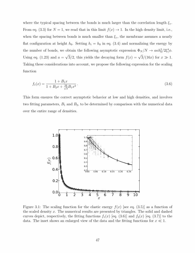
where the typical spacing between the bonds is much larger than the correlation length ξγ.
From eq. (3.3) for N = 1, we read that in this limit f(x)→ 1. In the high density limit, i.e.,
when the spacing between bonds is much smaller than ξγ, the membrane assumes a nearly
flat configuration at height h0. Setting hi = h0 in eq. (3.4) and normalizing the energy by
the number of bonds, we obtain the following asymptotic expression ΦN/N → aκh20/2ξ
4γφ.
Using eq. (1.23) and a =√
3/2, this yields the decaying form f(x) =√
3/(16x) for x � 1.
Taking these considerations into account, we propose the following expression for the scaling
function
f1(x) =1 +B1x
1 +B2x+ 16√3B1x2
. (3.6)
This form ensures the correct asymptotic behavior at low and high densities, and involves
two fitting parameters, B1 and B2, to be determined by comparison with the numerical data
over the entire range of densities.
Figure 3.1: The scaling function for the elastic energy f(x) [see eq. (3.5)] as a function ofthe scaled density x. The numerical results are presented by triangles. The solid and dashedcurves depict, respectively, the fitting functions f1(x) [eq. (3.6)] and f2(x) [eq. (3.7)] to thedata. The inset shows an enlarged view of the data and the fitting functions for x� 1.
47

In Fig. 3.1 we plot the computational results (triangles) for the elastic energy per bond,
normalized by the energy of a single isolated bond E1 = 4κ(h0/ξγ)2 [see eq. (1.27)], which
defines f(x) in eq. (3.5). The data, which is plotted against the scaled density x = ξ2γφ,
exhibits an excellent data collapse over the entire range x ≤ 10. The solid curve represents
the fitting of the data to the form f1(x) given by eq. (3.6), with the parameters B1 ' 5.08
and B2 ' 9.87 that give the best fit. The scatter of the computational data is due to the
randomness of the simulated configurations. As expected, the scatter is larger for small
values x � 1, where the interaction between the closer pairs of adhesion bonds dominates
the energy of the configuration. In fact, for some configurations in this regime, we find
f(x) to be slightly larger than unity. This feature is to be expected, and follows from the
non-monotonicity of the Kelvin function defining the elements of the coupling matrix M [see
eq. (1.36)]. For x � 1, the PMF between the bonds can be approximated by a sum of
pair potentials, as was assumed in ref. [45] (see also discussion in Appendix A). By setting
N = 2 in eqs. (1.36) and (1.37), it is easy to confirm that the pair PMF is slightly repulsive
at large bond separations. We, therefore, conclude that the scaling function f(x) should be
non-monotonic; it first increases for very small values of x before dropping to zero at larger
values. Furthermore, from the fact that the Kelvin function converges exponentially to zero
for large arguments, one can also conclude that the derivative of the scaling function satisfies
df/dx = 0 at x = 0. These features of f(x) in the x → 0 limit are not accounted for by
the scaling form f1(x) proposed by eq. (3.6). Therefore, we also consider the three fitting
parameter scaling function
f2(x) =1 + C1x+ C2x
2
1 + C1x+ C3x2 + 16√3C2x3
, (3.7)
which, in contrast to f1(x), correctly captures the behavior of f(x) near x = 0. The fit of
the scaling function f2(x) to the computational data is also plotted in Fig. 3.1 (dashed line)
with C1 ' 74.8, C2 ' 2174 and C3 ' 1836 that produce the best fit. The difference between
f1(x) and f2(x) is visible only for x ' 0, as seen in the inset in Fig. 3.1. Interestingly, even
though f2(x) is better suited to represent the scaling function close to the origin than f1(x),
the latter seems to provide a better fit to the numerical data. In any case, we expect these
two functions to yield similar binodal and spinodal curves, except for x ' 0. This will turn
48

out to be in the vicinity of the critical point, which is where the validity of the mean-field
picture is questionable anyhow.
3.2.2 Phase diagram
Plugging eq. (3.5) into eq. (3.2), the mean-field free energy, F , of a system with adhesion
bond concentration φ and correlation length ξγ reads
aF
ApkBT' φ lnφ+ (1− φ) ln(1− φ) +
1
2ξ2γ
(h0
∆
)2
g(x), (3.8)
where g(x) = xf(x). With this expression for F , we analytically obtain the spinodal curve
enclosing the region of thermodynamic instability by solving ∂2F/∂φ2 = 0, which yields
(∆
h0
)2
=x(x− ξ2
γ
)2ξ2γ
∂2g
∂x2. (3.9)
The binodal curve, which defines the thermodynamic coexistence line, is obtained numer-
ically using a common tangent construction for F . Figs. 3.2(A) and (B) show the phase
diagrams calculated using the scaling functions f1(x) and f2(x), respectively. In each of
these figures, we plot the spinodal curve for ξγ = 5 (solid line) and ξγ = 10 (dotted line),
which turn out to be practically indistinguishable. The binodal curves for ξ = 5 and ξγ = 10
are given by squares and circles, respectively. As for the spinodal lines, the binodal curves
for different values of ξγ also overlap each other. Comparing the phase diagrams presented
in Figs. 3.2(A) [for f(x) = f1(x)] and (B) [for f(x) = f2(x)], we conclude that the phase
diagrams appear to be similar, expect for x . 0.6. This is to be expected because only in
this regime, the scaling functions are essentially different (see inset in Fig. 3.1). Fig. 3.2(C)
presents an enlargement of the low density regime, showing the binodal [squares for f1(x),
and circles for f2(x)] and spinodal [solid line for f1(x), and dotted line for f2(x)] curves, for
ξγ = 10. Note that the critical point is located at low densities, and the two scaling functions
place it at somewhat different values.
49

(C)
(A) (B)
Figure 3.2: (A) The phase diagram corresponding to the free energy eq. (3.8) with f(x) =f1(x) given by eq. (3.6). The binodal curve is represented by the symbols (with dashedlines serving as guides to the eye), where squares and circles represent data for ξγ = 5 andξγ = 10, respectively. The two binodal curves nearly overlap each other. The spinodalcurves, which are presented by the solid (for ξ = 5) and dotted (for ξγ = 10) lines, are alsoindistinguishable. (B) Same as (A), but for f(x) = f2(x) in eq. (3.8). (C) A zoom on thevicinity of the critical point, where the differences between the scaling functions f1(x) andf2(x) are visible. The phase diagrams are calculated for ξγ = 10. Binodal curves are plottedby squares for f1(x) and circles for f2(x). The spinodal lines are presented by the solid anddotted lines for f1(x) and f2(x), respectively.
3.3 Semi-dilute domains
Looking at the phase diagram depicted Fig. 3.2, the one feature that stands out is that the
critical point is found at very low densities. The precise value of the critical scaled density
xc is, of course, unknown since it depends on the form of the scaling function f(x) [see
Fig. 3.2(C)], and because the mean-field picture is not adequate in the vicinity of the critical
point. Nevertheless, it is fair to conclude from the data in Fig. 3.2 that xc < 0.1, which
implies that φc = xc/ξ2γ � 10−2 (unless the correlation length is microscopically small, i.e.,
ξγ ∼ 1). The critical temperature Tc can be related to the elastic deformation energy due to
50

a single bond E1/kBT = 0.5 (h0/∆)2. From Fig. 3.2 we read that the critical temperature
satisfies E1/kBTc ' 2−3. Another noticeable feature in Fig. 3.2 is the fact that the spinodal
and binodal curves of membranes with different values of ξγ overlap each other when plotted
against the scaled density x. This does not a priori follow from the data collapse exhibited in
Fig. 3.1, because of the mixing entropy contribution to the free energy. The latter depends
on the density φ rather than the scaled density x. At low densities, however, we can use
the approximation (1− φ) ln (1− φ) ' −φ in eq. (3.2), and then it can be easily shown that
the spinodal line [r.h.s. of eq. (3.9)] becomes only a function of x. Thus, the observation in
Fig. 3.2 that the phase diagram depends on the scaled density is related to the fact that our
investigation focuses on membrane with low densities of bonds.
The fact that the critical point is located at very low densities means that, slightly
below Tc, we expect phase coexistence between two low-density phases. From Fig. 3.2 we also
notice that for x & 1, phase separation occurs only when the temperature drops significantly
to roughly T . 0.2Tc. This implies that low density systems with large ξγ will not phase
separate unless the bonds considerably deform the membrane (h0 � ∆). In the two phase
region of such a system, the scaled density of the condensed phase satisfies x & 1 which,
depending on the value of ξγ, could mean that the density φ is quite low. We term low-density
(φ � 1) regions with scaled density x ∼ 1 as semi-dilute, and conclude that the elasticity-
mediated interactions may indeed lead to the formation of such semi-dilute domains.
The “weakness” of the elasticity-mediated effect and its inability to induce formation
of dense adhesion domains, can be understood by looking at the variation of the total elastic
deformation energy [second term on the r.h.s. of eq. (3.8)] with the density of the bonds φ.
The elastic energy E, normalized per unit area, is plotted in Fig. 3.3 for membranes with
(h0/∆)2 = 20 (corresponding to T ∼ 0.2Tc), and ξγ = 10. Also shown in Fig. 3.3 is the
free energy of mixing −TS (S denotes the mixing entropy), per unit area, given by the first
term on the r.h.s. of eq. (3.8). Both contributions to the free energy are given in units of the
thermal energy kBT . We observe that total elastic deformation energy increases with φ but,
somewhat surprisingly, saturates at extremely low densities. The dashed-dotted vertical line
in Fig. 3.3 at φ = 0.01 corresponds to x = ξ2γφ = 1, and one can read from the data that
the elastic energy of the membrane barely increases for x & 0.5. The interpretation of this
finding is that one needs a semi-dilute distribution of about one bond per area ξ2γ to cause
51
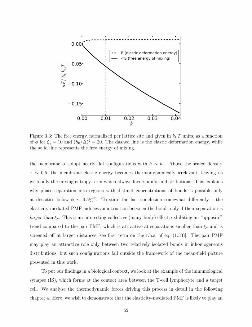
0.00 0.01 0.02 0.03 0.04φ
−0.15
−0.10
−0.05
0.00
aF/A
PkBT
E (elastic deformation energy)-TS (free energy of mixing)
Figure 3.3: The free energy, normalized per lattice site and given in kBT units, as a functionof φ for ξγ = 10 and (h0/∆)2 = 20. The dashed line is the elastic deformation energy, whilethe solid line represents the free energy of mixing.
the membrane to adopt nearly flat configurations with h ∼ h0. Above the scaled density
x ∼ 0.5, the membrane elastic energy becomes thermodynamically irrelevant, leaving us
with only the mixing entropy term which always favors uniform distributions. This explains
why phase separation into regions with distinct concentrations of bonds is possible only
at densities below φ ∼ 0.5ξ−2γ . To state the last conclusion somewhat differently – the
elasticity-mediated PMF induces an attraction between the bonds only if their separation is
larger than ξγ. This is an interesting collective (many-body) effect, exhibiting an “opposite”
trend compared to the pair PMF, which is attractive at separations smaller than ξγ and is
screened off at larger distances [see first term on the r.h.s. of eq. (1.33)]. The pair PMF
may play an attractive role only between two relatively isolated bonds in inhomogeneous
distributions, but such configurations fall outside the framework of the mean-field picture
presented in this work.
To put our findings in a biological context, we look at the example of the immunological
synapse (IS), which forms at the contact area between the T-cell lymphocyte and a target
cell. We analyze the thermodynamic forces driving this process in detail in the following
chapter 4. Here, we wish to demonstrate that the elasticity-mediated PMF is likely to play an
52

important role in IS formation. In this specific example, the cell-cell adhesion is mediated
via binding between T-cell receptors (TCR) and MHC-peptide (pMHC) complexes, and
between integrin LFA1 and its ligand ICAM1 [73]. These two types of adhesion bonds
form a unique structure, in which TCR-pMHC bonds are clustered in its center, while
the LFA1-ICAM1 bonds aggregate in the periphery of synapse. It is believed that the
central domain, i.e., the TCR-pMHC rich area, plays a pivotal role in regulating T-cell
activation [74]. Typically, the bond density within the synapse is around 100 bonds per
µm2, and the bond lengths are 14 nm and 41 nm for TCR-pMHC and LFA1-ICAM1 bonds,
respectively [75, 76]. We recall that in the model presented here, h0 represents the local
membrane deformation imposed by a bond relative to the resting height of the membrane.
Thus, if we consider the resting separation between the two membranes in the IS to be
dictated by the longer bonds, we can estimate the deformation to simply be the difference
between the two bond lengths, h0 ' 27 nm. Taking the membrane bending rigidity to be
κ ' 15 kBT and the harmonic potential strength as γ ' 6 · 105kBT µm−4 [77], we arrive
to the values x ' 0.5 and (∆/h0)2 ' 0.057 for the coordinates of this point in the phase
diagram displayed in Fig. 3.2. Remarkably, the point lies in the two-phase region of the
phase diagram, close to the binodal line. This raises the possibility that the TCR-pMHC
rich domain may be the semi-dilute phase coexisting with a dilute phase of vanishingly small
density. Thus, we speculate that the elasticity-mediated interactions may play an important
role in the condensation of the TCR-pMHC signaling domain. They provide attraction
which enables the TCR-pMHC bonds to spontaneously aggregate into domains with density
comparable to that existing in the IS central zone.
3.4 Summary
In this chapter we analyzed the formation of adhesion domains in the van der Waals regime.
In this regime, the thermal fluctuations of the membrane are small and, therefore, the system
free energy is dominated by the energy, which is determined by the elastic bending energy and
the harmonic confining potential. We have presented a novel mean-field approach to study
the condensation transition of the system. Our approach is based on an empirical expression
for the average elastic deformation energy of a membrane as a function of the density of
53
adhesion bonds, which was obtained numerically for random distributions of adhesion bonds.
We found that the phase diagrams of dilute systems with different values of densities φ and
healing lengths ξγ are practically identical when the inverse deformation energy (∆/h0)2 ∼
T/Tc is plotted against the rescaled density x = ξ2γφ. Our results show that the phase
coexistence regime is largely concentrated in a region where 0 < x . 1, and that the critical
point is located at very small of x. Therefore, the curvature-mediated interactions may cause
the system to phase separate into an extremely dilute phase with x ' 0 and a semi-dilute
phase x ' 1. Highly condensed domains with x� 1 can only be formed if the membrane is
considerably deformed, but in such cases one can no longer use the Monge representation to
study the system. Thus, we conclude that the curvature-induced interactions are too weak to
promote formation of tightly packed domains, but can lead to semi-dilute adhesion domains
where the typical separation between the adhesion bonds is set by the healing length ξγ.
Interestingly, we find that the mature immunological synapse (IS) between a T cell
and an antigen-presenting cell is characterized by an adhesion domain of TCR-pMHC bonds
with densities of roughly x ' 1. For the system parameters of the IS, we find that our
mean-field model for domain formation driven by membrane curvature is indeed able to
generate domains with comparable densities to those found in the IS. This finding is in line
with several recent studies suggesting that passive thermodynamic processes can describe the
short-time condensation of adhesion clusters of the IS, without evoking any active processes
in the cytoskeleton (see, e.g., [78], and refs. therein). Forces stemming from cytoskeletal
activity may be essential during the later stages of IS pattern formation and stabilization [79,
80]. In the following chapter, we take a closer look at the manner by which active and
thermodynamic (passive) mechanisms govern the process of IS formation.
54
Chapter 4
Passive and active mechanisms in the
formation of the immunological
synapse
4.1 Introduction
The adaptive immune system heavily relies upon the ability of T cells to properly interact
with antigen-presenting cells (APCs). The contact area between the two cells is established
by specific receptor-ligand bonds that crosslink the plasma membranes of the T cell and
the APC. The key players in this cellular recognition process are the two T cell membrane
proteins T cell receptor (TCR) and lymphocyte function-associated antigen 1 (LFA1) that
respectively bind to peptide displaying major histocompatibility complex (pMHC) and in-
tercellular adhesion molecule 1 (ICAM1) embedded in the APC’s plasma membrane [2, 81].
During T cell activation, the TCR-pMHC and the LFA1-ICAM1 receptor-ligand bonds are
redistributed and form a unique geometric pattern of concentric supra-molecular activation
centers (SMACs) within approximately 15-30 minutes of initial contact [73,82]. In this special
arrangement, which is commonly referred to as the immunological synapse (IS), TCR-pMHC
bonds are concentrated into a central SMAC (cSMAC), while the LFA1-ICAM1 adhesion
bonds form a surrounding ring termed the peripheral SMAC (pSMAC) [74,83]. This molec-
ular redistribution is thought to play an important role in signal regulation [84], T cell
55
proliferation [85], and focalized secretion of lytic granules and cytokines [86].
Extensive research effort has been devoted to understanding the mechanisms govern-
ing the formation of the special architecture of the IS. Mounting evidence from experimental
studies point to the actin cytoskeleton as a vital element in controlling the centripetal mo-
tion of TCR-pMHC and LFA1-ICAM1 bonds towards their final locations [87]. In the early
stages of IS formation, the T cell’s actin meshwork reorganizes into actin-free and actin-rich
zones in the center and periphery of the contact area, respectively. Later on in the process,
the actin-depleted area constitutes the location of the cSMAC of the IS, while the pSMAC
is located at the peripheral actin-rich zone [88]. Moreover, actin polymerization occurring
at the periphery of the contact area results in centripetal actin retrograde flow that is cru-
cial for protein translocation [89, 90]. It has been hypothesized that actin retrograde flow
produces viscous forces on the intracellular part of TCRs, which lead to their centripetal
motion [91–94]. Furthermore, directed transport by dynein motor proteins along the cy-
toskeletal microtubules has been identified as another adenosine triphosphate (ATP)-driven
process contributing to protein localization in the IS [95, 96]. These experimental evidences
have led to the notion that IS formation is governed by active cellular processes related to
the cytoskeleton activity.
Interestingly, several theoretical studies have suggested that passive (non-active) mech-
anisms may also be involved in the formation process of the IS [78, 97, 98]. Membrane-
mediated attraction emerges as one of these mechanisms, as demonstrated by our calcula-
tions in the previous chapter, and by other theoretical studies also considering the binding
kinetics of the adhesion bonds [99]. It has been argued that membrane-mediated interactions
between adhesion bonds in the IS may result in patterns that are not only extremely similar
to those observed experimentally, but also form on biologically relevant timescales [78,97,98].
In chapter 3, we have analyzed the curvature-induced interactions between adhesion
bonds and demonstrated that TCR-pMHC bonds can phase separate from LFA1-ICAM1
bonds and form domains with similar densities to those in the IS [100]. However, conven-
tional phase separation theories are insufficient to explain the bullseye pattern of the IS,
i.e., the aggregation of TCR-pMHC bonds at the central contact area and the accumula-
tion of LFA1-ICAM1 bonds at the periphery. This very particular structure seems to be
directly linked to the activity of the actin cytoskeleton, especially to directed transport of
56

TCR-pMHC by dynein motors along microtubules [96], and the actin retrograde flow which
induces a centripetal force on the TCR-pMHC bonds [79,101]. It may also be related to the
depletion of actin from the center of the contact area, which occurs at the very beginning
of the IS formation process. The cytoskeleton is also expected to have a direct influence
on the membrane-mediated interactions between the TCR-pMHC bonds. This follows from
the attachment of the T cell’s membrane to the actin cytoskeleton by various molecules,
such as proteins from the ezrin-mesoin-radixin (ERM) family [102], phosphatidylinositol
4,5- bisphosphate (PIP2) [103, 104] and coronin 1 [105]. This coupling between the mem-
brane and the cytoskeleton directly impacts the shape of the membrane and, thus, may
modify the membrane-mediated PMF. Here, we extend the analysis of chapter 3 on the
membrane elasticity-induced formation of TCR-pMHC domains, by including the aforemen-
tioned cytoskeleton-related effects, and studying the interplay between the passive and active
mechanisms.
In order to follow the process of IS formation, we develop and simulate a simple lattice
model that constitutes a discrete representation of the contact region between the T cell
and the APC. A schematic picture depicting the contact area and its lattice representa-
tion is shown in Figs. 4.1(A) and (B), respectively. The lattice sites can be: (i) empty, or
singly occupied by either (ii) a mobile point representing a TCR-pMHC bond, or by (iii)
an immobile point representing an attachment protein between the membrane and the cy-
toskeleton. The latter are absent from the actin-depleted central region of the contact area.
In this coarse-grained physical framework, the cell membranes are implicitly accounted for
by nearest-neighbor interactions that represent the PMF originating from the membrane
deformation energy. The cell cytoskeleton is not modeled explicitly in our coarse-grained
simulations, but is implicitly introduced via an effective potential that generates the active
cytoskeleton forces. Moreover, since we focus on the aggregation dynamics of TCR-pMHC
bonds, we do not study the very rapid remodeling process of the actin cytoskeleton (which
is completed within less than a minute from the initial contact between the T cell and the
APC [88]), but consider a system where a central actin-depleted region has already been
formed. Below, we elaborate on the specifics of the model and simulations.
57

Actin cytoskeletonLFA1-ICAM1
Actin pinning pointsTCR-pMHC
T cell
APC41 nm
14 nm
~ 2
(A) (B)
Figure 4.1: (A) Schematics of the contact area between the membranes of the T cell and theAPC. The two membranes are connected by two types of adhesion proteins: LFA1-ICAM1and TCR-pMHC with bond lengths of 41 nm and 14 nm, respectively. The T cell’s membraneis attached to the cytoskeleton by a set of actin pinning proteins. A central region in thecontact region of diameter ' 4 µm is devoid of actin filaments. (B) Schematics of the latticerepresentation of the contact area shown in (A). The lattice sites can be either empty (inwhich case they are marked by the “x” symbols), or occupied by a single TCR-pMHC bond(black circles) or by a single actin pinning point (red circles). The latter are immobile andare excluded form the actin-depleted central region of the system, represented by the dashedcircle.
4.2 Model and simulations
4.2.1 Nearest-neighbor approximation
The membrane-mediated interactions between TCR-pMHC bonds can be studied within
the van der Waals regime. We focus on dilute systems, where the many-body PMF is well-
approximated by the sum of pairwise interactions that depend only on the distance r between
the adhesion bonds (see section 1.2.2.3). From eq. (1.33), the pairwise curvature-induced
attraction between a pair of TCR-pMHC is given by
Φatt(r)
kBT=
Φ2(r)− Φ2(r →∞)
kBT=
(h0
∆
)2
4
πkei
(r
ξγ
)1− 4
πkei
(r
ξγ
) , (4.1)
where we have shifted the PMF such that it vanishes for large separations (r → ∞). For
T cells, the values of the healing length and the thermal roughness are roughly given by
ξγ ' 100 nm and ∆ ' 8 nm [65], respectively, while the value of the deformation caused by a
58
TCR-pMHC bond is set by the mismatch in bond length with respect to the LFA1-ICAM1
bonds, h0 = 41 − 14 = 27 nm [75, 76]. The pair PMF, Φatt/kBT (expressed in units of the
thermal energy), is depicted by the solid line in Fig. 4.2 as a function of the normalized pair
distance r/ξγ, for the aforementioned values of the systems parameters ξγ, ∆ and h0. In
addition to the membrane-mediated interactions between TCR-pMHC bonds, we also need
to calculate the pair PMF between the TCR-pMHC bonds and proteins that pin the T cell
membrane to the actin cytoskeleton. These interactions are obviously repulsive due to the
large differences in the height of the membrane at the locations of these two proteins (h = 0
at the pinning sites compared to h = h0 = 27 nm at the sites of the TCR-pMHC bonds).
The repulsive pair PMF can be derived from the partition function
ZB =
∫D [h(r)] e−βHδ (h(0)− h0) δ (h(r)) , (4.2)
which differs from the partition function (1.32) by one height constraint. The resulting
repulsive pair PMF is given by
Φrep(r)
kBT=
1
2
(h0
∆
)2
(4
π
)2
kei2(r
ξγ
)1−
(4
π
)2
kei2(r
ξγ
) (4.3)
and is depicted by the dashed line in Fig. 4.2 for similar values of system parameters. From
Fig. 4.2 it is clear that Φrep is a purely repulsive potential of range r ' ξγ that quickly decays
to zero at larger separations.
At separations smaller than ξγ, one has to take into account direct excluded volume
(hard core) interactions between the adhesion proteins. Since these are missing in the cal-
culation of the partition functions, a purely repulsive potential diverging for r → 0 must be
added to Φatt. The full pair potential between TCR-pMHC bonds is, thus, reminiscent of a
Lennard-Jones potential, i.e., repulsive at very short distances and attractive at an interme-
diate finite range. Conversely, from Fig. 4.2 we see that for r < ξγ, Φrep sharply increases
similarly to an excluded volume potential. We thus conclude that the membrane curvature
itself serves as a source of repulsion between TCR-pMHC bonds and actin pinning proteins,
which renders the addition of explicit excluded volume (hard core) interactions unnecessary
59

in this case.
0 1 2 3 4 5 6 7 8r/ξγ
6
4
2
0
2
4
6
8
10
Φ(r
)/k
BT
Φatt
Φrep
Figure 4.2: Curvature-induced interactions: the solid line depicts the curvature-inducedattraction between two TCR-pMHC bonds, while the dashed line stands for the curvature-induced repulsion between a TCR-pMHC bond and a membrane-cytoskeleton pinning point.
4.2.2 Lattice-gas model
Domain formation under the influence of short-range pairwise attractive interactions can be
conveniently studied within the framework of the classical discrete lattice-gas (Ising) model
with a nearest-neighbor attraction of strength ε (see also our discussion on the WF model in
section 1.2.1.3). In this framework, each lattice site can be occupied by at most one lattice
point, in order to account for the short-range excluded volume repulsion. Here, we study the
IS formation process by considering a triangular lattice with lattice spacing ξγ = 100 nm,
which is comparable to the range of the membrane-mediated interactions between the TCR-
pMHC bonds, and also to the typical spcaing between them in the central domain of the IS.
This sets the spatial resolution of our model, and allows us to ignore direct (e.g., van der
Waals and diploar [28]) and lipid-mediated [109] interactions between the various proteins in
the system since, typically, the range of these interactions does not extend beyond . 10 nm.
The linear size of the system is roughly L = 10 µm (we simulate a lattice of 99 × 114
60

sites with an aspect ratio close to unity), which is representative of the dimensions of the
contact area. The model includes two types of lattice points representing the TCR-pMHC
bonds (type A) and the membrane-cytoskeleton pinning proteins (type B). The former are
mobile, while the latter are located at fixed lattice sites. Both A-A and A-B interactions
are purely repulsive at short separations (see section 4.2.1), and this feature is accounted for
by prohibiting multiple occupancy of a lattice site. At a distance ξγ, the A-A interactions
are attractive [see eq. (4.1) and Fig. 4.2], and this is represented in the model via a nearest-
neighbor interaction energy of strength ε = Φatt(r = ξγ) = −4.5 kBT . We do not consider
next-nearest-neighbor interactions, despite of the fact that the Φatt does not fully decay at
r = ξ. The reason for this decision is the many-body nature of the membrane-mediated PMF,
which becomes important at the onset of the formation of adhesion clusters. In high density
domains, each adhesion bond interacts with the proximal bonds in the first surrounding
shell, whose very presence screens the interactions with the slightly more distant bonds in
the next shells [46]. The model does not include explicit representation of the LFA1-ICAM1
bonds. The effect of these bonds is incorporated in the statistical-mechanical calculations of
the PMFs through the parameter h0 = 27 nm [see eqs. (4.1) and (4.3)], which is the bond
length mismatch between LFA1-ICAM1 and TCR-pMHC bonds1.
The densities of the TCR-pMHC bonds (type A lattice points) and the membrane-
cytoskeleton pinning proteins (type B) greatly vary between different experimental works
(if reported at all). We therefore set their values in the simulations based on the following
considerations: Type A points aggregate to form the cSMAC, which is a circular domain
of radius ∼ 2 µm that almost fills (at the end of the process) the actin-depleted central
region. The latter includes about 1200 lattice sites, and the number of type A points is
set to a slightly smaller value NA = 1000. We note that since the lattice spacing is set to
ξγ = 100 nm, the density of the TCR-pMHC bonds in a cluster is ' 80 bonds/µm2, which
is indeed above the threshold density required for full T cell activation [73,74]. A reasonable
estimate for the spacing, lP, between membrane-cytoskeleton pinning proteins is a distance
of a few hundred nm [110]. In what follows, we show results for systems where the average
1More accurately, the effect of the LFA1-ICAM1 bonds is represented by the uniform harmonic potentialin Hamiltonian (1.20). The utility of this approach was demonstrated in chapter 3, where we employed amore fine-grained lattice model with a microscopic spacing of 5 nm and an explicit representation of themembrane.
61
spacing between type B points is set to lP = 300 nm. To avoid further complication of our
model, we neglect the binding/unbinding kinetics of the membrane-cytoskeleton linkers and
assume that the number of type B points is fixed. Nevertheless, we note here that results of
simulations with lP = 500 nm (i.e., with a fewer number of type B points) exhibit negligible
differences. The type B points can be found everywhere on the lattice, except for the central
actin-depleted region. They are randomly distributed to account for local variations in lP,
but we do not allow two type B points to occupy nearest-neighbor lattice sites in order to
ensure a fairly uniform distribution and avoid type B clusters.
4.2.3 Monte Carlo simulations
We perform Monte Carlo (MC) simulations to study the evolution of the lattice model. While
MC simulations are designed for statistical ensemble sampling at equilibrium, they can be
also used to effectively generate Brownian dynamics in lattice models. The simulations
consist of move attempts of a randomly chosen type A point to a nearest lattice site, which
is accepted according to the standard Metropolis criterion [62]. During a single MC time
unit, τMC, each type A point experiences (on average) one move attempt. Mapping the
MC time unit to real time, t, can be achieved by considering the two-dimensional diffusion
relation 〈r2〉 = 4Dt, where 〈r2〉 denotes the mean square displacement and D is the diffusion
coefficient. For the MC simulations, each point moves a distance of the lattice spacing
ξγ = 100 nm and, therefore, τMC = ξ2γ/4D. This can be compared to typical values for
the diffusion coefficient of T cell membrane proteins D ' 0.1 µm2/sec [111], which yields
τMC ' 25 msec.
4.2.4 Active cytoskeleton forces
Active cytoskeleton processes are not modeled explicitly in our simulations, but are instead
represented by the effective forces that they induce on the TCR-pMHC bonds. Two active
processes are considered, namely (i) actin retrograde flow, and (ii) dynein minus-end directed
transport toward the microtubule organizing center (MTOC) that is translocated towards
the central actin-free area [88]. These may be viewed as complementary mechanisms since
they produce the same net effect of transport toward the center, while operating at different
62
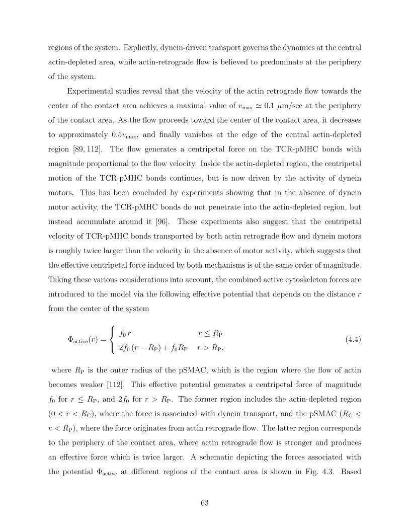
regions of the system. Explicitly, dynein-driven transport governs the dynamics at the central
actin-depleted area, while actin-retrograde flow is believed to predominate at the periphery
of the system.
Experimental studies reveal that the velocity of the actin retrograde flow towards the
center of the contact area achieves a maximal value of vmax ' 0.1 µm/sec at the periphery
of the contact area. As the flow proceeds toward the center of the contact area, it decreases
to approximately 0.5vmax, and finally vanishes at the edge of the central actin-depleted
region [89, 112]. The flow generates a centripetal force on the TCR-pMHC bonds with
magnitude proportional to the flow velocity. Inside the actin-depleted region, the centripetal
motion of the TCR-pMHC bonds continues, but is now driven by the activity of dynein
motors. This has been concluded by experiments showing that in the absence of dynein
motor activity, the TCR-pMHC bonds do not penetrate into the actin-depleted region, but
instead accumulate around it [96]. These experiments also suggest that the centripetal
velocity of TCR-pMHC bonds transported by both actin retrograde flow and dynein motors
is roughly twice larger than the velocity in the absence of motor activity, which suggests that
the effective centripetal force induced by both mechanisms is of the same order of magnitude.
Taking these various considerations into account, the combined active cytoskeleton forces are
introduced to the model via the following effective potential that depends on the distance r
from the center of the system
Φactive(r) =
f0 r r ≤ RP
2f0 (r −RP) + f0RP r > RP,(4.4)
where RP is the outer radius of the pSMAC, which is the region where the flow of actin
becomes weaker [112]. This effective potential generates a centripetal force of magnitude
f0 for r ≤ RP, and 2f0 for r > RP. The former region includes the actin-depleted region
(0 < r < RC), where the force is associated with dynein transport, and the pSMAC (RC <
r < RP), where the force originates from actin retrograde flow. The latter region corresponds
to the periphery of the contact area, where actin retrograde flow is stronger and produces
an effective force which is twice larger. A schematic depicting the forces associated with
the potential Φactive at different regions of the contact area is shown in Fig. 4.3. Based
63

Figure 4.3: Effective active cytoskeleton forces acting on the TCR-pMHC bonds in differentregions of the T cell-APC’s contact area. The inner dashed circle marks the edge of thecentral actin-depleted region with radius RC = 2µm, while the outer dotted circle marksthe edge of the pSMAC, and has a radius RP = 4µm. The black and blue arrows representeffective centripetal forces arising from the actin retrograde flow and directed transport bydynein motors, respectively. For r < RP, the magnitude of the active centripetal force is setto f0 = 0.1 pN (small arrows), while for r > RP the force is set to a twice larger value of2f0 = 0.2 pN, and is indicated by large arrows.
on confocal images, we set RC = 2 µm and RP = 4 µm [91]. In order to determine the
value of f0, we use the observation that the actin retrograde flow causes a small peripheral
microcluster (a few hundred nanometers in size) to move toward the cell’s center at a velocity
of vc ' 20 nm/sec [113]. We, thus, performed short MC simulations for a single microcluster
composed of 10 TCR-pMHC bonds, which is initially located at the outer region of the
system. We measured the velocity of the centripetal movement of the microcluster as a
function of f0, and found that it attains the value of vc for f0 = 0.1 pN. Comparable
forces have been measured in experiments of optically trapped microbeads coupled to actin
retrograde flow of similar velocity [114].
4.3 Results
The spatio-temporal evolution of the system has been analyzed from 10 independent MC
runs starting with different random distributions of both type A and B points. Typical
snapshots from different stages of the process are shown in Fig. 4.4, with the type A and
B points presented by black and red dots, respectively. At t = 0, the type A points are
64

randomly distributed outside of the inner actin-depleted regime (Fig. 4.4a). Within less than
one minute, the type A points coalesce and form small peripheral microclusters consisting
of . 20 points (Fig. 4.4b), which begin to move centripetally. Several type A points have
already reached the center system at this stage. The peripheral microclusters are believed
to play a vital role in initiating and sustaining TCR signaling [106, 115]. As time proceeds,
the clusters further coarsen, which decreases their mobility and slows down their centripetal
motion (Figs. 4.4c-e). The increase in the size of the clusters also causes them to be more
affected by the presence of the type B points that act as repelling obstacles. This further
restrains the centripetal movement of the microclusters, which have to “navigate” their way
through the “curvature corrals” that the type B points form. We also observe in Figs. 4.4c-e
the gradual increase in the size of the central domain, which is the destination where the
microclusters accumulate. After 45 minutes (Fig. 4.4f), almost all type A points reside
in the central domain. The dynamics of the MC simulations, as depicted in Figs. 4.4a-e,
closely resembles epifluorescence and total internal reflection fluorescence (TIRF) microscopy
images of the IS formation process. Specifically, the microscopy images show the generation
of similar microclusters, their drift to the center of the contact area, and their accumulation
at the center of the contact area [94, 116]. The simulations exhibit a very good agreement
with the experimental observations not only with regard to spatial evolution of the system,
but also with respect to the time scales of the different stages of the process. Fig. 4.4g
depicts the percentage of type A points located in the central domain at the actin-depleted
area. About 90% of the lattice points have been accumulated at the center after roughly
40 minutes, which agrees very well with the times reported in the literature for cSMAC
formation.
Figs. 4.5a-c show snapshots from simulations in which dynein activity is turned off.
This is done by modifying the effective potential (4.4) such that Φactive (r < RC) = f0RC =
Constant, and thus, the associated force vanishes at the actin-depleted area where the dynein
motors operate. For this model system, we observe formation of peripheral microclusters
that move centripetally, but do not enter the actin-depleted area. Instead of forming a
central quasi-circular domain, the type A points now accumulate in a ring-shaped domain
at the edge of the actin-depleted region. Interestingly, very similar ring-like structures have
been observed in experiments where dynein activity was inhibited by dynein heavy chain
65
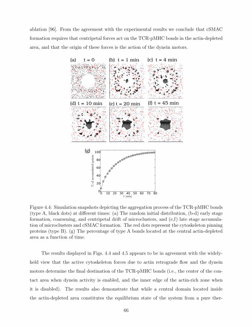
ablation [96]. From the agreement with the experimental results we conclude that cSMAC
formation requires that centripetal forces act on the TCR-pMHC bonds in the actin-depleted
area, and that the origin of these forces is the action of the dynein motors.
(a) (b) (c)
(d) (e) (f)
(g)
t = 20 min t = 45 min
t = 4 min
t = 10 min
t = 0 t = 1 min
0 10 20 30 40 50 60 70 80t [min]
0
20
40
60
80
100
%ofaccumulatedpoints
Figure 4.4: Simulation snapshots depicting the aggregation process of the TCR-pMHC bonds(type A, black dots) at different times: (a) The random initial distribution, (b-d) early stageformation, coarsening, and centripetal drift of microclusters, and (e,f) late stage accumula-tion of microclusters and cSMAC formation. The red dots represent the cytoskeleton pinningproteins (type B). (g) The percentage of type A bonds located at the central actin-depletedarea as a function of time.
The results displayed in Figs. 4.4 and 4.5 appears to be in agreement with the widely-
held view that the active cytoskeleton forces due to actin retrograde flow and the dynein
motors determine the final destination of the TCR-pMHC bonds (i.e., the center of the con-
tact area when dynein activity is enabled, and the inner edge of the actin-rich zone when
it is disabled). The results also demonstrate that while a central domain located inside
the actin-depleted area constitutes the equilibrium state of the system from a pure ther-
66
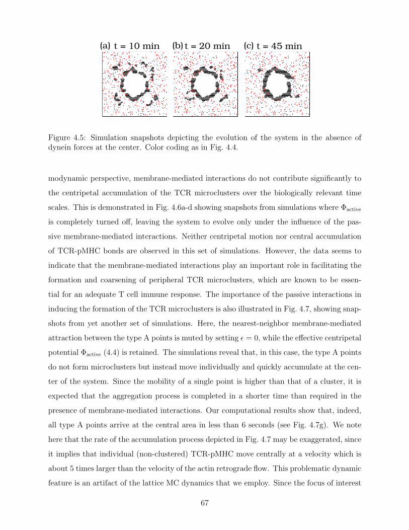
(a) t = 20 min t = 45 mint = 10 min (b) (c)
Figure 4.5: Simulation snapshots depicting the evolution of the system in the absence ofdynein forces at the center. Color coding as in Fig. 4.4.
modynamic perspective, membrane-mediated interactions do not contribute significantly to
the centripetal accumulation of the TCR microclusters over the biologically relevant time
scales. This is demonstrated in Fig. 4.6a-d showing snapshots from simulations where Φactive
is completely turned off, leaving the system to evolve only under the influence of the pas-
sive membrane-mediated interactions. Neither centripetal motion nor central accumulation
of TCR-pMHC bonds are observed in this set of simulations. However, the data seems to
indicate that the membrane-mediated interactions play an important role in facilitating the
formation and coarsening of peripheral TCR microclusters, which are known to be essen-
tial for an adequate T cell immune response. The importance of the passive interactions in
inducing the formation of the TCR microclusters is also illustrated in Fig. 4.7, showing snap-
shots from yet another set of simulations. Here, the nearest-neighbor membrane-mediated
attraction between the type A points is muted by setting ε = 0, while the effective centripetal
potential Φactive (4.4) is retained. The simulations reveal that, in this case, the type A points
do not form microclusters but instead move individually and quickly accumulate at the cen-
ter of the system. Since the mobility of a single point is higher than that of a cluster, it is
expected that the aggregation process is completed in a shorter time than required in the
presence of membrane-mediated interactions. Our computational results show that, indeed,
all type A points arrive at the central area in less than 6 seconds (see Fig. 4.7g). We note
here that the rate of the accumulation process depicted in Fig. 4.7 may be exaggerated, since
it implies that individual (non-clustered) TCR-pMHC move centrally at a velocity which is
about 5 times larger than the velocity of the actin retrograde flow. This problematic dynamic
feature is an artifact of the lattice MC dynamics that we employ. Since the focus of interest
67

(a) t = 0 t = 30 min
t = 1 hour t = 5 hours
(b)
(c) (d)
Figure 4.6: Simulation snapshots depicting the aggregation process in the absence of activecytoskeleton forces. Color coding as in Fig. 4.4. The type A points form microclusters that,on time scales of hours, barely grow and do not exhibit a drift toward the central area.
is the evolution of the system over durations of minutes and hours, the actin retrograde
flow force has been calibrated to produce correctly the centripetal velocity of small clusters
on these temporal scales (see detailed discussion in section 4.2.4). The consequence of this
choice is that the velocity of the individual TCR-pMHC bonds turns out to be too high. A
reasonable estimation for the centripetal velocity of individual bonds at the periphery of the
system is 20 < v < 100 nm/sec, i.e., higher than the velocity of a single small microcluster
but lower than the actin retrograde flow velocity. The accumulation time of bonds moving
at such speeds is 0.5-2 minutes, which is still more than an order of magnitude smaller than
the IS formation time. The comparison between the results of Fig. 4.7 from simulations that
miss the membrane-mediated attraction and Fig. 4.4 highlights two important aspects of
membrane elasticity: First, it facilitates early microcluster formation that play a vital role
in T cell activation. Second, it leads to coarsening dynamics that results in a decrease in the
mobility of the clusters and, thereby, regulates the duration of the aggregation process.
68
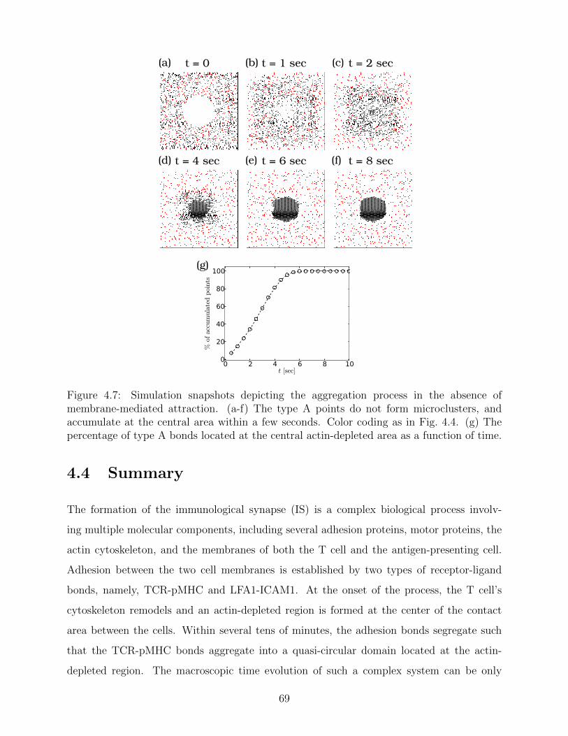
(a) t = 0 t = 1 sec t = 2 sec
t = 6 sec
(b) (c)
(e)t = 4 sec(d) t = 8 sec(f)
0 2 4 6 8 10t [sec]
0
20
40
60
80
100
%ofaccumulatedpoints
(g)
Figure 4.7: Simulation snapshots depicting the aggregation process in the absence ofmembrane-mediated attraction. (a-f) The type A points do not form microclusters, andaccumulate at the central area within a few seconds. Color coding as in Fig. 4.4. (g) Thepercentage of type A bonds located at the central actin-depleted area as a function of time.
4.4 Summary
The formation of the immunological synapse (IS) is a complex biological process involv-
ing multiple molecular components, including several adhesion proteins, motor proteins, the
actin cytoskeleton, and the membranes of both the T cell and the antigen-presenting cell.
Adhesion between the two cell membranes is established by two types of receptor-ligand
bonds, namely, TCR-pMHC and LFA1-ICAM1. At the onset of the process, the T cell’s
cytoskeleton remodels and an actin-depleted region is formed at the center of the contact
area between the cells. Within several tens of minutes, the adhesion bonds segregate such
that the TCR-pMHC bonds aggregate into a quasi-circular domain located at the actin-
depleted region. The macroscopic time evolution of such a complex system can be only
69
addressed through coarse-grained models employing simplified molecular representation and
focusing on the most dominant biophysical mechanisms. In this chapter, we present a min-
imal computational lattice model aiming to study the dynamics of the TCR-pMHC bonds
(represented as lattice points). The model takes into account several forces that emerge in
the literature as key factors in TCR-pMHC localization. One is a passive thermodynamic
force associated with the membrane curvature energy, which induces attraction between the
TCR-pMHC bonds, and provides repulsion between the TCR-pMHC bonds and the pro-
teins connecting the actin cytoskeleton to the membrane. The others are centripetal forces
exerted on the TCR-pMHC bonds due to the actin retrograde flow and directed transport
by dynein motors along microtubules, which constitute active (non-equilibrium) forces, since
they originate from processes consuming ATP chemical energy. Only in simulations where
all the driving forces are present, the signature features of the IS formation process are
observed. These include microclusters formation and coarsening dynamics, their transport
to the central actin-depleted area, and their aggregation into a single quasi-circular domain
(the cSMAC). Moreover, the simulated system evolves at the experimentally observed rates;
microclusters are formed early in the process within roughly a minute from the beginning of
the process, and the final bullseye structure is formed within 15-30 minutes.
Our coarse-grained simulations provide a clear and intuitive picture for the respec-
tive roles played by active and passive forces and into the intricate interplay between them
that regulates the spatio-temporal pattern formation of the IS. Membrane-mediated attrac-
tion facilitates the formation of TCR-pMHC microclusters at the periphery of the contact
area. These peripheral microclusters, which initiate biological cues necessary for T cell ac-
tivation, are rapidly formed and continue to grow by coarsening dynamics over a period of
approximately 10 minutes. The TCR-pMHC microclusters are corralled by the membrane-
cytoskeleton binding proteins of the T cell, with which they have repulsive membrane-
mediated interactions. As the microclusters grow in size, the corralling effect becomes more
significant and their diffusivity decreases. This inhibits their accumulation in a central
quasi-circular domain, despite the fact that this configuration constitutes the equilibrium
state of the system. What speeds up the dynamics of the microclusters and directs their
movement toward the center of the contact area is a centripetal active force induced by
actin retrograde flow at the periphery of the synapse. This observation corroborates the
70

largely consensual view about the importance of actin remodeling and retrograde flow for
the centripetal translocation of TCR-pMHC bonds at the IS. It is, however, important to
emphasize that in simulations without membrane-mediated attraction, a central domain
forms very rapidly without exhibiting intermediate microclusters. This observation high-
lights the, rather overlooked, important role played by the membrane-mediated interactions
in regulating the rate of the IS formation process. This novel conclusion drawn from our
model and simulations has not been addressed experimentally thus far. A possible setup by
which it can be tested is a model system consisting of all the molecular ingredients of the
system, except for the LFA1-ICAM1 bonds. This would shut down the membrane-mediated
interactions whose origin is the mismatch in bond length between the TCR-pMHC and the
LFA1-ICAM1 bonds.
At the central actin-depleted region, a centripetal force (of magnitude similar to the
actin retrograde flow induced force) is effectively generated by dynein motors that walk
toward the minus-end of the microtubules. An interesting observation from our simulations is
the formation of a ring-shaped domain at the edge of the actin-depleted region in simulations
where this force is turned off. This result is in line with a recent study, in which similar
structures were observed when dynein activity was genetically impaired, but actin retrograde
flow was maintained. The inability of the system to produce a central circular domain
in the absence of dynein activity highlights the role played by these motors at the final
stage of the process, where they enable the TCR microclusters to enter the actin-depleted
region. Furthermore, the agreement between our simulation results and the experimental
data supports the notion presented here of considering two different concentric regions for
the active forces, namely a central area dominated by the dynein motors, and a more distant
one where the actin retrograde flow is the main origin of the centripetal movement of TCR
microclusters.
71

Chapter 5
Conclusion
Cellular adhesion is mediated through specific receptor-ligand bonds and constitutes a vital
feature of biological systems. The formation of macroscopic adhesion domains that attach
the plasma membrane to the extracellular matrix and/or other neighboring cell membranes
not only provides mechanical stability to the cell, but is also involved in promoting im-
portant biological cues and in regulating inter-cellular communication. Gaining a thorough
understanding of the biophysical principles driving the aggregation process not only provides
insights into the biological mechanisms of life, but is also important for the development of
biosensing applications and the design of drug delivery systems that rely on efficient adhesion
between the carrier and the target plasma membrane.
This thesis aimed to develop a better theoretical understanding of membrane-mediated
mechanisms for protein clustering. Membrane thermal fluctuations and elastic curvature
energy effectively induce attractive forces between adhesion bonds. These forces originate
from the decrease in the system’s free energy upon aggregation, which enhances the thermal
fluctuations (and, therefore, the configurational entropy) of the membrane, and reduces its
bending energy. These two thermodynamic mechanisms have been studied in two distinct
regimes of the elasticity theory for lipid bilayers. In the Helfrich regime, where the membrane
undulations are strong, the thermal fluctuations induce a long-range potential of mean force
(PMF) between adhesion bonds. In contrast, in the van der Waals regime, where thermal
fluctuations are small, the membrane curvature deformations induce a finite-range PMF.
The most challenging aspect of the analysis of adhesion domains formation is the
72
many-body nature of the membrane-mediated PMF. In this work, we used an arsenal of
theoretical methods, including computer simulations, statistical-mechanical calculations, and
novel mean-field theories to study the condensation transition of adhesion bonds, and to
elucidate the role played by membrane-mediated interactions in this process. We analyzed
the aggregation behavior in both the Helfrich and van der Waals regimes of strong and weak
thermal fluctuations, respectively. The former is discussed in chapter 2, where we present
results of computer simulations of a coarse-grained solvent-free model for supported lipid
bilayers. The results for non-constrained membranes were found to be in excellent agreement
with the recently proposed Weil-Farago (WF) lattice model. The WF model is based on the
notion that the local suppression of membrane thermal undulations is mainly determined
by the distance from the nearest adhesion point and can, therefore, be determined from the
Voronoi diagram corresponding to a given distribution of adhesion points. Our molecular
simulations results are in line with the conclusion emerging from lattice simulations of the
WF model, that thermal fluctuations alone are insufficient to promote cluster formation,
yet they have a considerable impact on the condensation transition. We arrive at this
conclusion by comparing simulations of supported membranes with those of non-fluctuating
flat membranes subjected to a strong physical confinement. In the latter case, the fluctuation-
induced interactions vanish; condensation may be induced by residual direct interactions (if
sufficiently strong), and the system can be described by the standard nearest-neighbor lattice-
gas (Ising) model for gas-liquid phase transitions. In the case of non-confined membranes, our
simulations demonstrate that the system remains in the Ising universality class. However,
the membrane-mediated interactions effectively reduce the thermodynamic temperature by
a factor of 2-3; i.e., they compensate for roughly half of the mixing entropy lost upon
condensation. This picture is consistent with the idea that in a distribution of adhesion
bonds, the long-range attractive PMF arising from the membrane-thermal fluctuations is
self-screened. We also performed simulations of membranes subjected to negative surface
tension that presumably enhances the thermal roughness of the membrane. We found that
the negative tension had no effect on the gas phase, where the scattered adhesion bonds
strongly suppress the thermal undulations. The influence of the negative tension is seen
in the condensed phase, where the membranes exhibit buckling instabilities and elongated
adhesion domains (stripes) are formed in the non-buckled portion of the system.
73

In chapter 3, we examine the condensation transition in the van der Waals regime.
As opposed to the entropic attraction between adhesion bonds in the Helfrich regime, the
curvature-induced pair PMF in the van der Waals regime decays over a characteristic correla-
tion length of ξγ ' 50−100 nm. This feature of the pair PMF implies that the condensation
transition is expected to be of the lattice-gas (Ising) universality class. Our investigation
of the curvature-mediated many-body PMF and the condensation transition are based on a
novel mean-field approach involving numerical evaluation of the membrane’s elastic energy
for random distributions of adhesion bonds on a triangular lattice. We found that the elastic
deformation energy per adhesion bond in systems with different healing lengths ξγ and bond
densities φ exhibits data collapse when plotted against the scaled density x = ξ2γφ. This
enabled us to obtain an empirical expression for the system energy, which is then used in
a mean-field expression for the system’s free energy. From the mean-field free energy, we
obtain the phase diagram of the system and identify the two-phase coexistence region. The
most important aspects of the phase diagram are the observations that: (i) the critical point
xc lies in regions where x attains extremely small values, and (ii) the binodal/spinodal curves
sharply decline with the deformation length, h0, imposed by a single bond for xc < x . 1.
The parameter h0 serves in the problem as a temperature variable. When assigning pa-
rameter values to the model that are characteristic of biological membranes, we show that
biological adhesion domains may, in some cases, be associated with the two-phase region
of the phase diagram. Specifically, the adhesion domains constitute a semi-dilute phase co-
existing with an extremely dilute phase that is essentially empty of bonds. The densities
in the semi-dilute domains are such that the adhesion bonds are typically separated by a
distance ξγ from each other. The formation of highly packed condensed domains requires
much stronger deformations (h0), that are often (but not always) unrealistically large. This
can be understood from the observation that for an adhesion bond density of φ ' ξ−2γ ,
the membrane is relatively flat and, therefore, the curvature-mediated attraction effectively
saturates at these densities.
In chapter 4, we take a closer look at a specific biological example of an adhesion do-
mains, which is the immunological synapse (IS) that forms upon contact between T cells
and antigen-presenting cells. This specialized cellular junction is established by two types
of receptor-ligand complexes namely, TCR-pMHC and LFA1-ICAM1 adhesion bonds, which
74
form a unique concentric pattern where the TCR-pMHC bonds are clustered in the center of
the contact area, while the LFA1-ICAM1 bonds accumulate at the periphery. This segrega-
tion process can be partially attributed to the marked mismatch in bond lengths between the
two types of adhesion bonds, which results in substantial membrane deformations leading to
curvature-induced interactions. For the system parameters characterizing the IS, our mean-
field theory (developed in chapter 3) predicts the formation of a semi-dilute TCR-pMHC
domain with a bond density of φ ' ξ−2γ ∼ 100µm2 that is comparable with their densities
in the IS. Nevertheless, domain formation purely driven by a spontaneous phase separation
mechanism cannot account for the concentric geometric pattern characteristic of the IS. In-
deed, active cytoskeleton processes stemming form actin polymerization/depolymerization
events and dynein motor activity are thought to be indispensable for this special molecular
arrangement. The peripheral actin retrograde flow and the more centrally located directed
transport of TCRs by dyneins generate additional centripetal forces, which are believed to
be responsible for the broken symmetry in the system. In order to investigate these issues,
we developed an implicit-membrane 2D lattice model for TCR-pMHC aggregation in the IS.
In this model, the elasticity-induced many-body PMF in the van der Waals regime is ap-
proximated by nearest-neighbor pair interactions. This approximation holds for semi-dilute
systems like the IS. The active cytoskeleton forces are modeled by an effective centripetal
potential. The spatio-temporal evolution of the system is studied using Monte Carlo simula-
tions, and the relationship between the Monte Carlo and the physical time units is established
from the diffusion coefficient of TCR proteins. The simulated aggregation process exhibits a
very good agreement with numerous experimental observations, and correctly captures the
signature features of IS formation. The process begins with the nucleation of small peripheral
TCR-pMHC microclusters. Over time, the microclusters grow by coarsening dynamics, and
exhibit centripetal motion towards the center of the system. Apart from the agreement on
the stages and the spatial evolution of the system, the simulations also show that the central
domain is formed within ∼ 30 min, which is indeed the experimentally observed timescale
for IS formation.
Consistent with previous findings, our lattice simulations point to the great importance
of active cytoskeleton forces, which drive the TCR-pMHC centripetally and allow their ag-
gregation to be completed over biologically relevant timescales. Our results also highlight
75
the rather overlooked importance of membrane-mediated interactions in T cell activation.
The central accumulation of TCR-pMHC in simulations lacking these interactions occurs
too rapidly and does not exhibit the formation of peripheral microclusters, which are only
observed in systems where the curvature-induced attraction is enabled. The formation of
microclusters decreases the mobility of TCR-pMHC bonds and slows down their centripetal
movement. This provides T cells with an appropriate time window to allow biological signal-
ing via peripheral TCR microclusters, which is vital for a proper immune response. Thus,
our lattice model and Monte Carlo simulations shed light on the respective roles of pas-
sive and active mechanisms in IS formation, and the delicate interplay between them that
regulates the spatio-temporal evolution of the process.
The present work provides a comprehensive biophysical basis for the understanding of
adhesion domain formation driven under the influence of membrane-mediated interactions.
The concepts and methods developed here can be applied to the study of other biological
systems exhibiting membrane adhesion clusters. In the example of the IS, we focused on
the formation of semi-dilute adhesion domains; however, it is important to note that our
mean-field treatment suggests that densely packed clusters can, in principle, also form in
membranes subjected to strong deformations. One should keep in mind that our analysis
is based on a linear theory, where the membrane is represented in the Monge gauge using
the small gradient approximation. This approximation may not hold in the case of strongly
deformed membranes, which calls for further theoretical studies. Such investigations are
especially interesting in light of recent evidence suggesting that curvature-mediated mecha-
nisms may be responsible for integrin clustering in cancerous cells [117]. Strong membrane
deformations caused by large glycoproteins, which are commonly overexpressed in malignant
cells and can extend up to 200 nm away from the membrane’s surface [118,119], were found
to facilitate integrin clustering, unlike short (3 nm long) glycoproteins. The formation of
focal adhesion domains in tumor cells enhances metastatic capabilities and promotes cellu-
lar growth and survival, which points to an interesting mechanical role of the glycocalyx in
cellular signaling. Interestingly, our mean-field phase diagram predicts that for a membrane
deformation of h0 & 100 nm (which is much larger than the typical thermal roughness), the
condensed phase is characterized by a scaled density of x & 10, corresponding roughly to
densities of the order of & 1000 bonds per µm2, which are comparable to the typical integrin
76

density in focal adhesion [120]. We hope that the present work will stimulate further bio-
physical research that will deepen our understating of nature’s ability to exploit the physical
properties of membranes to execute biological functions.
77
Appendix A
With the Helfrich Hamiltonian in the van der Waals regime (1.20), the membrane’s height-
height correlations in the absence of adhesion bonds are given by
〈h(r)h(r′)〉 =
(l
L
)4∑q
〈∣∣h2
q
∣∣〉e−iq·(r−r′) ≈ − 4
π∆2kei
(|r− r′|ξγ
), (A.1)
For very small distances (r → r′), eq. (A.1) reduces to the thermal roughness ∆2 given
by eq. (1.23). Let us now look at a collection of N height variables at different positions
{ri}Ni=1 creating the vector h = [h(r1), . . . , h(rN)]T. The vector h is N−variate Gaussian
distributed, with a probability density function
p(h) =1
(2π)N/2√
det Cexp
(−1
2hTC−1h
), (A.2)
where Cij = 〈h(ri)h(rj)〉 is the covariance matrix of h according to eq. (A.1). To describe a
membrane locally fixed by N points at {ri}Ni=1 to a height h0, one could calculate the proba-
bility density p(h = h0 [1, . . . , 1]T = h0
)that a free membrane attains such a configuration,
p(h = h0) =1
(2π)N/2√
det Cexp
(−h
20
2
N∑i,j=1
C−1ij
)(A.3)
=1
(2π∆2)N/2√
det Mexp
{−1
2
(h0
∆
)2 N∑i,j=1
M−1ij
},
where we have introduced the matrix M that satisfies C = ∆2M. Eq. (A.3) exactly equals the
ratio between the partition functions of the attached and the unbound membranes ZN/Z0,
which is reconciled with eq. (1.35) for ZN . For dilute distributions of attachments points,
78

where the typical distance between them |r− r′| � ξγ, the matrix M can be approximated
by
Mij ≈ 1 + ηij, (A.4)
with ηii = 0 and ηi 6=j = − 4πkei (|ri − rj| /ξγ) � 1. The inverse matrix M−1 can be approxi-
mated by M−1ij ≈ 1− ηij, and the partition function ZN reads
ZN ≈ exp
{−1
2
(h0
∆
)2(N −
∑i 6=j
ηij
)}. (A.5)
This form of the partition function ZN is isomorphic to that of a lattice-gas model in the
canonical ensemble, with short-range interactions of range ∼ ξγ. Thus in the dilute limit,
the many-body nature of the membrane-mediated PMF between the adhesion bonds is elim-
inated and, instead, the PMF can be described as a simple sum of pairwise interactions. If
the lattice spacing of the discrete model is set to ≥ ξγ, the interactions can be limited to
nearest-neighbor sites.
79
Bibliography
[1] G. M. Cooper, The Cell (Sinauer Associates, Massachusetts, 2000).
[2] B. Alberts, D. Bray, J. Lewis, M. Raff, K. Roberts, and J. D. Watson, Molecular
Biology of the Cell (Garland, New York, 1994).
[3] G. L. Nicolson, The Fluid-Mosaic Model of Membrane Structure: Still relevant to
understanding the structure, function and dynamics of biological membranes after
more than 40 years. Biochim. Biophys. Acta 1838, 1451 (2014).
[4] A. Kusumi, Y. Sako and M. Yamamoto, Confined lateral diffusion of membrane re-
ceptors as studied by single particle tracking (nanovid microscopy). Effects of calcium-
induced differentiation in cultured epithelial cells. Biophys. J. 65, 2021 (1993).
[5] A. J. Ridley et al., Cell migration: integrating signals from front to back. Science 302,
1704 (2003).
[6] J. M. Halbleib and W. J. Nelson, Cadherins in development: cell adhesion, sorting,
and tissue morphogenesis. Genes Dev. 20, 3199 (2006).
[7] S. J. Singer, Intercellular communication and cell-cell adhesion. Science 255, 1671
(1992).
[8] T. A. Springer, Adhesion receptors of the immune system. Nature 346, 425 (1990).
[9] N. M. Kumar and N. B. Gilula, The gap junction communication channel. Cell 3, 381
(1996).
[10] M. A. Schwartz, Integrins and extracellular matrix in mechanotransduction. Cold
Spring Harb. Perspect. Biol. 2, a005066 (2010).
80
[11] K. Burridge and M. Chrzanowska-Wodnicka, Focal adhesions, contractility, and sig-
naling. Annu. Rev. Cell Dev. Biol. 12, 463 (1996).
[12] K. Burridge et al., Focal adhesions: transmembrane junctions between the extracellular
matrix and the cytoskeleton. Annu. Rev. Cell Dev. Biol. 4, 487 (1988).
[13] M. A. Wozniak, K. Modzelewska, L. Kwong and P. J. Keely, Focal adhesion regulation
of cell behavior. Biochim. Biophys. Acta 1692, 103 (2004).
[14] F. G. Giancotti and E. Ruoslahti, Integrin signaling. Science 285, 1028 (1999).
[15] A. S. Yap, W. M. Brieher and B. M. Gumbiner, Molecular and functional analysis of
cadherin-based adherens junctions. Annu. Rev. Cell Dev. Biol. 13, 119 (1997).
[16] B. M. Gumbiner, Regulation of cadherin-mediated adhesion in morphogenesis. Nat.
Rev. Mol. Cell Biol. 6, 622 (2005).
[17] J. T. Groves, L. K. Mahal and C. R. Bertozzi, Control of cell adhesion and growth
with micropatterned supported lipid membranes. Langmuir 17, 5129 (2001).
[18] M. Tanaka and E. Sackmann, Polymer-supported membranes as models of the cell
surface. Nature 437, 656 (2005).
[19] A. S. Smith and U. Seifert, Vesicles as a model for controlled (de-) adhesion of cells: a
thermodynamic approach. Soft Matter 3, 275 (2007).
[20] A. S. Smith and E. Sackmann, Progress in mimetic studies of cell adhesion and the
mechanosensing. Chem. Phys. Chem. 10, 66 (2009).
[21] A. Kloboucek, A. Behrisch, J. Faix and E. Sackmannn, Adhesion-induced receptor
segregation and adhesion plaque formation: a model membrane study. Biophys. J. 77,
2311 (1999).
[22] A. S. Smith, B. G. Lorz, U. Seifert and E. Sackmann, Antagonist-induced deadhesion
of specifically adhered vesicles. Biophys. J. 90, 1064 (2006).
[23] M. Tanaka and E. Sackmann, Supported membranes as biofunctional interfaces and
smart biosensor platforms. Phys. Stat. Sol. (a) 203, 3452 (2006).
81

[24] W. Helfrich, Elastic properties of lipid bilayers: theory and possible experiments. Z.
Naturforsch. C. 28, 693 (1973).
[25] J. F. Faucon, M. D. Mitov, P. Meleard, I. Bivas and P. Bothorel, Bending elasticity and
thermal fluctuations of lipid membranes. Theoretical and experimental requirements.
J. Phys. France 50, 2389 (1989).
[26] G. Khelashvili, B. Kollmitzer, P. Heftberger, G. Pabst and D. Harries, Calculating the
bending modulus for multicomponent lipid membranes in different thermodynamic
phases. J. Chem. Theory Comput. 9, 3866 (2013).
[27] N. Mohandas and E. Evans, Mechanical properties of the red cell embrane in relation
to Molecular structure and genetic defects. Annu. Rev. Biophys. Biomol. Struct. 23,
787 (1994).
[28] J. N. Israelachvili, Intermolecular and Surface Forces (Academic Press, San Diego,
1992).
[29] B. Geiger, J. P. Spatz and A. D. Bershadsky, Environmental sensing through focal
adhesions. Nat. Rev. Mol. Cell Biol. 10, 21 (2009).
[30] R. Bruinsma and P. Pincus, Protein aggregation in membranes. Curr. Opin. Solid St.
M. 1, 401 (1996).
[31] O. Farago, in Advances in Planar Lipid Bilayers and Liposomes, 14 Chap. 5, pp. 129-
155, edited by. A. Iglic (Elsevier 2011).
[32] B. Rozycki, R. Lipowsky, and T. R. Weikl, Segregation of receptorligand complexes
in cell adhesion zones: phase diagrams and the role of thermal membrane roughness.
New J. Phys. 12, 095003 (2010).
[33] M. Goulian, R. Bruinsma and P.A. Pincus, Long-range forces in heterogeneous fluid
membranes. Europhys. Lett. 22, 145 (1993).
[34] T. R. Weikl, M. M. Kozlov and W. Helfrich, Interaction of conical membrane inclusions:
Effect of lateral tension. Phys. Rev. E 57, 6988 (1998).
82
[35] P. G. Dommersnes, J. B. Fournier and P. Galatola, Long-range elastic forces between
membrane inclusions in spherical vesicles. Europhys. Lett. 42, 233 (1998).
[36] M. Kardar and R. Golestanian, The “friction” of vacuum, and other fluctuation-
induced forces. Rev. Mod. Phys. 71, 1233 (1999).
[37] R. Golestanian, M. Goulian and M. Kardar, Fluctuation-induced interactions between
rods on a membrane. Phys. Rev. E 54, 6725 (1996).
[38] R. Bruinsma, M. Goulian and P. Pincus, Self-assembly of membrane junctions. Bio-
phys. J. 67, 746 (1994).
[39] J. O. Radler, T. J. Feder, H. H. Strey and E. Sackmann, Fluctuation analysis of
tension-controlled undulation and solid substrates. Phys. Rev. E 51, 4526 (1995).
[40] O. Farago, Membrane fluctuations near a plane rigid surface. Phys. Rev. E 78, 051919
(2008).
[41] W. Helfrich, Steric interaction of fluid membranes in multilayer systems. Z. Natur-
forsch. A 33a, 305 (1978).
[42] O. Farago, Fluctuation-induced attraction between adhesion sites of supported mem-
branes. Phys. Rev. E 81, 050902(R) (2010).
[43] R. Lipowsky, Adhesion of membranes via anchored stickers. Phys. Rev. Lett. 77, 1652
(1996).
[44] T. R. Weikl and R. Lipowsky, Adhesion-induced phase behavior of multicomponent
membranes. Phys. Rev. E 64, 011903 (2001).
[45] T. Speck and R. L. C. Vink, Random pinning limits the size of membrane adhesion
domains. Phys. Rev. E 86, 031923 (2012).
[46] N. Weil and O. Farago, Entropy-driven aggregation of adhesion sites of supported
membranes. Eur. Phys. J. E 33, 81 (2010).
[47] H. Noguchi, Entropy-driven aggregation in multilamellar membranes. Europhys. Lett.
102, 68001 (2013).
83

[48] M. Abramowitz and I. A. Stegun, Handbook of Mathematical Functions: with formulas,
graphs, and mathematical tables (Dover, New York, 1972).
[49] T. Bihr, U. Seifert and A. S. Smith, Multiscale approaches to protein-mediated inter-
actions between membranesrelating microscopic and macroscopic dynamics in radially
growing adhesions. New J. Phys. 18, 083016 (2015).
[50] T. Speck, E. Reister and U. Seifert, Specific adhesion of membranes: Mapping to an
effective bond lattice gas. Phys. Rev. E 82, 021923 (2010).
[51] D. Schmidt, T. Bihr, U. Seifert and A.S. Smith, Coexistence of dilute and densely
packed domains of ligand-receptor bonds in membrane adhesion. Europhys. Lett. 99,
38003 (2012).
[52] F. L. H. Brown, Elastic modeling of biomembranes and lipid bilayers. Ann Rev. Phys.
Chem. 59, 685 (2008).
[53] E. Lindahl and O. Edholm, Mesoscopic undulations and thickness fluctuations in lipid
bilayers from molecular dynamics simulations. Biophys. J. 79, 426 (2000).
[54] Z. A. Levine et al., Determination of biomembrane bending moduli in fully atomistic
simulations. J. Am. Chem. Soc. 136, 13582 (2014).
[55] F. Schmid, D. Duchs, O. Lenz and B. West, A generic model for lipid monolayers,
bilayers, and membranes. Comp. Phys. Comm. 177, 168 (2007).
[56] S. J. Marrink, A. H. de Vries and A. E. Mark, Coarse grained model for semiquanti-
tative lipid simulations. J. Phys. Chem. B 108, 750 (2004).
[57] S. J. Marrink, H. J. Risselada, S. Yefimov, D. P. Tieleman and A. H. de Vries, The
MARTINI force field: coarse grained model for biomolecular simulations. J. Phys.
Chem. B 111, 7812 (2007).
[58] M. Venturoli, B. Smit, and M. M. Sperotto, Simulation studies of protein-induced
bilayer deformations, and lipid-induced protein tilting, on a mesoscopic model for lipid
bilayers with embedded proteins. Biophys. J. 88, 1778 (2005).
84

[59] O. Farago, “Water-free” computer model for fluid bilayer membranes. J. Chem. Phys.
119, 596 (2003).
[60] J. M. Drouffe, A. C. Maggs and S. Leibler, Computer simulations of self-assembled
membranes. Science 254, 1353 (1991).
[61] I. R. Cooke and M. Deserno, Solvent-free model for self-assembling fluid bilayer mem-
branes: stabilization of the fluid phase based on broad attractive tail potentials. J.
Chem. Phys. 123, 224710 (2005). M. Deserno, Mesoscopic membrane physics: con-
cepts, simulations, and selected applications. Macromol. Rapid Commun. 30, 752
(2009).
[62] Metropolis N., Rosenbluth A. W. , Rosenbluth M. N., Teller A. H. and Teller E.
Equation of state calculations by fast computing machines. J. Chem. Phys. 21, 1087
(1953).
[63] O. Farago, Mode excitation Monte Carlo simulations of mesoscopically large mem-
branes. J. Chem. Phys. 128, 184105 (2008).
[64] B. I. Halperin and D. R. Nelson, Theory of two-dimensional melting. Phys. Rev. Lett.
41, 121 (1978).
[65] N. Dharan and O. Farago, Coarse-grained molecular simulations of membrane adhesion
domains. J. Chem. Phys. 141, 024903 (2014).
[66] O. Farago and N. Grønbech-Jensen, Computational and analytical modeling of cationic
lipid-DNA complexes. Biophys. J. 92, 3228 (2007).
[67] H. Noguchi, Anisotropic surface tension of buckled fluid membranes. Phys. Rev. E 83,
061919 (2011).
[68] W. K. den Otter, Area compressibility and buckling of amphiphilic bilayers in molec-
ular dynamics simulations. J. Chem. Phys. 123, 214906 (2005).
[69] Y. Y. Avital and O. Farago, Small membranes under negative surface tension. J. Chem.
Phys. 142, 124902 (2015).
85

[70] M. Staykova, M. Arroyo, M. Rahimi, and H. A. Stone, Confined bilayers passively
regulate shape and stress. Phys. Rev. Lett. 110, 028101 (2013).
[71] O. Farago, Fluctuation-induced attraction between adhesion sites of supported mem-
branes. Phys. Rev. E 81, 050902R (2010).
[72] N. Grønbech-Jensen and O. Farago, A simple and effective Verlet-type algorithm for
simulating Langevin dynamics. Mol. Phys. 111, 983 (2013).
[73] A. Grakoui et al., The immunological synapse: a molecular machine controlling T cell
activation. Science 285, 221 (1999).
[74] S. K. Bromley et al., The immunological synapse. Annu. Rev. Immunol. 19, 375 (2001).
[75] D. Coombs, M. Dembo, C. Wofsy and B. Goldstein, Equilibrium thermodynamics
of cell-cell adhesion mediated by multiple ligand-receptor pairs. Biophys. J. 86, 1408
(2004).
[76] S. J. E. Lee, Y. Hori, J. T. Groves, M. L. Dustin and A. K. Chakraborty, Correlation
of a dynamic model for immunological synapse formation with effector functions: two
pathways to synapse formation. Trends Immunol 23, 492 (2002).
[77] A. K. Chattopadhyay and N. J. Burroughs, Close contact fluctuations: the seeding of
signalling domains in the immunological synapse. Europhys. Lett. 77, 48003 (2007).
[78] A. Carlson and L. Mahadevan, Elastohydrodynamics and kinetics of protein patterning
in the immunological synapse. PLoS Comput. Biol. 11, e1004481 (2015).
[79] N. J. Burroughs and C. Wulfing, Differential segregation in a cell-cell contact interface:
the dynamics of the immunological synapse. Biophys. J. 83, 1784 (2002).
[80] T. R. Weikl, M. Asfaw, H. Krobath, B. Rozycki and R. Lipowsky, Adhesion of mem-
branes via receptorligand complexes: Domain formation, binding cooperativity, and
active processes. Soft Matter 5, 3213 (2009).
[81] J. B. Huppa and M. M. Davis, T-cell-antigen recognition and the immunological
synapse. Nat. Rev. Immunol. 3, 973 (2003).
86
[82] K. G. Johnson, S. K. Bromley, M. L. Dustin and M. L. Thomas, A supramolecular
basis for CD45 tyrosine phosphatase regulation in sustained T cell activation. Proc.
Natl. Acad. Sci. U. S. A. 97, 10138 (2000).
[83] C. R. F. Monks, B. A. Freiberg, H. Kupfer, N. Sciaky and A. Kupfer, Three-dimensional
segregation of supramolecular activation clusters in T cells. Nature 395, 82 (1998).
[84] K. D. Mossman, G. Campi, J. T. Groves and M. L. Dustin, Altered TCR signaling
from geometrically repatterne. Science 310, 1191 (2005).
[85] J. Storim, E. B. Brocker and P. Friedl, A dynamic immunological synapse mediates
homeostatic TCR-dependent and -independent signaling. Eur. J. Immunol. 40, 2741
(2010).
[86] G. M. Griffiths, A. Tsun and J. C .Stinchcombe, The immunological synapse: a focal
point for endocytosis and exocytosis. J. Cell Biol. 189, 399 (2010).
[87] M. L. Dustin and J. A. Cooper, The immunological synapse and the actin cytoskeleton:
molecular hardware for T cell signaling. Nat. Immunol. 1, 23 (2000).
[88] A. T. Ritter et al., Actin depletion initiates events leading to granule secretion at the
immunological synapse. Immunity 42, 864 (2015).
[89] J. Yi, X. S. Wu, T. Crites and J. A. Hammer III, Actin retrograde flow and actomyosin
II arc contraction drive receptor cluster dynamics at the immunological synapse in
Jurkat T cells. Mol. Biol. Cell 23, 834 (2012).
[90] A. Babich et al., F-actin polymerization and retrograde flow drive sustained PLCγ1
signaling during T cell activation. J. Cell Biol. 197, 775 (2012).
[91] Y. Kaizuka, A. D. Douglass, R. Varma, M. L. Dustin and R. D. Vale, Mechanisms
for segregating T cell receptor and adhesion molecules during immunological synapse
formation in Jurkat T cells. Proc. Natl. Acad. Sci. U. S. A. 104, 20296 (2007).
[92] C. H. Yu, H. J. Wu, Y. Kaizuka Y. R. D. Vale and J. T. Groves, Altered actin cen-
tripetal retrograde flow in physically restricted immunological synapses. PLoS ONE 5,
e11878 (2010).
87
[93] I. Tskvitaria-Fuller, A L. Rozelle, H. L. Yin and C. Wulfing, Regulation of sustained
actin dynamics by the TCR and costimulation as a mechanism of receptor localization.
J. Immunol. 171, 2287 (2003).
[94] N. C. Hartman, J. A. Nye and J. T. Groves, Cluster size regulates protein sorting in
the immunological synapse. Proc. Natl. Acad. Sci. U. S. A. 106, 12729 (2009).
[95] T. Schnyder et al., B cell receptor-mediated antigen gathering requires ubiquitin ligase
Cbl and adaptors Grb2 and Dok-3 to recruit dynein to the signaling microcluster.
Immunity 34, 905 (2011).
[96] A. Hashimoto-Tane et al., Dynein-driven transport of T cell receptor microclusters
regulates immune synapse formation and T cell activation. Immunity 34, 919 (2011).
[97] S. Y. Qi, J. T. Groves and A. K. Chakraborty, Synaptic pattern formation during
cellular recognition. Proc. Natl. Acad. Sci. U. S. A. 98, 6548 (2001).
[98] M. T. Figge and M. Meyer-Hermann, Modeling receptor-ligand binding kinetics in
immunological synapse formation. Eur. Phys. J. D 51, 153 (2009).
[99] H. Krobath, B. Rozycki, R. Lipowsky and T. R. Weikl, Binding cooperativity of mem-
brane adhesion receptors. Soft Matter 5, 3354 (2009).
[100] N. Dharan and O. Farago, Formation of semi-dilute adhesion domains driven by weak
elasticity-mediated interactions. Soft Matter 12, 6649 (2016).
[101] T. R. Weikl and R. Lipowsky, Pattern formation during T-cell adhesion. Biophys. J.
87, 3665 (2004).
[102] A. S. Shaw, FERMing up the synapse. Immunity 15, 683 (2001).
[103] C. M. Johnson and W. Rodgers, Spatial segregation of phosphatidylinositol 4,5-
bisphosphate (PIP2) signaling in immune cell functions. Immunol. Endocr. Metab.
Agents Med. Chem. 8, 349 (2008).
88

[104] Y. Sun, R. D. Dandekar, Y. S. Mao, H. L. Yin and C. Wulfing, Phosphatidylinositol
(4,5) bisphosphate controls T cell activation by regulating T cell rigidity and organi-
zation. PLoS ONE 6, e27227 (2011).
[105] J. Gatfield, I. Albrecht, B. Zanolari, M. O. Steinmet and J. Pieters, Association of the
leukocyte plasma membrane with the actin cytoskeleton through coiled coil-mediated
trimeric coronin 1 molecules. Mol. Biol. Cell 16, 2786 (2005).
[106] R. Varma, G. Campi, T Yokosuka, T. Saito and M. L. Dustin, T cell receptor-
proximal signals are sustained in peripheral microclusters and terminated in the central
supramolecular activation cluster. Immunity 25, 117 (2006).
[107] E. Molnar, S. Deswal, W. W. A. Schamel, Pre-clustered TCR complexes. FEBS Lett.
584, 4832 (2010).
[108] D. M. Davis and M. L. Dustin, What is the importance of the immunological synapse?
Trends Immunol. 25, 323 (2004).
[109] J. Gomez-Llobregat, J. Buceta and R. Reigada, Interplay of cytoskeletal activity and
lipid phase stability in dynamic protein recruitment and clustering. Sci. Rep. 3, 2608
(2013).
[110] A. Kusumi et al., Dynamic organizing principles of the plasma membrane that regulate
signal transduction: commemorating the fortieth anniversary of Singer and Nicolson’s
fluid-mosaic model. Annu Rev Cell Dev Biol 28, 215 (2012).
[111] B. Favier, N. J. Burroughs, L. Wedderburn and S. Valitutti, TCR dynamics on the
surface of living T cells. Int. Immunol. 13, 1525 (2001).
[112] S. Kumari, S. Curado, V. Mayya and M. L. Dustin, T cell antigen receptor activation
and actin cytoskeleton remodeling. Biochim. Biophys. Acta 1838, 546 (2014).
[113] A. L. DeMond, K. D. Mossman, T. Starr, M. L. Dustin and J. T. Groves, T cell receptor
microcluster transport through molecular mazes reveals mechanism of translocation.
Biophys. J. 94, 3286 (2008).
89

[114] C. O. Mejean et al., Elastic coupling of nascent apCAM adhesions to flowing actin
networks. PLoS One 8, e73389 (2013).
[115] G. Campi, R. Varma, M. L. Dustin, Actin and agonist MHC-peptide complex-
dependent T cell receptor microclusters as scaffolds for signaling. J. Exp. Med 202,
1031 (2005).
[116] T. Yokosuka et al., Newly generated T cell receptor microclusters initiate and sustain
T cell activation by recruitment of Zap70 and SLP-76. Nat. Immunol 6, 1253 (2005).
[117] Paszek M. J. et al., The cancer glycocalyx mechanically primes integrin-mediated
growth and survival. Nature 511, 319 (2014).
[118] D. W. Kufe, Mucins in cancer: function, prognosis and therapy. Nat. Rev. Cancer 9,
874 (2009).
[119] H. Vink and B. R. Duling, Identification of distinct luminal domains for macro-
molecules, erythrocytes, and leukocytes within mammalian capillaries. Circ. Res. 79,
581 (1996).
[120] P. W. Wiseman et al., Spatial mapping of integrin interactions and dynamics during
cell migration by image correlation microscopy. J. Cell Sci. 117, 5521 (2004).
90

תקציר
כיווני -ממברנות ביולוגיות מהוות חוצץ טבעי בין תוכן התא וסביבתו החיצונית, ומאפשרות מעבר דו
מבוקר של יונים, מולקולות ואותות כימיים שונים. הממברנות עשויות משכבה כפולה של מולוקלות שומן בפאזה
חלבוני אדהזיה הינם משפחה יחודית של ידים מגוונים. קנוזלית, ומכילות סוגים שונים של חלבונים בעלי תפ
חלבוני ממברנה, שיוצרים קשרים ספציפיים עם המטריקס החוץ תאי ו/או חלבונים אחרים בממברנות של תאים
שכנים. במקרים רבים, קשרי האדהזיה הללו מתגבשים ליצירת צמתי אדהזיה המכילים מאות עד אלפי קשרי
ם חשובים כגון, נדידת תאים, תקשורת בין יה של תהליכים ביולוגיאדהזיה. צבירי אדהזיה אלו נחוצים לבקר
תאית, היווצרות רקמות והפעלת התגובה החיסונית. אחד המנגנונים המניעים את התעבותם של קשרי אדהזיה
לכדי צבירים גדולים נובע מאינטרקציות אפקטיביות בינהם המושרות על ידי הממברנה. מקור האינטקרציות
האלה טמון בתנודות התרמיות של הממברנה והמעוותים האלסטיים שהיא חווה. הרווח האנטרופי האפקטיביות
המתקבל מהגדלת הפלקטואציות התרמיות כאשר קשרי האדהזיה מתקבצים, כמו גם הקטנת האנרגיה
אנו האלסטית המלווה בתהליך זה, מהווים מנגון תרמודינמי ליצירת צבירי אדהזיה. בעבודת המחקר הנוכחית,
סטיסטיים, סימולציות מחשב מולקולריות ותאוריות שדה ממוצע חדשניות על מנת -משתמשים בחישובים מכנו
לחקור את האינטרקציות האפקטיביות בין קשרי אדהזיה המושרות על ידי הממברנה, לקבל הבנה עמוקה יותר
.עבותהזיה, ולאפיין את מעבר הפאזה להתלגבי תפקידם בתהליך היווצרות צמתי אד
בכדי לחקור את המנגנון האנטרופי למשיכה בין קשרי אדהזיה, אנו מבצעים סימולציות מחשב של מודל
אדהזיה. תוצאות הסימולציות מראות כי הפלקטואציות אתרירנות המצומדות למשטח בעזרת מופשט לממב
התעבות על שפעה חזקהה התרמיות לבדן אינן חזקות דיו בכדי לגרום ליצירת צבירי אדהזיה, אך יש להן
המערכת. מסקנה זו נובעת מסימולציות של ממברנות שטוחות שאינן מסוגלות להתנודד כלל, על ידי הגבלתם בין
שני משטחים. מצב זה מדכא את הפלקטואציות התרמיות ולכן מכבה את האינטרקציות המושרות על ידי תנודות
נות ללא אילוצים מתרחשת בטמפרטורה קריטית רבמשרי האדהזיה במהממברנה. אנו מוצאים כי התעבות ק
ים ימזו של ממברנות שאינן חוות פלקטואציות, בהתאמה מעולה עם מסקנות ממודלים תיאורט 2-3הקטנה פי
קודמים. בניגוד לסימולציות המגבילות את תנודות הממברנה, סימולציות של ממברנות תחת מתח פנים שלילי,
כי מתח הפנים השלילי כמעט שאינו משפיע על מעבר הפאזה. אולם, מתחת המגביר את תנודתיותן, מראות
הטמפרטורה הקריטית מתח הפנים השלילי עלול להתבטא באי יציבות מכנית והופעתם של צבירי אדהזיה בעלי
גיאומטריות מאורכות.
ידי על מנת לחקור את המכניזם האנרגטי להתעבות קשרי אדהזיה, בו האינטרקציות המושרות על
ננומטרים, אנו 50-100של קורלציה אופייני ות משיכה קצרת טווח על פני מרחקהאלסטיות של הממברנה יוצר
מציגים תאוריית שדה ממוצע המבוססת על חישובים נומריים של אנרגית הממברנה כפונקציה של ריכוז קשרי
י אמפירי לאנרגיה רכת מתוך ביטואדהזיה. גישת השדה הממוצע מאפשרת גזירה של דיאגרמת הפאזות של המע
של המעוותים הטיפוסיים לנקודה הקריטית בסמוךמראה כי של המערכת. דיאגרמת הפאזותהחופשית
גורמים להפרדת פאזות במערכת, וליצירת פאזה דלילה למחיצה בה ריכוז קשרי שרי האדהזיה ממברנות עקב ק
100µm-2~ האדהזיה הוא כ TCR-pMHCנפסה האימונולוגית, שבה קשרי . ריכוזים דומים נמצאים בסי
LFA1-ICAM1לתאים מציגי אנטיגנים, בעוד שקשרי אדהזיה מסוג Tמתקבצים במרכז הממשק בין תאי
דיאגרמת הפאזות חוזה יצירת, סינפסה האימונולוגיתפרמטרים הביולוגיים של היבם. עבור המתגבשים סב
.צבירים דלילים למחצה עם ריכוזים דומים לאלו הנמדדים בסינפסה
על אף כי האנליזה שאנו מבצעים מדגישה את חשיבות אלסטיות הממברנה ליצירת הסינפסה
, דורשת מנגון שובר תאי-לכדי צביר במרכז הצומת הבין TCR-pMHC האימונולוגית, התקבצותם של קשרי

מנגנון שכזה מיוצר על ידי תהליכים קשרים אלו למרכז המערכת. סימטריה המכוון את התגבשותם של
מפעיליםחלבוני מנוע מסוג דינאין, הזרימה רטרוגרדית של אקטין ופעילות עים מבהנושל שלד התא אקטיביים
תפקידן של תהליכים פאסיביים מול . על מנת לחקור את TCR-pMHCכוחות צנטריפטליים על קשרי ה
עם ייצוג בלתי מפורש של אנו מציגים מודל שריג פשוט ת הסינפסה האימונולוגית,אקטיביים בהיווצרו
והתהליכים האקטיביים של שלד התא מברנהות המאלסטיהממברנה, בו האינטרקציות המושרות על ידי
. תוצאות הסימולציות של מודל השריג מראות התאמה טובה מאוד עם מיוצגים בעזרת פוטנציאלים פשוטים
-הבולטים המאפיינים את תהליך היווצרות הסינפסה. הסימולציות מראות כי תחילה נוצרים מיקרו הסממנים
צבירים קטנים בפריפריה של המערכת, הגדלים עם הזמן, נעים לכיוון מרכז המערכת ומתגבשים ליצירת הצבר
ני של ומה לזמן האופיידקות, הד 30כ סקאלת הזמן להיווצרות הצביר המרכזי היא של כזי בסינפסה. בנוסף, המר
על הבקרה ההדדית בין תהליכים פאסיביים ואקטיביים בהיווצרות שופכות אור היווצרות הסינפסה. התוצאות
אלסטיות הממברנהעל תקפידה הקריטי של אלסטיות הממברנה בתהליך זה. ותהסינפסה האימונולוגית, ומצביע
. T -וצים להפעלת אותות ביולוגיים לשפעולם של תאי הצבירים בפריפריית המערכת, הנח-יצירת מיקרו מעודדת
-לפני הגעתם של המיקרו זמן של כמה עשרות בודדות של דקותבנוסף, היא מאפשרת לאותות אלה לפעול לפרקי
צבירים למרכז הסינפסה, שמדכא את יצירת האותות הללו.
שריג, מודלים -י אדהזיה, מודל גזמילות מפתח: אלסטיות של ממברנות, תנודות תרמיות, צמתי תאים, צביר
מופשטים, סימולציות מונטה קרלו, מעברי פאזה, תיאוריית שדה ממוצע, סינפסה אימונולוגית, כוחות אקטיביים
ופסיביים.

הצהרת תלמיד המחקר עם הגשת עבודת הדוקטור לשיפוט
אני החתום מטה מצהיר/ה בזאת: )אנא סמן(:
תי את חיבורי בעצמי, להוציא עזרת ההדרכה שקיבלתי מאת מנחה/ים.___ חיבר
.מתקופת היותי תלמיד/ת מחקר___ החומר המדעי הנכלל בעבודה זו הינו פרי מחקרי
הנהוגה בעבודה ___ בעבודה נכלל חומר מחקרי שהוא פרי שיתוף עם אחרים, למעט עזרה טכנית
תרומתי ותרומת שותפי למחקר, שאושרה על ידם ניסיונית. לפי כך מצורפת בזאת הצהרה על
ומוגשת בהסכמתם.
חתימה ___________ דהרן נדיבשם התלמיד/ה: 2017ביולי, 24: תאריך

פרגו פרופ' עודד העבודה נעשתה בהדרכת
ביורפואית להנדסה במחלקה
להנדסה בפקולטה

מימטיות-וביו ביולוגיות בממברנות אדהזיה אתרי של התגבשות
לפילוסופיה" "דוקטור תואר לקבלת הדרישות של חלקי מילוי לשם מחקר
מאת
דהרן נדיב
בנגב גוריון בן אוניברסיטת לסינאט הוגש
____________________ המנחה אישור
____________________ קרייטמן ע"ש מתקדמים מחקר ללימודי הספר בית דיקן אישור
2017יולי, ב 24 א' באב, תשע"ז
שבע באר

מימטיות-וביו ביולוגיות בממברנות אדהזיה אתרי של התגבשות
לפילוסופיה" "דוקטור תואר לקבלת הדרישות של חלקי מילוי לשם מחקר
מאת
דהרן נדיב
בנגב גוריון בן אוניברסיטת לסינאט הוגש
2017יולי, ב 24 א' באב, תשע"ז
שבע באר
![Jean-Pierre Dupuy IMETIC THEORY AS SCIENCE+Mimetic+Theory.pdf · Jean-Pierre Dupuy 1. MIMETIC THEORY AS SCIENCE This paper is about Mimetic Theory [MT] and its efforts to constitute](https://static.fdocuments.in/doc/165x107/5e17950bec51206ecd3d09f1/jean-pierre-dupuy-imetic-theory-as-mimetictheorypdf-jean-pierre-dupuy-1-mimetic.jpg)