Concentration jump studies of intracellularly dialysed Xenopus oocytes show desensitization of...
4
Neuroscience Letters, 129 (1991) 201-204 ~(" 1991 Elsevier Scientific Publishers Ireland Ltd. 0304-3940/91/$ 03.50 A DO NIS 030439409100430J NSL 07951 201 Concentration jump studies of intracellularly dialysed Xenopus oocytes show desensitization of kainate receptors V.L. Arvanov* and P.N.R. Usherwood Department of Zoology, University of Nottingham, Nottingham ( U.K. ) (Received 26 April 1991; Revised version received 14 May 1991: Accepted 14 May 1991) Key words." Xenopus oocyte; Intracellular dialysis; Concentration jump; L-Kainate receptor; Desensitization The techniques of intracellular dialysis and 'concentration jump' have been applied to Xenopus oocytes injected with rat brain RNA. Changes in the internal ionic environment of the oocyte through intracellular perfusion via patch pipettes, in whole cell recording mode, led to expected changes in the reversal potential for current induced during application of L-kainic acid. The responses to this amino acid were also studied using the agonist 'concentration jump' technique. Dose-response relationships for c-kainate were similar to those obtained with oocyte superfusion, but the I,-kainate-induced currents exhibited desensitization. It is well-documented that the Xenopus oocyte can be employed as a vehicle for expressing membrane-bound proteins of a variety of excitable tissues [3, 6]; the phar- macological properties of the exogenous receptors expressed in this manner being similar to those obtaining in the host tissues [7, 12]. Although Xenopus oocytes have been used in this way in many laboratories for stud- ying ligand-gated receptors and voltage-sensitive chan- nels of nerve, muscle and other tissues, their value in these respects has not yet been fully realised. In this re- port we describe two technical developments, namely intracellular dialysis and 'concentration jump', which should extend the usefulness of the Xenopus oocyte for studying exogenous and endogenous membrane-bound proteins. The technique of intracellular dialysis originally de- scribed for mollusc (snail) neurones [9] allows contin- uous control of the internal environment of a cell and, as such, it provides important additional information on the ionic bases for transmembrane currents and poten- tials. Fig. 1 is a diagrammatic representation of the sys- tem that we have employed internally to dialyse the Xenopus oocyte. Terminal stage oocytes were obtained from adult Xenopus [3, 6] and maintained in sterile medium with daily changes of the latter [4]. Before being placed in the *Present address: Institute of Experimental Biology, Yerevan, Arme- nia; U.S.S.R. Correspondence." P.N.R. Usherwood, Department of Zoology, Univer- sity of Nottingham, Nottingham NG7 2RD, U.K. experimental chamber (B) they were defoliculated either enzymically with collagenase or mechanically. The experimental chamber, which contained standard extra- cellular saline viz; NaCI 115 raM, KC1 2.5 mM, NaC1 1.8 raM, MgCI2 1 mM, HEPES 10 mM, pH 7.5, was immersed in a bath (C) containing the same saline. V- shaped plastic patch pipettes (Db D:) (outside diam. 1.2 ram; wall thickness 100 /tm) with inlet and outlet branches [9] were fabricated from Teflon catheters in a stream of hot air (c. 200°C). A hole 15-20/lm diameter was made in the tip of each pipette using a tungsten nee- dle covered with a mixture of parafilm and vaseline [9]. The pipettes were filled with standard intracellular saline viz; Tris-HCl 60 mM, KCI 35 mM, NaC1 10 mM, CaCI: 1.5 raM, EGTA 3 mM, HEPES 10 mM, pH 7.3. The patch pipettes were manipulated to either side of the oocyte and with slight internal negative pressure thcx readily formed giga-ohm seals with the surface mem- brane of the oocyte. Once this was achieved, additional negative pressure led to both pipettes operating in the whole cell recording mode, such that their contents were confluent with the internal contents of the oocyte. Bx manipulating the heights of the saline columns (E~. li,l attached to the outlets of the pipettes it was possible 1o control the rate and direction of flow of saline through the oocyte. Changes in composition of the intracellular saline were readily achieved via the pipette inlet reser- voirs (FI, F2). Judged by their responsiveness to c-kai- nate (see below), dialysed oocytes maintain their proper- ties for many hours.
Transcript of Concentration jump studies of intracellularly dialysed Xenopus oocytes show desensitization of...
Neuroscience Letters, 129 (1991) 201-204 ~(" 1991 Elsevier Scientific Publishers Ireland Ltd. 0304-3940/91/$ 03.50 A DO NIS 030439409100430J
NSL 07951
201
Concentration jump studies of intracellularly dialysed Xenopus oocytes show desensitization of kainate receptors
V.L. Arvanov* and P.N.R. Usherwood
Department of Zoology, University of Nottingham, Nottingham ( U.K. )
(Received 26 April 1991; Revised version received 14 May 1991: Accepted 14 May 1991)
Key words." Xenopus oocyte; Intracellular dialysis; Concentration jump; L-Kainate receptor; Desensitization
The techniques of intracellular dialysis and 'concentration jump' have been applied to Xenopus oocytes injected with rat brain RNA. Changes in the internal ionic environment of the oocyte through intracellular perfusion via patch pipettes, in whole cell recording mode, led to expected changes in the reversal potential for current induced during application of L-kainic acid. The responses to this amino acid were also studied using the agonist 'concentration jump' technique. Dose-response relationships for c-kainate were similar to those obtained with oocyte superfusion, but the I,-kainate-induced currents exhibited desensitization.
It is well-documented that the Xenopus oocyte can be employed as a vehicle for expressing membrane-bound proteins of a variety of excitable tissues [3, 6]; the phar- macological properties of the exogenous receptors expressed in this manner being similar to those obtaining in the host tissues [7, 12]. Although Xenopus oocytes have been used in this way in many laboratories for stud- ying ligand-gated receptors and voltage-sensitive chan- nels of nerve, muscle and other tissues, their value in these respects has not yet been fully realised. In this re- port we describe two technical developments, namely intracellular dialysis and 'concentration jump', which should extend the usefulness of the Xenopus oocyte for studying exogenous and endogenous membrane-bound proteins.
The technique of intracellular dialysis originally de- scribed for mollusc (snail) neurones [9] allows contin- uous control of the internal environment of a cell and, as such, it provides important additional information on the ionic bases for transmembrane currents and poten- tials. Fig. 1 is a diagrammatic representation of the sys- tem that we have employed internally to dialyse the Xenopus oocyte.
Terminal stage oocytes were obtained from adult Xenopus [3, 6] and maintained in sterile medium with daily changes of the latter [4]. Before being placed in the
*Present address: Institute of Experimental Biology, Yerevan, Arme- nia; U.S.S.R. Correspondence." P.N.R. Usherwood, Department of Zoology, Univer- sity of Nottingham, Nottingham NG7 2RD, U.K.
experimental chamber (B) they were defoliculated either enzymically with collagenase or mechanically. The experimental chamber, which contained standard extra- cellular saline viz; NaCI 115 raM, KC1 2.5 mM, NaC1 1.8 raM, MgCI2 1 mM, HEPES 10 mM, pH 7.5, was immersed in a bath (C) containing the same saline. V- shaped plastic patch pipettes (Db D:) (outside diam. 1.2 ram; wall thickness 100 /tm) with inlet and outlet branches [9] were fabricated from Teflon catheters in a stream of hot air (c. 200°C). A hole 15-20/lm diameter was made in the tip of each pipette using a tungsten nee- dle covered with a mixture of parafilm and vaseline [9]. The pipettes were filled with standard intracellular saline viz; Tris-HCl 60 mM, KCI 35 mM, NaC1 10 mM, CaCI: 1.5 raM, EGTA 3 mM, HEPES 10 mM, pH 7.3. The patch pipettes were manipulated to either side of the oocyte and with slight internal negative pressure thcx readily formed giga-ohm seals with the surface mem- brane of the oocyte. Once this was achieved, additional negative pressure led to both pipettes operating in the whole cell recording mode, such that their contents were confluent with the internal contents of the oocyte. Bx manipulating the heights of the saline columns (E~. li,l attached to the outlets of the pipettes it was possible 1o control the rate and direction of flow of saline through the oocyte. Changes in composition of the intracellular saline were readily achieved via the pipette inlet reser- voirs (FI, F2). Judged by their responsiveness to c-kai- nate (see below), dialysed oocytes maintain their proper- ties for many hours.
202
I Fig. 1. Diagrammatic representation of technique employed (a) internally to perfuse the voltage-clamped Xenopu s oocyte, and (b) to apply agonist by 'concentration jump' to the oocyte through rapid exchange of its bathing medium. B, experimental chamber for oocyte; C, saline bath shown by dotted line; D~ and D2, plastic patch pipettes; E1 and E2, outlets of patch pipettes with adjustable heights (arrows) to control hydrostatic pressure at tips of pipettes; G, outlet for oocyte chamber; A l, A2 and A3, amplifiers of axoclamp connected to experimental system by Ag/AgC1 macrodec-
trodes for controlling membrane potential, holding potential and transmembrane current. See text for further explanation.
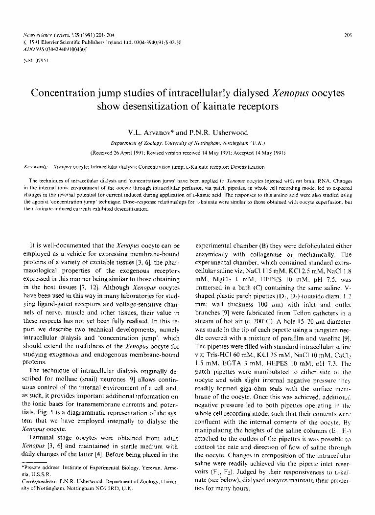
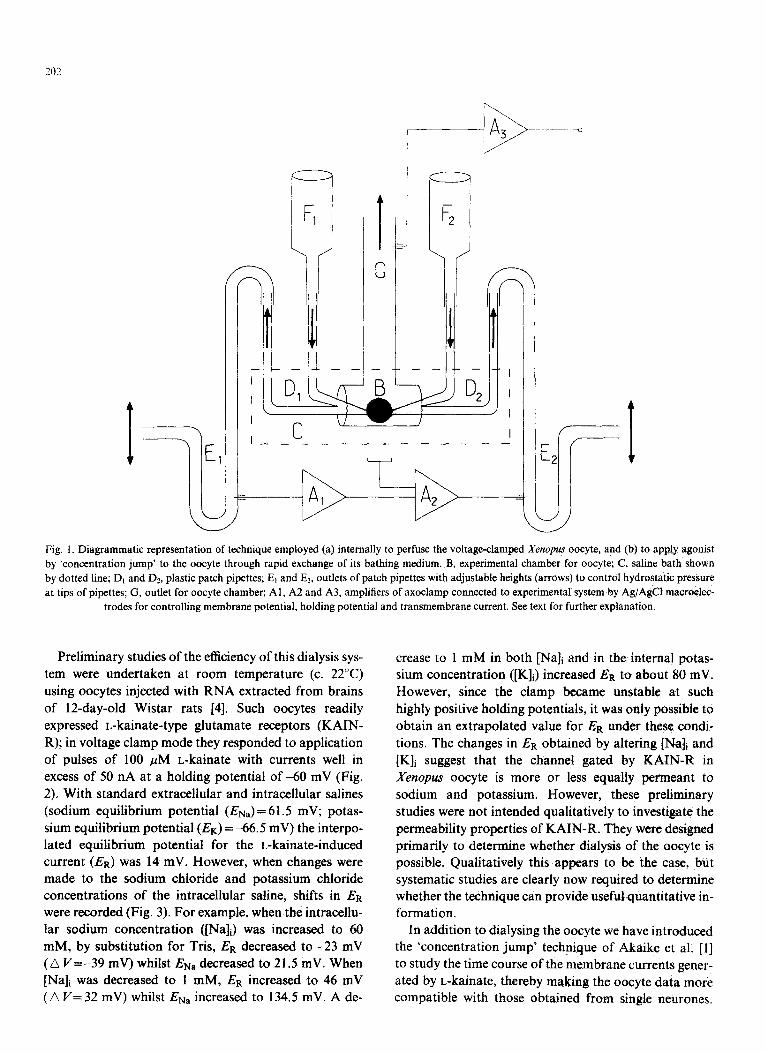
Preliminary studies of the efficiency of this dialysis sys- tem were undertaken at room temperature (c. 22°C) using oocytes injected with RNA extracted from brains of 12-day-old Wistar rats [4]. Such oocytes readily expressed L-kainate-type glutamate receptors (KAIN- R); in voltage clamp mode they responded to application of pulses of 100 /tM L-kainate with currents well in excess of 50 nA at a holding potential o f - 6 0 mV (Fig. 2). With standard extracellular and intracellular salines (sodium equilibrium potential (ENa)=61.5 mV; potas- sium equilibrium potential (EK)=-66.5 mV) the interpo- lated equilibrium potential for the L-kainate-induced current (ER) was 14 mV. However, when changes were made to the sodium chloride and potassium chloride concentrations of the intracellular saline, shifts in ER were recorded (Fig. 3). For example, when the intracellu- lar sodium concentration ([Na]i) was increased to 60 mM, by substitution for Tris, ER decreased to -23 mV (G V=-39 mV) whilst ENa decreased to 21.5 mV. When [Na]i was decreased to 1 mM, ER increased to 46 mV (A V=32 mV) whilst ENa increased to 134.5 mV. A de-
crease to 1 mM in both [Na]i and in the internal potas- sium concentration ([K]i) increased ER to about 80 mV. However, since the clamp became unstable at such highly positive holding potentials, it was only possible to obtain an extrapolated value for ER under these condi- tions. The changes in ER obtained by altering [Na]i and [K]i suggest that the channel gated by KAIN-R in Xenopus oocyte is more or less equally permeant to sodium and potassium. However, these preliminary studies were not intended qualitatively to investigate the permeability properties of KAIN-R. They were designed primarily to determine whether dialysis of the oocyte is possible. Qualitatively this appears to be the case, but systematic studies are clearly now required to determine whether the technique can provide usefulquantitative in- formation.
In addition to dialysing the oocyte we have introduced the 'concentration jump' technique of Akaike et al. [1] to study the time course of the membrane currents gener- ated by L-kainate, thereby making the oocyte data more compatible with those obtained from single neurones.

A. 50wM 100pM 200WM
' R . SOpM lO 0~,~ 2 0 0 m',~
2 0 s
Fig. 2. Responses of dialysed oocytes during 'concentration jump' ap- plication of L-kainate. Duration of L-kainate application shown by bars above current traces. Data recorded from 2 oocytes (A and B, re- spectively), 6 (A) and 8 days (B) after injection with rat brain RNA. Total RNA was obtained from brains of I l-day-old Wistar rats (4).
Similar results were obtained with non-dialysed neurones.
203
The responses to L-kainate of rat cortical or hippocam- pal neurones studied using 'concentration jump' have been described as non-desensitizing [8, 10] like those recorded from Xenopus oocytes injected with rat brain R N A and studied without 'concentration jump' [4]. However, L-kainate-activated responses of rat dorsal root neurones [7] and ventral spinal cord cells [11] exhi- bit marked desensitization. In our studies, a 'concentra- tion jump' technique was used as follows. For applica- tion of L-kainate, the bath (C) in Fig. 1 was lowered. Electrical connections were maintained during this pro- cedure and the oocyte remained surrounded by saline in the recording chamber (B). The bath was then filled with saline containing L-kainate and raised rapidly to its for- mer position, at which time suction was applied to the outlet tube (G). Tests made with solutions of markedly different conductances indicated that the contents of the
150
c
E-
L L D
(_]
Holding Potential (mV) I I I, L / f e ' ~ ~ ' ~ j
3 . •
4.
100
50
J
j ~ I t - ~ 1 f ~ 60
-50 ~ /
@
I I 80 ]00
- I00
- 150
Fig. 3. Current -voltage characteristics for L-kainate-induced currents in dialysed Xenopus oocyte injected with rat brain RNA. Data recorded 6 days post-injection. Agonist applied by 'concentration jump' technique. L-kainate-induced responses obtained at different holding potentials during ap- plication of 50 ~M agonist. The extracellular and intracellular salines were as follows. (1) Extracellular, standard extracellular saline; intracellular, standard intracellular saline (see text for details of both of these salines); (2) extracellular, standard extracellular saline; intracellular, NaCI 60 mM, Kci 35 mM, CaCI2 1.5 raM, EGTA 3 raM, Tris-HC1 10 mM, HEPES 10 mM, pH 7.3; (3) extracellular, standard extracellular saline; intracellular, NaCI 1 mM, KCI 35 mM, CaCI2 1.5 raM, EGTA 3 raM, Tris-HC1 69 mM, HEPES 10 mM, pH 7.3; (4) extracellular, standard extracellular saline; intracellular, NaC1 1 mM, KCI 1 mM, CaCI2 1.5 mM, EGTA 3 mM, Tris-HCl 103 mM, HEPES 10 mM pH 7.3. Lines through data points drawn
by linear regression.
204
chamber (C) were replaced within 20 ms using this tech- nique.
Dose-response relationships for t.-kainate obtained
using 'concentration jump' were similar to those
obtained with a slower superfusion technique [4]. How-
ever, the time course of responses to L-kainate obtained
were more complex. With superfusion, the responses to
L-kainate are usually slow rising and do not exhibit any
obvious fade, at least during brief (10 s) applications of
this amino acid, although with high concentrations (10 --~
M) some evidence for desensitization has been seen (P.L.
Brackley and P.N.R. Usherwood, in preparation). With
'concentration jump', even during application of rela- tively low concentrations (e.g. 50 pM) of L-kainate, the
responses of some oocytes were characterised by a slow
rising phase bearing a small lip (Fig. 2A). In other
oocytes the responses quickly reached a peak before
desensitizing to a lower current level (Fig. 2B). Finally, in some oocytes the responses were intermediate between
these two extremes, i.e. first peaking, then transiently
fading and then rising to a plateau, which was either
slightly less than or slightly greater than the initial cur-
rent peak (data not illustrated). Changes in the extracel-
lular environment of an oocyte made under 'concentra-
tion jump' conditions, but in the absence of L-kainate,
did not cause any changes in base-line current. These re-
sults suggest that with rapid application of L-kainate,
fading of the response to this agonist does occur,
although the basis for this phenomenon has not been
established. In fact, even with agonist exchange times of
around 20 ms some desensitization could still be masked. At face value the results suggest that oocytes injected
with rat brain R N A express two types of KAIN-R,
desensitising and non-desensitizing respectively. How-
ever, homomeric GIu-R1 receptors expressed in Xenopus oocytes exhibit responses during rapid L-kainate super-
fusion similar to those described herein [5]. This report shows that cell dialysis could be used rou-
tinely to assist investigations of the chemosensitive and ionic properties of the Xenopus oocyte. This approach
will be of particular value to those interested in studying drugs which might act on intracellular domains of mem-
brane bound proteins and in studying ligand-gated receptors which operate secondary messenger systems. For example, through intracellular application of enzymes, enzyme inhibitors, ADP, ATP, 3',5'-AMP, 3' ,5 '-GMP and Ca 2÷ etc. it will be possible more directly
to study the influence of phosphorylation on membrane-
bound signalling proteins expressed from exogenous
RNA. The effects of phosphorylation on specific ion
pumps may also be studied in this manner to gain further
insight into the relationship between activity of these
proteins and membrane chemosensitivity [2]. This report
also shows that the "concentration jump' technique
further extends the usefulness of the Xenopus oocyte as an expression vehicle for exogenous, membrane-bound proteins.
V.L.A. thanks the Royal Society of London for financial
support. Chris Standley is thanked for assistance with
the illustrations.
1 Akaike, N., Inoue, M. and Kristhal, O.A., "Concentration clamp" study of GABA-induced chloride current kinetics in frog sensory neurones, J. Physiol., 397 (1986), 171-185.
2 Arvanov, V.A. and Usherwood, P.N.R., Effects of ouabain on volume and chemosensitivity of Xenopus oocytes injected with rat brain mRNA, Neurosci. Lett., 125 (1991), 9-11.
3 Barnard, E.A. and Bitbe, G., Functional expression in the Xenopus oocyte of mRNAs for receptors and ion channels. In A.L Turner and H.S. Batchelard (Eds.), Neurochemistry; A Practical Ap- proach, I.R.L. Press, Oxford, 1989, pp. 243 270.
4 Brackley, P., Goodnow, Jr., R., Nakanishi, K., Sudan, H.L. and Usherwood, P.N.R., Spermine and philanthotoxin potentiate exci- tatory amino acid responses of Xenopus oocytes injected with rat and chick brain RNA, Neurosci. Lett., 114 (1990) 51-56.
5 Dawson, T.L., Nicholas, R.A. and Dingledine, R., Homomeric GluR1 excitatory amino acid receptors expressed in Xenopus oocytes, Mol. Pharm., 38 (1990), 779--784.
6 Gundersen, C.B., Miledi, R. and Parker, I., Glutamate and kainate receptors induced by rat brain messenger RNA in Xenopus oocytes, Proc. Roy. Soc. Lond. B., 221 (1984) 127--143.
7 Huettner, J.E., Glutamate receptor channels in rat dorsal root ganglion neurones; activation by kainate and quisqualate and blockade of desensitization by concanavalin A, Neuron, 5 (1990) 255-266.
8 Kiskin, N.I., Kristal, O.A. and Tsyndrenko, A,, Excitatory amino acid receptors in hippocampal neurones: kainate fails to desensitize them, Neurosci. Lett., 63 (1986) 225-230.
9 Kostyuk, P.G., Kristhal, O.A. and Pidoplichko, V.L., Effects of in- ternal fluoride and phosphate on the membrane currents during in tracellular dialyses of nerve cells, Nature, 257 (1975) 69 l q593.
10 Mayer, M.L., Vyklicky, Jr. L. and Clements, J., Regulation of NMDA receptor desensitization in mouse hippocampal neurons, Nature, 338 (1989) 425-427.
I I Smith, D.O., Franke, Ch., Rosenheimer, J.L., Zufall, F. and Hart, H., Glutamate-activated channels in adult rat ventral spinal cord cells, J. Neurophysiol., in press.
12 Verdoorm T.A. and Dingledine, R., Excitatory amino acid recep- tors expressed in Xenopus oocytes: agonist pharmacology, Mol. Pharmacol., 34 (1988) 298-307.


![Renal anaemia treatment in haemodialysis patients in the Central … · 2017-08-26 · (CKD) and dialysed patients [3, 4]. Silverberg et al. [5] proposed the term ‘cardio-renal](https://static.fdocuments.in/doc/165x107/5f78876c990a4719c709230a/renal-anaemia-treatment-in-haemodialysis-patients-in-the-central-2017-08-26-ckd.jpg)


![· 2019-07-08 · ing erythropoiesis both in patients on dialysis [10], and in patients not dialysed with chronic renal failure [11]. Several studies have demonstrated that patients](https://static.fdocuments.in/doc/165x107/5ea80824d07b732cbc502055/2019-07-08-ing-erythropoiesis-both-in-patients-on-dialysis-10-and-in-patients.jpg)